association between the low levels of vitamin d and treg...
TRANSCRIPT

26
ORIGINAL ARTICLE
Acta Medica Indonesiana - The Indonesian Journal of Internal Medicine
Association Between the Low Levels of Vitamin D and Treg Function in Systemic Lupus Erythematosus Patients
Kusworini Handono1, Dona Marisa2, Handono Kalim3
1 Department of Clinical Pathology, Faculty of Medicine, Brawijaya University. Jl. Veteran Malang, Malang, Indonesia.2 Department of Physiology, Faculty of Medicine, Lambung Mangkurat University, Banjarmasin, Indonesia.3 Department of Internal Medicine, Faculty of Medicine, Brawijaya University, Malang, Indonesia.Correspondence mail: [email protected].
ABSTRAKTujuan: mengetahui hubungan kadar vitamin D dan ekspresi CCR4 sel Treg serta kapasitas migrasi sel Treg
pada pasien SLE di Indonesia. Metode: pengukuran kadar vitamin D, ekspresi CCR4 sel Treg dan kapasitas migrasi sel Treg dilakukan pada 41 pasien SLE dan 20 subjek sehat sebagai kontrol. Kadar vitamin D diukur dengan teknik ELISA (Cusabio). Ekspresi CCR4 sel Treg dideteksi dengan sitometri alir (flow cytometry) dan kapasitas migrasi sel Treg dilakukan dalam ruang kemotaksis (chemotaxis chamber) menggunakan ligand CCR4, TARC, dan MDC yang selanjutnya dianalisis dengan menggunakan flow cytometry menggunakan FACS Calibur (Becton Dickinson). Jumlah sel Treg yang mengalami migrasi merupakan rasio jumlah sel CD4+CD25+CCR4+ pada ruang kemotaksis bawah dibandingkan jumlah keseluruhan sel CD4+CD25+CCR4+ sebelum migrasi dan dinyatakan dalam bentuk persentase. Hasil: kadar vitamin D secara bermakna ternyata lebih rendah pada pasien SLE dibandingkan subjek kontrol yang sehat (p=0,000). Kadar vitamin D berkaitan positif dengan jumlah persentase sel Treg yang bermigrasi menuju TARC (p=0,015) dan MDC (p=0,000), namun tidak ditemukan perbedaaan dalam hal ekspresi CCR4 sel Treg antara pasien SLE dan subjek kontrol yang sehat. Kesimpulan: kadar vitamin D yang rendah menyebabkan penurunan kapasitas migrasi sel Treg pada pasien SLE dan orang sehat. Pengaruh ini dapat terjadi lebih karena faktor lainnya dan cenderung bukan melalui mekanisme ekspresi CCR4.
Kata kunci: vitamin D, fungsi CCR4 sel Treg, MDC, TARC.
ABSTRACTAim: to determine the relationships between vitamin D and CCR4 expression on Treg, and Treg migratory
capacity in SLE patients in Indonesia. Methods: vitamin D level assesment, CCR4 expression on Treg and Treg migratory capacity were performed on 41 SLE patients and 20 healthy controls. Serum vitamin D levels were measured by ELISA (Cusabio). The expression of CCR4 on Treg was detected by flow cytometry and Treg migratory capacity was performed in chemotaxis chamber using CCR4 ligands, TARC and MDC, and subsequently analyzed by flow cytometry using FACS Calibur (Becton Dickinson). The number of migrated Treg is the ratio of CD4+CD25+CCR4+ cells number in lower chamber of chemotaxis compared to the total number of CD4+CD25+CCR4+ cells before migration, and stated in percentage. Results: vitamin D levels were significantly lower in SLE patients compared with healthy controls (p=0.000). The vitamin D levels were positively correlated to the percentage of migrated Treg toward TARC (p=0.015) and MDC (p=0.000), but there was no difference in Treg CCR4 expression between SLE patients and healthy controls. Conclusion: low vitamin D levels cause reduced Treg migratory capacity in SLE patients and healthy people. This influence occurs through other factors rather than CCR4 expression.
Key words: vitamin D, treg function CCR4, MDC, TARC.

Vol 45 • Number 1 • January 2013 Association between the low levels of vitamin D and Treg function in SLE
27
INTRODUCTIONSystemic lupus erythematosus (SLE) is a
systemic autoimmune disease which occurs more frequently in the last decade. One mechanism involved in the pathogenesis of loss of tolerance in autoimmune disease including SLE is Treg dysfunctions. A decreased in Treg numbers or their functional deficiency seem to be associated with the active stage of the disease.1 The suppressive function of Treg on effector cells largely depends on their migratory capacity to the site of inflammation. Treg expresses chemokine receptors type 4 (CCR4) that respond to the macrophage derived chemokine (MDC/CCL22) and activation-regulated chemokine (TARC/CCL17) to migrate toward APC and activated T cells. By this mechanism they regulate ongoing inflammatory responses and control these cells to prevent it becoming hyperactive. Treg migratory capacity therefore plays a critical role in the maintenance of self-tolerance.2 Thus, Treg are of considerable interest as targets for the treatment of autoimmune disease.3 Indeed, induction of Treg in experimental lupus mice can prevent the development of lupus manifestation.4
Vitamin D in general exerts an inhibitory action on the the adaptive immune reponse by several mechanisms, including inhibition of dendritic cell (DC) maturation, Th1 activity, B cel maturation and differentiation and Treg function.5 Treatment of DC with 1,25 (OH)2D can induce Treg development, as shown by increased FoxP3+ expression and Treg function by altering homing (migration) properties of the cells.6-7 Hence, by impoving Treg cell function, vitamin D may be have beneficial effect in the heatment of SLE and treatment with vitamin D may improved patients response to immnusuppresant medication.8 Recent studies have reported an associations between vitamin D deficiency with the risk of SLE and disease activity of SLE.8-11
A recent study showed that in the Indonesian population, a country located on the equator with year-long sun exposure, there were a significant number of subjects with low levels vitamin D.12 The clinical manifestations of Indonesian SLE patients are different from those of Caucasians origins; Indonesian SLE patients have shown more severe clinical manifestations, higher levels of anti-ds DNA antibodies, and photosensitivity.1 These facts raise the question whether the severity of their clinical manifestations is affected
by vitamin D levels result in increased Treg dysfunction in our patients. The aim of this study is to determine the associations of vitamin D, expression of CCR4 on Treg and Treg function (migratory capacity) in SLE patients in Indonesia.
METHODSThe subjects were female SLE patients
(based on 1997 ACR criteria), had experienced flare, with systemic lupus erythematosus disease activity index (SLEDAI) score >3, and had not taken vitamin D supplement from Rheumato-Immunology Clinic, Department of Internal Medicine, Saiful Anwar Hospital, Malang. The controls were healthy female matched in age, and body weight, and also did not take vitamin D supplement. This study met the ethical clearance by Ethics Commission of Faculty of Medicine, Brawijaya University. Informed consents were obtained from all subjects.
Isolation of Peripheral Blood Mononuclear Cells (PBMCs)
Four ml of fresh blood from each subject collected in serum separator tube was centrifuged in 4°C for 20 minutes. The supernatant was collected into fresh tubes in aliquot and stored in 70°C until the next process for ELISA. Twelve ml of heparinized blood from each subject was diluted 1:1 with phosphate buffer solution (PBS) in a canonical tube. Then, this mixture was layered over Ficoll® (Amersham Biosciences) in the ratio of 2:1 in tubes. These tubes were centrifuged (1000xg) in 4°C for 30 minutes. PBMCs harvested from the interface between Ficoll® and plasma (the buffy coat) were collected into fresh tubes and washed twice with PBS. These PBMCs were divided into 2 tubes in the ratio of 1:5 for detection of CCR4 (direct/fresh process) and Treg migratory capacity test, respectively.
Immunofluorecent Staining Procedure for CCR4 Expression on Treg
All reagents for this procedure were produced by Biolegend. Fresh PBMCs were re-suspended with 0.5 ml of cell staining buffer. 20 μl of FITC anti-human CD4 antibody, 20 μl of PE anti-human CD25 antibody and 5 μl of PerCP/Cy5.5 anti-human CCR4 antibody were added, incubated at room temperature for 15-20 minutes in the dark, and then washed twice with 1.5 ml of cell staining buffer with a centrifuge at 350xg

Kusworini Handono Acta Med Indones-Indones J Intern Med
28
for 5 minutes. The cells were re-suspended in 0.5 ml of cell staining buffer and analyzed by flow cytometry.
Treg FunctionTo asses the function of Treg we assayed
the migratory capacity of Treg conducted in 24-well chemotaxis chambers (Costar) with polyvinylpyrrolidine-free polycarbonate membranes (5 μm pore size). The bottom chamber of each well was filled with 600 μl of agonist at the appropriate concentration (diluted in RPMI 1640 and 0.1% BSA) and carefully overlaid with the polycarbonate membrane. Human chemokines TARC (CCL17, R&D Systems) and MDC (CCL22, R&D Systems) were used, 100 and 50 ng/ml, respectively. PBMCs were re-suspended in RPMI 1640 medium and 0.1% BSA at 5x10⁵ cells/ml, and 100 μl of the cell suspension was added to the top chambers. The chambers were incubated for 2 hours in a 5% CO2 humidified incubator at 37°C, and the cells migrated across the membrane into the lower chamber were counted with flow cytometry.
Flow Cytometry AnalysisExpression of CCR4 on Treg was the
percentage of CD4+CD25+CCR4+ cells number from all CD4+CD25+ cells number. The number of migrated Treg is the ratio of CD4+CD25+CCR4+ cells number in lower chamber of chemotaxis compared to the total number of CD4+CD25+CCR4+ cells before migration, and stated in percentage. These analyses were using FACS Calibur (Becton Dickinson).
Quantitative Detection of 25(OH) Vitamin D325(OH)D3 was quantitatively detected by
ELISA kit produced by Cusabio (cat. CSB-E07900h). A hundred micro liters of standard, blank, or sample were added to well, covered, incubated for 2 hours at 37°C and the liquid of each well was removed without washing. A hundred micro liters of biotin-antibody working solution was added to each well and incubated for 1 hour at 37°C, and then washed three times. A hundred micro liters of HRP-avidin working solution was added to each well, covered, incubated for 1 hour at 37°C, then washed three times. Ninety micro liters of TMB substrate was added to each well, covered, incubated for 30 minutes at 37°C in the dark. Fifty micro liters of stop solution was added to each well. The optical density of each well was determined within 30 minutes using a microplate reader at 450 nm. Normal level of vitamin D is 30 ng/ml or more.
Statistical Analysis All results were expressed as mean±SD. The
independent t-test was performed to compare variables between groups and correlations between two clinical parameters were evaluated with Pearson correlation test using SPSS program. The differences were significant if p-values were <0.05.
RESULTSThe mean age of SLE patients was not
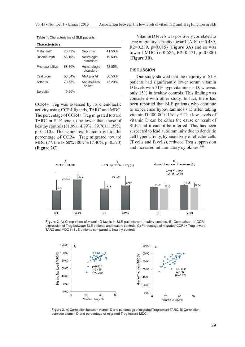
significantly different compared with healthy control subject (29.35±9.99 v.s. 32.91±5.92, p=0.02), with duration of illness 2-54 months. Most of the clinical manifestations were nephritis, photosensitivity, and arthritis (Table 1). Vitamin D levels in SLE patients were significantly lower than those in healthy controls (23.0±11.9 ng/mL v.s. 36.0±5,7 ng/mL, p=0.000). Hipovitaminosis D (<30 ng/ml) were observed in 29 SLE patients (71%) and three healthy controls (15%), whereas the frequency of subjects with normal levels of vitamin D (levels ≥30 ng/ml) was significantly lower compared with healthy controls (29% : 85%, p=0.001).
Expression of CCR4 on Treg and Treg Migratory Capacity
The expression of CCR4 on Treg in SLE was not significantly different compared to healthy controls (49.33±17.00% v.s. 57.87±20.89%, p=0.230) (Figure 2B). Migratory capacity of
Figure 1. Expression of CCR4 on Treg in healthy controls (A) and SLE patients (B). PBMCs were isolated from fresh blood and stained with immunofluorescent for Treg surface markers (CD4 and CD25) and its chemokine receptor marker (CCR4). Dots in right upper quadrants show each cells which was stained with CD4, CD25 and CCR4.
Vol 45 • Number 1 • January 2013 Association between the low levels of vitamin D and Treg function in SLE
29
CCR4+ Treg was assessed by its chemotactic activity using CCR4 ligands, TARC and MDC. The percentage of CCR4+ Treg migrated toward TARC in SLE tend to be lower than those of healthy controls (81.99±14.79% : 89.76±11.39%, p=0.110). The same result occurred to the percentage of CCR4+ Treg migrated toward MDC (77.15±18.60% : 80.74±17.40%, p=0.590) (Figure 2C).
Vitamin D levels was positively correlated to Treg migratory capacity toward TARC (r=0.489, R2=0.239, p=0.015) (Figure 3A) and so was toward MDC (r=0.686, R2=0.471, p=0.000) (Figure 3B).
DISCUSSIONOur study showed that the majority of SLE
patients had significantly lower serum vitamin D levels with 71% hypovitaminosis D, whereas only 15% in healthy controls. This finding was consistent with other study. In fact, there has been reported that SLE patients who continue to experience hypovitaminosis D after taking vitamin D 400-800 IU/day.13 The low levels of vitamin D can be either the cause or result of SLE, and it cannot be inferred. This has been suspected to lead autoimmunity due to dendritic cell hyperactivity, hyperactivity of effector cells (T cells and B cells), reduced Treg suppression and increased inflammatory cytokines.8-11
Table 1. Characteristics of SLE patients
Characteristics
Malar rash 70.73% Nephritis 41.50%
Discoid rash 56.10% Neurologic disorders
19.50%
Photosensitive 68.30% Hematologic disorders
78.05%
Oral ulcer 58.54% ANA positif 80.50%
Arthritis 70.73% Anti ds-DNA positif
73.20%
Serositis 19.50%
Figure 2. A) Comparison of vitamin D levels in SLE patients and healthy controls. B) Comparison of CCR4 expression of Treg between SLE patients and healthy controls. C) Percentage of migrated CCR4+ Treg toward TARC and MDC in SLE patients compared to healthy controls.
Figure 3. A) Correlation between vitamin D and percentage of migrated Treg toward TARC. B) Correlation between vitamin D and percentage of migrated Treg toward MDC.

Kusworini Handono Acta Med Indones-Indones J Intern Med
30
Previous studies have shown a decrease in circulating Treg number in SLE patients, and escepicially during the flare, their suppressive functions become normal in vitro. Generally, researchers have found a decrease in suppressive function of T cells, however few others have found the opposite; therefore the role of Treg cells in the pathogenesis of SLE still remains to be investigated further.14-15 The ability of Treg to suppress other cells depends on their migratory capability to inflammatory sites.2 Treg migration is regulated by specific signals emanating from the chemokine and integrin.16 Treg expresses CCR4 and CCR8 and respond to macrophage derived chemokine (MDC/CCL22), thymus activation-regulated chemokine (TARC/CCL17), I-309/CCL1, and vMIP-virokine I. Expression of CCR4 and CCR8 on Treg cells cause the cells to migrate toward APC and T cells, and further suppress the function of APC or T cells. CCL17 and CCL22 produced by a variety of cells in inflammatory tissues, and recruit Treg to down regulate ongoing inflammatory process. CCR4 is the major chemokine receptor expressed by Treg.2
A number of studies suggested the existence of vitamins that regulate the expression of chemokine receptors, such as vitamin A and D, as well as several cytokines such as IL-12.17 In the rat study model of multiple sclerosis (MS) and experimental autoimmune encephalomyelitis (EAE), it was found that 1,25(OH)2D3 inhibited CCR6 expression on T cells that had been activated by transforming growth factor-β (TGF-β) and interleukin-6 (IL-6).18 Further studies conducted by Sigmundsdottir et al. showed that vitamin D3 induced CCR10 expression on CD4+ T cells that had been activated IL-12.19
Finaly, the mechanism of immunosuppression requires contact between effector cells and Treg. Twenty percent of circulating Treg have CCR4, a chemokine receptor which allow Treg to migrate to inflammatory area to do their regulatory function.20 Research conducted by Lee et al. showed that CCR4 expression on Treg in SLE patients was significantly lower in comparison to healthy controls.20 In contrast to previous studies, this study shows no significant difference in CCR4 expression on Treg and their migratory capacity between SLE patients and healthy controls. However, when we made correlation from all subjects (SLE patients and healthy controls), there were significant
positive correlation between vitamin D and Treg migratory capacity. Vitamin D affects Treg migratory capacity toward TARC as much as 23.9% and toward MDC as much as 47.1%. These results indicated that vitamin D affects Treg migratory capacity through other factors rather than CCR4.
The current treatment for SLE is aimed at suppressing the immune response and excessive inflammation by prescribing immunosuppressive medications. The use of these medications has shown positive results in developed countries, however in developing countries there has been a financial constrain in administering these types of medications. This situation has led to the initiative of using supplement in SLE patients in developing countries. By improving Treg cells funtion, vitamin D is a good option to supplement immuno suppresant drugs in the treatment of SLE.
CONCLUSIONLow vitamin D levels cause the reduction of
Treg migratory capacity in SLE patients and the healthy controls. This influence occurs through factors other than CCR4 expression.
REFERENCES1. Kleczynska W, Bogdan J, Hanna P, et al. Imbalance
between Th17 and regulatory T-cells in systemic lupus erythematosus. Folia Histochemica Et Cytobiologica. 2011;49(4):646-53.
2. Tischner D, Weishaupt A, van den Brandt J, et al. Polyclonal expansion of regulatory T cells interferes with effector cell migration in a model of multiple sclerosis. Brain. 2006;129:2635–47.
3. Fort MM and Padma KN. Review manipulation of regulatory T-cell function by immunomodulators: a boon or a curse? Toxicological Sciences. 2010;117(2):253–62.
4. Lan Q, Huimin F, Valerie Q, et al. Induced Foxp3+ regulatory T cells: a potential new weapon to treat autoimmune and inflammatory diseases? J Molecular Cell Biology. 2012;4:22–8.
5. Bickle D. Nonclassic actions of vitamin D.J Clin Endocrinol Metab. 2009;94(1):26–34.
6. Gregori S, Casorati M, Amuchastegui S, et al. Regulatory T cells induced by 1 alpha,25-dihydroxyvitamin D3 and mycophenolate mofetil treatment mediate transplantation tolerance. J Immunol. 2001;167:1945–53.
7. Sakaguchi S, Yamaguchi T, Nomura T, et al. Regulatory T-cells and immune tolerance. Cell. 2008;133:775–87.
8. Toubi E, Shoenfeld Y. The role of vitamin D in regulating immune responses. IMAJ. 2010;12:174-5.
9. Irastorza GR, Egurbide MV, Olivares N, et al. Vitamin D deficiency in systemic lupus erythematosus:

Vol 45 • Number 1 • January 2013 Association between the low levels of vitamin D and Treg function in SLE
31
prevalence, predictors and clinical consequences. Rheumato. 2008;47:920-3.
10. Ginanjar E, Sumariyono, Setiati S, et al. Vitamin D and autoimmune disease. Acta Med Indones-Indones J Intern Med. 2007;39(3):133-41.
11. Mouyis M, Ostor AJK, Crisp AJ, et al. Hypovitaminosis D among rheumatology outpatients in clinical practice. Rheumato. 2008;47:1348-51.
12. Setiati S. Vitamin D status among Indonesian elderly woman living in institutionalized care unit. Acta Med Indones-Indones J Intern Med. 2008;40(2):78-82.
13. Toloza SM, Cole DE, Gladman DD, et al. Vitamin D insufficiency in a large female SLE cohort. Lupus. 2010;19(1):13-9.
14. Horwitz DA. Regulatory T cells in systemic lupus erythematosus: past, present and future. Arthritis Research & Therapy. 2008;10:227.
15. Valencia X, Yarboro C, Illei G, et al. Deficient CD4+CD25high T regulatory cell function in patients with active systemic lupus erythematosus. J Immunol. 2007;178:2579–88.
16. Wei S, Kryczec I, and Zou W. Regulatory T-cell compartmentalization and trafficking. Blood. 2006;108:426-31.
17. Mebius RE. Vitamins in control of lymphocyte migration. Nat Immunol. 2007;8:229–30.
18. Chang JH, Cha HR, Lee DS, et al. 1,25-dihydroxyvitamin D3 inhibits the differentiation and migration of TH17 cells to protect against experimental autoimmune encephalomyelitis. Plos One. 2010;5(9):e12925.
19. Sigmundsdottir H, Pan J, Debes GF, et al. DCs metabolize sunlight-induced vitamin D3 to ‘program’ T cell attraction to the epidermal chemokine CCL27. Nat Immunol. 2007;8:285–93.
20. Lee HY, Hong YK, Yun HJ, et al. Altered frequency and migration capacity of CD4+CD25+regulatory T cells in systemic lupus erythematosus. Rheumato. 2008;47:789-94.