assessment of investigation techniques for scyphozoan
TRANSCRIPT

MARINE ECOLOGY PROGRESS SERIESMar Ecol Prog Ser
Advance Viewhttps://doi.org/10.3354/meps12292
Available online:October 6, 2017
INTRODUCTION
Scyphozoan and cubozoan statocysts contain sta-toliths (crystals) composed of calcium sulfate hemi -hydrate (bassanite) (Becker et al. 2005, Sötje et al.2011). Adult cubozoan medusae bear a single stato -lith in each of 4 statocysts (Sötje et al. 2011), where asthe statocysts of investigated adult scyphomedu saecontain numerous single statoliths (Holst et al. 2007,
Sötje et al. 2011). Because the statoliths are the onlysolid, inorganic structures in the medusa bell, theyprovide the potential for age determination compara-ble to the calcified body parts of other marine organ-isms, for example squid and gastropod stato liths (e.g.Rodhouse & Hatfield 1990, Barroso et al. 2005) andfish otoliths (e.g. Panfili et al. 2009). Age determina-tion is an important tool for studies of populationdynamics and could improve backtracking models of
© The authors 2017. Open Access under Creative Commons byAttribution Licence. Use, distribution and reproduction are un -restricted. Authors and original publication must be credited.
Publisher: Inter-Research · www.int-res.com
*Corresponding author: [email protected]
Assessment of investigation techniques for scyphozoan statoliths, with focus on early
development of the jellyfish Sanderia malayensis
Anneke Heins1,2,*, Ilka Sötje3, Sabine Holst2
1University of Oldenburg, Institute for Biology and Environmental Sciences, 26129 Oldenburg, Germany2Senckenberg am Meer, German Center for Marine Biodiversity Research, 20146 Hamburg, Germany
3University of Hamburg, Biocenter Grindel, Zoological Institute, 20146 Hamburg, Germany
ABSTRACT: Statolith ageing techniques provide a useful tool in the study of age, growth, andpopulation structure of several marine organisms. Previous investigations on scyphozoan stato -liths demonstrated increased statolith numbers and size with increasing medusa size, as well astaxon-specific variations in statolith shape. Thus, statoliths are potentially useful for age determi-nation and taxonomic studies on medusae. Unfortunately, studies on statoliths are difficultbecause of the fragile structure of the numerous tiny crystals and their hygroscopic nature. Weevaluated the potential of various modified study techniques for scyphozoan statocysts and stato -liths applied to early medusa stages of Sanderia malayensis Goette, 1886. Light microscopy wassuitable to follow the increase of statolith numbers and sizes with age and allowed differentiationof various statolith shape types, which were confirmed by scanning electron microscopy. The useof the fluorescent marker calcein provided direct evidence for measurable statolith growth. Ourresults indicated that statoliths stopped growing after reaching a certain size while additionalstato liths developed in the basal region of the statocyst. Micro-computed tomography dataallowed visualization and morphometric measurements of statoliths, revealing interspecific differ-ences that may be useful for phylogenetic studies. Statolith numbers were positively correlatedwith medusa age and bell diameter. Thus, statolith analyses could provide information on medusaage or size, both of which are important parameters for population structure analyses that are difficult to assess with currently applied techniques. Our study provides methodological bases forfuture studies to reveal further potential of scyphozoan statolith investigations for phylogeneticand ecological studies.
KEY WORDS: Cnidaria · Statocyst · Medusa · Slide scanner · Micro-tomography · Calcein · Scanning electron microscopy · SEM · Energy-dispersive X-ray spectroscopy · EDX
Contribution to the Theme Section ‘Jellyfish bloom research: advances and challenges’ OPENPEN ACCESSCCESS

Mar Ecol Prog Ser · Theme Section · Advance View
jellyfish pathways and thus discovery of polyp habi-tats (Dulière et al. 2014, Gambill 2016), which play akey role in determining the timing and intensity ofjellyfish blooms (Schiariti et al. 2015).
Scyphozoan statoliths have been described as trigo-nal-trapezohedral crystals composed of 6 trapezoids(side faces) and 3 pentagons on each side of the rod(head faces) (Sötje et al. 2017). The size, growth, andnumber of scyphozoan statoliths increase with in -creasing medusa size (e.g. Holst et al. 2007, Sötje et al.2011, 2017), indicating the potential of these crystalsfor medusa age determination, as previously shownfor cubozoans (Gordon et al. 2004). Moreover, previ-ous investigations on scyphozoan statocysts have re-vealed taxa-specific differences in the morphology ofthe statolith arrangement and in statolith shapes.Thus, statolith analyses may be useful for taxo nomicand phylogenetic studies (Becker et al. 2005, Holst etal. 2007, 2016, Sötje et al. 2011, 2017). Difficulties inscyphozoan statolith analyses lie in the extremely hygroscopic nature of bassanite, which quickly trans-forms to dehydrate (gypsum) in contact with moisture(Becker et al. 2005, Boßelmann et al. 2007). Because ofthis and the fragility of the numerous tiny crystals, thefew studies conducted on statolith development relat-ing to medusa age are often based on few test animalsor a unilateral spectrum of methods (Sötje et al. 2011,2017, Hopf & Kingsford 2013).
In the present study, we conducted the first com-prehensive study on statoliths of a scyphomedusabelonging to the taxon Pelagiidae. We provide anextensive dataset of statocysts and statolith analyseson ephyrae and medusae of different ages. The de -velopment of ephyra to early medusa stages ofSanderia malayensis Goette, 1886 was documentedwith focus on the morphology and development ofstatocysts, on the analysis of statocyst number perspecimen, statolith number per statocyst, statolithand statocyst shape and growth, and statolith chem-istry. We used a combination of previously estab-lished and new methods, especially modified for theparticular requirements of statoliths composed ofbasanite, in order to evaluate the utility of differenttechniques in future studies. Our methods included(1) light microscopic (LM) studies with a slide scan-ner, (2) application of the fluorescent marker calceinon statoliths combined with confocal laser scanningmicro scopy (cLSM), (3) 3-dimensional reconstruc-tions of statocysts from X-ray micro-tomographicdata, (4) scanning electron microscopy (SEM) ofstato liths from different de velopmental stages, and(5) determination of the statolith chemical composi-tion by energy-dispersive X-ray spectroscopy (EDX).
MATERIALS AND METHODS
Cultivation of Sanderia malayensis
Ephyrae were obtained from strobilating S. ma lay -ensis polyps (Fig. 1A−F) (for origin of polyp culture,see Adler & Jarms 2009) cultured in darkness, at aconstant temperature of 20°C in natural seawater(5−13 µm filtered, salinity 35, pH 8.1 ± 0.1). The polypswere fed with 0 to 1 d old Artemia salina naupliiweekly. Detached ephyrae were removed from theculture daily and photo graphed individually in theextended state (Fig. 1G). Ephyrae released in the first2 mo were used to define the normal morphology andidentifiy specimens with malformations. Normalephyrae released from the third month on were se-lected for the experiments. Some of these specimenswere preserved directly after detachment (0 d old, n =11), whereas others were reared for 7, 14, 21, 28, 35, or42 d (n = 9, 5, 8, 9, 7, or 10, respectively). The ephyraewere cultured separately in slightly aerated Erlen-meyer flasks (100 ml for 0 to 28 d old specimens,200 ml for >28 to 45 d old specimens) in seawater at20°C. The cultured animals were fed with Artemiasalina nauplii and mashed Aurelia aurita medusaeevery third day starting on the day of their release (fordetails, see Table S1 in the Supplement at www. int-res. com/ articles/ suppl/m12292_supp. pdf).
Growth of cultured specimens
Ephyrae with 15 or 16 rhopalia without malforma-tions (reared to 0, 7, 14, 21, 28, 35, and 42 d; n = 6, 3,3, 6, 6, 4, and 6, respectively) were selected for stato-cyst and statolith analyses. Stereomicroscopic photo-graphs were taken on the day of their release (0 dold) and on the day of their fixation in 100% ethanolto document their growth. Four diameters and 3 mar-ginal lappet parameters were measured (for detailssee Fig. 1G and caption of Table S2 in the Supple-ment). The percentage increase of each parameter wascalculated using the following equation:
increase% = 100*(second measurement –first measurement)/first measurement
(1)
Carbon weight (CW; mg) and the instantaneousgrowth rate (μ; d−1) were calculated using the equa-tions based on Lilley et al. (2014a):
CW = 0.24 × Dt3 (2)
and
μ = ln(CW2/CW1)/(t2 − t1) (3)
2

Heins et al.: Investigation techniques for scyphozoan statoliths
where Dt is the total ephyra diameter measuredbetween opposite lappet tips on the day of release (t1)and on the day of preservation of the same specimen(t2), respectively. Resulting carbon weights (CW1,CW2) were used for the calculation of growth rates μ(in %) between t1 and t2.
Light microscopic statolith analyses
One specimen per age with 16 developed stato-cysts was chosen for the LM statolith analyses of eachof its statocysts. Differences in the mean statolithnumbers were compared between all statocysts and
3
Fig. 1. Strobilation and development of Sanderia malayensis. (A−C) Process of monodisc strobilation. (A) Circular constriction (ar-row) of the polyp calyx below the tentacle region. (B) Developing ephyra with thinning connection to the polyp and beginning re-duction of tentacles. (C) Advanced ephyra and polyp with regenerating tentacles. (D−F) Close up of the developing ephyra show-ing (D) the development of marginal lappet pairs between the former polyp tentacles, (E) the appearance of statocysts betweenthe lappet pairs, and (F) the thin connection of ephyra and polyp in advanced strobilation. (G) Ephyra after release from the polyp(0 d old) in aboral view showing the parameters measured (for details see Table S2 in the Supplement at www. int-res. com/articles/ suppl/m12292_supp. pdf). (H) Side view of a 42 d old medusa with long oral arms and tentacles. Da: adradial diameter; ca:calyx; Dc: central disc diameter; con: connection; gf: gastral filament; Ls: lappet stem length; ma: manubrium; ml: marginal lappet(Lml = length); mlp: marginal lappet pair; nte: new developed tentacles; oa: oral arm; Dr: rhopalar diameter;
Lr: rhopalar lobe length; sc: statocyst; sk: stalk; st: stomach; Dt: total body diameter; te: tentacles. Scale bars = 500 µm

Mar Ecol Prog Ser · Theme Section · Advance View
4 randomly selected statocysts from the same speci-men. Because no significant differences were found(see ‘Results’), 4 statocysts were randomly chosen forfurther statolith analyses of additional specimens ateach age. The marginal lappet pairs were dissectedunder a stereomicroscope (Leica M125) in pure etha -nol. The subsequent removal of tissues surroundingthe statocyst was conducted in water-free glycerol. Ahigh-precision microscope cover glass (22 × 22 mm2,thickness 170 ± 5 µm) was carefully applied, andimages of the statocysts were taken using a micro-scope (Leica DM2500) equipped with a camera(Leica DFC295) for later size measurements.
Subsequently, the cover glass was moved in a slow,circular motion until the tension between the 2 glassplanes opened the statocyst and statoliths were dis-tributed in 1 layer on the slide. The sample wassealed with transparent nail polish before furtheranalyses were conducted 12−48 h after preparation.The samples were scanned with a slide scanner(Leica DM6000 B) equipped with a camera (LeicaDFC 425) at 200-fold magnification with 8-bit mono-chrome acquisition channel and an image auto focus.The system was adjusted to the specific characteris-tics of the translucent statolith crystals by a LeicaMicrosystems technician to achieve an optimizedimage auto focus and to avoid measurement errorsdue to image overlaps in the stitched images. Theresulting tiff-images were stitched together (LeicaMM AF, edition 1.5.0), and the stitched image wassearched for insufficiently focused statoliths, whichwere photographed again.
Each statolith that appeared on the images wasmeasured and its orientation to the slide was docu-mented. Three statolith orientations to the micro-scopic slide were defined: (1) side-face orientation(axes approximately parallel to the slide), (2) head-face orientation (approximately orthogonal to theslide), and (3) tilted orientation (inclined positionbetween cover glass and slide). Due to this prepara-tion method, the statoliths were kept in good condi-tion, and broken statoliths appeared only in excep-tional cases. Tilted or broken statoliths were countedbut not measured. The length and width of eachside-face-oriented statolith were measured and thelength: width (aspect) ratio was calculated. Three sta-tolith shapes were defined according to the termsused by Sötje et al. (2017): (1) disc-shaped withaspect ratio <1 (Fig. 2A), (2) rod-shaped with aspectratio ≥1 and ≤5 (Fig. 2B), (3) needle-shaped withaspect ratio >5 (Fig. 2C). The length × width wasused to calculate the approximate statolith cross-sectional area for each statolith.
In head-face-oriented statoliths, the side lengthsand the diameters of the statolith cross profiles weremeasured (Fig. 2D−G). These parameters were con-sidered equal if they differed by less than 15%. Foursymmetries of the hexagonal statolith cross profilewere defined: (1) ‘equilateral’ hexagon with equalside lengths and diameters (Fig. 2D), (2) ‘isogonal’hexagon with 3 short alternating with 3 long sidesand 3 equal diameters (Fig. 2E), (3) ‘parallelogonal’hexagon with 2 opposite pairs of equally long sidesand 2 equal diameters, combined with 1 oppositepair of longer or shorter side length and diameter(Fig. 2F), (4) ‘irregular’ hexagon with unequally longopposite sides, at least 3 different side lengths, and 2equal diameters (Fig. 2G).
Calcein experiments
Statoliths labelled with calcein (Sigma-Aldrich,product number C0875) were used to estimate growthfollowing the methods of Sötje et al. (2017). Four cal-cein treatments were performed with 4 normalephyrae using 2 calcein concentrations: (1) 2 incuba-tions with 100 µmol l−1 calcein, (2) 2 incubations with200 µmol l−1 calcein, (3) 3 incubations with 100 µmoll−1 calcein, and (4) 3 incubations with 200 µmol l−1 cal-cein. The specimens were cultured in Erlenmeyerflasks in calcein-seawater solutions for 3 d of each in-cubation. The first incubation was conducted with 1 to4 d old specimens, and the second incubation wasstarted 28 d after the end of the first incubation. Thethird incubation, conducted with 2 specimens (Treat-ments 3 and 4), was started 14 d after the end of thesecond incubation. After the last incubations, the testanimals were photographed in the relaxed state. Sta-tocyts were scanned using cLSM. Statoliths in head-face orientation showing 2 fluorescent hexagonallines inside the statolith (Fig. 2J,K) were analyzed(Treatment 3: n = 5, Treatment 4: n = 13). The stato lithdiameters and increments were measured on 2 virtualcut-planes generated with the software AVIZO (9.0)(for details, see Sötje et al. 2017).
Scanning electron microscopy and energy- dispersive X-ray spectroscopy
Statoliths of one 0 d old and one 42 d old specimenwere investigated with SEM after Sötje et al. (2017).Two large statoliths from the 0 d old ephyra and 5statoliths from the 42 d old medusa were analyzed byEDX (Becker et al. 2005).
4

Heins et al.: Investigation techniques for scyphozoan statoliths
Micro-computed tomography
One statocyst from each of 3 S. malayensis spe -cimens (0, 14, and 42 d old) fixed in pure ethanolwas used for micro-computed tomographic analyses(Xradia MicroXCT-200 Xray imaging system, CarlZeiss Microscopy). The statocysts were embedded inLR-White, scanned at the highest possible resolution(40× objective), and analyzed with the softwareAVIZO (9.0) after Holst et al. (2016). The statolithswere automatically counted and measured by thelabel analysis module of the software providing 3length measurements in x-, y-, and z-axes for eachstatolith (‘Bounding Box’ values), maximum lengthmeasurements (‘Length3D’ values), as well as vol-ume measurements (‘Volume3D’ values, see Holst et
al. 2016). The highest and the lowest Bounding Boxvalues for each statolith were used for the evaluationof the approximate statolith cross-sectional areas.
Statistics
The test statistics were conducted with the soft-ware SigmaPlot (12.5). Correlations with the speci-men sizes and ages were tested by the Pearson prod-uct moment correlation or a Spearman rank ordercorrelation. The Kruskal-Wallis 1-way analysis ofvariance (ANOVA) on ranks was used for compar-isons between age groups. Comparisons between 2groups were conducted with a t-test or a Mann-Whit-ney rank sum test.
5
Fig. 2. Statoliths of Sanderia malayensis in various shapes and orientations. (A−C) Statoliths with axes oriented approximatelyparallel to the microscope slide (side-face orientation) showing the dimensions of head faces (white arrows) and side faces(black arrows): (A) disc shape, (B) rod shape, (C) needle shape. (D−G) Statoliths with axes oriented approximately orthogonalto the microscope slide (head-face orientation), showing the dimensions of statolith cross profiles: (D) equilateral, (E) isogonal,(F) parallelogonal, (G) irregular. (H,I) 1−2 µm wide ring structures inside head-face-oriented crystals at 1000-fold magnifica-tion. (J,K) Statolith after treatment with the fluorescent marker calcein under excitation by confocal laser scanning microscopy:(J) side view; parallel white lines indicate the cut-planes generated for measurements of statolith increments, (K) view ofcut-plane showing the measurements of statolith diameters (I−III) and increments (1−6). cp: cut-plane; D: diameter; il: inner
fluorescent line; ol: outer fluorescent line; S: side. Scale bars = 5 µm

Mar Ecol Prog Ser · Theme Section · Advance View
RESULTS
Ephyra development
All observed strobilations were monodisc (Fig. 1A−F)and took 2 to 4 d. The polyp tentacles were reduced,and the rhopalia with statocysts appeared in theclefts between 2 rhopalar lobes (Fig. 1E). The 0 d old
ephyrae had Dt = 3.8 ± 0.2 mm and Dr = 2.83 ± 0.2 mm(mean ± SD, n = 6; Table S2 in the Supplement). Mor-phological malformations were documented in 33%(n = 184) of newly-detached ephyrae (n = 560),mostly at the marginal lappets (Fig. 3A,B).
The rhopalia numbers varied between 9 and 21(Fig. 3C). Specimens with 15 (25.0%, n = 140) and 16(23.8%, n = 133) rhopalia represented 48.8% of all
6
Fig. 3. Types of malformations and recorded rhopalia numbers in Sanderia malayensis. (A) Observed malformation types. I:absence of 1 rhopalar lobe and rhopalium; II: absence of rhopalium in the cleft of normally developed lobe pair; III: rhopaliumwith 1 missing rhopalar lobe; IV: absence of marginal lappet pair and rhopalium; V: more than 2 rhopalar lobes at 1 marginallappet stem; VI: incompletely reduced polyp tentacles (arrow); VII: 2 marginal lappet pairs on the same lappet stem; VIII: ex-ceptionally large statocyst (arrow); IX: white-opaque coloration; X: degenerated body parts (arrow shows degeneratedrhopalar lobe); XI: slim marginal lappets with narrow rhopalar canal (arrow); XII: 2 rhopalia in the cleft between 1 marginallappet pair. Scale bars = 100 µm. (B) Percentages of malformation types in newly released ephyrae (n = 184). (C) Percentages
of rhopalia numbers in newly released ephyrae (n = 560)

Heins et al.: Investigation techniques for scyphozoan statoliths
observed ephyrae, and both were considered to benormally developed. Four of the 0 d old ephyrae cho-sen for statolith analysis (15 rhopalia: n = 9, 16 rho -palia: n = 29) later developed malformations. Twoephyrae cultured for 7 d developed exceptionallyslim marginal lappets with abnormally narrow rho -palar canals (Fig. 3A, type XI); 2 specimens, culturedfor 35 d, developed additional rhopalia in at least 1 oftheir marginal lappet pairs (Fig. 3A, type XII). Thesespecimens were analyzed separately.
The time of the appearance of 6 morphological fea-tures (Table 1) documented during developmentfrom ephyrae to 42 d old medusae (Fig. 1H) wasrather variable. All measures (Fig. 1G) on specimenswithout malformations increased significantly withthe age of the specimens (Pearson correlations for Dt,Dr, Da, Dc, Lml, Lr [see legend of Fig. 1G for defini-tions]: p ≤ 0.001; Spearman correlation for Ls: p ≤0.001; Fig. 4A). Specimens reared to 42 d had a meanDr of 10.5 ± 1.1 mm (n = 6) corresponding to 327 ±76% of the mean Dr of the same specimens at 0 d(2.5 ± 0.3 mm; Table S2, Fig. 4B). Al though the dia -meters of 42 d old specimens were smaller thanthose of 35 d old specimens (Fig. 4A), the percentagegrowth was highest in 42 d old specimens (Fig. 4B),probably due to the smaller initial diameters of thelatter at 0 d (Table S2). The highest growth of bodyparts in the 42 d old specimens was measured in thecentral disc (363 ± 58%) and the adradial diame-ters (361 ± 72%), whereas the lowest growth was documented for the marginal lappets (158 ± 52%).These results reflect the transformation from the star-shaped ephyra bell with deep marginal clefts to themore circular medusa bell. The instantaneous growthrate (μ) was 17.0 ± 3.5% d−1 for 7 d old specimens anddecreased with the age of specimens to 9.3 ± 0.9%d−1 in 42 d old specimens (Table S2).
Statolith numbers
The mean value of statolith numbers from all 16statocysts from 1 specimen did not significantly differfrom the mean value of statolith numbers from 4 ran-domly chosen statocysts from that specimen at anyage (0−35 d: t-test, p ≥ 0.05, df 18, nsmall = 4, nbig =16; 42 d: Mann-Whitney rank sum test p ≥ 0.05, T =40, nsmall = 4, nbig = 16), indicating that statoliths from4 statocysts were sufficient for analysis.
The LM analyses of 220 statocysts from 34 speci-mens without malformations revealed higher num-bers of statoliths in older specimens. Fewer than 100statoliths per statocyst were observed in 0 to 14 d old
specimens, whereas as many as 250 statoliths wereanalyzed in 35 and 42 d old specimens. The stato -liths per statocyst showed high variability withineach age group, leading to overlaps between thegroups (Fig. 4C). Nevertheless, differences in statolith num bers among the 7 age groups (Kruskal-WallisANOVA on ranks, H = 205.032, p ≤ 0.001, n = 220)and the increase in the statoliths per statocyst withage of the specimens (Spearman correlation, r2 =0.962, p ≤ 0.001, n = 220; Fig. 4D) were significant.Thus, statoliths per statocyst increased significantlywith age. Statoliths per statocyst also increased sig-nificantly with the size (Dr and Dt-diameters) of thespecimens (Spearman correlation, r2 = 0.932 and0.938, p ≤ 0.001, n = 220; Fig. 4E,F). The specimenswhich had developed malformations had significantlylower mean statoliths per statocyst than normal onesat the same age (t-test specimens with ad ditionalrhopalia: p < 0.001, df 34, nnormal = 28, nmalformed = 8;Mann-Whitney rank sum test specimens with slimmarginal lappets: p < 0.05, nnormal = 24, nmalformed = 8).
The label analyses of tomographic data revealed41, 68, and 194 statoliths in the statocysts of 0, 14,and 42 d old specimens, respectively. The statolithnumbers determined by LM investigations were insimilar magnitudes of 24−30 in 0 d, 50−74 in 14 d,and 217−241 in 42 d old specimens (Table S3 in theSupplement).
Statolith shapes
The majority of statoliths (70.6%, n = 17553) inves-tigated with the LM method appeared in almost par-allel orientation to the slice (side-face orientation,Fig. 5A). The percentage of the unmeasurable tiltedstatoliths was low at all ages (1.8−11.0%).
7
Time n NC Tentacles Brown Trans-(d) Bell Oral Buds Elon- pigmented lucent
arms gated NC bell
0 11 72.7 − − − − −7 9 44.4 − − − − −
14 5 100 60 − − − −21 8 100 100 60 − − −28 9 100 100 100 44.4 22.2 −35 7 100 100 100 85.7 85.7 14.342 10 100 100 100 100 100 60
Table 1. Percentages of Sanderia malayensis specimens (n)in which morphological features were present 0 to 42 d aftertheir release. Time (d): days after release; NC: nematocyst
clusters

Mar Ecol Prog Ser · Theme Section · Advance View8
Fig. 4. Growth of body parts and increase of statoliths with age in Sanderia malayensis. (A) Absolute growth of different bodyparts (see legend of Fig. 1 for definitions). (B) Percentage growth of different body parts. (C) Statolith numbers. Lines: medians;vertical boxes: 25th and 75th percentiles; whiskers: 10th and 90th percentiles; dots: outliers. (D) Means (± SD) of statolith numbers per statocyst. (E,F) Individual means (± SD) of statoliths per statocyst in specimens of different sizes. Black symbols:n = 16 statocysts; white symbols: n = 4 statocysts. Grey numbers in (A): number of measured individuals; in (C,D): number of
analyzed statoliths

Heins et al.: Investigation techniques for scyphozoan statoliths
In side-face-oriented statoliths (n = 17553),rod-shaped was the most common type through-out all ages from 0 to 42 d (mean 74.3 ± 4.6%,range 68.2− 80.6%; Fig. 5B), representing thetypical statolith shape of Sanderia malayensis.The disc-shaped stato lith was the second mostfrequent type (17.9 ± 2.2%, 13.9−20.3%) in thestatocysts of the 7 to 42 d old specimens, whichcontained few needle-shaped statoliths (6.8 ±2.4%, 3−8.9%). In contrast, needle-shaped stato -liths were more frequent (31.3%) than the disc-shaped statoliths (0.5%) in statocysts of 0 d oldspecimens. Each shape type represented an in -tact crystal with straight edges (Fig. 2A−C) anddiffered from broken fragments which appearedas splinter-like particles in very few prepara-tions. Most stato liths in head-face orientationhad an equilateral symmetry (65.8 ± 6.4%,53.4−74.4%; Fig. 5C). At 1000× magnification,LM photographs of head-face-orien ted statolithsshowed fine hexagonal ring structures of1−2 µm per ring inside the crystals, which ap -peared parallel to the hexagonal statolith crossprofile (Fig. 2H,I).
In contrast to the LM images (up to 4.0 pixelµm−1 at 20× magnification, Fig. 2A−G), thetomo graphic re constructions (about 2.0 pixelµm−1) did not reflect the real statolith shapes,with planar surfaces and straight edges(Fig. 6A,B). The analysis of labeled statocysts(Fig. 6C) allowed visualization and automatedmorphometric measurements of single statolithsin 3 dimensions (Fig. 7A,B), but did not allowconclusions on the actual statolith shape typesand dimensions, which were not accurately displayed in the tomographic reconstructions.Hence, it was not possible to distinguish be -tween disc-shaped and rod-shaped statolithsbecause the orientation of head faces and sidefaces was not determinable as was possible inthe LM photographs (Fig. 2A−C). However, thereconstructions revealed that small elongatedstatoliths were more abundant in the proximalthan in the distal region of the statocyst(Fig. 6A−C).
Although the statoliths were often partly cov-ered with tissue remnants, the high resolution ofSEM micrographs (up to 191.7 pixels µm−1) pro-vided good depictions of statolith shapes, de -monstrating high variability of geometric forms(Fig. 6D−I). However, the pointed tips of the sta-tolith head faces often were not accurately dis-played in the SEM micrographs. In addition to
9
Fig. 5. Statolith orientations and shapes from Sanderia malayensisof different ages. (A) Orientation of statoliths to the microscopicslide. (B) Shape types of side-face-oriented statoliths. (C) Shapetypes of head-face-oriented statoliths. See Fig. 2 for illustrationsof the different shapes. Numbers above bars: number of statoliths

Mar Ecol Prog Ser · Theme Section · Advance View
the rod-, disc-, and needle-shaped statoliths docu-mented by LM photographs (Fig. 5B), the SEMshowed statoliths with almost equilateral sides in alldimensions (Fig. 6I).
Statolith growth
The statolith volumes revealed by the label analy-ses of tomographic data (Fig. 7C), as well as the cal-culated mean cross-sectional areas (length × width)of the statoliths in side-face orientation investigatedby LM (Fig. 7D) confirmed a correlation of statolith
size with age of the specimens (Spearman correlationmicro-CT: p < 0.001, r2 = 0.508, n = 303; Spearmancorrelation LM: p < 0.001, r2 = 0.112, n = 17553).However, the mean values, medians, and maximumvalues of LM measurements showed clear growthuntil the second (14 d) to third (21 d) week only(Fig. 7D), suggesting that newly developed statolithsmay have reached their final size during that period.
Both calcein concentrations (100 and 200 µmol l−1)caused clearly visible fluorescence hexagonal linesin side the statolith (Fig. 2K), which allowed re peat -ed scanning with the cLSM without considerablebleach ing effects. Statoliths after 3 incubations with
10
Fig. 6. Statocysts and statoliths of Sanderia malayensis. (A−C) Tomographic reconstructions of statocysts from a 42 d oldmedusa. White arrows indicate the basal region. (A) Statocyst in side view, white lines indicate the level of the generated vir-tual cut. (B) View of lower half of statocyst from the basal region after the virtual cut of the upper half (as shown in A) high-lighting the cavity in the central basal area (arrow). (C) Statocyst in side view after the label analysis of separated statoliths.(D−I) Scanning electron micrographs of statoliths (hf: head face; sf: side face). (D,E) Statoliths of 0 d old ephyra showing variations in rod shape. (F−I) Shapes of statoliths from 42 d old medusa: (F) needle, (G) disc and long rod, (H) short rod, (I) equi-
lateral. Scale bars = (A−C) 10 µm, (D−I) 1 µm

Heins et al.: Investigation techniques for scyphozoan statoliths 11
Fig. 7. Morphometric measurements of Sanderia malayensis statoliths and statocysts. (A−C) Automated measurements on tomographic statocyst reconstructions after label analysis of statoliths. (A) Length measurements on 3 axes on statoliths of 42 dold medusae. (B) Length vs. volume measurements on statoliths of 42 d old medusae. (C) Statolith volumes at 3 ages (1 stato-cyst each). (D−F) Light microscopic measurements on statoliths from ephyrae at different ages. (D) Statolith cross-sectional areas and means (± SD) of (E) statocyst cross-sectional areas and (F) individual statocyst cross-sectional areas in specimens ofdifferent sizes. In (C) and (D) dashed lines: mean values; lines: medians; vertical boxes: 25th and 75th percentiles; whiskers:
10th and 90th percentiles; dots: outliers. Grey numbers in (D) and (E): numbers of statocysts
Mar Ecol Prog Ser · Theme Section · Advance View
calcein (Treatments 3 and 4) showed 1 to 2 fluores-cent hexagonal rings under excitation by cLSM, but 3fluorescent rings were not detected in any of the sta-toliths. Statoliths after 2 incubations (Treatments 1and 2) showed only 1 fluorescent hexagonal ring.
The statolith increments (distances between theinner and the outer hexagonal lines after 3 incuba-tions) ranged from 1.6 to 4.9 µm (mean 3.1 ± 1.0 µm,n = 60) in statoliths from Treatment 3 (100 µmol l−1)and from 1.4 to 3.8 µm (mean 2.6 ± 0.6 µm, n = 156) instatoliths from Treatment 4 (200 µmol l−1; Table S4 inthe Supplement). Based on the assumption that thedistance between the 2 rings reflected a growthphase of 2 wk, daily increments of 0.1 to 0.3 µm(mean 0.2 ± 0.1 µm) and of 0.1 to 0.3 µm (mean 0.2 ±0.0 µm) on 1 statolith side face were calculated forTreatments 3 and 4, respectively.
Statolith chemistry
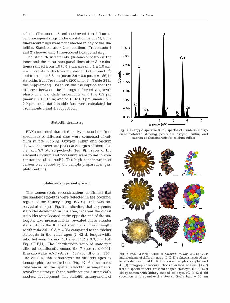
EDX confirmed that all 6 analyzed statoliths fromspecimens of different ages were composed of cal-cium sulfate (CaSO4). Oxygen, sulfur, and calciumshowed characteristic peaks at energies of about 0.4,2.3, and 3.7 eV, respectively (Fig. 8). Traces of theelements sodium and potassium were found in con-centrations of <1 mol%. The high concentration ofcarbon was caused by the sample preparation (gra -phite coating).
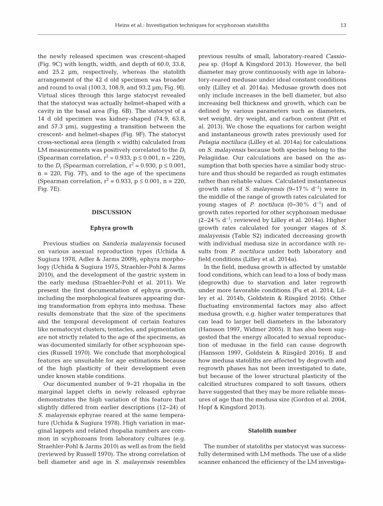
Statocyst shape and growth
The tomographic reconstructions confirmed thatthe smallest statoliths were detected in the proximalregion of the statocyst (Fig. 6A−C). This was ob -served at all ages (Fig. 9), indicating that tiny youngstatoliths developed in this area, whereas the oldeststatoliths were located at the opposite end of the sta-tocysts. LM measurements revealed more slenderstatocysts in the 0 d old specimens (mean length:width ratio 2.5 ± 0.5, n = 36) compared to the thickerstatocysts in the other ages (7−42 d, length:widthratio between 0.7 and 1.8, mean 1.2 ± 0.3, n = 184;Fig. 9B,E,H). The length:width ratio of statocystsdiffe red significantly among the 7 ages (p ≤ 0.001,Kruskal-Wallis ANOVA, H = 127.483, df 6, n = 220).The visualization of statocysts on different ages bytomographic reconstructions (Fig. 9C,F,I) confirmeddifferences in the spatial statolith arrangements,revealing statocyst shape modifications during earlymedusa development. The statolith arrangement of
12
Fig. 8. Energy-dispersive X-ray spectra of Sanderia malay -en sis statoliths showing peaks for oxygen, sulfur, and
calcium as characteristic for calcium sulfate
Fig. 9. (A,D,G) Bell shapes of Sanderia malayensis ephyraeand medusae of different ages; (B, E, H) related shapes of sta-tocysts demonstrated by light microscopic photographs; and(C,F,I) tomographic reconstructions after label analysis. (A−C)0 d old specimen with crescent-shaped statocyst. (D−F) 14 dold specimen with kidney-shaped statocyst. (G−I) 42 d oldspecimen with round-oval statocyst. Scale bars = 10 µm
Heins et al.: Investigation techniques for scyphozoan statoliths
the newly released specimen was crescent-shaped(Fig. 9C) with length, width, and depth of 60.0, 33.8,and 25.2 µm, respectively, whereas the statolitharrangement of the 42 d old specimen was broaderand round to oval (100.3, 108.9, and 93.2 µm; Fig. 9I).Virtual slices through this large statocyst revealedthat the statocyst was actually helmet-shaped with acavity in the basal area (Fig. 6B). The statocyst of a14 d old specimen was kidney-shaped (74.9, 63.8,and 57.3 µm), suggesting a transition between thecrescent- and helmet-shapes (Fig. 9F). The statocystcross-sectional area (length × width) calculated fromLM measurements was positively correlated to the Dt
(Spearman correlation, r2 = 0.933, p ≤ 0.001, n = 220),to the Dr (Spearman correlation, r2 = 0.930, p ≤ 0.001,n = 220, Fig. 7F), and to the age of the specimens(Spearman correlation, r2 = 0.933, p ≤ 0.001, n = 220,Fig. 7E).
DISCUSSION
Ephyra growth
Previous studies on Sanderia malayensis focusedon various asexual reproduction types (Uchida &Sugiura 1978, Adler & Jarms 2009), ephyra morpho -logy (Uchida & Sugiura 1975, Straehler-Pohl & Jarms2010), and the development of the gastric system inthe early medusa (Straehler-Pohl et al. 2011). Wepresent the first documentation of ephyra growth,including the morphological features appearing dur-ing transformation from ephyra into medusa. Theseresults demonstrate that the size of the specimensand the temporal development of certain featureslike nematocyst clusters, tentacles, and pigmentationare not strictly related to the age of the specimens, aswas documented similarly for other scyphozoan spe-cies (Russell 1970). We conclude that morphologicalfeatures are unsuitable for age estimations becauseof the high plasticity of their development evenunder known stable conditions.
Our documented number of 9−21 rhopalia in themarginal lappet clefts in newly released ephyraedemonstrates the high variation of this feature thatslightly differed from earlier descriptions (12−24) ofS. malayensis ephyrae reared at the same tempera-ture (Uchida & Sugiura 1978). High variation in mar-ginal lappets and related rhopalia numbers are com-mon in scyphozoans from laboratory cultures (e.g.Straehler-Pohl & Jarms 2010) as well as from the field(reviewed by Russell 1970). The strong correlation ofbell diameter and age in S. malayensis resembles
previous results of small, laboratory-reared Cassio-pea sp. (Hopf & Kingsford 2013). However, the belldiameter may grow continuously with age in labora-tory-reared medusae under ideal constant conditionsonly (Lilley et al. 2014a). Medusae growth does notonly include increases in the bell diameter, but alsoincreasing bell thickness and growth, which can bedefined by various parameters such as diameters,wet weight, dry weight, and carbon content (Pitt etal. 2013). We chose the equations for carbon weightand instantaneous growth rates previously used forPelagia noctiluca (Lilley et al. 2014a) for calculationson S. malayensis because both species belong to thePelagiidae. Our calculations are based on the as -sumption that both species have a similar body struc-ture and thus should be regarded as rough estimatesrather than reliable values. Calculated instantaneousgrowth rates of S. malayensis (9−17% d−1) were inthe middle of the range of growth rates calculated foryoung stages of P. noctiluca (0−30% d−1) and ofgrowth rates reported for other scyphozoan medusae(2−24% d−1; reviewed by Lilley et al. 2014a). Highergrowth rates calculated for younger stages of S.malayensis (Table S2) indicated decreasing growthwith individual medusa size in accordance with re -sults from P. noctiluca under both laboratory andfield conditions (Lilley et al. 2014a).
In the field, medusa growth is affected by unstablefood conditions, which can lead to a loss of body mass(degrowth) due to starvation and later regrowthunder more favorable conditions (Fu et al. 2014, Lil-ley et al. 2014b, Gold stein & Riisgård 2016). Otherfluctuating environmental factors may also affectmedusa growth, e.g. higher water temperatures thatcan lead to larger bell diameters in the laboratory(Hansson 1997, Widmer 2005). It has also been sug-gested that the energy allocated to sexual reproduc-tion of medusae in the field can cause degrowth(Hansson 1997, Goldstein & Riisgård 2016). If andhow medusa statoliths are af fec ted by degrowth andregrowth phases has not been investigated to date,but because of the lower structural plasticity of thecalcified structures compared to soft tissues, othershave suggested that they may be more reliable meas-ures of age than the medusa size (Gordon et al. 2004,Hopf & Kingsford 2013).
Statolith number
The number of statoliths per statocyst was success-fully determined with LM methods. The use of a slidescanner enhanced the efficiency of the LM investiga-
13

Mar Ecol Prog Ser · Theme Section · Advance View
tions, but system adjustments by a specialist arehighly recommended to avoid measurement errors.Automated counting was not possible because stato -liths could not be differentiated from tissue remnantsand air inclusions by the system, and adjacent stato -liths were not identified as separate objects. Micro-CT provided an alternative method that allowedautomated determination of statolith numbers; how-ever, the required label analysis of tomographic datais time-consuming (Holst et al. 2016). Statocysts oflarge scyphozoan medusae can contain several thou-sand statoliths (Sötje et al. 2011), and neither of the 2methods tested allows a rapid automated counting ofsuch large statolith numbers. Improved resolutionand optical recognition in future imaging systemscould reduce this problem.
Our results demonstrate high overlaps of statolithsper statocyst between the investigated size groups.Similar overlaps were also documented for other spe-cies (Table 2). Nevertheless, strong correlations ofstatolith numbers with medusa age and size were
shown for normal specimens of S. malayensis andconfirmed previous investigations on the scyphozoanspecies Rhizostoma octopus (Holst et al. 2007), Peri-phylla periphylla (Sötje et al. 2011), and Cassiopeasp. (Hopf & Kingsford 2013). It is still unclear if thestatolith number of scyphozoan jellyfish can be corre-lated to age or if the number is strictly dependent onthe bell size. However, in both cases, statolith analy-ses can provide a promising tool for investigations onjellyfish population structures. Age determination ofscyphozoan jellyfish would allow more precise back-tracking models for jellyfish pathways in order tolocate likely polyp habitats (Dulière et al. 2014, Gam-bill 2016). Little is known about the settlement areasof scyphozoan polyps, and their location is unknownfor many species (van Walraven et al. 2016, Widmeret al. 2016). For this reason, only few in situ studieson scyphozoan polyps provide information on theirseasonal abundances (e.g. Ishii & Katsukoshi 2010)and the effects of environmental factors on the pro-duction of young jellyfish (strobilation) (e.g. Purcell
14
Species Diameter Age (d) Statoliths per Predominant Reference(mm) statocyst (n) statolith shape
Sanderia malayensis 2.6−5.0 0−7 15−55 (7) Rod Present study5.1−7.5 7−14 21−99 (6)
7.6−10.0 21−28 54−157 (8)10.1−12.5 35−42 96−288 (9)12.6−15.0 42 85−260 (4)
Rhizostoma octopus 0.1−2.5 nd 17−98 (3) Isodiametric Holst et al. (2007)2.6−5.0 32−68 (2)5.1−7.5 85−215 (4)7.6−10.0 97−264 (3)
10.1−12.5 443−465 (1)12.6−15.0 329−448 (1)
Periphylla periphylla 0.1−2.5 nd 12−39 (3) Rod Sötje et al. (2011)2.6−5.0 35−40 (3)5.1−7.5 nd7.6−10.0 65−162 (4)
10.1−12.5 127 (1)12.6−15.0 158−240 (4)
Cassiopea andromeda nd 0−7 18−120a (18) Disc Hopf & Kingsford (2013)8−14 60−140a (8)15−21 120−220a (9)22−28 135−265a (5)29−35 100−285a (7)36−42 200−315a (6)
Aurelia aurita 2.6−5.0 0−3 17−19 (3) Rod Sötje et al. (2017)5.1−7.5 nd 70
aMean values, estimated from graphics
Table 2. Statoliths counted in the statocysts of young scyphozoans and species-specific predominant statolith shapes. Speci-mens were grouped by 2.5 mm groups up to 15 mm diameter or by 7 d age groups up to 42 d. n: number of investigated speci-mens; nd: no data. Diameter refers to the total bell diameter, except in Periphylla periphylla, for which the coronal diameter
was measured

Heins et al.: Investigation techniques for scyphozoan statoliths
et al. 2009). The strobilation process is linked to thedevelopment of jellyfish blooms (Boero et al. 2008,Holst 2012), and thus the realization of more in situstudies on polyps could help to understand thecauses of blooming events.
The bell diameter composition and the related bio-mass are important parameters for conclusions onpopulation dynamics and their functional effects ofjellyfish (Makabe et al. 2012). Bell sizes also allowconclusions on the predatory impact and the fecun-dity of medusae (Goldstein & Riisgård 2016). How-ever, the determinations of bell diameters on livingmedusae from net hauls on board are challengingsince each specimen has to be spread flat on a platebefore measurement (Makabe et al. 2012). More-over, the specimens can be damaged by nets (Båm -stedt et al. 2003), leading to difficulties in correctmea surements. Measurements on complete forma-lin-preserved medusae are possible in the laboratory(e.g. Morandini et al. 2017), but the fixation of com-plete medusae requires adequate storage space andlarge amounts of poisonous fixative which causessevere shrinkage (Holst & Laakmann 2014, Laak-mann & Holst 2014), impeding the later determina-tion of the original bell diameters. Statolith analysesin the laboratory could replace measurements onboard or on preserved material if future studies con-firm clear correlations of statolith numbers and bellsizes. The preservation of small bell rim pieces dur-ing collection would be sufficient for later statolithanalyses, but high-proof ethanol is required for a suf-ficient preservation of statoliths (Holst et al. 2016).The ethanol-preserved bell rim pieces could also beused in parallel for molecular genetic analyses allow-ing conclusions on population genetic structure (Leeet al. 2013, Dawson et al. 2015, Glynn et al. 2016).
Scyphomedusan degrowth can occur synchronouslywith sexual reproduction (Goldstein & Riisgård 2016)and was also reported in overwintering populations(Makabe et al. 2012). Positive correlations amongstatolith numbers and bell dia meters are probablylimited to growth phases. In conclusion, analyses ofstatolith numbers of degrown and overwintered pop-ulations could allow conclusions on the earlier maxi-mum bell size on an individual level, which may pro-vide more precise data on the overwinteringpopulation structure than analyses of bell diametercompositions (Makabe et al. 2012).
S. malayensis statocysts contained fewer statolithsthan R. octopus medusae of similar sizes (Holst et al.2007) or Cassiopea sp. medusae of similar ages (Hopf& Kingsford 2013) (Table 2). We found fewer stato -liths in specimens with malformations than in nor-
mally developed ones. Because malformations andvariable numbers of marginal lappets are frequent inyoung scyphozoans in the laboratory (e.g. Holst et al.2007, Straehler-Pohl & Jarms 2010, Algueró-Muñiz etal. 2016, this study), possible effects on statolith num-bers should be considered in future studies.
Statolith shapes
Statolith measurements by light microscopic meth-ods provided reliable data on S. malayensis statolithmorphometry. Our results revealed differences in thepercentages of statolith shape types (Fig. 5B), de -fined by the statolith aspect ratios, among the inves-tigated ages. It was suggested that the growth ofindividual statoliths may be affected by the sur-rounding statoliths and that statolith shapes maydepend on the growth and development of the wholestatocyst (Sötje et al. 2017). Needle-shaped statolithswere most abundant in newly released S. malayensisephyrae and in the proximal end of the statocystleading to the assumption that the needle shapecould be the initial shape of newly developed stato -liths which later transform to other shapes duringstato lith growth. However, needle-shaped statolithswere not found in the statocysts of newly releasedAurelia aurita ephyrae (Sötje et al. 2017), indicatingthat the needle shape is not necessarily the initialstato lith shape and that statolith development couldbe species-specific. We revealed the rod shape as thetypical statolith type in S. malayensis, which was alsotypical for A. aurita and P. periphylla, whereas thetypical shape of R. octopus statoliths was approxi-mately equilateral and that of Cassiopea sp. wasdisc-shaped (Table 2). Even though a rod shapeappears to be the predominant shape in the taxaCoronatae (P. periphylla), Cyaneidae (Cyanea capil-lata, C. lamarckii), Cepheidae (Cotylorhiza tubercu-lata), Ulmaridae (A. aurita), and Pelagiidae (S. mala -yensis), different species show differences in statolithshapes and in shape compositions (Tiemann et al.2002, Becker et al. 2005, Boßelmann et al. 2007). Rod-and needle-shaped statoliths, but no isodiametric ordisc-shaped crystals, were detected in A. aurita (Sötjeet al. 2017), whereas isodiametric, rod-, needle-, anddisc-shaped crystals were present in S. malayensis(this study).
SEM micrographs provided a good additional toolfor depiction of statolith shapes of some statoliths, butcould not reflect the broad variability of statolithshapes and sizes in the statocyst. The dissolution ofthe pointed tips at the statolith head-faces in the SEM
15

Mar Ecol Prog Ser · Theme Section · Advance View
micrographs probably occurred after death of thespecimens (Sötje et al. 2017). The extent of the disso-lution process may depend on the amount of water inthe tissue surrounding the statocyst during fixation,and thus, it is important to minimize the amount ofsurrounding tissue and to keep the amount and proofof ethanol high during the fixation of statoliths.
A limitation of LM and SEM was that the statolithswere visualized only in 2 dimensions. Therefore, onlyside-face-oriented statoliths were useful for the LMevaluation of aspect ratios, whereas measurementson head-face-oriented statoliths reflected variationsin the statolith cross profile only (Fig. 5C). In contrast,micro-CT analyses provided statolith measurementsof all statoliths in 3 dimensions. However, the methodwas not suitable to display the shapes and sizes ofstatoliths as accurately as was possible by LM, prob-ably because of the limited image resolution. Thus,the method was not suitable to display the appear-ance of different shape types in different stages.Nevertheless, the scatter plots of automated meas-urements are potentially useful for intra- and inter-specific comparisons of scyphozoan statoliths (Holstet al. 2016). The distribution pattern shown in ourplots for S. malayensis (Fig. 7A,B) resemble the pat-tern in the plots of micro-CT data for statoliths of C.lamarckii (compare Fig. 8B,C in Holst et al. 2016).These similarities may reflect a close phylogeneticrelationship of the 2 species, which both belong tothe scyphozoan taxon Semeostomeae. The poten-tial of interspecific statolith shape comparisons wasshown for the single large statoliths in the stato-cysts of Cubozoa (Mooney & Kingsford 2016a, 2017).Micro-CT provides a suitable tool for interspecificcomparisons of statolith shapes. Improvement of thespatial resolution and efficiency in future micro-CTsystems will lead to enhanced 3-dimensional visuali-zation of statoliths and will allow comparisons amongmany species from various taxonomic levels usefulfor phylogenetic comparisons.
Statolith size and growth
As in previous studies (Holst et al. 2007, Sötje et al.2011), our results revealed a positive correlation ofstatolith size and medusa age. However, closerexamination of the data indicates significant statolithsize increase in younger medusa stages only. Forexample, maximum statolith widths of the 5 largeststatoliths measured increased in P. periphylla from15 to around 45 µm in medusae of 2−23 mm in dia -meter, but the mean widths did not increase consid-erably in larger medusae with diameters up to
185 mm (with 1 exception of 53 µm mean width; seeFig. 5C in Sötje et al. 2011). Increments in statolithswere used to back-date the age of the cubomedusaChironex fleckeri (Gordon & Seymour 2012). Ourdata now indicate that in scyphozoans, the statolithsize does not allow conclusions about medusa dia -meter or age because statoliths stop, or at least considerably slow growth, when a certain size isreached. Instead, the determination of statolith num-bers is a promising parameter for the age determina-tion or bell size correlations of scyphozoan medusae(see discussion on statolith number above). Thelargest statolith volumes measured on micro-CT dataof Cyanea lamarckii were around 10000 µm3 (Holstet al. 2016), more than 3 times larger compared to thestatolith volumes in S. malayensis (around 3000 µm3;Fig. 7B), suggesting that maximum statolith sizecould be species-specific. Studies on the statoliths ofA. labiata epyhrae (Winans & Purcell 2010) andyoung cubomedusae (Klein et al. 2014) indicate thatdifferent pH conditions could affect the growth ofstatoliths, but this may be caused by secondaryeffects of low pH on feeding and thus energy avail-able to build the bassanite crystals (Tills et al. 2016).High intraspecific variation in statolith sizes werealso documented for specimens cultured under thesame laboratory conditions (Winans & Purcell 2010,this study). Further analyses are required to detect ifdifferences in statolith size are caused by environ-mental effects or if they represent inter- or evenintraspecific variation.
Differences in the statolith composition in differ-ent ages of S. malayensis were represented by dif-ferent percentages of shape types (Fig. 5B). Sta-toliths with isogonal or irregular hexagonal statolithcross profiles were not present in the 0 d old speci-mens, but appeared in older ones (Fig. 5C). Theseresults indicate a change in crystal shapes by sta-tolith growth, as previously suggested for A. auritastatoliths (Sötje et al. 2017). As shown by Sötje et al.(2017), our study demonstrated that statolith growthcan be visualized by using calcein as a fluorescentmarker. This me thod allowed calculations of thedaily statolith size increase of 0.2 µm for S. malayen-sis. The lower mean daily increment of 0.1 µm cal-culated for A. aurita statoliths was probably causedby the lower culture temperature of 12°C (Sötje etal. 2017). The hexa gonal ring structures detected inS. malayensis statoliths by LM in 1000-fold magnifi-cation (Fig. 2H,I) showed rings of 1−2 µm, whichwas about a factor of 10 wider than the daily sta-tolith increment calculated from the calcein experi-ments. In conclusion, it is unlikely that these ring
16

Heins et al.: Investigation techniques for scyphozoan statoliths
structures represented daily statolith growth, as as -sumed for the ring structures in cubozoan statoliths(Kawamura et al. 2003, Gordon et al. 2004, Gordon& Seymour 2012).
In our methods, the time interval between the firstand the second calcein incubations (4 wk) differedfrom the interval between the second and the thirdincubations (2 wk), which led to difficulties in inter-pretation of our results. The application of 3 calceintreatments could cause various scenarios of result-ing fluorescent markings in the statoliths dependingon the degree of statolith development (Fig. S1). Wedetected only 1 fluorescent ring after 2 incubationsand only 2 rings after 3 incubations, probably be -cause the statoliths had reached their final sizewithin 3 wk as indicated by our LM measurements(Fig. 7D). We concluded that the distances meas-ured between the 2 fluorescent rings (after 3 treat-ments) represented a growth phase of 2 wk becausefew stato liths were present in the newly detachedephyrae used for the first incubation, and those fewstatoliths probably were not detected among themany statoliths that had developed by the thirdincubation in 42 d old specimens. For the same rea-son, only statoliths with 1 fluorescent hexagonalring were found in specimens after 2 calcein incu-bations (Fig. S1 in the Supplement). For successfulcalcein treatment, the calcein molecules are sur-rounded by bassanite while the statolith grows;thereby the fluorescent calcium is incorporated intothe crystal (Sötje et al. 2017). Conversely, calceinhas no effect if the statoliths do not grow by bassan-ite increments after the incubation. The statolithsthat appeared without fluorescent rings in our sam-ples had probably developed after the first incuba-tion and stopped growing before the second incu -bation (scenario VII in Fig. S1). To avoid suchinter pretation problems, we recommend keepingthe same time intervals of 2 wk or less between cal-cein incubations. Because only head-face-orientedcrystals can be used for the analyses of calcein-labeled statoliths (Sötje et al. 2017), it may beadvantageous to use specimens older than 1 wk thatmay contain higher percentages of head-face-oriented (disc-shaped) statoliths in their statocyststhan newly released ephyrae (Fig. 5A). Treatmentswith a calcein concentration of 100 µmol l−1 for 3 dare recommended for future studies, be cause theyled to sufficient fluorescent markings in the bassan-ite statoliths (Sötje et al. 2017, this study). Lowercalcein concentration (50 µmol l−1) was insufficientto label A. aurita statoliths (Sötje et al. 2017), andhigher concentration (200 µmol l−1) did not improve
the results over 100 µmol l−1 in our study on S.malayensis statoliths.
Statolith chemistry
The EDX analyses of S. malayensis statolithsshowed mainly calcium, sulfur, and oxygen, indica-ting the presence of calcium sulfate (Becker et al.2005). It is likely that the statoliths were composed ofbassanite, a calcium sulfate subhydrate, as previous -ly shown for other scyphozoan and cubozoan stato -liths by single X-ray diffraction (Becker et al. 2005,Tiemann et al. 2006, Sötje et al. 2011). EDX, however,is not suitable to distinguish calcium sulfate dehy-drate (gypsum) from the subhydrate, which led tomisinterpretations of the statolith material in earlierstudies on some scyphozoan and cubozoan species(reviewed by Becker et al. 2005). Unequivocal identi-fication of the calcium sulfated subhydrate is possibleonly by single-crystal X-ray analysis. The drawbackof the method is the requirement of special diffrac-tometer technologies, whereas EDX can be con-ducted with an SEM system, which is available to abroad group of scientists.
Statocyst size and shape
Previous positive correlations between scyphozoanstatocyst sizes and medusa diameters (Holst et al.2007, Sötje et al. 2011) were confirmed for S. mala -yensis (Fig. 7F). The statocyst cross-sectional areawas also correlated with the age of the specimens(Fig. 7E), suggesting that this parameter could beuseful for an age determination. However, because ofthe complex structure of the statocyst, the simpledetection of the statocyst length and width from 2-dimensional photographs is insufficient to reflect theactual statocyst volumes. Measurements on tomo-graphic data could provide more exact determina-tions of statocyst volumes and shapes, but currentmicro-CT techniques are unsuitable for large-scaleinvestigations. Instead, tomographic data are usefulfor interspecific comparisons on the arrangement ofstatoliths reflecting the statocyst shapes (Holst et al.2016). Our results on statocyst shape modificationsduring the early medusa development of S. malay -ensis (Fig. 9) indicated that statocyst shapes can dif-fer considerably from young specimens to adults,which should be considered in future investigationson interspecific differences of statocyst shapes andvolumes.
17
Mar Ecol Prog Ser · Theme Section · Advance View
Environmental effects on medusa and statolithdevelopment
Our results on ephyra development demonstratethat differences in the appearance of certain morpho-logical features are not necessarily linked to differentenvironmental conditions and thus are usually notuseful indicators for environmental effects. Labora-tory experiments on A. aurita ephyrae suggest thatthey are not likely to suffer from environmentalchanges related to climate change (ocean acidifica-tion, warming, and deoxygenation) in the near future(Algueró-Muñiz et al. 2016). However, the few avail-able data on environmental and nutritional effects onstatoliths indicate that these parameters can affectstatolith numbers or sizes. Statolith numbers of thescyphozoan Cassiopea sp. were affected by tempera-ture, but not by salinity or food in experimental stud-ies (Hopf & Kingsford 2013), and pH affected statolithsizes but not numbers in A. labiata (Winans & Purcell2010). Although reduced pH has no direct dissolutioneffects on calcium sulfate subhydrate composed stato -liths, A. aurita ephyrae exposed to reduced seawaterpH showed a decrease in pulsation rate probablybecause of secondary effects of low pH on feedingand thus energy available for the growth of statoliths(Tills et al. 2016). More experimental research in -cluding the combination of laboratory and field data,might lead to more reliable conclusions on environ-mental effects on statolith and statocyst develop-ment. Studies on cubozoan medusae indicated a highpotential for the reconstruction of medusa move-ments by statolith elemental chemistry in areas withknown salinity and temperature gradients (Mooney& Kingsford 2016b) which may also be true forscyphozoans. In turn, statolith analyses could be usedto conclude on environmental conditions.
CONCLUSIONS
Statoliths represent the only hard structures in thegelatinous bodies of scyphozoan jellyfish and thuscould be a very useful tool for age determination andphylogenetic analyses. Our methodical approachprovides a comparison of various investigation tech-niques for scyphozoan statoliths, revealing the pos -sibilities and advantages of certain techniques tosupport a successful experimental design of futurestudies on scyphozoan statoliths. Reliable determina-tion of statolith numbers and sizes is possible byLM and micro-CT analyses. Both methods are time- consuming with the currently available techniques,
but the optimization of optical systems may acceler-ate the processing time in future studies. Calceinlabeling is a suitable method to follow statolithgrowth and to calculate daily increments, which maydiffer with culture temperature. Statolith numberscould be useful for age estimation of scyphozoanjelly fish because the numbers increase continuouslywith medusa size and age in the laboratory, unlikestatolith size, which seems to reach a maximum andcannot be linked to a certain developmental stage orage. Statolith and statocyst morphometry can be doc-umented by LM and SEM analyses, but those dataare based on 2-dimensional measurements that donot reflect the actual volumes of the structures.Micro-CT allows 3-dimensional depictions andmeasurements of statoliths and statocysts, but thelimited resolution of the system we used did notallow for sufficient representation of stato lith shapes.
In conclusion, a combination of microscopic andmicro-tomographic techniques is recommended forinvestigations on interspecific comparisons on stato -lith shapes and statocyst shapes, which may provideuseful tools for phylogenetic studies on scyphozoans.Additional studies comparing specimens reared inthe laboratory and from the field, experimental stud-ies on the effects of environmental conditions onstato liths, as well as studies on a broad range of taxain various developmental stages are necessary todetermine the scope of applications of stato liths indemographic and phylogenetic studies on scypho-zoan jellyfish. Further work would contribute torefine methodological approaches and to reduce thetime and cost expense of statolith analyses, whichwould help to establish statolith analyses as a stan-dard method in jellyfish taxonomy and ecology.
Acknowledgements. We thank Frank Friedrich, PascalMeyer, Janina Fiebig, Stephan Henne, Sabine Gaude,Renate Walter, and Svenja Warnke for their technical sup-port in sample preparation and helpful advice on micro -scopy and software application, and Peter Michalik andJakob Krieger for their help in micro-CT processing. Wethank Sea Pen Scientific Writing for editing services and 2anonymous reviewers for their comments improving themanuscript. This manuscript is based on the MSc thesis ofA.H. conducted at the University of Oldenburg. We thankPedro Martínez-Arbizu for co-supervising the thesis. Thisstudy was funded by the German Science Foundation (DFGSO 1238/2-1, DFG HO 4097/3-1).
LITERATURE CITED
Adler L, Jarms G (2009) New insights into reproductivetraits of scyphozoans: special methods of propagation inSanderia malayensis GOETTE, 1886 (Pelagiidae, Sema -eostomeae) enable establishing a new classification of
18

Heins et al.: Investigation techniques for scyphozoan statoliths
asexual reproduction in the class Scyphozoa. Mar Biol156: 1411−1420
Algueró-Muñiz M, Meunier CL, Holst S, Alvarez-FernandezS, Boersma M (2016) Withstanding multiple stressors: ephyrae of the moon jellyfish (Aurelia aurita, Scyphozoa)in a high-temperature, high-CO2 and low-oxygen envi-ronment. Mar Biol 163: 185
Båmstedt U, Kaartvedt S, Youngbluth M (2003) An evalua-tion of acoustic and video methods to estimate the abun-dance and vertical distribution of jellyfish. J PlanktonRes 25: 1307−1318
Barroso CM, Nunes M, Richardson CA, Moreira MH (2005)The gastropod statolith: a tool for determining the age ofNassarius reticulatus. Mar Biol 146: 1139−1144
Becker A, Sötje I, Paulmann C, Beckmann F and others(2005) Calcium sulfate hemihydrate is the inorganic mineral in statoliths of Scyphozoan medusae (Cnidaria).Dalton Trans 1545−1550
Boero F, Boullion J, Gravili C, Miglietta MP, Parsons T,Piraino S (2008) Gelatinous plankton: irregularities rulethe world (sometimes). Mar Ecol Prog Ser 356: 299−310
Boßelmann F, Epple M, Sötje I, Tiemann H (2007) Statolithsof calcium sulfate hemihydrate are used for gravity sens-ing in rhopaliophoran medusae (Cnidaria). In: BäuerleinE (ed) Handbook of biomineralization: biological aspectsand structure formation. Wiley-VCH Verlag, Weinheim,p 262–272
Dawson MN, Cieciel K, Decker MB, Hays GC, Lucas CH,Pitt KA (2015) Population-level perspectives on globalchange: genetic and demographic analyses indicate var-ious scales, timing, and causes of scyphozoan jellyfishblooms. Biol Invasions 17: 851−867
Dulière V, Kerckhof F, Lacroix G (2014) Where is my jelly?De Strandvlo 34: 48−65
Fu Z, Shibata M, Makabe R, Ikeda H, Uye S (2014) Body sizereduction under starvation, and the point of no return, inephyrae of the moon jellyfish Aurelia aurita. Mar EcolProg Ser 510: 255−263
Gambill M (2016) Ecophysiology and life cycle dynamics ofNorth Sea gelatinous zooplankton. PhD dissertation,University of Hamburg
Glynn F, Houghton JDR, Bastian T, Doyle TK, Fuentes V, Lilley MKS, Provan J (2016) High-resolution geneticanalysis reveals extensive gene flow within the jellyfishPelagia noctiluca (Scyphozoa) in the North Atlantic andMediterranean Sea. Biol J Linn Soc 117: 252−263
Goldstein J, Riisgård HU (2016) Population dynamics andfactors controlling somatic degrowth of the commonjelly fish, Aurelia aurita, in a temperate semi-enclosedcove (Kertinge Nor, Denmark). Mar Biol 163: 33
Gordon M, Seymour J (2012) Growth, development andtemporal variation in the onset of six Chironex fleckerimedusae seasons: a contribution to understanding jelly-fish ecology. PLOS ONE 7: e31277
Gordon M, Hatcher C, Seymour J (2004) Growth and agedetermination of the tropical Australian cubozoan Chiro -psalmus sp. Hydrobiologia 530-531: 339−345
Hansson LJ (1997) Effect of temperature on growth rate ofAurelia aurita (Cnidaria, Scyphozoa) from Gullmars -fjorden, Sweden. Mar Ecol Prog Ser 161: 145−153
Holst S (2012) Effects of climate warming on strobilation andephyra production of North Sea scyphozoan jellyfish.Hydrobiologia 690: 127−140
Holst S, Laakmann S (2014) Morphological and moleculardiscrimination of two closely related jellyfish species,
Cyanea capillata and C. lamarckii (Cnidaria, Scyphozoa),from the northeast Atlantic. J Plankton Res 36:48–63
Holst S, Sötje I, Tiemann H, Jarms G (2007) Life cycle of therhizostome jellyfish Rhizostoma octopus (L.) (Scyphozoa,Rhizostomeae), with studies on cnidocysts and statoliths.Mar Biol 151: 1695−1710
Holst S, Michalik P, Noske M, Krieger J, Sötje I (2016) Poten-tial of X-ray micro-computed tomography for soft-bodiedand gelatinous cnidarians with special emphasis onscyphozoan and cubozoan statoliths. J Plankton Res 38: 1225−1242
Hopf JK, Kingsford MJ (2013) The utility of statoliths andbell size to elucidate age and condition of a scypho -medusa (Cassiopea sp.). Mar Biol 160: 951−960
Ishii H, Katsukoshi K (2010) Seasonal and vertical distribu-tion of Aurelia aurita polyps on a pylon in the innermostpart of Tokyo Bay. J Oceanogr 66: 329−336
Kawamura M, Ueno S, Iwanaga S, Oshiro N, Kubota S(2003) The relationship between fine rings in the statolithand growth of the cubomedusa Chiropsalmus quadri -gatus (Cnidaria: Cubozoa) from Okinawa Island, Japan.Plankton Biol Ecol 50: 37−42
Klein SG, Pitt KA, Rathjen KA, Seymour JE (2014) Irukandjijellyfish polyps exhibit tolerance to interacting climatechange stressors. Glob Change Biol 20: 28−37
Laakmann S, Holst S (2014) Emphasizing the diversity ofNorth Sea hydromedusae by combined morphologicaland molecular methods. J Plankton Res 36: 64−76
Lee PLM, Dawson M, Neill SP, Robins PE, Houghton JDR,Doyle TK, Hays GC (2013) Identification of geneticallyand oceanographically distinct blooms of jellyfish. J RSoc Interface 10: 20120920
Lilley MKS, Ferraris M, Elineau A, Berline L and others(2014a) Culture and growth of the jellyfish Pelagia noc-tiluca in the laboratory. Mar Ecol Prog Ser 510: 265−273
Lilley MKS, Elineau A, Ferraris M, Thiery A, Stemmann L,Gorsky G, Lombard F (2014b) Individual shrinking toenhance population survival: quantifying the reproduc-tive and metabolic expenditures of a starving jellyfish,Pelagia noctiluca. J Plankton Res 36: 1585−1597
Makabe R, Kurihara T, Uye SI (2012) Spatio-temporal distri-bution and seasonal population dynamics of the jellyfishAurelia aurita s.l. studied with Dual-frequency IDentifi-cation SONar (DIDSON). J Plankton Res 34: 936−950
Mooney CJ, Kingsford MJ (2016a) Statolith morphometricscan discriminate among taxa of cubozoan jellyfishes.PLOS ONE 11: e0155719
Mooney CJ, Kingsford MJ (2016b) The influence of salinityon box jellyfish (Chironex fleckeri, Cubozoa) statolithelemental chemistry. Mar Biol 163: 103
Mooney CJ, Kingsford MJ (2017) Statolith morphometrics asa tool to distinguish among populations of three cubo-zoan species. Hydrobiologia 787: 111−121
Morandini AC, Stampar SN, Maronna MM, Da Silveira FL(2017) All non-indigenous species were introduced re -cently? The case study of Cassiopea (Cnidaria: Scypho-zoa) in Brazilian waters. J Mar Biol Assoc UK 97: 321−328
Panfili J, Tomás J, Morales-Nin B (2009) Otolith microstruc-ture in tropical fish. In: Green BS, Mapstone BD, CarlosG, Begg GA (eds) Tropical fish otoliths: information forassessment, management and ecology. Reviews: meth-ods and technologies in fish biology and fisheries, Vol 11.Springer, New York, NY, p 212–248
Pitt KA, Duarte CM, Lucas CH, Sutherland KR and others(2013) Jellyfish body plans provide allometric advan-
19

Mar Ecol Prog Ser · Theme Section · Advance View
tages beyond low carbon content. PLOS ONE 8: e72683Purcell JE, Hoover RA, Schwarck NT (2009) Interannual
variation of strobilation by the scyphozoan Aurelia labi-ata in relation to polyp density, temperature, salinity, andlight conditions in situ. Mar Ecol Prog Ser 375: 139−149
Rodhouse PG, Hatfield EMC (1990) Age determination insquid using statolith growth increments. Fish Res 8: 323−334
Russell FS (1970) The medusae of the British Isles II. PelagicScyphozoa with a supplement to the first volume onhydromedusae, Vol 2. Cambridge University Press, Cam-bridge
Schiariti A, Melica V, Kogovšek T, Malej A (2015) Density-dependent effects control the reproductive strategy andpopulation growth of Aurelia aurita s.l. scyphistomae.Mar Biol 162: 1665−1672
Sötje I, Neues F, Ludwig W, Rack A, Gordon M, Boese R,Tiemann H (2011) Comparison of the statolith structuresof Chironex fleckeri (Cnidaria, Cubozoa) and Periphyllaperiphylla (Cnidaria, Scyphozoa): a phylogenetic ap -proach. Mar Biol 158: 1149−1161
Sötje I, Dishon T, Hoffmann F, Holst S (2017) New methodsof morphometric analyses on scyphozoan jellyfish stato -liths including the first direct evidence for statolithgrowth using calcein as a fluorescent marker. MicroscMicroanal 23: 553−568
Straehler-Pohl I, Jarms G (2010) Identification key for youngephyrae: a first step for early detection of jellyfishblooms. Hydrobiologia 645: 3−21
Straehler-Pohl I, Widmer CL, Morandini AC (2011) Charac-terizations of juvenile stages of some semaeostomeScyphozoa (Cnidaria), with recognition of a new family
(Phacellophoridae). Zootaxa 2741: 1−37Tiemann H, Sötje I, Jarms G, Paulmann C, Epple M, Hasse B
(2002) Calcium sulfate hemihydrate in statoliths of deep-sea medusae. J Chem Soc Dalton Trans 2002: 1266−1268
Tiemann H, Sötje I, Becker A, Jarms G, Epple M (2006) Cal-cium sulfate hemihydrate (bassanite) statoliths in thecubozoan Carybdea sp. Zool Anz 245: 13−17
Tills O, Sun X, Rundle SD, Heimbach T and others (2016)Reduced pH affects pulsing behaviour and body size inephyrae of the moon jellyfish, Aurelia aurita. J Exp MarBiol Ecol 480: 54−61
Uchida T, Sugiura Y (1975) On the ephyra and postephyra ofa semaeostome medusa, Sanderia malayensis Goette.J Fac Sci Hokkaido Univ Ser VI Zool 19: 879−881
Uchida T, Sugiura Y (1978) On the polyp of the scypho -medusa, Sanderia malayensis and its reproduction. J FacSci Hokkaido Univ Ser VI Zool 21: 279−286
van Walraven L, Driessen F, van Bleijswijk J, Bol A and oth-ers (2016) Where are the polyps? Molecular identifica-tion, distribution and population differentiation of Aure-lia aurita jellyfish polyps in the southern North Sea area.Mar Biol 163: 172
Widmer CL (2005) Effects of temperature on growth ofnorth-east Pacific moon jellyfish ephyrae, Aurelia labiata(Cnidaria: Scyphozoa). J Mar Biol Assoc UK 85: 569−573
Widmer CL, Fox CJ, Brierley AS (2016) Effects of tempera-ture and salinity on four species of northeastern Atlanticscyphistomae (Cnidaria: Scyphozoa). Mar Ecol Prog Ser559: 73−88
Winans AK, Purcell JE (2010) Effects of pH on asexualreproduction and statolith formation of the scyphozoan,Aurelia labiata. Hydrobiologia 645: 39−52
20
Editorial responsibility: Fabien Lombard (Guest Editor),Villefranche-sur-Mer, France
Submitted: December 30, 2016; Accepted: August 3, 2016Proofs received from author(s): September 18, 2017