assessment of diastolic function in single-ventricle
TRANSCRIPT

Children's Mercy Kansas CitySHARE @ Children's Mercy
Manuscripts, Articles, Book Chapters and Other Papers
11-1-2016
Assessment of Diastolic Function in Single-Ventricle Patients After the Fontan Procedure.Renee Margossian
Lynn A. Sleeper
Gail D. Pearson
Piers C. Barker
Luc Mertens
See next page for additional authors
Follow this and additional works at: https://scholarlyexchange.childrensmercy.org/papers
Part of the Cardiology Commons, Cardiovascular System Commons, Congenital, Hereditary,and Neonatal Diseases and Abnormalities Commons, Investigative Techniques Commons,Pediatrics Commons, and the Surgical Procedures, Operative Commons
This Article is brought to you for free and open access by SHARE @ Children's Mercy. It has been accepted for inclusion in Manuscripts, Articles, BookChapters and Other Papers by an authorized administrator of SHARE @ Children's Mercy. For more information, please [email protected].
Recommended CitationMargossian, Renee; Sleeper, Lynn A.; Pearson, Gail D.; Barker, Piers C.; Mertens, Luc; Quartermain, Michael D.; Su, Jason T.; Shirali,Girish S.; Chen, Shan; Colan, Steven D.; and Pediatric Heart Network Investigators, "Assessment of Diastolic Function in Single-Ventricle Patients After the Fontan Procedure." (2016). Manuscripts, Articles, Book Chapters and Other Papers. 896.https://scholarlyexchange.childrensmercy.org/papers/896

Creator(s)Renee Margossian, Lynn A. Sleeper, Gail D. Pearson, Piers C. Barker, Luc Mertens, Michael D. Quartermain,Jason T. Su, Girish S. Shirali, Shan Chen, Steven D. Colan, and Pediatric Heart Network Investigators
This article is available at SHARE @ Children's Mercy: https://scholarlyexchange.childrensmercy.org/papers/896

Assessment of Diastolic Function in Single Ventricle Patients Following the Fontan Procedure
Renee Margossian, MD1, Lynn A. Sleeper, ScD2, Gail D. Pearson, MD ScD3, Piers C. Barker, MD4, Luc Mertens, MD5, Michael D. Quartermain, MD6, Jason T. Su, DO7, Girish Shirali, MD8, Shan Chen, MS2, Steven D. Colan, MD1, and for the Pediatric Heart Network Investigators1Boston Children’s Hospital and Harvard Medical School, Boston, MA
2New England Research Institutes, Watertown, MA
3National Heart, Lung, and Blood Institute, Bethesda, MD
4Duke University Medical Center, Durham NC
5The Hospital for Sick Children, Toronto, Canada
6The Children’s Hospital of Philadelphia, Philadelphia, PA
7University of Utah, Salt Lake City, UT
8Medical University of South Carolina, Charleston, SC
Abstract
Objectives—Patients with functional single ventricles (FSV) following the Fontan procedure
have abnormal cardiac mechanics. We sought to determine factors that influence diastolic function
and to describe associations of diastolic function with current clinical status.
Methods—Echocardiograms were obtained as part of the Pediatric Heart Network Fontan Cross-
Sectional Study. Diastolic function grade (DFG) was assessed as normal (grade 0), impaired
relaxation (grade 1), pseudonormalization (grade 2), restrictive (grade 3). Studies were also
classified dichotomously (restrictive pattern present or absent). Relationships between DFG and
pre-Fontan variables (e.g., ventricular morphology, age at Fontan, history of volume-unloading
surgery), and current status (e.g., systolic function, valvar regurgitation, exercise performance)
were explored.
Results—DFG was calculable in 326/546 subjects (60%); mean age = 11.7±3.3 years. Overall,
32% of patients had grade 0, 9% grade 1, 37% grade 2, and 22% grade 3. Although there was no
association between ventricular morphology and DFG, there was an association between
Corresponding Author: Renee Margossian, MD, Department of Cardiology, Boston Children’s Hospital, 300 Longwood Avenue, Boston, MA 02115, [email protected], Tel: 617 355-6429, Fax: 617 739-6282.
Publisher's Disclaimer: This is a PDF file of an unedited manuscript that has been accepted for publication. As a service to our customers we are providing this early version of the manuscript. The manuscript will undergo copyediting, typesetting, and review of the resulting proof before it is published in its final citable form. Please note that during the production process errors may be discovered which could affect the content, and all legal disclaimers that apply to the journal pertain.
Clinical Trials Registration #: NCT00132782
HHS Public AccessAuthor manuscriptJ Am Soc Echocardiogr. Author manuscript; available in PMC 2017 November 01.
Published in final edited form as:J Am Soc Echocardiogr. 2016 November ; 29(11): 1066–1073. doi:10.1016/j.echo.2016.07.016.
Author M
anuscriptA
uthor Manuscript
Author M
anuscriptA
uthor Manuscript

ventricular morphology and E’, which was lowest in those with right ventricular morphology (p<.
001); this association remained significant when using z-scores adjusted for age (p=<.001). DFG
was associated with achieving maximal effort on exercise testing (p=.004); the majority (64%) of
those not achieving maximal effort had DFG 2 or 3.No additional significant associations of DFG
with laboratory or clinical measures were identified.
Conclusion—Assessment of diastolic function by current algorithms results in a high percentage
of patients with abnormal DFG, but we found few clinically or statistically significant
associations. This may imply a lack of impact of abnormal diastolic function upon clinical
outcome in this cohort, or may indicate that the methodology may not be applicable to pediatric
FSV patients.
INTRODUCTION
Although surgical palliation of patients with functional single ventricles by the Fontan
procedure has resulted in improved survival during childhood, long-term morbidity and
mortality remain major concerns. In particular, the relationships of pre-Fontan variables to
diastolic function and of diastolic function to long term morbidity and mortality in this
cohort are still largely unknown. In the NHLBI-sponsored Pediatric Heart Network Fontan
Cross-Sectional Study of 546 Fontan survivors, the majority of patients (73%) had normal
ejection fractions by echocardiography, but only 28% had normal indices of diastolic
function (1). While patients with single ventricle physiology have been described as having
diastolic dysfunction with both reduced compliance and impaired relaxation (2), associations
with anatomic, clinical and historical factors have not been described. Additionally, because
an acutely increased mass-to-volume ratio immediately following a Fontan operation has
been postulated by some to be detrimental to diastolic function (3, 4), we sought to assess
correlations between diastolic function and current mass-to-volume ratio, and determine
whether mass-to-volume ratio correlates with clinical characteristics.
METHODS
The NHLBI-sponsored Pediatric Heart Network Fontan Cross-Sectional Study characterized
the health status of 546 Fontan survivors aged 6 to 18 years, enrolled by seven clinical
centers in North America. Prospective data collection included two-dimensional (2D) and
Doppler echocardiography, exercise testing, health status questionnaires, and laboratory
tests. Full details of the study have been published (1, 5, 6). Written informed consent and
assent were obtained according to local guidelines, and the study protocol was approved by
the Institutional Review Board/Research Ethics Board of each center.
Echocardiograms were obtained according to a predetermined study protocol. None were
performed under sedation. The echocardiograms were submitted to the data coordinating
center for initial quality control, and then forwarded to the echocardiographic core lab for
analysis. Ventricular morphology was characterized as left ventricular (LV) dominant (e.g.,
tricuspid atresia), right ventricular (RV) dominant (e.g., hypoplastic left heart syndrome) or
mixed (e.g., unbalanced atrioventricular canal defect). A single representative beat was
selected for all systolic and diastolic measures. To determine ventricular size and ejection
fraction, the ventricle was analyzed from the apical transverse plane (ventricular long-axis)
Margossian et al. Page 2
J Am Soc Echocardiogr. Author manuscript; available in PMC 2017 November 01.
Author M
anuscriptA
uthor Manuscript
Author M
anuscriptA
uthor Manuscript

and the parasternal short-axis views. End-diastolic (EDV) and end-systolic volumes (ESV)
were calculated using the biplane modified Simpson algorithm (7). Percent ejection fraction
(EF %) was then determined as ([EDV-ESV]/EDV) x 100. Ventricular mass was calculated
as myocardial end-diastolic volume (epicardial volume - endocardial volume) x myocardial
density (1.05 g/ml).
Assessment of diastolic function typically involves a multi-parameter approach,
predominantly utilizing spectral Doppler velocities and tissue Doppler velocities. Lester et al
described a practical approach to diastolic function assessment (8) which utilized
assessments of mitral inflow characteristics, tissue Doppler early diastolic velocities, atrial
size, pulmonary vein flow, isovolumic relaxation time and mitral inflow propagation velocity
(Vfp). Some of these parameters were not assessable in single ventricle anatomic variants,
particularly atrial size, due to the markedly abnormal and variable geometries of the atrial
chambers in the various iterations of single ventricle circulations.
Therefore, our diastolic assessment was based on the following measurements: Doppler
parameters of atrioventricular valve (AVV) inflow [ratio of early mitral inflow velocity (E)
to atrial wave inflow (A) velocity (E/A ratio) and deceleration time (DT) of early inflow],
tissue Doppler assessment of peak AVV annulus velocities in early diastole (E’), and Vfp
using M-mode color Doppler across the AVV inflow. For E’ values, we used the average of
the velocities of the two walls bounding the systemic ventricle (septum and AVV annulus for
single ventricles, right and left lateral walls of unbalanced atrioventricular canal systemic
ventricle). Two grading systems were used. First, a diastolic function grade (DFG) was
assigned to each study based on an algorithm proposed by others (8, 9). For application to a
pediatric cohort, an E/E’ z-score of 3 was used, the equivalent of an absolute value E/E’ of
10 in adults :
0. Normal = [(1<E/A≤ 2) and (DT ≥ 140 msec) and (E/E’ z ≤ 3)]
1. Impaired relaxation = [E/A ≤ 1]
2. Pseudonormalization = [(1<E/A≤2) and [(DT < 140 msec) or (E/E’z >3) or (Vfp
< 55 cm/sec)]]
3. Restrictive = [E/A > 2]
Second, studies were classified as having restrictive pattern present or absent; restrictive
pattern was considered to be present if E/A>2 or if 1<E/A<2 and DT<140 msec. Because
determination of both the DFG and presence/absence of a restrictive pattern required E/A
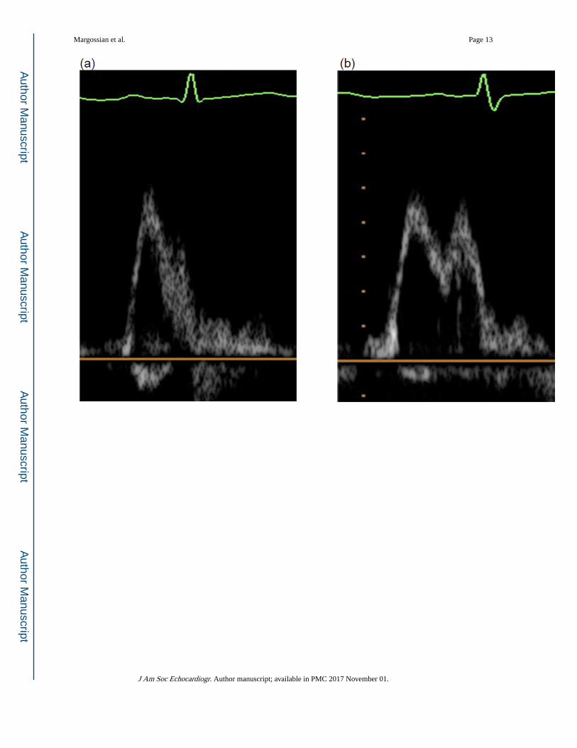
ratios, 84 studies with E and A wave fusion (either partially or fully, as demonstrated in
Figure 1) were excluded from analysis.
Historical factors were obtained from medical chart review, including pre-Fontan
catheterization variables, history of coarctation requiring intervention at the time of the pre-
Fontan catheterization, history of superior cavopulmonary anastomosis (SCPA, e.g.,
bidirectional Glenn shunt, hemi-Fontan), and type of Fontan procedure (e.g., lateral tunnel
connection, direct right atrial to pulmonary artery connection). Clinical data obtained at the
time of the study echocardiogram included BNP level (B-type natriuretic peptide), Child
Health Questionnaire (CHQ) parental report (CHQ-PF50) (10) Physical and Psychosocial
Margossian et al. Page 3
J Am Soc Echocardiogr. Author manuscript; available in PMC 2017 November 01.
Author M
anuscriptA
uthor Manuscript
Author M
anuscriptA
uthor Manuscript

summary scores, and medications, including history of and current angiotensin converting
enzyme (ACE) inhibitor usage. Subjects also underwent exercise stress testing and the
following parameters were recorded: peak oxygen consumption (VO2), ventilatory anaerobic
threshold (VAT), %predicted peak VO2, % predicted VAT, maximal O2 pulse/body surface
area (BSA), maximal work rate, %predicted maximal O2 pulse, %predicted maximal work
rate and %predicted maximal heart rate.
Statistical Methods
Descriptive statistics were reported as mean ± standard deviation and median (interquartile
range). Logistic regression was used to model the presence vs. absence of a restrictive
pattern as a function of age, and a generalized additive model was used to assess potential
nonlinearity between age and restrictive pattern. Spearman correlation coefficients were
used to examine the association between continuous diastolic function measures and age.
Cumulative logistic regression was used to model the relationship between DFG and age. An
interaction was fit between age and ventricular morphology to determine whether
associations between DFG and age were dependent on anatomy. Similarly, tests of
interaction were fit to assess whether associations of DFG and other clinical measurements
depended on ventricular morphology. We used a chi-square test to assess the association of
DFG and categorical patient and clinical variables. ANOVA and Kruskal-Wallis tests were
used to compare parameters of systolic function (EF, ESV, EDV and mass:volume z-scores
(11)), current AVV or semilunar valve regurgitation, current use of ACE inhibition, exercise
performance, and functional status by DFG. Multivariable modeling was not conducted
because few variables were associated with DFG. Analysis of variance was used to compare
the distributions of E/E’ ratio and other continuous diastolic function parameters and their z-
scores indexed to age (11) by age group and by ventricular morphology. To determine
whether mass-to-volume ratio differed according to DFG and ACE inhibitor use, we used
the Kruskal-Wallis test. A p-value of less than 0.05 was considered significant. All analyses
were conducted in SAS version 9.2 (Statistical Analysis System, Inc., Cary, NC) and S-Plus
(Insightful Corp., Seattle, WA).
RESULTS
A summary of the parameters by DFG, with z-scores where applicable, is included in Table
1, and baseline demographic characteristics for the cohort are presented in Table 2. Of the
546 subjects who underwent a study echocardiogram, 326 (60%) had echocardiograms
adequate to assign a DFG, and in 343 (63%) it was possible to determine the presence or
absence of a restrictive pattern. The 220 subjects for whom a DFG could not be calculated
were similar to those with a calculated grade with respect to time since the Fontan
procedure, gender, and ventricular morphology, but differed slightly with regard to race
(84% vs. 77% white). The distribution of DFG in the overall cohort was: DFG 0, 32%; DFG
1, 9%; DFG 2, 37%; and DFG 3, 22%.
Age-Related Patterns of Diastolic Function
DFG was not associated with time since Fontan or age at echo (Table 2). There were few
associations with age among the individual variables used to determine diastolic function
Margossian et al. Page 4
J Am Soc Echocardiogr. Author manuscript; available in PMC 2017 November 01.
Author M
anuscriptA
uthor Manuscript
Author M
anuscriptA
uthor Manuscript
grade. Mitral valve DT was positively correlated with age (Spearman R = 0.15, p=0.007) but
this was not significant after adjustment for heart rate (p=0.14). None of the other variables
had statistically significant associations with age after adjustment for heart rate.
Diastolic Function Grade Associations with Pre-Fontan Variables
Ventricular morphology—E’ was lowest in RV morphology subjects (mean±SD: RV,
8.0±2.8; LV, 10.0±3.2; Mixed 10.4±3.9, p <.001) (Table 3) and this finding remained
statistically significant when using z-scores– for-age (p <.001). The E/E’ ratio was higher in
the RV group than in the others, due to the lower E’. (Figure 2).
Age at Fontan Surgery—We stratified the cohort by age at Fontan surgery as <2 years,
2-<3 years, 3-<4 years and ≥ 4 years. DFG did not differ significantly among the groups (p=.
50); nor did E/E’ z-score (p=0.45; Table 4).
Other Pre-Fontan Medical History Factors—Neither the individual diastolic function
variables nor the DFG differed between subjects who underwent a prior SCPA surgery
compared to those who did not, even after adjusting for age (Table 5). Among those who
underwent volume unloading surgery, the age at surgery was very mildly negatively
correlated with E (R= − 0.13, p=.02) and E/E’(R = −0.15, p = .01) and positively correlated
with E’ (R=0.15, p=.002). The DFG also did not differ by type of Fontan procedure
performed, presence or absence of a fenestration, presence of coarctation intervention at the
time of the pre-Fontan catheterization or pre-Fontan cardiac catheterization pressures (Table
5).
Diastolic Function Grade Associations with Measures of Current Status
No association was seen between DFG and parameters of systolic function (EDV z-score,
ESV z-score, EF, EF z-score, end-systolic global average fiber stress, or stroke volume z-
score), nor between DFG and functional health status (CHQ Physical summary score or
Psychosocial summary score). There was no significant association between DFG and
atrioventricular valve regurgitation (p=0.20) or semilunar valve regurgitation (p=0.52).
There was no difference in DFG between patients currently taking ACE inhibitor therapy
versus those who were not (p=0.70)
Of the 326 subjects for whom DFG was calculable, 254 underwent an exercise test, with 99
subjects achieving maximum effort defined as respiratory exchange ratio ≥1.1. The
percentage achieving maximal effort differed by DFG (p=0.004; Figure 3). Of the nine
individual exercise parameters, only maximal O2 pulse/BSA (p=.03) and maximal work rate
differed by DFG (p=.04). The DFG 0 and 3 groups had the highest mean maximal O2
pulse/BSA (5.8±1.4 and 58±1.8 ml O2/BSA), while the other two groups had means of 5.03
and 5.35 ml O2/BSA. For maximal work rate, the DFG 0 group had the highest mean
(2.0±0.5 W), while the other groups had means between 1.8 and 1.9 W. When the analysis
was restricted to the maximal effort group no significant association was seen in maximal O2
pulse/BSA (p=.28) or maximal work rate (p=.874). For maximal O2 pulse/BSA, the loss of
significance was in part due to the restricted sample size; the DFG group mean was 6.1 ml
O2/BSA while the other group means were 5.4 to 5.5 ml O2/BSA.
Margossian et al. Page 5
J Am Soc Echocardiogr. Author manuscript; available in PMC 2017 November 01.
Author M
anuscriptA
uthor Manuscript
Author M
anuscriptA
uthor Manuscript
We next examined whether E’ alone was associated with clinical outcome. A total of 452
subjects had a measurable annular E’ velocity, and 24% had an abnormal value (z-score <
−2). No association was seen between E’ and CHQ summary score, BNP or exercise study
parameters.
Associations with Presence of a Restrictive Pattern
We sought to determine whether a simplified approach of assessment of diastolic function,
i.e., presence or absence of a restrictive pattern, would reveal associations not seen with the
0–3 grading scale of DFG. Over half (52%) had a restrictive pattern. This pattern was
associated with not achieving maximal effort on exercise testing (32% in the restrictive
pattern group achieved maximal effort vs. 48% in those without a restrictive pattern, p=.
012). Catheterization intervention for coarctation was marginally associated (7.3% in the
restrictive pattern group vs. 3.0% in those without a restrictive pattern, p=.08). Maximal
work rate was also associated, but not within the subset who achieved maximal effort on
testing. No significant associations with age, pre-Fontan variables or current outcomes were
identified with this approach.
Associations with Mass-to-Volume Ratio
Of the 326 subjects with a calculable DFG, 258 had a determination of mass-to-volume ratio
by echocardiography. There was no significant difference in mass-to-volume ratio or mass-
to-volume ratio z-score by DFG (Kruskal-Wallis p>0.3). Mass-to-volume ratio z-score was
positively associated with the number of years since the Fontan procedure was performed
(R=0.20, p<.001). Mass-to-volume ratio was not associated with current or historical use of
ACE inhibitors. Mass-to-volume ratio did not correlate with exercise performance or CHQ
summary scores.
DISCUSSION
The surgical strategy of early ventricular volume unloading in patients with functional single
ventricles has been developed to mitigate the myocardial changes which occur with chronic
volume overload. However, this strategy results in an abnormal physiological state with low
resting cardiac output, elevated systemic vascular resistance, abnormal ventricular-vascular
coupling, and increased total ventricular work due to the absence of a second ventricle and
other power losses throughout the Fontan circulation (12).
Our finding that the majority of subjects have preserved systolic function with abnormal
diastolic function is consistent with prior studies (2, 12, 13). In the current era, systolic
dysfunction is seldom a major problem until late after the Fontan procedure, especially when
it is assessed at the ages in our cohort. Noninvasive evidence of diastolic dysfunction,
however, is likely to be present early, primarily due to the hemodynamic abnormalities that
result from the Fontan surgical pathway (12, 14). Although the mechanisms are not
completely clear, our data from this large cohort confirm the high prevalence of diastolic
dysfunction after the Fontan procedure, as determined by standard algorithms. Noninvasive
diastolic function indices do not seem to have the same implications for the single ventricle
as they do for a two-ventricle circulation. Compared to the single ventricle cohort presented
Margossian et al. Page 6
J Am Soc Echocardiogr. Author manuscript; available in PMC 2017 November 01.
Author M
anuscriptA
uthor Manuscript
Author M
anuscriptA
uthor Manuscript

here, correlation between E/E’ and EDP is stronger in children with aortic stenosis, where
there are two ventricles that interact in series and in parallel and the disease affects only the
left ventricle (15).
Overall, there are numerous factors in this population that would diminish the coupling
between E/E’ and EDP, as we observed. Mechanistically, E/E’ reflects the combined effects
of the pressure gradient between the left atrium and the left ventricle at the time of mitral
valve opening (this gradient is the determinant of E) and the speed of myocardial relaxation
(E’ correlates with Tau, the time constant of myocardial relaxation). The peak early
transmitral gradient (and hence E) depends directly on left atrial end-systolic pressure which
is in turn dependent on the combination of end-diastolic pressure and left atrial compliance.
E/E’ therefore increases in conjunction with higher EDP or lower left atrial compliance and
also increases with a lower value of Tau. Differential trends in any of these variables will
disrupt the correlation between EDP and E/E’. Impaired relaxation that does not result in
elevated EDP or is associated with a compliant atrium may fail to manifest a significant
elevation in E/E’. Similarly, normal relaxation rate may mask the effect of elevated EDP on
E/E’. It is important to note that the finding of elevated E/E’ was more prevalent in the RV
group, where the “normal” value for E/E’ may be quite different than the normal value in a
LV. There is, of course, no “normal” value for E’ in a single RV but in the study population
the RV E’ of 8.0±2.8 is lower than the LV E’ of 10.0±3.2. Due to its relatively under-
developed conduction system the RV may have more diastolic dyssynchrony than the LV
and in general, the single ventricles may have more diastolic dyssynchrony than normal
ventricles, which would result in an elevated Tau and hence lower E’. Ventricular volume
overload is always present in these patients prior to the Fontan and is associated with
variably increased ventricular compliance. Higher ventricular compliance results in a higher
peak E value, which would mask the impact of a lower E’ on the E/E’.
Our study is the first to attempt to establish predictors or determine the clinical correlations
of abnormal diastolic function in a large cohort of Fontan patients. We found that subjects
with RV morphology were more likely to have diastolic dysfunction as measured by
standard guidelines (8, 9), even when adjusted for heart rate. We found few significant
associations with anatomic variables or current status, and no associations were found
between mass-to-volume ratio and DFG, ACE inhibitor use or exercise performance.
The group of subjects with systemic RVs demonstrated more abnormalities in DFG than
other groups. Similar findings have been described in other smaller series (16, 17), which
reported lower S’ and E’ velocities as well as higher E/E’ in subjects with RV morphology
compared to those with LV morphology. Systemic RVs in biventricular circulations have
also been shown to have decreased longitudinal velocities (18) compared to systemic LVs. It
should be noted that E’ measures peak velocity of longitudinal lengthening. Because the
fiber structure of the RV has fewer longitudinally oriented muscle fibers (19), it is not
surprising that longitudinal velocity is lower. In the present cohort, AVV regurgitation was
worse in the RV subgroup (reported previously (1)). Along with other factors intrinsic to the
RV, this may have contributed to the overall higher DFG in the RV group, since DGF groups
2 and 3 were more likely to have higher degrees of AVVR.
Margossian et al. Page 7
J Am Soc Echocardiogr. Author manuscript; available in PMC 2017 November 01.
Author M
anuscriptA
uthor Manuscript
Author M
anuscriptA
uthor Manuscript
The use of standard algorithms to assess diastolic function takes advantage of multiple
parameters to define abnormalities. However, most other studies in this population have
relied on single variables to describe diastolic function. In a small series of 32 patients
(mean age 30 months ± 22 months) with single ventricle palliation, which included pre- and
post-Glenn as well as post-Fontan palliation, Menon et al found modest correlations between
mean ventricular end-diastolic pressure with pulmonary vein A wave reversal duration and
E’ (20). Along similar lines, Vitarelli and colleagues described lower E’ velocities in patients
following Fontan compared to healthy controls regardless of whether or not the patient had
systolic dysfunction (21). One morphologic reason that individual variables may not be
adequate to assess associations in patients with single ventricles is that the size of the AVV
in single ventricles is quite variable, influencing AVV inflow patterns. For the same atrium-
to-ventricle pressure differential, flow velocity through a larger valve will be lower.
The overall exercise capacity of this cohort has been previously reported (22), demonstrating
a reduced maximal aerobic capacity. No echocardiographic measure of systolic or diastolic
function correlated with %predicted VO2, supporting the notion that exercise capacity in this
population is restricted by factors other than the properties of the single ventricle. The
principal factor influencing the ability to exercise after the Fontan procedure appears to be
the known inability of these patients to increase transpulmonary blood flow appropriately
with exercise (23). A fenestration, when present, can also increase cardiac output, but
limitation of transpulmonary blood flow remains an issue. Following this line of reasoning,
Goldberg and colleagues have recently reported a proof-of-concept placebo-controlled trial
of an oral pulmonary vasodilator, sildenafil, to test this question (24). They were able to
demonstrate improved ventilatory efficiency and exercise performance at the anaerobic
threshold. Even though VO2 max was unchanged, these early results merit further
investigation and provide promise for a therapeutic option.
The potential impact of diastolic dysfunction on exercise capacity in Fontan patients remains
uncertain. During exercise, there is normally a rise in stroke volume related to a rise in end-
diastolic volume (preload reserve) and a fall in end-systolic volume (contractile reserve)
(25). It has been reported that utilization of preload reserve is subnormal in adult patients
with diastolic dysfunction with normal systolic function (26) and with systolic dysfunction
(27). In a recent study by Schmitt et al (14) evaluating the response of the Fontan circulation
to dobutamine stress testing, pulmonary vascular resistance was found to drop appropriately
but the expected preload reserve response was not observed secondary to a dobutamine-
induced rise in the end-diastolic pressure-volume curve (decreased compliance). In normal
subjects, the exercise-associated rise in cardiac output is mediated primarily by heart rate
response, with a much smaller effect due to a rise in stroke volume (28). In contrast, analysis
of the factors influencing exercise response in the cohort of Fontan patients presented here
(previously published by Paridon et al (22)) found that oxygen pulse (a surrogate measure of
stroke volume) had the closest association with the rise in maximum oxygen consumption
during treadmill exercise. Nonetheless, a relationship between the usual echocardiographic
indices of diastolic function and maximum oxygen consumption was not observed. There
are a number of potential explanations for the apparent differences in the Schmitt and
Paridon observations, including differences in the response to supine dobutamine stress
compared to upright treadmill exercise, but an important possibility is that the currently
Margossian et al. Page 8
J Am Soc Echocardiogr. Author manuscript; available in PMC 2017 November 01.
Author M
anuscriptA
uthor Manuscript
Author M
anuscriptA
uthor Manuscript

available techniques for noninvasive detection of abnormal compliance are unreliable in the
single ventricle heart. Insofar as most of these indices are highly correlated with end-
diastolic pressure, diminished sensitivity of these indices may well relate to the limited
ability of the Fontan circulation to substantially elevate end-diastolic pressure.
Most of the diastolic function indices have been developed through study of adult patients
with very different cardio-pulmonary systems than those of our patients. In congenital heart
disease patients who have increased pressure loads (e.g., aortic stenosis) similar patterns of
diastolic indices are seen in children (29) as have been described in adults. However,
increased pressure load is less common in the single ventricle population. Altered diastolic
indices likely have a fundamentally different etiology, and for multifactorial reasons.
Diastolic behavior of the ventricle relates to both chamber factors (e.g.shape and mass to
volume ratio) and myocardial factors (e.g. relaxation and muscle compliance). When
diastolic dysfunction occurs in the presence of hypertrophy, the impact of this chamber
property must be accounted for before it is possible to ascertain the impact of myocardial
properties. We hypothesized that the chamber factor mass to volume ratio may make an
important contribution to diastolic function in this population: the force required to expand
the ventricle (diastolic wall stress) is proportional to pressure x (radius/thickness). This
means a lower radius/thickness (which is equivalent to a higher mass/volume) requires a
higher pressure to achieve the same wall stress. Therefore, geometric relationships dictate
that a higher pressure is required to fill thicker ventricles even if the compliance of the
myocardium remains the same. The failure of this relationship to be important in these
patients implies that factors other than the geometric influence of hypertrophy dominate the
variation in diastolic function.
From a technical standpoint, of the 546 echocardiograms submitted to the core lab, we were
able to calculate a DFG on 60%. The presence of a summation wave, common at pediatric
heart rates, precluded assessment on 84 studies (16% of the total and 38% of those without
an assignable DFG). Determination of whether a restrictive pattern was present was able to
be made in 344 patients or 63% of the total group, indicating that although a majority of
single ventricles can be assessed in such a manner, the technical difficulties in obtaining on-
axis Doppler signals in a significant fraction of the patients additionally restricts the data.
Limitations
Our data have important limitations. Age association analysis was restricted by the relatively
young age of the cohort studied (maximum age 18 years). As this group reaches the third
and fourth decades of life, more significant differences may appear. Additionally, this is a
descriptive study of the outcome of a constantly evolving treatment strategy. Patients with
single ventricle physiology born today will not be managed in the same manner as our
cohort, thereby limiting the generalizability of the results. Assessment of the impact of
individual medications on diastolic function was limited due to small sample sizes. For
example only 19 subjects cohort-wide were taking beta blockers at the time of evaluation.
Margossian et al. Page 9
J Am Soc Echocardiogr. Author manuscript; available in PMC 2017 November 01.
Author M
anuscriptA
uthor Manuscript
Author M
anuscriptA
uthor Manuscript

Conclusions
Echocardiographic assessment of diastolic function in patients with single ventricle
physiology using current algorithms is technically possible and a high percentage of patients
have abnormal results. However, we found few clinically or statistically significant
associations with diastolic dysfunction within our cohort. While this may imply a lack of
impact of abnormal diastolic function upon clinical outcome in this cohort, our results could
also indicate that the methodology developed for echocardiographic assessment of diastolic
function in adults with biventricular hearts may not be applicable to pediatric single ventricle
patients. Further efforts are needed to develop non-invasive methods to evaluate diastolic
function in this unique circulation.
Acknowledgments
Supported by U01 grants from the National Heart, Lung, and Blood Institute (HL068269, HL068270, HL068279, HL068281, HL068285, HL068292, HL068290, HL068288). This work is solely the responsibility of the authors and does not necessarily represent the official views of the NIH/NHLBI.
References
1. Anderson PA, Sleeper LA, Mahony L, Colan SD, Atz AM, Breitbart RE, et al. Contemporary outcomes after the Fontan procedure: a Pediatric Heart Network multicenter study. J Am Coll Cardiol. 2008; 52(2):85–98. [PubMed: 18598886]
2. Cheung YF, Penny DJ, Redington AN. Serial assessment of left ventricular diastolic function after Fontan procedure. Heart. 2000; 83(4):420–424. [PubMed: 10722541]
3. Rychik J, Jacobs ML, Norwood WI Jr. Acute changes in left ventricular geometry after volume reduction operation. Ann Thorac Surg. 1995; 60(5):1267–1273. discussion 74. [PubMed: 8526611]
4. Kirklin JK, Blackstone EH, Kirklin JW, Pacifico AD, Bargeron LM Jr. The Fontan operation. Ventricular hypertrophy, age, and date of operation as risk factors. J Thorac Cardiovasc Surg. 1986; 92(6):1049–1064. [PubMed: 2946901]
5. McCrindle BW, Zak V, Sleeper LA, Paridon SM, Colan SD, Geva T, et al. Laboratory measures of exercise capacity and ventricular characteristics and function are weakly associated with functional health status after Fontan procedure. Circulation. 121(1):34–42.
6. Sleeper LA, Anderson P, Hsu DT, Mahony L, McCrindle BW, Roth SJ, et al. Design of a large cross-sectional study to facilitate future clinical trials in children with the Fontan palliation. Am Heart J. 2006; 152(3):427–433. [PubMed: 16923408]
7. Margossian R, Schwartz ML, Prakash A, Wruck L, Colan SD, Atz AM, et al. Comparison of echocardiographic and cardiac magnetic resonance imaging measurements of functional single ventricular volumes, mass, and ejection fraction (from the Pediatric Heart Network Fontan Cross-Sectional Study). Am J Cardiol. 2009; 104(3):419–428. [PubMed: 19616678]
8. Lester SJ, Tajik AJ, Nishimura RA, Oh JK, Khandheria BK, Seward JB. Unlocking the mysteries of diastolic function: deciphering the Rosetta Stone 10 years later. J Am Coll Cardiol. 2008; 51(7):679–689. [PubMed: 18279730]
9. Nishimura RA, Tajik AJ. Evaluation of diastolic filling of left ventricle in health and disease: Doppler echocardiography is the clinician’s Rosetta Stone. J Am Coll Cardiol. 1997; 30(1):8–18. [PubMed: 9207615]
10. Landgraf, JMAL., Ware, JE. The Child Health Questionnaire (CHQ) User’s Manual. Boston, MA: HealthAct; 1999.
11. Sluysmans T, Colan SD. Theoretical and empirical derivation of cardiovascular allometric relationships in children. J Appl Physiol. 2005; 99(2):445–457. [PubMed: 15557009]
12. Senzaki H, Masutani S, Kobayashi J, Kobayashi T, Sasaki N, Asano H, et al. Ventricular afterload and ventricular work in Fontan circulation: comparison with normal two-ventricle circulation and
Margossian et al. Page 10
J Am Soc Echocardiogr. Author manuscript; available in PMC 2017 November 01.
Author M
anuscriptA
uthor Manuscript
Author M
anuscriptA
uthor Manuscript

single-ventricle circulation with Blalock-Taussig shunts. Circulation. 2002; 105(24):2885–2892. [PubMed: 12070118]
13. Olivier M, O’Leary PW, Pankratz VS, Lohse CM, Walsh BE, Tajik AJ, et al. Serial Doppler assessment of diastolic function before and after the Fontan operation. J Am Soc Echocardiogr. 2003; 16(11):1136–1143. [PubMed: 14608284]
14. Schmitt B, Steendijk P, Ovroutski S, Lunze K, Rahmanzadeh P, Maarouf N, et al. Pulmonary vascular resistance, collateral flow, and ventricular function in patients with a Fontan circulation at rest and during dobutamine stress. Circ Cardiovasc Imaging. 2010; 3(5):623–631. [PubMed: 20631032]
15. Friedman KG, McElhinney DB, Colan SD, Porras D, Powell AJ, Lock JE, et al. Left ventricular remodeling and improvement in diastolic function after balloon aortic valvuloplasty for congenital aortic stenosis. Circ Cardiovasc Interv. 2012; 5(4):549–554. [PubMed: 22739787]
16. Hershenson JA, Zaidi AN, Texter KM, Moiduddin N, Stefaniak CA, Hayes J, et al. Differences in tissue Doppler imaging between single ventricles after the Fontan operation and normal controls. Am J Cardiol. 2010; 106(1):99–103. [PubMed: 20609655]
17. Menon SC, Dearani JA, Cetta F. Long-term outcome after atrioventricular valve surgery following modified Fontan operation. Cardiol Young. 2011; 21(1):83–88. [PubMed: 20977827]
18. Pettersen E, Helle-Valle T, Edvardsen T, Lindberg H, Smith HJ, Smevik B, et al. Contraction pattern of the systemic right ventricle shift from longitudinal to circumferential shortening and absent global ventricular torsion. J Am Coll Cardiol. 2007; 49(25):2450–2456. [PubMed: 17599609]
19. Friedberg MK, Redington AN. Right versus left ventricular failure: differences, similarities, and interactions. Circulation. 2014; 129(9):1033–1044. [PubMed: 24589696]
20. Menon SC, Gray R, Tani LY. Evaluation of ventricular filling pressures and ventricular function by Doppler echocardiography in patients with functional single ventricle: correlation with simultaneous cardiac catheterization. J Am Soc Echocardiogr. 2011; 24(11):1220–1225. [PubMed: 21962450]
21. Vitarelli A, Conde Y, Cimino E, D’Angeli I, D’Orazio S, Ventriglia F, et al. Quantitative assessment of systolic and diastolic ventricular function with tissue Doppler imaging after Fontan type of operation. Int J Cardiol. 2005; 102(1):61–69. [PubMed: 15939100]
22. Paridon SM, Mitchell PD, Colan SD, Williams RV, Blaufox A, Li JS, et al. A cross-sectional study of exercise performance during the first 2 decades of life after the Fontan operation. J Am Coll Cardiol. 2008; 52(2):99–107. [PubMed: 18598887]
23. Gewillig M, Brown SC, Eyskens B, Heying R, Ganame J, Budts W, et al. The Fontan circulation: who controls cardiac output? Interact Cardiovasc Thorac Surg. 2010; 10(3):428–433. [PubMed: 19995891]
24. Goldberg DJ, French B, McBride MG, Marino BS, Mirarchi N, Hanna BD, et al. Impact of oral sildenafil on exercise performance in children and young adults after the Fontan operation: a randomized, double-blind, placebo-controlled, crossover trial. Circulation. 2011; 123(11):1185–1193. [PubMed: 21382896]
25. Levine BD, Lane LD, Buckey JC, Friedman DB, Blomqvist CG. Left ventricular pressure-volume and Frank-Starling relations in endurance athletes. Implications for orthostatic tolerance and exercise performance. Circulation. 1991; 84(3):1016–1023. [PubMed: 1884438]
26. Kitzman DW, Higginbotham MB, Cobb FR, Sheikh KH, Sullivan MJ. Exercise intolerance in patients with heart failure and preserved left ventricular systolic function: failure of the Frank-Starling mechanism. J Am Coll Cardiol. 1991; 17(5):1065–1072. [PubMed: 2007704]
27. Dahan M, Aubry N, Baleynaud S, Ferreira B, Yu J, Gourgon R. Influence of preload reserve on stroke volume response to exercise in patients with left ventricular systolic dysfunction: a Doppler echocardiographic study. J Am Coll Cardiol. 1995; 25(3):680–686. [PubMed: 7860913]
28. Hosenpud JD, Morton MJ, Wilson RA, Pantely GA, Norman DJ, Cobanoglu MA, et al. Abnormal exercise hemodynamics in cardiac allograft recipients 1 year after cardiac transplantation. Relation to preload reserve. Circulation. 1989; 80(3):525–532. [PubMed: 2670315]
Margossian et al. Page 11
J Am Soc Echocardiogr. Author manuscript; available in PMC 2017 November 01.
Author M
anuscriptA
uthor Manuscript
Author M
anuscriptA
uthor Manuscript

29. Friedman KG, McElhinney DB, Rhodes J, Powell AJ, Colan SD, Lock JE, et al. Left ventricular diastolic function in children and young adults with congenital aortic valve disease. Am J Cardiol. 2013; 111(2):243–249. [PubMed: 23102884]
Margossian et al. Page 12
J Am Soc Echocardiogr. Author manuscript; available in PMC 2017 November 01.
Author M
anuscriptA
uthor Manuscript
Author M
anuscriptA
uthor Manuscript
Margossian et al. Page 13
J Am Soc Echocardiogr. Author manuscript; available in PMC 2017 November 01.
Author M
anuscriptA
uthor Manuscript
Author M
anuscriptA
uthor Manuscript

Figure 1. Mitral Valve E/A and TDI E’/A’ partial-complete fusionExamples of mitral valve inflow E/A complete (a) and partial (b) fusion and TDI E’/A’
complete (c) and partial (d) fusion.
Margossian et al. Page 14
J Am Soc Echocardiogr. Author manuscript; available in PMC 2017 November 01.
Author M
anuscriptA
uthor Manuscript
Author M
anuscriptA
uthor Manuscript

Figure 2. E/E’ ratio and E/E’ ratio z-score by ventricular morphology; p-value <0.001, LV = left
ventricle, RV = right ventricle. The width of each box is proportional to the square root of
the group size.
Margossian et al. Page 15
J Am Soc Echocardiogr. Author manuscript; available in PMC 2017 November 01.
Author M
anuscriptA
uthor Manuscript
Author M
anuscriptA
uthor Manuscript

Figure 3. Percentage of subjects in each diastolic function grade, by those who achieved maximal
effort (defined as achieving respiratory exchange ratio ≥ 1.1) and those achieving non-
maximal effort.
Margossian et al. Page 16
J Am Soc Echocardiogr. Author manuscript; available in PMC 2017 November 01.
Author M
anuscriptA
uthor Manuscript
Author M
anuscriptA
uthor Manuscript

Author M
anuscriptA
uthor Manuscript
Author M
anuscriptA
uthor Manuscript
Margossian et al. Page 17
Table 1
Summary of Parameters by Diastolic Function
Variable Normal(Grade 0)
ImpairedRelaxation(Grade 1)
Pseudo-normalization
(Grade 2)
Restrictive(Grade 3)
N 104 30 120 72
E:E’ 7.3±2.2 6.8±3.0 9.6±5.0 9.0±3.7
Z-score 0.6±1.1 0.4±1.4 1.7±2.4 1.4±1.8
E:A 1.5±0.3 0.9±0.1 1.4±0.3 2.5±0.4
Z-score −1.2±0.4 −2.1 ±0.2 −1.3±0.5 0.4±0.6
DT (msec) 204±56 125±34 120±39 193±87
Z-score 1.6±1.6 −0.7±1.0 −0.8±1.2 1.3±2.6
Vfp (cm/sec)* 63±22 67±17 64 ±20 64±17
Z-score −0.3±1.0 −0.1 ±0.7 −0.2±0.9 −0.2±0.8
EF (%)** 59±10 56±12 61 ±11 60±10
Z-score −1.0±2.3 −1.7±2.7 −0.6±2.2 −0.7±2.1
Mass:Volume*** 1.1±0.3 1.3±0.6 1.2±0.4 1.2±0.4
Z-score 2.1 ±2.1 3.2±5.2 2.5±3.4 2.9±3.0
DT = deceleration time; EF = ejection fraction; Vfp = ventricular flow propagation
*Vfp sample sizes by grade are 39; 7; 57; 38
**EF sample sizes by grade are 84; 25; 94; 61
***Mass:volume sample sizes by grade are 80; 25; 92; 61
J Am Soc Echocardiogr. Author manuscript; available in PMC 2017 November 01.

Author M
anuscriptA
uthor Manuscript
Author M
anuscriptA
uthor Manuscript
Margossian et al. Page 18
Tab
le 2
Patie
nt C
hara
cter
istic
s by
Dia
stol
ic F
unct
ion
Gra
de
Var
iabl
eN
oas
sign
able
DF
G
All
wit
has
sign
edD
FG
P1
Nor
mal
(Gra
de 0
)Im
pair
edR
elax
atio
n(G
rade
1)
Pse
udo-
norm
aliz
atio
n(G
rade
2)
Res
tric
tive
(Gra
de 3
)P
2
N22
032
610
430
120
72
Tim
e si
nce
Font
an, y
r8.
6±3.
68.
5±3.
3.8
38.
9±3.
37.
8±3.
88.
2±3.
28.
9±3.
20.
18
Age
at e
cho,
yr12
.1±
3.5
11.7
±3.
3.3
112
.3±
3.5
11.4
±3.
411
.2±
3.1
12.0
±3.
40.
09
Hea
rt r
ate,
bpm
*79
±15
71 ±
13<
.001
68±
1180
±9
77±
1364
±12
<.0
01
Mal
e57
%62
%.2
461
%50
%66
%64
%0.
43
Ven
tric
ular
Typ
e.7
50.
30
LV
48%
49%
53%
47%
41%
58%
RV
35%
33%
28%
37%
39%
26%
Mix
ed17
%18
%19
%17
%20
%15
%
* For
hear
t rat
e N
=16
0 w
ithou
t DFG
, N=
277
with
DFG
; N=
88 D
FG0,
N=
25 D
FG1,
N=
100
DFG
2; N
=64
DFG
3. D
FG=
Dia
stol
ic F
unct
ion
Gra
de
P1=
p-va
lue
com
pari
ng s
ubje
cts
with
vs.
with
out a
n as
sign
able
DFG
P2=
p-va
lue
com
pari
ng th
e 4
DFG
gro
ups
J Am Soc Echocardiogr. Author manuscript; available in PMC 2017 November 01.

Author M
anuscriptA
uthor Manuscript
Author M
anuscriptA
uthor Manuscript
Margossian et al. Page 19
Tab
le 3
Com
pari
son
of E
, E’
and
E/E
’ by
ven
tric
ular
type
LVR
VM
ixed
nM
ean
± SD
nM
ean
± SD
nM
ean
± SD
P
E z-sc
ore*
169
0.7
± 0
.2−
1.4
±1.
110
80.
8 ±
0.2
−0.
9 ±
1.1
660.
7 ±
0.1
−1.
3 ±
0.9
<.0
01<
.001
E’
z-sc
ore*
222
10.0
± 3
.2−
1.3
± 0
.715
58.
0 ±
2.8
−1.
8 ±
0.6
7510
.4 ±
3.9
−1.
2 ±
0.9
<.0
01<
.001
E/E
’
z-sc
ore*
147
7.2
± 2
.30.
6 ±
1.1
9310
.9 ±
5.1
2.4
± 2
.456
7.4
± 3
.00.
6 ±
1.4
<.0
01<
.001
* z-sc
ore
for
age
J Am Soc Echocardiogr. Author manuscript; available in PMC 2017 November 01.

Author M
anuscriptA
uthor Manuscript
Author M
anuscriptA
uthor Manuscript
Margossian et al. Page 20
Tab
le 4
Mea
n±SD
of
E/E
’ R
atio
Z-s
core
by
Age
at F
onta
n, a
nd V
entr
icul
ar T
ype
Age
at
Fon
tan,
yr
Ven
tric
ular
Typ
e (n
)<
2(n
=66)
2 to
< 3
(n=1
09)
3 to
< 4
(n=5
2)≥
4(n
=69)
P-v
alue
*
LV (
147)
0.67
±1.
07 (
27)
0.42
±1.
18 (
57)
0.79
±1.
17 (
29)
0.71
±1.
00 (
34)
0.27
RV
(93
)3.
58±
3.19
(25
)2.
03±
1.90
(36
)1.
91±
1.61
(12
)1.
82±
2.01
(20
)0.
15
Mix
ed (
56)
0.48
±1.
57 (
14)
0.81
±1.
56 (
156)
0.73
±1.
28 (
11)
0.54
±1.
45 (
15)
0.79
All
(296
)1.
73±
2.61
(66
)1.
01±
1.66
(10
9)0.
97±
1.41
(52
)0.
99±
1.53
(69
)0.
45
* P-va
lues
wer
e ca
lcul
ated
by
AN
OV
A. S
imila
r in
fere
nces
wer
e dr
awn
usin
g no
n-pa
ram
etri
c te
stin
g (K
rusk
al-W
allis
test
).
J Am Soc Echocardiogr. Author manuscript; available in PMC 2017 November 01.

Author M
anuscriptA
uthor Manuscript
Author M
anuscriptA
uthor Manuscript
Margossian et al. Page 21
Tab
le 5
Oth
er P
re-F
onta
n M
edic
al H
isto
ry F
acto
rs b
y D
iast
olic
Fun
ctio
n G
rade
Var
iabl
eN
orm
alIm
pair
edR
elax
atio
nP
seud
o-no
rmal
izat
ion
Res
tric
tive
chis
q p
N10
430
120
72
SCPA
per
form
ed68
%73
%78
%69
%0.
43
In
LVs
56%
64%
67%
71%
0.45
In
RV
s86
%91
%91
%84
%0.
81
In
Mix
ed75
%60
%71
%36
%0.
15
Typ
e of
Fon
tan
0.60
Atr
iopu
lmon
ary
conn
ectio
n17
%10
%13
%13
%
Intr
acar
diac
late
ral t
unne
l50
%70
%65
%63
%
Ext
raca
rdia
c co
ndui
t29
%21
%20
%22
%
Ext
raca
rdia
c la
tera
l tun
nel
1%0%
2%0%
Oth
er3%
0%1%
3%
Fene
stra
ted
Font
an65
%67
%70
%65
%0.
80
Pre-
Font
an c
oarc
tatio
n ca
thet
eriz
atio
nin
terv
entio
n3%
3%8%
7%0.
42
N w
ith c
athe
teri
zatio
n10
430
120
72
End
-dia
stol
ic p
ress
ure,
mm
Hg
(med
ian)
7.1±
4.4
(8.0
)7.
4±3.
6 (8
.0)
7.0±
4.2
(7.0
)7.
4±6.
0 (7
.0)
0.95
/0.6
7*
PA p
ress
ure,
mm
Hg
(med
ian)
9.6±
6.8
(11.
0)11
.0±
4.5
(11.
0)8.
9±7.
6 (1
1.0)
8.7±
7.6
(10.
0)0.
65/0
.13*
O2
satu
ratio
n, %
(med
ian)
81±
21 (
85)
83±
5 (8
4)80
±19
(85
)78
±22
(83
)0.
57/0
.75*
* AN
OV
A p
-val
ue/K
rusk
al-W
allis
test
p-v
alue
SCPA
= s
uper
ior
cavo
pulm
onar
y an
asto
mos
is; P
A=
pulm
onar
y ar
tery
J Am Soc Echocardiogr. Author manuscript; available in PMC 2017 November 01.