echocardiographic evaluation of lv diastolic function dysfunction and diastolic heart failure (dhf)...
TRANSCRIPT
Diastolic Dysfunction
Gary W. Lewis, M.D.
• Diastolic heart failure (DHF) is a clinical syndrome in which patients have symptoms and signs of HF, normal or near normal left ventricular ejection fraction (LVEF), and evidence of diastolic dysfunction (eg, abnormal left ventricular filling and elevated filling pressures).
Diastolic dysfunction and diastolic heart failure (DHF) are not synonymous. The term diastolic HF is reserved for patients with clinical HF, in the setting of a normal or near-normal EF, and abnormalities in diastolic function

HFPEF
HFPEF Heart Failure with
Preserved Ejection Fraction

DHF is associated with remodeling that affects left ventricular and left atrial chambers, the cardiomyocytes, and extracellular matrix with impact on diastolic as well as systolic function. Nearly all patients with diastolic HF have a normal LV end diastolic volume; most have increased LV wall thicknesses, mass and relative wall thickness
Asymptomatic diastolic dysfunction is much more common than symptomatic disease..
The prevalence of DHF increases with age. DHF is more common in women than in men.
The major causes of DHF are chronic hypertension with left ventricular hypertrophy, hypertrophic cardiomyopathy, coronary heart disease, and restrictive cardiomyopathy. Diastolic function is determined by two factors: the process of myocardial relaxation (which is an active process that requires metabolic energy) and the elasticity or distensibility of the left ventricle (which is a passive process).
Diastolic and systolic HF have similar symptoms. Exercise intolerance seen in DHF may be caused by elevation in left atrial and pulmonary venous pressures and/or impaired stroke volume leading to dyspnea and fatigue.

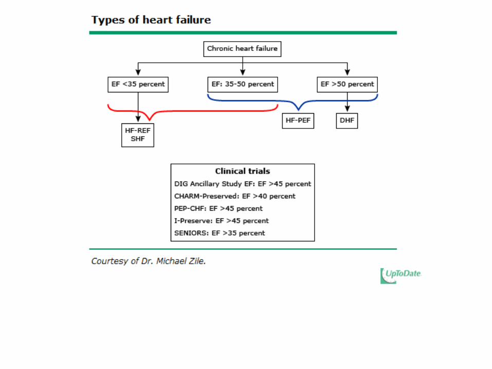
In clinical practice, the diagnosis of DHF is typically based upon finding signs and symptoms of HF, normal or mildly abnormal LVEF (LVEF >50 percent), and evidence of diastolic dysfunction on Doppler echocardiography.
DHF is one of several causes of cardiogenic pulmonary edema in patients with a normal LVEF.
Occult coronary heart disease is a potentially reversible cause of DHF.
Plasma BNP and N-terminal pro-BNP are elevated in patients with DHF but cannot effectively distinguish DHF from SHF

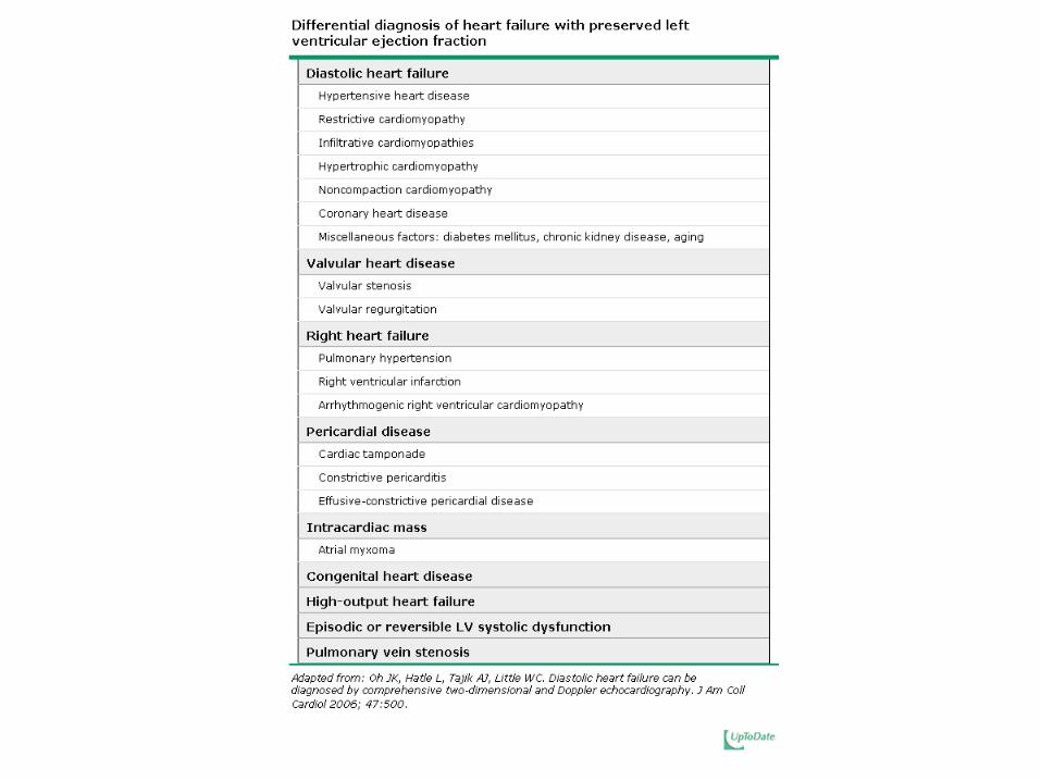
The key distinguishing feature between systolic and diastolic HF is whether the ejection fraction is reduced (indicating systolic HF) or preserved, meaning normal or near-normal (indicating diastolic HF). Diastolic dysfunction is not the only cause of HF in patients with preserved LVEF

During exercise, physiologic mechanisms normally ensure that cardiac input keeps pace with cardiac output with preservation of a low pulmonary venous pressure.

Since both afterload (systolic pressure) and diastolic load (left atrial diastolic pressure) can affect measurement of diastolic function, these factors must be considered in assessing the intrinsic relaxation rate.
The two most common pathways to DHF are left ventricular hypertrophy and ischemia.
In patients with diastolic heart failure (DHF), certain types of hemodynamic stress including atrial fibrillation; tachycardia; abrupt, severe, or refractory elevations in systemic blood pressure, and myocardial ischemia are associated with worsening of diastolic dysfunction.
Echocardiography is the recommended imaging modality for the assessment of left ventricular (LV) diastolic function.

Doppler measurements provide incremental prognostic information to clinical and anatomic findings
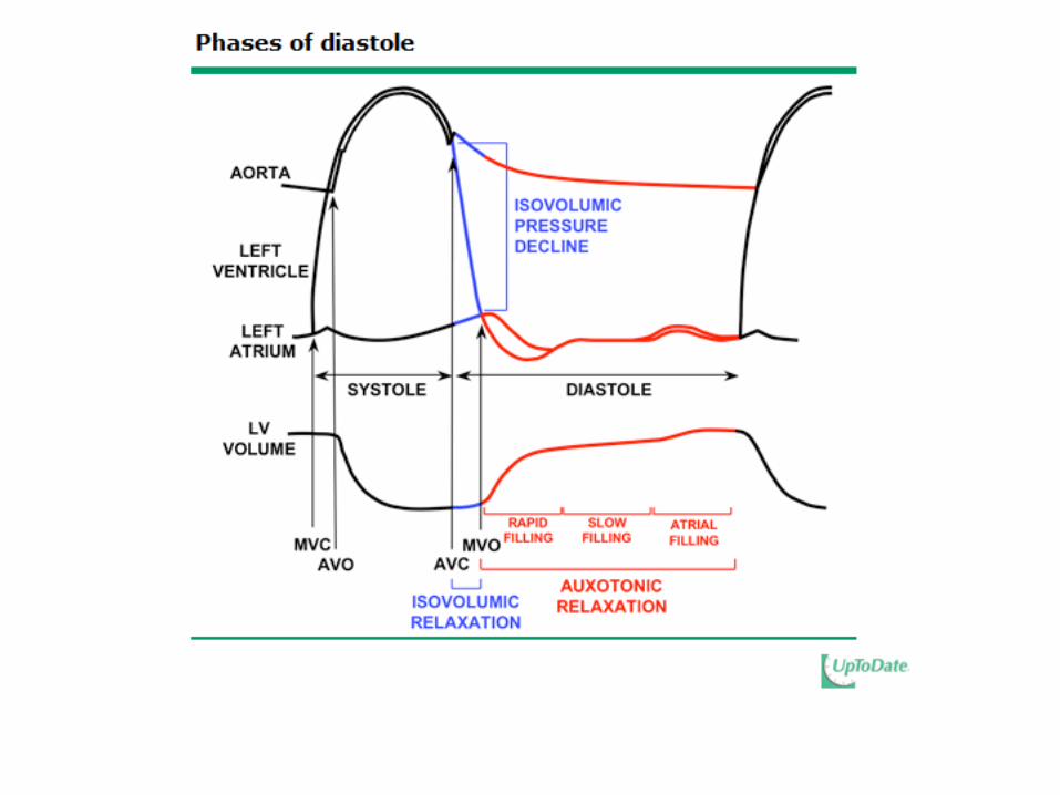
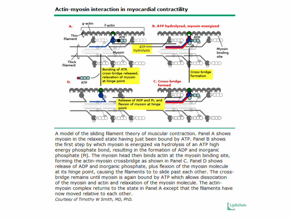
Diastole begins with isovolumic relaxation followed by auxotonic relaxation and continues until atrial contraction is complete. During the later phases of diastolic HF, the LV is readily distensible. Atrial contraction normally contributes 20 to 30 percent to total LV filling volume but usually increases diastolic pressures by less than 5 mmHg.
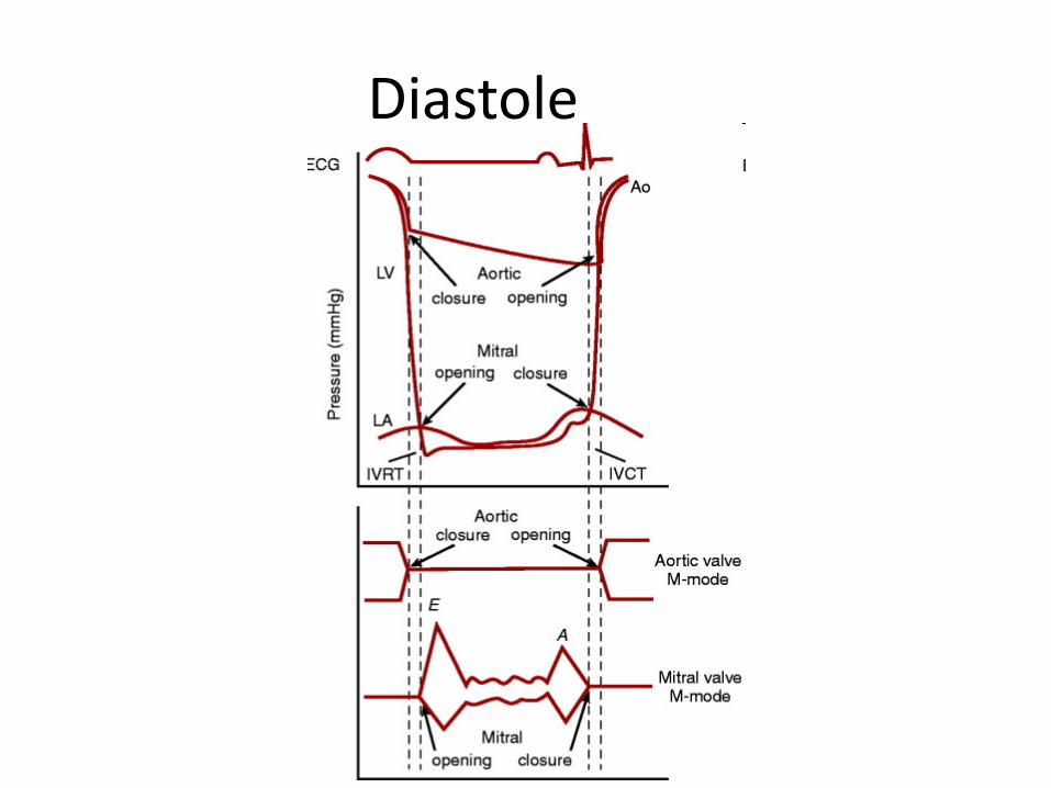
Diastole
Insert Otto Figure 7-1 +/- 7-2
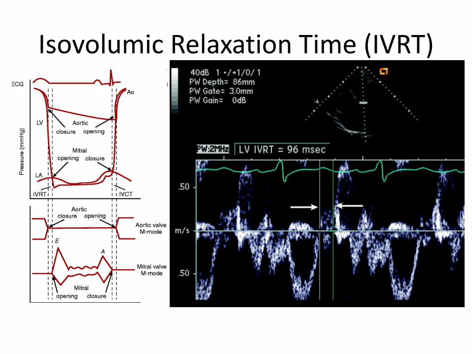
Isovolumic Relaxation Time (IVRT)
Normal IVRT 70-90ms. IVRT lengthens w/ impaired LV relaxation and shortens when LV compliance is decreased and LV filling pressures increase. IVRT varies with HR, preload and ventricular function.
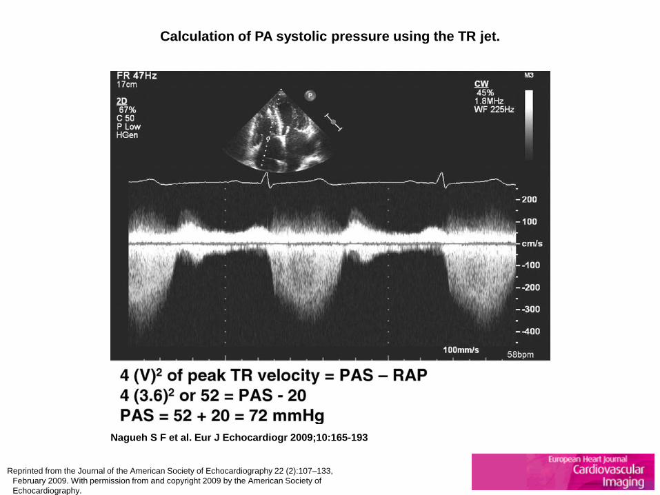
Calculation of PA systolic pressure using the TR jet.
Nagueh S F et al. Eur J Echocardiogr 2009;10:165-193
Reprinted from the Journal of the American Society of Echocardiography 22 (2):107–133, February 2009. With permission from and copyright 2009 by the American Society of Echocardiography.
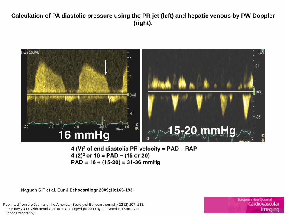
Calculation of PA diastolic pressure using the PR jet (left) and hepatic venous by PW Doppler (right).
Nagueh S F et al. Eur J Echocardiogr 2009;10:165-193
Reprinted from the Journal of the American Society of Echocardiography 22 (2):107–133, February 2009. With permission from and copyright 2009 by the American Society of Echocardiography.
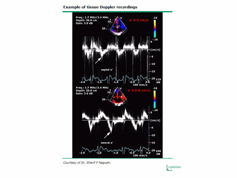
In patients with normal LV EF, the initial step is calculating the E/e' ratio
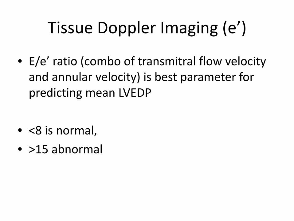
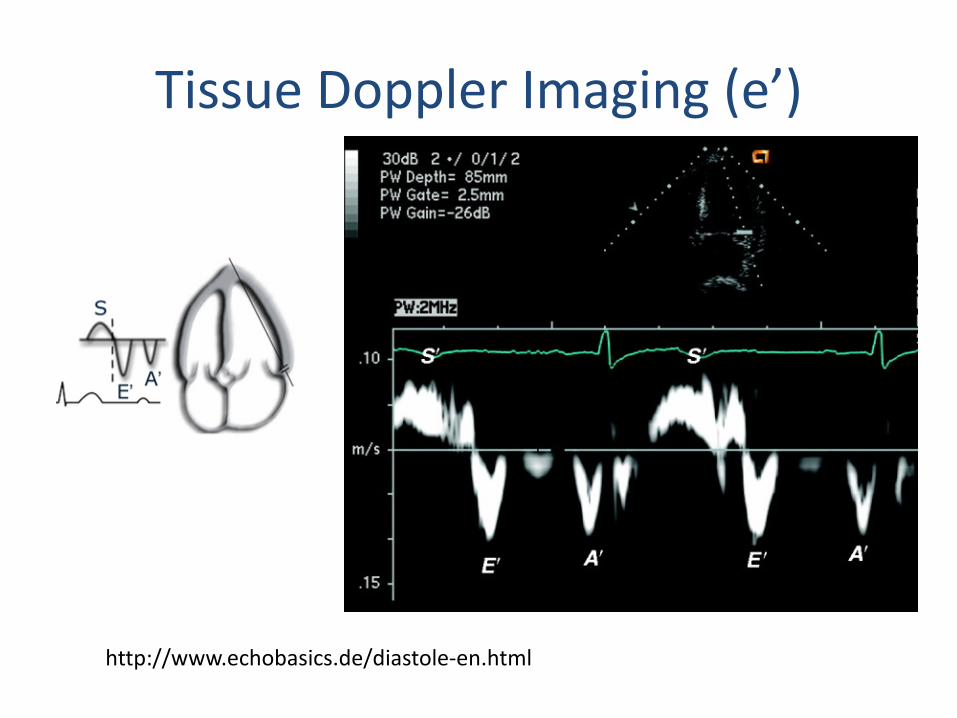
Tissue Doppler Imaging (e’)
• E/e’ ratio (combo of transmitral flow velocity and annular velocity) is best parameter for predicting mean LVEDP
• <8 is normal, • >15 abnormal
http://www.echobasics.de/diastole-en.html
Tissue Doppler Imaging (e’)
Septal and lateral velocities should be
acquired by PW Doppler
Sample volume should be placed at or 1 cm within
septal and lateral insertion sites of mitral leaflets

Identify e’ from Isovolumic Relaxation velocities
Avoid angulation
E/e’ not accurate in mitral valve disease, heavy annular
calcification, constrictive pericarditis, and abnormal septal motion
Average E/e’ ratio in patients with regional dysfunction
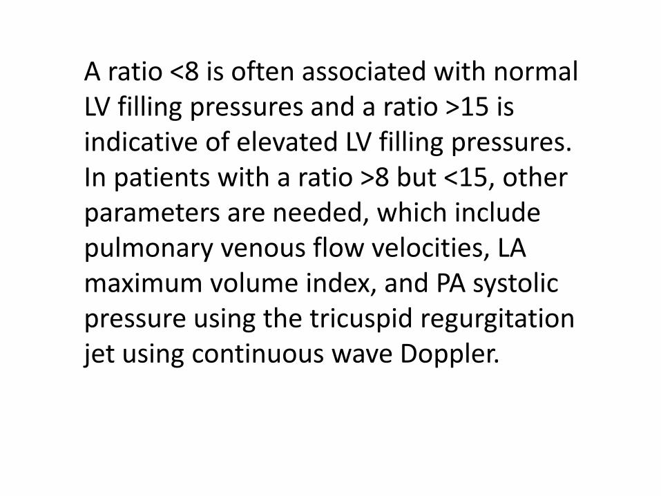
A ratio <8 is often associated with normal LV filling pressures and a ratio >15 is indicative of elevated LV filling pressures. In patients with a ratio >8 but <15, other parameters are needed, which include pulmonary venous flow velocities, LA maximum volume index, and PA systolic pressure using the tricuspid regurgitation jet using continuous wave Doppler.
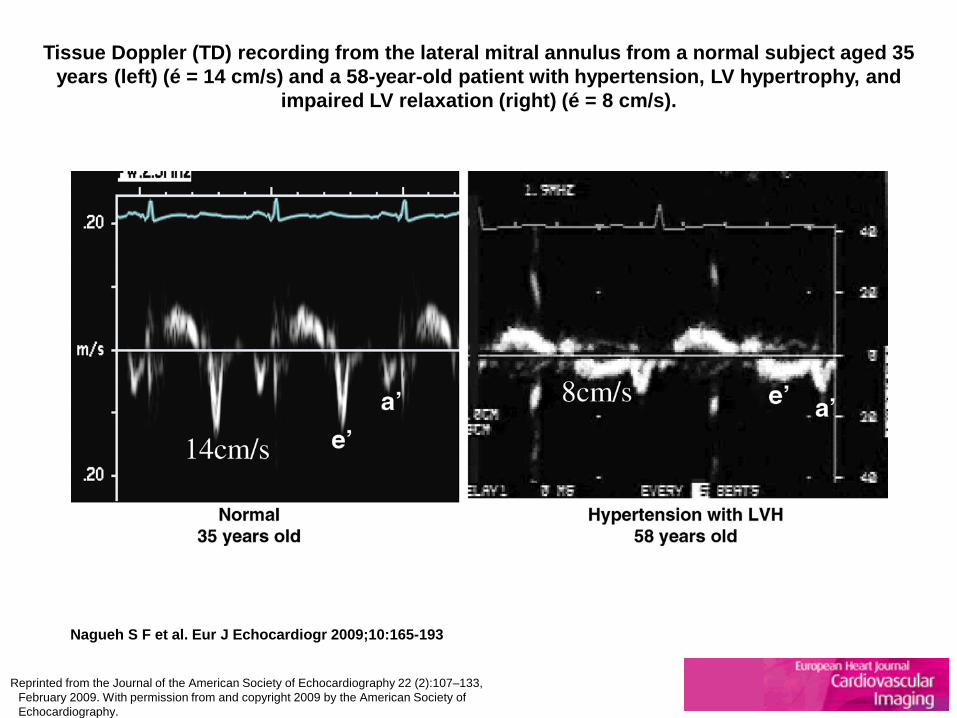
Tissue Doppler (TD) recording from the lateral mitral annulus from a normal subject aged 35 years (left) (é = 14 cm/s) and a 58-year-old patient with hypertension, LV hypertrophy, and
impaired LV relaxation (right) (é = 8 cm/s).
Nagueh S F et al. Eur J Echocardiogr 2009;10:165-193
Reprinted from the Journal of the American Society of Echocardiography 22 (2):107–133, February 2009. With permission from and copyright 2009 by the American Society of Echocardiography.
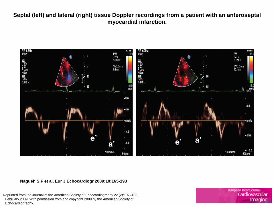
Septal (left) and lateral (right) tissue Doppler recordings from a patient with an anteroseptal myocardial infarction.
Nagueh S F et al. Eur J Echocardiogr 2009;10:165-193
Reprinted from the Journal of the American Society of Echocardiography 22 (2):107–133, February 2009. With permission from and copyright 2009 by the American Society of Echocardiography.
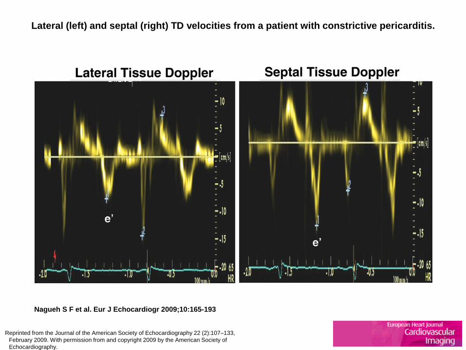
Lateral (left) and septal (right) TD velocities from a patient with constrictive pericarditis.
Nagueh S F et al. Eur J Echocardiogr 2009;10:165-193
Reprinted from the Journal of the American Society of Echocardiography 22 (2):107–133, February 2009. With permission from and copyright 2009 by the American Society of Echocardiography.
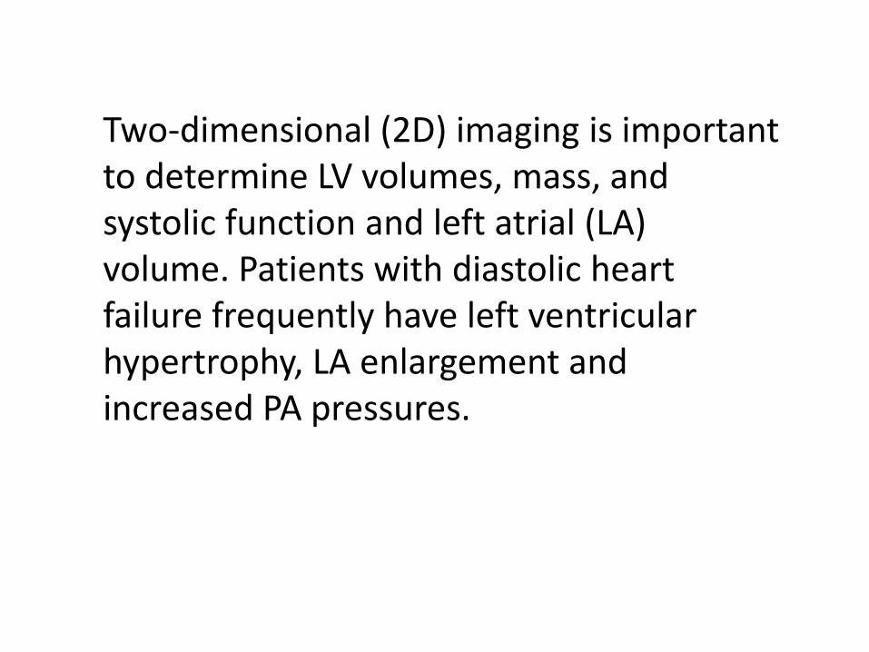
Two-dimensional (2D) imaging is important to determine LV volumes, mass, and systolic function and left atrial (LA) volume. Patients with diastolic heart failure frequently have left ventricular hypertrophy, LA enlargement and increased PA pressures.
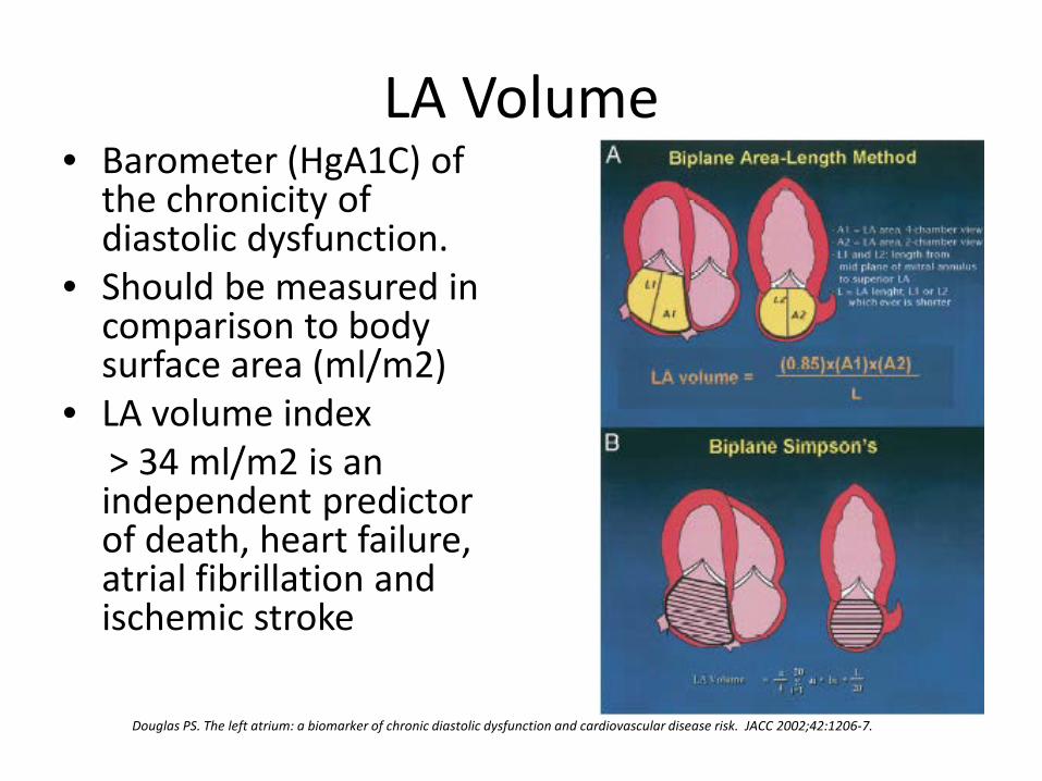
LA Volume • Barometer (HgA1C) of
the chronicity of diastolic dysfunction.
• Should be measured in comparison to body surface area (ml/m2)
• LA volume index > 34 ml/m2 is an
independent predictor of death, heart failure, atrial fibrillation and ischemic stroke
Douglas PS. The left atrium: a biomarker of chronic diastolic dysfunction and cardiovascular disease risk. JACC 2002;42:1206-7.

(Left) End-systolic (maximum) LA volume from an elite athlete with a volume index of 33 mL/m2.
Nagueh S F et al. Eur J Echocardiogr 2009;10:165-193
Reprinted from the Journal of the American Society of Echocardiography 22 (2):107–133, February 2009. With permission from and copyright 2009 by the American Society of Echocardiography.
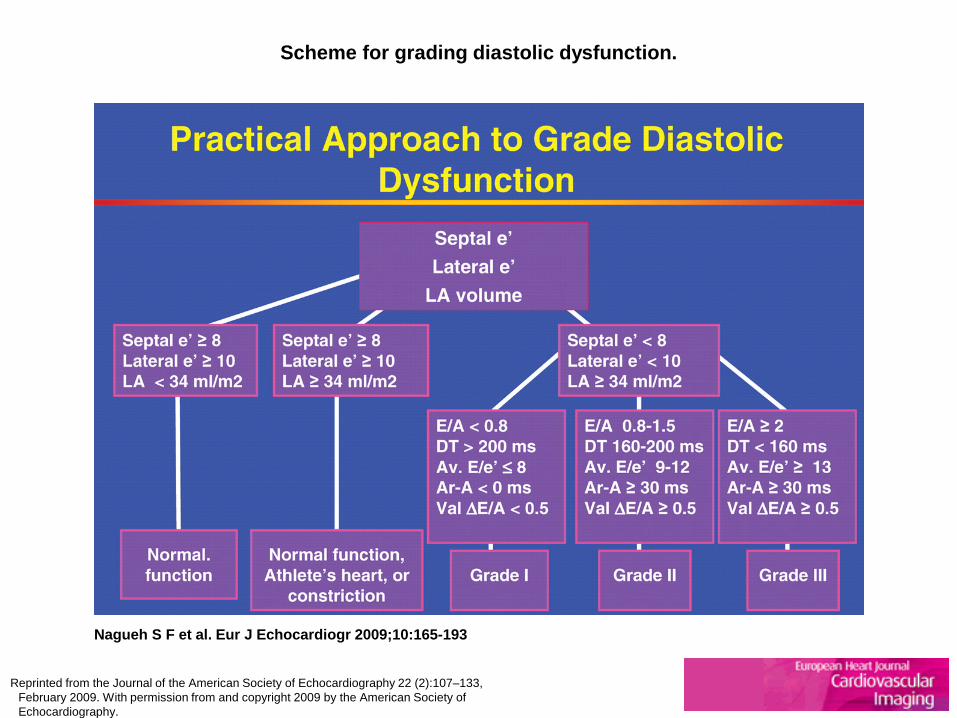
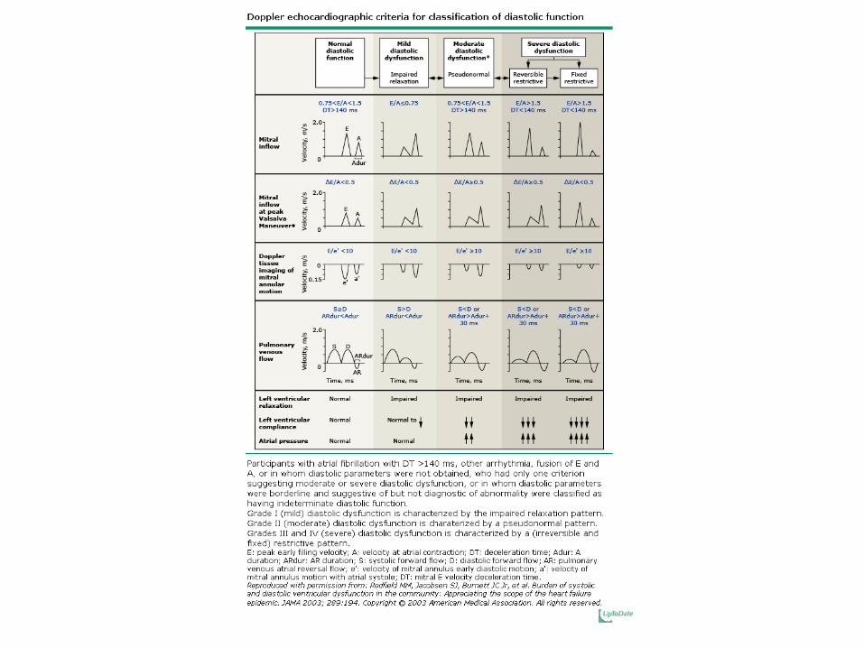
Scheme for grading diastolic dysfunction.
Nagueh S F et al. Eur J Echocardiogr 2009;10:165-193
Reprinted from the Journal of the American Society of Echocardiography 22 (2):107–133, February 2009. With permission from and copyright 2009 by the American Society of Echocardiography.
Copyright © The American College of Cardiology. All rights reserved.
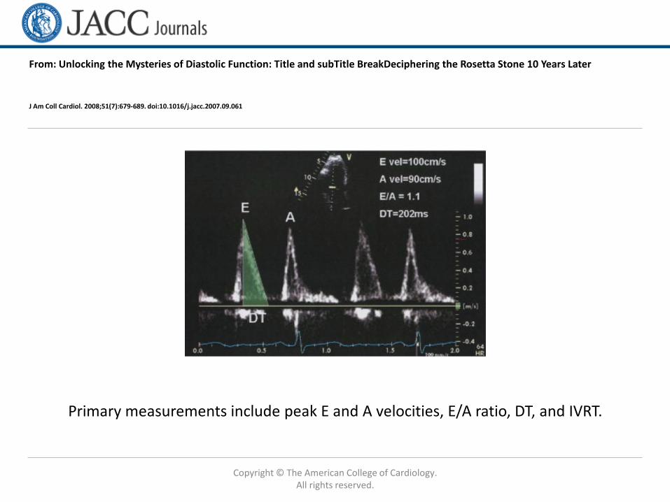
From: Unlocking the Mysteries of Diastolic Function: Title and subTitle BreakDeciphering the Rosetta Stone 10 Years Later
J Am Coll Cardiol. 2008;51(7):679-689. doi:10.1016/j.jacc.2007.09.061
Primary measurements include peak E and A velocities, E/A ratio, DT, and IVRT.
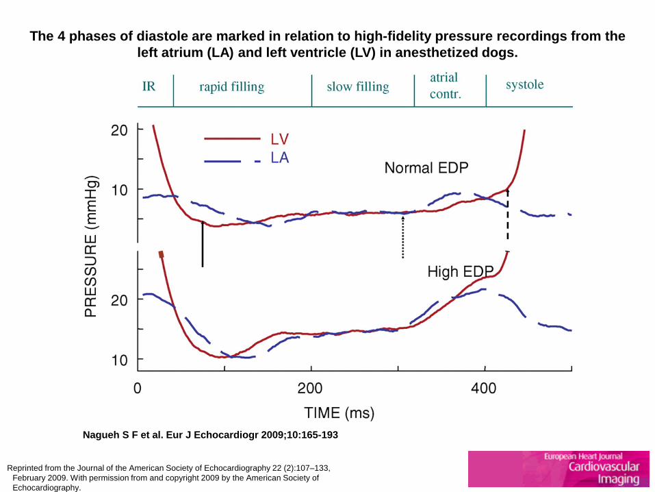
The 4 phases of diastole are marked in relation to high-fidelity pressure recordings from the left atrium (LA) and left ventricle (LV) in anesthetized dogs.
Nagueh S F et al. Eur J Echocardiogr 2009;10:165-193
Reprinted from the Journal of the American Society of Echocardiography 22 (2):107–133, February 2009. With permission from and copyright 2009 by the American Society of Echocardiography.

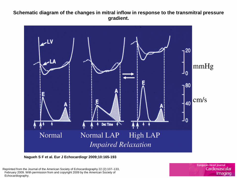
Schematic diagram of the changes in mitral inflow in response to the transmitral pressure gradient.
Nagueh S F et al. Eur J Echocardiogr 2009;10:165-193
Reprinted from the Journal of the American Society of Echocardiography 22 (2):107–133, February 2009. With permission from and copyright 2009 by the American Society of Echocardiography.
Date of download: 1/27/2013
Copyright © The American College of Cardiology. All rights reserved.
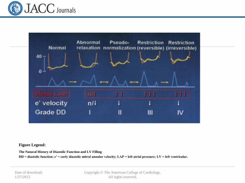
The Natural History of Diastolic Function and LV Filling DD = diastolic function; e′ = early diastolic mitral annular velocity; LAP = left atrial pressure; LV = left ventricular.
Figure Legend:
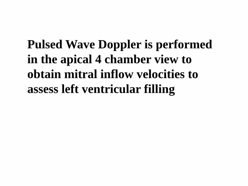
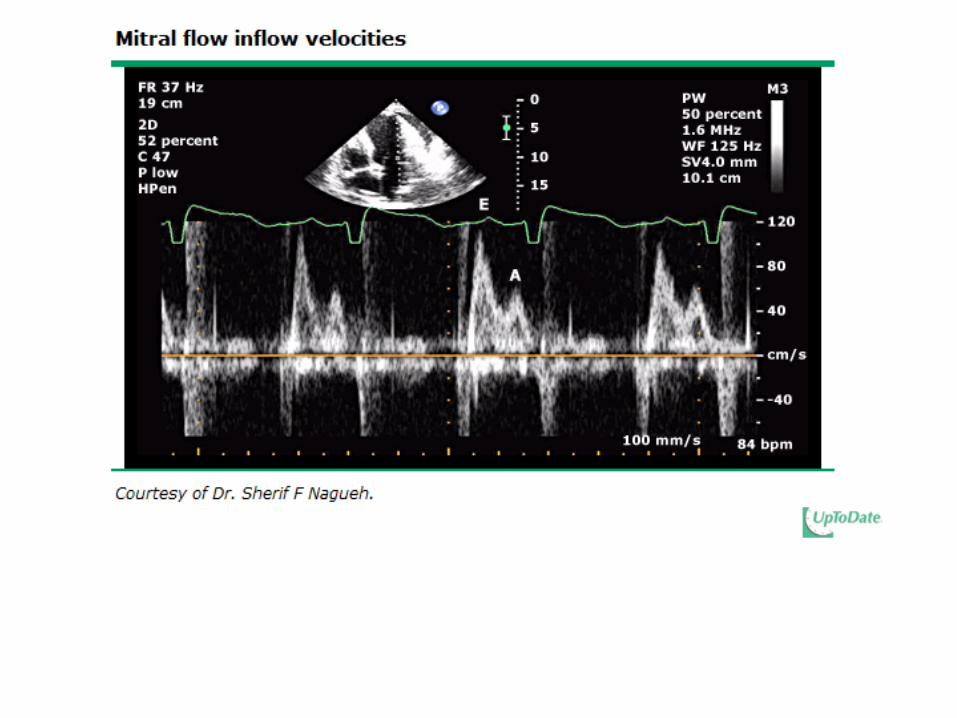
Pulsed Wave Doppler is performed in the apical 4 chamber view to obtain mitral inflow velocities to assess left ventricular filling
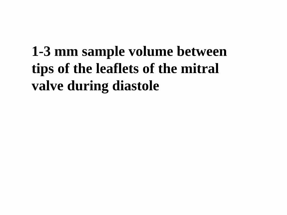
1-3 mm sample volume between tips of the leaflets of the mitral valve during diastole
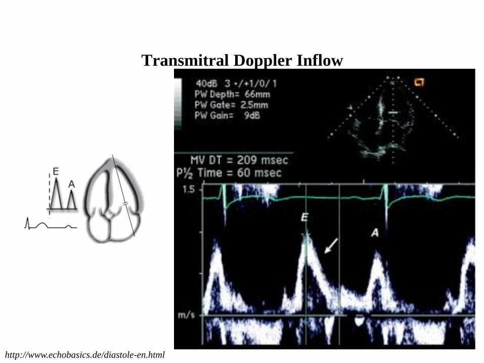
Transmitral Doppler Inflow
http://www.echobasics.de/diastole-en.html
Valsalva maneuver can be performed to
identify pseudonormal filling
The Valsalva maneuver is performed by forceful expiration (about 40 mm Hg) against a closed nose and mouth. The patient must generate a sufficient increase in intrathoracic pressure, and the sonographer needs to maintain the correct sample volume location between the mitral leaflet tips during the maneuver. A decrease of 20 cm/s in mitral peak E velocity is usually considered an adequate effort in patients without restrictive filling.
In cardiac patients, ≥ 50% decrease in E/A ratio
has high specificity for increased filling
pressures, but smaller changes do not always
indicate normal diastolic function
In Systolic Heart Failure, mitral velocities and
time intervals correlate better with filling pressures, and
prognosis than Ejection Fraction
In patients with depressed left ventricular systolic function, mitral inflow velocities can be used as the first step in an algorithm to estimate LV filling pressure

When the E/A ratio is <1 and pulmonary venous flow shows predominant systolic filling, LV filling pressures are usually normal. On the other hand, a restrictive LV filling pattern (transmitral E/A ratio (≥2), IVRT (<70 ms) and DT (<150 ms)) indicates elevated LA pressure. LA pressure elevation can be confirmed with a systolic filling fraction <40 percent.
While most patients with pseudonormal filling have elevated filling pressures, it is preferable to confirm that conclusion by additional Doppler findings. These include the following: change in E/A ratio with Valsalva, E/e' ratio (average >15), E/Vp ratio (≥2.5), and PA systolic pressures (>35 mmHg).
In Diastolic Heart Failure, mitral inflow velocities do not relate well with
LV filling pressures


Mitral Inflow Propagation Velocity (Vp)
http://www.echobasics.de/diastole-en.html
M-mode scan line should be placed in centre
of LV inflow column

Baseline shift so central highest velocity jet is
blue

Color M-mode Vp from a patient with depressed EF and impaired LV relaxation.
Nagueh S F et al. Eur J Echocardiogr 2009;10:165-193
Reprinted from the Journal of the American Society of Echocardiography 22 (2):107–133, February 2009. With permission from and copyright 2009 by the American Society of Echocardiography.
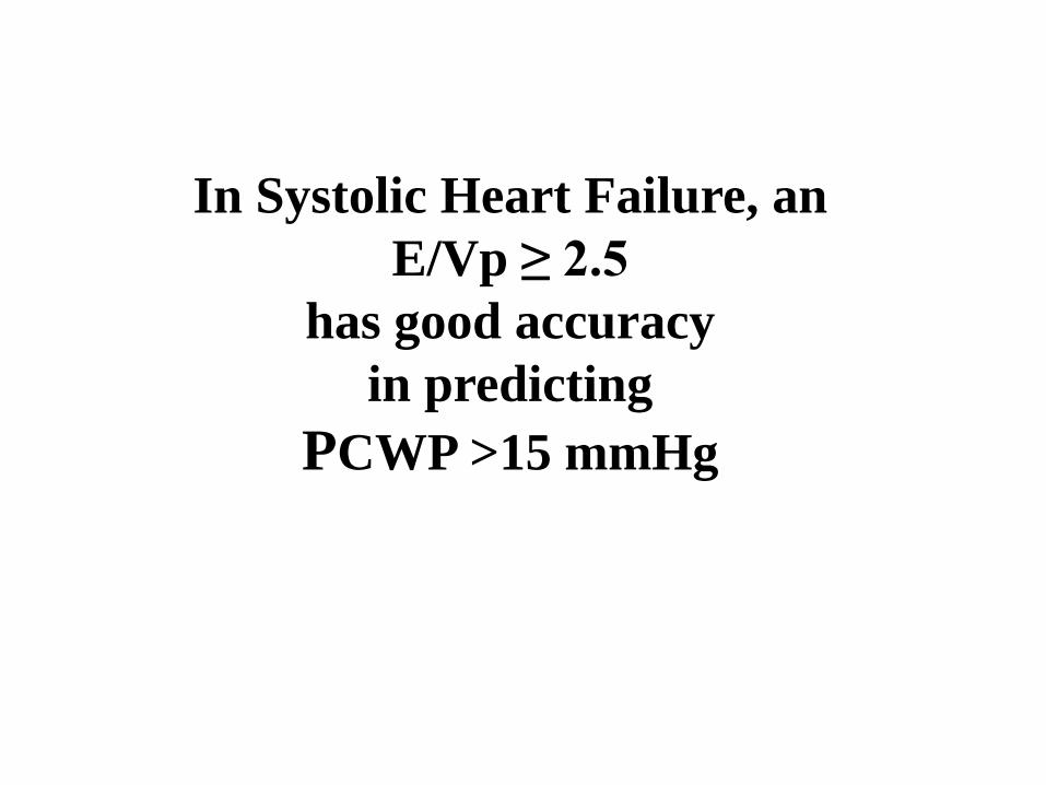
In Systolic Heart Failure, an E/Vp ≥ 2.5
has good accuracy in predicting
PCWP >15 mmHg
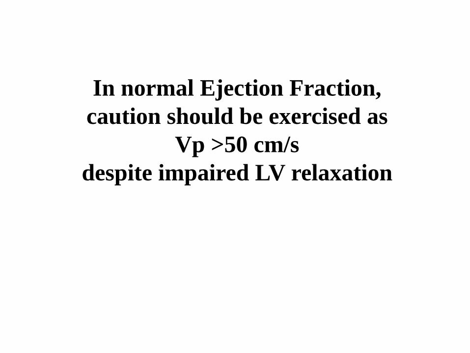
In normal Ejection Fraction, caution should be exercised as
Vp >50 cm/s despite impaired LV relaxation

Recording of mitral inflow at the level of the annulus (left) and pulmonary venous flow (right) from a patient with increased LVEDP. Notice the markedly increased pulmonary venous Ar velocity at 50 cm/s and its prolonged duration at >200 ms in comparison with mitral A (late
diastolic) velocity.
Nagueh S F et al. Eur J Echocardiogr 2009;10:165-193
Reprinted from the Journal of the American Society of Echocardiography 22 (2):107–133, February 2009. With permission from and copyright 2009 by the American Society of Echocardiography.

http://www.echobasics.de/diastole-en.html
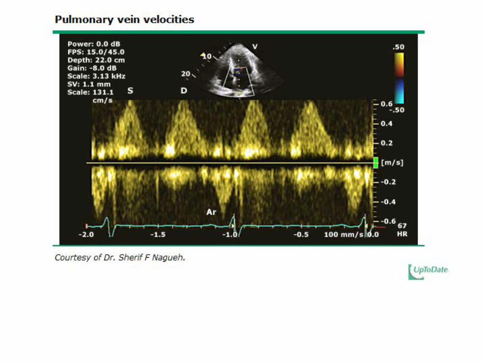
Pulmonary Venous Doppler Flow
PW Doppler of pulmonary venous flow is performed in the apical 4-chamber View. A 2-mm to 3-mm sample volume is placed 0.5 cm into the pulmonary vein for optimal recording of the spectral waveforms.
Measurements include peak S and D velocities, the S/D ratio, systolic filling fraction, and peak Ar velocity in late diastole. Another measurement is the time difference between Ar duration and mitral A-wave duration (Ar -A).
With increased LVEDP, Ar velocity and duration
increase, as well as Ar-A duration
In Systolic Heart Failure, a SFF <40% is associated
with increased LA Pressure
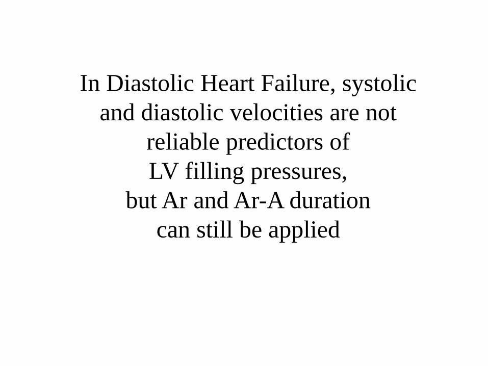
In Diastolic Heart Failure, systolic and diastolic velocities are not
reliable predictors of LV filling pressures,
but Ar and Ar-A duration can still be applied
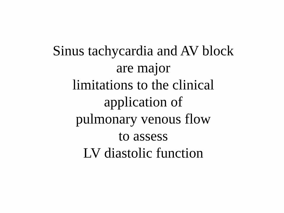
Sinus tachycardia and AV block are major
limitations to the clinical application of
pulmonary venous flow to assess
LV diastolic function
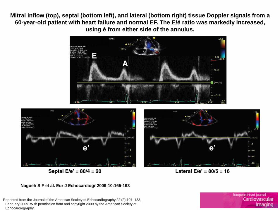
Mitral inflow (top), septal (bottom left), and lateral (bottom right) tissue Doppler signals from a 60-year-old patient with heart failure and normal EF. The E/é ratio was markedly increased,
using é from either side of the annulus.
Nagueh S F et al. Eur J Echocardiogr 2009;10:165-193
Reprinted from the Journal of the American Society of Echocardiography 22 (2):107–133, February 2009. With permission from and copyright 2009 by the American Society of Echocardiography.

Exercise Doppler recordings from a patient with reduced diastolic reserve.
Nagueh S F et al. Eur J Echocardiogr 2009;10:165-193
Reprinted from the Journal of the American Society of Echocardiography 22 (2):107–133, February 2009. With permission from and copyright 2009 by the American Society of Echocardiography.
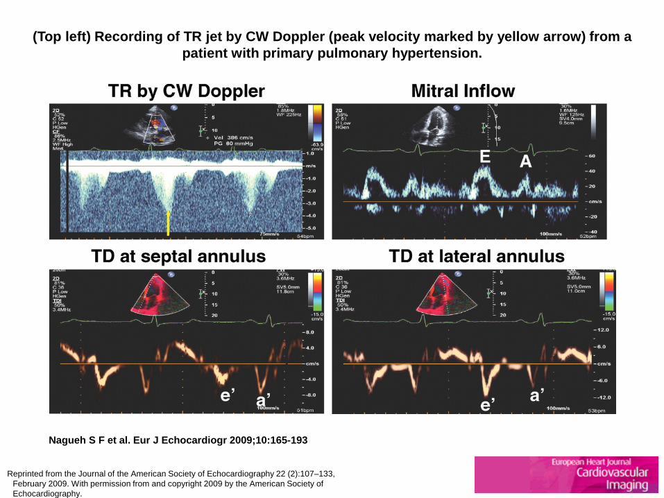
(Top left) Recording of TR jet by CW Doppler (peak velocity marked by yellow arrow) from a patient with primary pulmonary hypertension.
Nagueh S F et al. Eur J Echocardiogr 2009;10:165-193
Reprinted from the Journal of the American Society of Echocardiography 22 (2):107–133, February 2009. With permission from and copyright 2009 by the American Society of Echocardiography.
Special Populations
• Atrial fibrillation • Sinus tachycardia • Hypertrophic cardiomyopathy • Constrictive pericarditis • Restrictive cardiomyopathy • Noncardiac pulmonary hypertension • Mitral Stenosis • Mitral Regurgitation
Copyright © The American College of Cardiology. All rights reserved.
From: Unlocking the Mysteries of Diastolic Function: Title and subTitle BreakDeciphering the Rosetta Stone 10 Years Later
J Am Coll Cardiol. 2008;51(7):679-689. doi:10.1016/j.jacc.2007.09.061
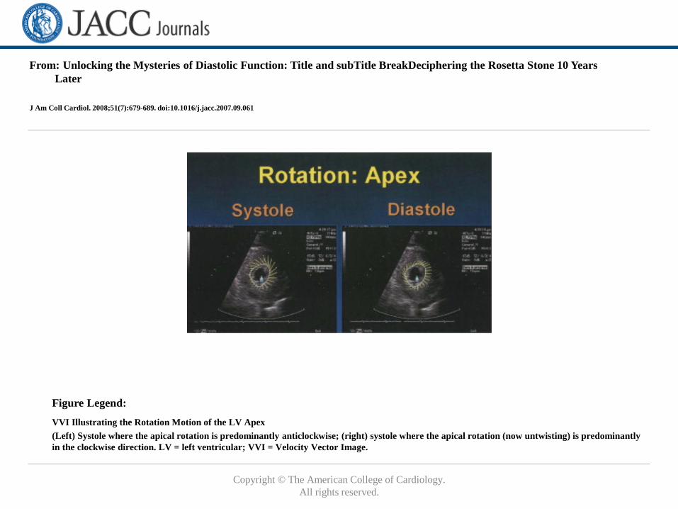
VVI Illustrating the Rotation Motion of the LV Apex (Left) Systole where the apical rotation is predominantly anticlockwise; (right) systole where the apical rotation (now untwisting) is predominantly in the clockwise direction. LV = left ventricular; VVI = Velocity Vector Image.
Figure Legend:
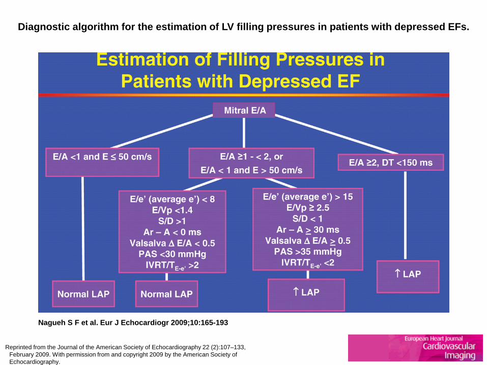
Diagnostic algorithm for the estimation of LV filling pressures in patients with depressed EFs.
Nagueh S F et al. Eur J Echocardiogr 2009;10:165-193
Reprinted from the Journal of the American Society of Echocardiography 22 (2):107–133, February 2009. With permission from and copyright 2009 by the American Society of Echocardiography.
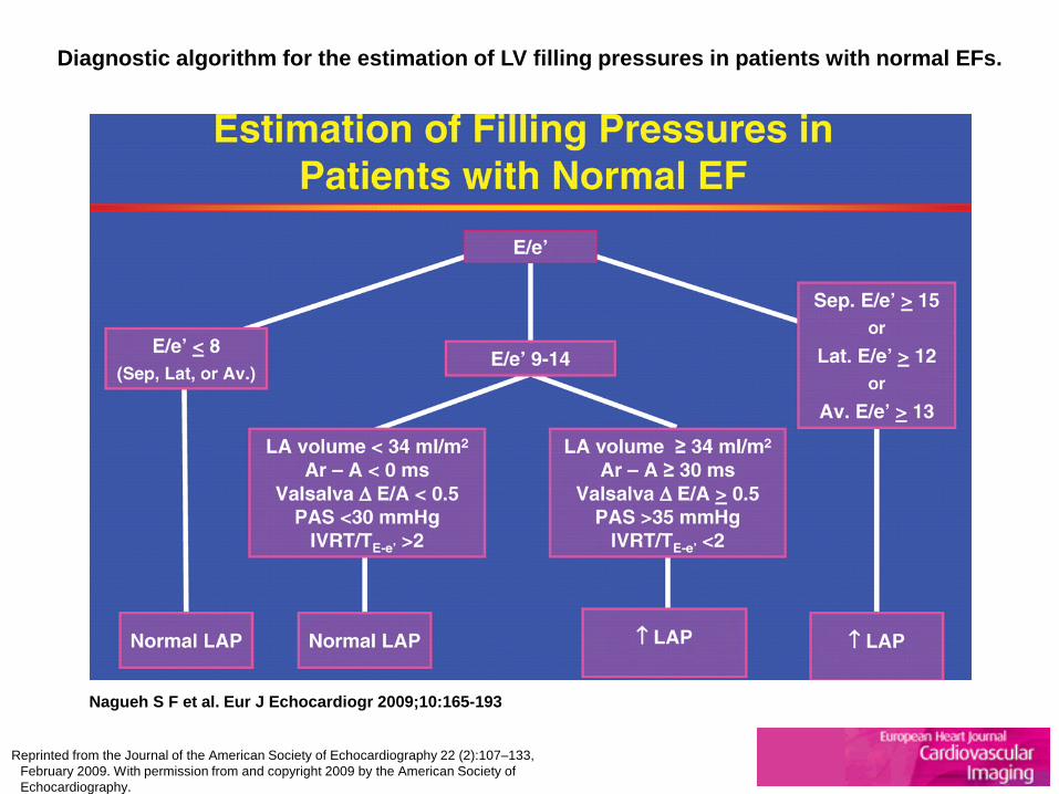
Diagnostic algorithm for the estimation of LV filling pressures in patients with normal EFs.
Nagueh S F et al. Eur J Echocardiogr 2009;10:165-193
Reprinted from the Journal of the American Society of Echocardiography 22 (2):107–133, February 2009. With permission from and copyright 2009 by the American Society of Echocardiography.

Scheme for grading diastolic dysfunction.
Nagueh S F et al. Eur J Echocardiogr 2009;10:165-193
Reprinted from the Journal of the American Society of Echocardiography 22 (2):107–133, February 2009. With permission from and copyright 2009 by the American Society of Echocardiography.
Treatment
Medications proven to be effective in the treatment of HFPEF

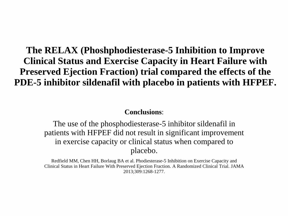
The RELAX (Phoshphodiesterase-5 Inhibition to Improve Clinical Status and Exercise Capacity in Heart Failure with
Preserved Ejection Fraction) trial compared the effects of the PDE-5 inhibitor sildenafil with placebo in patients with HFPEF.
Conclusions:
The use of the phosphodiesterase-5 inhibitor sildenafil in patients with HFPEF did not result in significant improvement
in exercise capacity or clinical status when compared to placebo.
Redfield MM, Chen HH, Borlaug BA et al. Phodiesterase-5 Inhibition on Exercise Capacity and Clinical Status in Heart Failure With Preserved Ejection Fraction. A Randomized Clinical Trial. JAMA
2013;309:1268-1277.
Asymptomatic LV diastolic dysfunction is a predictor of future cardiovascular morbidity. Symptomatic patients with DHF experience morbidities (eg, hospitalization for heart failure) at a rate that is virtually the same as that seen in patients with SHF. Mortality rates in both DHF and SHF are high; published data on differences in mortality rates are conflicting.
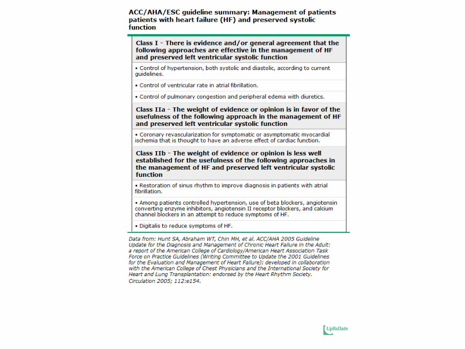
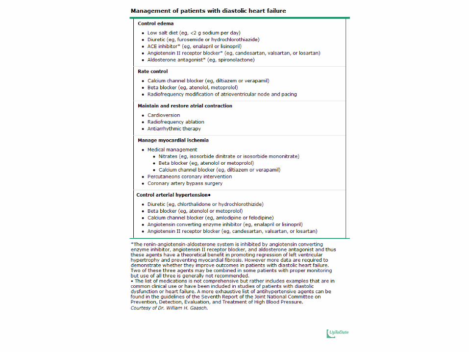
The treatment of DHF remains empiric since trial data are limited. The general principles for treatment of DHF are control of systolic and diastolic hypertension, control of ventricular rate, particularly in patients with atrial fibrillation, control of pulmonary congestion and peripheral edema with diuretics, and coronary revascularization in patients with coronary heart disease with ischemia judged to impair diastolic function.
An important caveat is that the patient who has LV diastolic dysfunction with a small, stiff left ventricular chamber is particularly susceptible to excessive preload reduction, which can lead sequentially to underfilling of the LV, a fall in cardiac output, and hypotension. In patients with severe left ventricular hypertrophy (LVH) due to hypertension or hypertrophic cardiomyopathy, excessive preload reduction can also create subaortic outflow obstruction.
For these reasons, the administration of diuretics or venodilators such as nitrates and dihydropyridine calcium channel blockers must be performed with caution.
Restoration and maintenance of sinus rhythm is preferred when AF occurs in patients with DHF. When this cannot be achieved, rate control becomes important.