ptemasters.com: echo assessment of diastolic function 13 basic and advanced diastolic fx... ·...
TRANSCRIPT

PTEmasters.com: Echo Assessment of Diastolic Function
For questions/reprints: [email protected] Page 1 of 21
Left ventricular (LV) diastolic function can be evaluated invasively and
noninvasively. Invasive measures of diastolic function include the peak instantaneous
rate of LV pressure decline (-dP/dt), the time constant of LV relaxation (tau), and the
stiffness modulus(1). Although echocardiography does not directly measure these
parameters, echocardiography is the most practical routine clinical approach for
evaluating LV diastolic function given clinical and experimental evidence supporting its
use as well as its safety, versatility, and portability(1,2). During this lecture we will
discuss the following metrics of diastolic function: transmitral pulsed-wave Doppler
analysis, pulmonary venous pulsed-wave Doppler analysis, transmitral color m-mode
flow propagation velocity (Vp) and tissue Doppler annular early and late diastolic
velocities.
Transmitral Pulsed-Wave Doppler Analysis of Diastolic Inflow
The midesophageal 4-chamber view is used for Pulsed-wave (PW) Doppler analysis of
mitral inflow velocities to assess left ventricular (LV) filling(1). Color flow imaging may
be helpful for optimal alignment of the Doppler beam, particularly in the setting of LV
dilation(1). Some authors advocate for initially performing CW Doppler prior to PW
Doppler to assess peak E (early diastolic) and A (late diastolic) velocities to ensure that
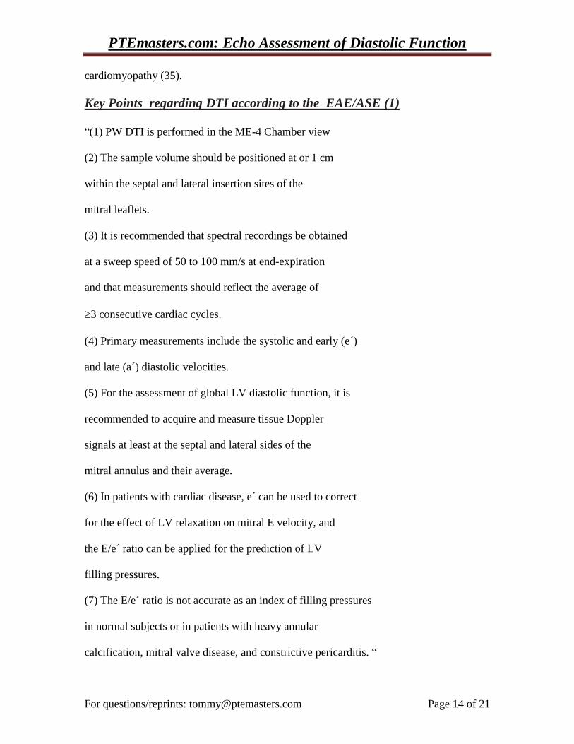
maximal velocities are obtained. Using PW Doppler, from a midesophageal 4-chamber
view a 1-mm to 3-mm sample volume is then placed between the mitral leaflet tips
during diastole to record a crisp spectral Doppler velocity profile (fig1). Spectral gain and
wall filter settings is important to clearly display the onset and cessation of transmitral
inflow. An adequate transmitral spectral Doppler profile may be obtained in most
PTEmasters.com: Echo Assessment of Diastolic Function
For questions/reprints: [email protected] Page 2 of 21
patients. Velocity recordings should initially be obtained at sweep speeds of 25 to 50
mm/s for the evaluation of respiratory variation of flow velocities, as seen in patients
with pulmonary or pericardial disease. If significant variation is not present, the sweep
speed is increased to 100 mm/s, and averaged over 3 consecutive cardiac cycles (3).
The following measurements are made (3):
Peak early filling (E-wave) velocity
Peak late filling (A-wave) velocity
E/A ratio
Deceleration time (DT) of the early filling velocity
Isovolumetric (Isovolemic) relaxation time (IVRT)
Other less common (secondary measurements) include:
A-wave duration (A-dur) (sample volume at annulus)
A-wave velocity time integral (VTI) (sample volume at the annulus)
Total mitral inflow VTI for calculation of the atrial filling fraction (sample
volume at the level of the MV annulus)
The IVRT is obtained from a deep transgastric long axis view by using a CW Doppler
beam in the LV outflow tract to simultaneously display the end of aortic ejection and the
onset of mitral inflow. Age must be considered when defining normal values of mitral
inflow velocities and time intervals. Slightly different normal values may be found in
multiple texts and articles, but the most recent guidelines (1,3) represents the best source
for these values.
Transmitral inflow patterns are primarily recognized based on IVRT, E/A ratio and DT.
These patterns include (figure 2) (3):
Normal (Normal IVRT, E/A >1, normal DT)
Impaired relaxation (Prolonged IVRT, E/A < 1, Prolonged DT)
Pseudonormal (Normal IVRT, E/A and DT look normal)
PTEmasters.com: Echo Assessment of Diastolic Function
For questions/reprints: [email protected] Page 3 of 21
Restrictive (Short IVRT, E/A >>1, Decreased (short) DT)
The distinction between pseudonormal and normal diastolic function requires measuring
other parameters of LV diastolic function, as these may not be distinguished by
transmitral inflow patterns alone.
There are multiple determinants of LV diastolic function and transmitral inflow.
Although this is an oversimplification, two parameters help determine transmitral filling:
1. Active LV relaxation, and
2. LV compliance (which determines LA pressure)
LV relaxation is an active energy dependent process. With the onset of diastolic
dysfunction, relaxation is impaired or delayed and an impaired relaxation pattern
develops with E/A <1, DT prolonged (Fig 2). As diastolic function worsens LV
relaxation is further impaired and there is a progression of filling patterns as follows:
Impaired relaxation pseudonormalrestrictive. The pseudonormal and restrictive
patterns result because the impaired relaxation (which tends to prolong IVRT, and DT
and decrease E/A) is overwhelmed by increased left atrial pressures, which tend to
shorten IVRT, and DT and increase E/A. With the initial impaired relaxation pattern the
LV fails to generate adequate diastolic suction and therefore the IVRT is prolonged
(takes a longer time to pop open the MV) and after the MV opens the decreased suction
generated causes a low peak E velocity and a prolonged DT (takes a long time for early
filling due to decreased suction from impairment of the active energy dependant
relaxation). Given the decreased volume of flow during early filling, the LA is relatively
full at the time of LA contraction and thus the A wave is larger (larger Peak A wave
velocity, A wave VTI, prolonged A wave duration) relative to the E wave (E/A<1). With
PTEmasters.com: Echo Assessment of Diastolic Function
For questions/reprints: [email protected] Page 4 of 21
continued worsening of diastolic function LV compliance decreases. Active relaxation is
still impaired, but the decreased LV compliance results in elevated left atrial pressures.
The pseudonormal and restrictive patterns result because the impaired relaxation (which
tends to prolong IVRT, and DT and decrease E/A) is overwhelmed by increased left atrial
pressures, which tend to shorten IVRT, and DT and increase E/A. (figure 2).
Transmitral inflow velocities are influenced by loading conditions, and rhythm
disturbances including: sinus tachycardia, conduction system disease, and arrhythmias.
Sinus tachycardia and first-degree AV block may cause partial or complete fusion of the
E and A waves.
Key Points regarding Transmitral PW Doppler according to EAE/ASE (1)
“(1) PW Doppler is performed in the ME 4-chamber view.
(2) A 1-mm to 3-mm sample volume is then placed between the mitral leaflet tips
(3) Primary measurements include peak E and A velocities, E/A ratio, DT, and IVRT.
(4) Mitral inflow patterns include normal, impaired LV relaxation, Pseudonormal, and
restrictive LV filling (fig 2).
(5) In patients with dilated cardiomyopathies, filling patterns correlate better with filling
pressures, functional class, and prognosis than LV EF.
(6) In patients with coronary artery disease and those with hypertrophic cardiomyopathy
in whom the LV EFs are ≥ 50%, mitral velocities correlate poorly with hemodynamics.”
PTEmasters.com: Echo Assessment of Diastolic Function
For questions/reprints: [email protected] Page 5 of 21
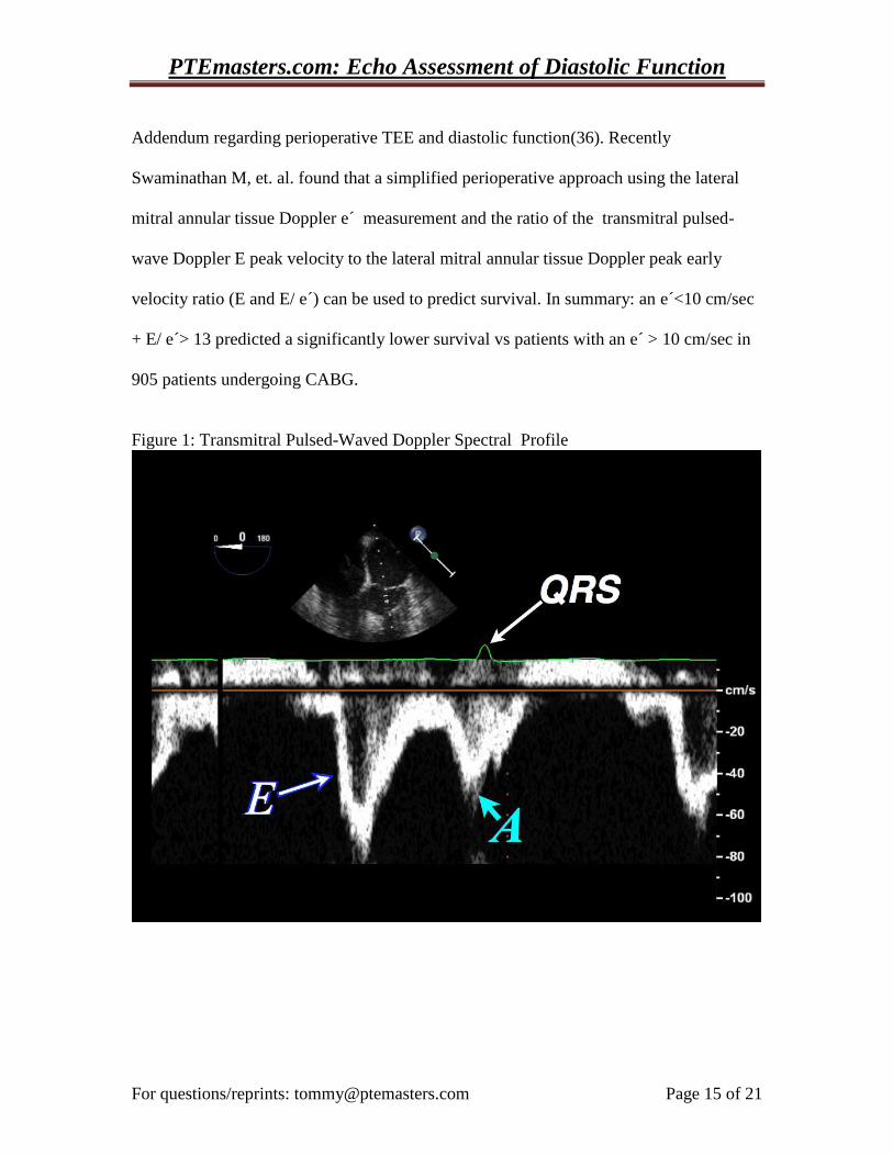
The pulsed-wave Doppler pulmonary venous flow waves are identified as follows:
Systolic wave (S-wave),
Diastolic wave (D-wave)
Atrial wave (PV A-wave) = Atrial reversal wave (PV AR-wave)
Note: A = atrial contraction, S = systole, D = diastole.
If one were to look for the corresponding left atrial pressure (LAP) tracing
components you will notice that anything that increases LA pressure will
decrease flow through the pulmonary veins to the LA.
Conversely, anything that decreases LAP will increase flow to the LA.
Below is a list of the LAP adjacent to its corresponding pulmonary venous
flow (PV) wave (figure 4):
LAP A-wavePV A-wave
LAP X-descentPV S-wave
LAP V-wavePV decline in velocity between S and D waves
LAP Y-descentPV D-wave
Notice as LAP increases, flow into the LA decreases and in some cases
reverses (A wave = AR wave = Atrial Reversal wave).
Notice there are two components to the pulmonary venous S wave: S1 and S2.
PTEmasters.com: Echo Assessment of Diastolic Function
For questions/reprints: [email protected] Page 6 of 21
The following are factors that influence the maximum velocity and timing of these two
waves:
S1: 1. Atrial relaxation in early systole
S2: 1. Right ventricular stroke volume
2. Left Atrial Compliance
3. Descent of the mitral valve annulus which lowers LA pressure The following cardiac disorders result in changes to the pulmonary venous flow pulsed-
wave Doppler spectral profile:
Elevated left atrial pressure (LAP) from decreased LV compliance as might be seen with
pseudonormal or restrictive diastolic dysfunction: S < D, PV A-wave duration >
transmitral A-wave duration (AR duration – A duration > 30 ms).
Mitral insufficiencyblunting or reversal of S wave (reversalsevere MR)
Large PV A-wave is seen with: Mitral Stenosis (MS) and complete heart
block (CHB). There are no valves in the pulmonary veins, so when the left
atrium contracts there will be forward flow into the left ventricle and
retrograde flow into the pulmonary veins creating the pulmonary venous A-
wave (PV A-wave = Atrial Reversal wave = AR wave). With MS there is
obstruction to forward flow through the stenotic mitral valve and therefore a
PTEmasters.com: Echo Assessment of Diastolic Function
For questions/reprints: [email protected] Page 7 of 21
predominance of retrograde pulmonary venous flow with atrial contraction.
CHB can be thought of as the worst MS ever, as the valve is closed when
atrial contraction occurs resulting in a large PV A-wave from retrograde
pulmonary venous flow. Decreased LV compliance (pseudonormal and
restrictive diastolic dysfunction) also may cause a large PV A-wave. With
restrictive diastolic dysfunction there is sometimes not a large A-wave and
this is thought to be because there is left atrial mechanical failure and the
atrium no longer has the ability to generate significant forward or retrograde
flow.
Key Points regarding PV PW Doppler according to EAE/ASE (1):
“(1) PW Doppler of pulmonary venous flow is performed in the ME 4-chamber view.
(2) A 2-mm to 3-mm sample volume is placed .0.5 cm into the pulmonary vein for
optimal recording of the spectral waveforms.
(3) Measurements include peak S and D velocities, the S/D ratio, systolic filling fraction,
and peak Ar velocity in late diastole. Another measurement is the time difference
between Ar duration and mitral A-wave duration (Ar - A).
(4) With increased LVEDP, Ar velocity and duration increase, as well as the (Ar – A)
duration.
(5) In patients with depressed EFs, reduced systolic filling fractions (< 40%) are related
to decreased LA compliance and increased mean LA pressure.”
PTEmasters.com: Echo Assessment of Diastolic Function
For questions/reprints: [email protected] Page 8 of 21
Color M-Mode Flow Propagation Velocity (Vp)
In the perioperative setting the Vp slope method(1,4,5) appears to have the least
variability(6). Acquisition via transesophageal echocardiography (TEE) is performed
with the ME 4-chamber view and with transthoracic echocardiography (TTE) it is
performed with the apical 4-chamber view. In both, color flow Doppler with a narrow
sector angle and gain adjusted to avoid noise is utilized with an M-mode scan line placed
through the center of the LV inflow column from the mitral valve to the LV apex (1,3).
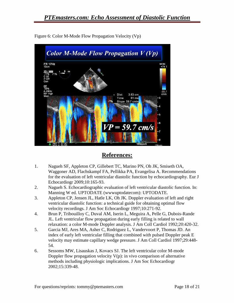
The color scale baseline is adjusted so that the central highest velocity jet is blue. Vp is
measured as the slope of the first aliasing velocity during early transmitral filling as
measured from the mitral valve plane to 4 cm distally into the LV cavity(1,5).
Alternatively the slope of the transition from no color to color can be measured(4).
Normal Vp is 45-50 cm/sec (1,5,7). Similar to the pulse-wave Doppler transmitral inflow
velocities, there is an early wave and a late atrial contraction wave (1). With normal
diastolic function, the early filling wave propagates rapidly toward the apex and is driven
by the pressure gradient from the LV base to apex(1,8). This suction force results from
energy-dependant active LV relaxation. With diastolic dysfunction, from ischemia or
heart failure, there is slowing of mitral-to-apical flow propagation consistent with a
reduction of apical suction (1,4,9,10). However, in clinical practice evaluation and
interpretation of intraventricular filling is complicated by the multitude of variables that
determine intraventricular flow(1). Despite the multiple variables affecting flow, the
slowing of mitral-to-apical flow propagation by color M-mode Doppler has proved to be
a semiquantitative marker of LV diastolic dysfunction(1). In addition, the ratio of the
PTEmasters.com: Echo Assessment of Diastolic Function
For questions/reprints: [email protected] Page 9 of 21
peak early transmitral inflow velocity (E) to Vp (E/Vp) can be used to predict LV filling
pressures (1,5). In patients with decreased systolic function (decreased LVEF) an E/Vp
2.5 predicts a PCWP 15 (1,11). However, in patients with normal systolic function
(normal LVEF) LV filling pressures can not be predicted by E/Vp (11). Also patients
with elevated filling pressures but a normal LVEF, and normal LV volumes can have an
erroneously normal Vp (12) (11,13,14). In addition, preload has been shown to have a
positive influence on Vp in patients with normal and depressed LVEF(1,13,15).
Key Points regarding Vp according to the EAE/ASE (1)
“1. Acquisition is performed in the 4-chamber view,
using color flow Doppler imaging.
2. The M-mode scan line is placed through the center of the
LV inflow blood column from the mitral valve to the
apex, with baseline shift to the color scale so
the central highest velocity jet is blue.
3. Vp is measured as the slope of the first aliasing velocity
during early filling, measured from the mitral valve
plane to 4 cm distally into the LV cavity, or the slope
of the transition from no color to color.
4. Vp 45-50 cm/s is considered normal.
5. In most patients with depressed EFs, Vp is reduced, and
should other Doppler indices appear inconclusive, an
E/Vp ratio 2.5 predicts PCWP 15 mm Hg with reasonable
accuracy.
6. Patients with normal LV volumes and EFs but elevated
filling pressures can have misleadingly normal Vp.”
PTEmasters.com: Echo Assessment of Diastolic Function
For questions/reprints: [email protected] Page 10 of 21
Mitral Annular Tissue Doppler Early (Em) and Late (Am) Diastolic Velocities (1,16):
Pulse wave (PW) Doppler tissue imaging (DTI) is performed with TEE in the ME-4-
chamber view and with TTE in the apical views, which allow acquisition of mitral
annular velocities (1,17). The sample volume should be placed at or 1 cm within the
septal and lateral insertion sites of the mitral leaflets and adjusted as necessary (usually 5-
10 mm) to cover the longitudinal excursion of the mitral annulus in both systole and
diastole (1). DTI velocities have higher amplitude and lower peak velocities when
compared with transmitral inflow velocities. Spectral gain settings can be manually
optimized for DTI, but most current ultraound systems have tissue Doppler presets for the
proper velocity scale and Doppler wall filter settings (1). Usually the velocity scale
should be set at about 10-20 cm/s above the zero-velocity baseline (1). Given the angle
dependance of all Doppler measurements, minimal angulation (< 20 degrees) should be
present between the ultrasound beam and the place of cardiac motion (16). Regardless of
the 2D image quality, DTI waveforms can be obtained in nearly all patients (>95%). The
recommended sweep speed is 50-100 mm/s at end expiration and measurements should
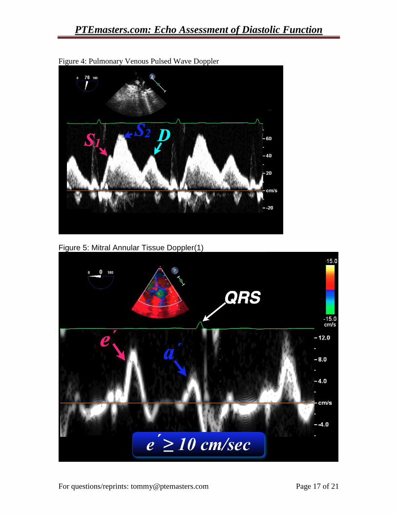
reflect the average of 3 cardiac cycles (1). Primary measurements included systolic (S),
early diastolic (e), and late diastolic velocities (a) (figure 5) (18). Early diastolic annular
tissue velocity has been expressed as Ea, Em, E and e’, in this syllabus we will use e and
E (1). Peak velocities alone are all that needs to be measured, as E deceleration time,
acceleration rates and deceleration rates, do not contain incremental information and need
not be performed (1,19).
E has been shown to have a significant association with LV relaxation in human
PTEmasters.com: Echo Assessment of Diastolic Function
For questions/reprints: [email protected] Page 11 of 21
and animal studies (1,20-24). E is related to LV diastolic properties, such as elastic recoil
and relaxation, regardless of filling pressures or systolic function but E is also influenced
by systolic function, preload, and LV minimal pressure (1,16,25). E changes in the same
direction as preload in patients with normal diastolic function(16). This effect is less
pronounced in ventricles with impaired relaxation where E remains decreased regardless
of changes in preload (16,21,26,27). Thus E is relatively preload independent in sick
patients, (those with significant diastolic dysfunction) including most of the patients
presenting for cardiac surgery.
The time interval between the QRS complex and the E onset is prolonged with
impaired LV relaxation and can provide incremental information in special patient
populations (1). Given the influence of regional function on tissue velocities and
intervals, it is recommended to acquire and measure tissue Doppler signals at least at the
septal and lateral sides of the mitral annulus and calculate their average, for assessment of
global LV diastolic function (1,2,11,28).
Once transmitral inflow PW flow, annular velocities and time intervals are
acquired, it is possible to compute additional time intervals and ratios (1,16). Important
ratios include: E/e, E/A and IVRT/TE-e. The ratio E/e, has been shown to help
estimate LV filling pressures in patients with LV diastolic dysfunction (18). An E/e >
12-15 is consistent with elevated LV filling pressures (2,18). In addition, E/e has been
shown to be a marker of severe cardiac disease. In a recent study of 205 patients an E/e
ratio 8 was shown to be associated with increased intensive care unit length of stay
(ICU-LOS) P = 0.037) and need for inotropic support (P = 0.002) (29). These results
were seen after making adjustments to account for other predictors (female gender,
PTEmasters.com: Echo Assessment of Diastolic Function
For questions/reprints: [email protected] Page 12 of 21
hypotension, diabetes, history of myocardial infarction, emergency surgery, renal failure,
procedure type, and length of aortic cross-clamp time) thereby implicating E/e as a
serious prognostic indicator (29,30). The TE-e interval is the time interval between the
QRS complex and the onset of the mitral E velocity subtracted from the time interval
between the QRS complex and the e onset (1). The TE-e interval is prolonged with
diastolic dysfunction, and animal and human studies have shown it to be strongly
dependent on the time constant of LV relaxation (tau) and minimal LV pressure
(1,31,32). Technically, it is essential to match the RR intervals for measuring both time
intervals (time to E and time to e) and to optimize the Doppler gain and filter settings,
because higher gain and filter settings interfere with correct identification of the onset of
e(1).
The main hemodynamic determinants of a include: LA systolic function and LVEDP. An
increase in LA contractility leads to an increase in a, and an increase in LVEDP leads to
a decrease in a(1,19). Normal values for DTI-derived velocities are influenced by age,
similar to other indices of LV diastolic function, but an but an e <8 cm/sec is generally
considered low(1,2). With age e decreases, a increases and E/e increases(1,33).
Clinical Application of DTI(1,16):
DTI mitral annular velocities assist in the evaluation of LV relaxation, and E/e can be
used to estimate LV filling pressures (1,2). Reliable conclusions require consideration of
multiple factors such as patient age, coexisting cardiovascular disease and other
echocardiographic abnormalities. Thus e and E/e should not be used in isolation. It is
PTEmasters.com: Echo Assessment of Diastolic Function
For questions/reprints: [email protected] Page 13 of 21
also important to use the average of e obtained from the septal and lateral sides of the
mitral annulus over several cardiac cycles. Skubas et al. (16) suggest utilizing the lateral
mitral annulus e in the E/e ratio for estimating filling pressures because the lateral mitral
annulus is rarely involved in ischemic disease and e measurements at this location will
usually reflect LV relaxation. An E/e < 8 indicates normal filling pressures and E/e >
12-15 indicates elevated filling pressures. The mean pulmonary capillary wedge pressure
can be estimated by the following formula: mean PCWP = (1.3 x E/e) + 2 (1,16).
Technical limitations to DTI include factors such as angle dependence, proper sample
size, gain, and Doppler filter settings. In addition, there are a number of clinical settings
in which e and E/e are misleading. In normal subjects e velocity is positively related to
preload and E/e can not be used to estimate filling pressures (1). E is also significantly
reduced in patients with significant mitral annular calcification, surgical rings, mitral
stenosis and prosthetic mitral valves(1). E is increased in patients with moderate to
severe MR and normal LV relaxation due to increased flow across the MV. E/e should
not be used in these patients, but the isovolumetric relaxation time to TE-e ratio
(IVRT/TE-e ) can be applied (an IVRT/ TE-e <2 is consistent with increased filling
pressures) (1,31,34). Patients with constrictive pericarditis usually have elevated e due
to preserved LV longitudinal expansion compensating for limited lateral and
anteroposterior diastolic excursion. Lateral e may be less than septal e in theis condtion
and the E/e should not be used to estimate filling pressures(1). However, a normal e in
the setting of restrictive transmitral inflow velocities can help distinguish constrictive
pericarditis from restrictive diastolic dysfunction due to an infiltrative restrictive
PTEmasters.com: Echo Assessment of Diastolic Function
For questions/reprints: [email protected] Page 14 of 21
cardiomyopathy (35).
Key Points regarding DTI according to the EAE/ASE (1)
“(1) PW DTI is performed in the ME-4 Chamber view
(2) The sample volume should be positioned at or 1 cm
within the septal and lateral insertion sites of the
mitral leaflets.
(3) It is recommended that spectral recordings be obtained
at a sweep speed of 50 to 100 mm/s at end-expiration
and that measurements should reflect the average of
3 consecutive cardiac cycles.
(4) Primary measurements include the systolic and early (e´)
and late (a´) diastolic velocities.
(5) For the assessment of global LV diastolic function, it is
recommended to acquire and measure tissue Doppler
signals at least at the septal and lateral sides of the
mitral annulus and their average.
(6) In patients with cardiac disease, e´ can be used to correct
for the effect of LV relaxation on mitral E velocity, and
the E/e´ ratio can be applied for the prediction of LV
filling pressures.
(7) The E/e´ ratio is not accurate as an index of filling pressures
in normal subjects or in patients with heavy annular
calcification, mitral valve disease, and constrictive pericarditis. “
PTEmasters.com: Echo Assessment of Diastolic Function
For questions/reprints: [email protected] Page 15 of 21
Addendum regarding perioperative TEE and diastolic function(36). Recently
Swaminathan M, et. al. found that a simplified perioperative approach using the lateral
mitral annular tissue Doppler e´ measurement and the ratio of the transmitral pulsed-
wave Doppler E peak velocity to the lateral mitral annular tissue Doppler peak early
velocity ratio (E and E/ e´) can be used to predict survival. In summary: an e´<10 cm/sec
+ E/ e´> 13 predicted a significantly lower survival vs patients with an e´ > 10 cm/sec in
905 patients undergoing CABG.
Figure 1: Transmitral Pulsed-Waved Doppler Spectral Profile

PTEmasters.com: Echo Assessment of Diastolic Function
For questions/reprints: [email protected] Page 16 of 21
Figure 2: Transmitral Pulsed Wave Doppler Profiles
Figure 3 Pulmonary Venous Flow
PTEmasters.com: Echo Assessment of Diastolic Function
For questions/reprints: [email protected] Page 17 of 21
Figure 4: Pulmonary Venous Pulsed Wave Doppler
Figure 5: Mitral Annular Tissue Doppler(1)
PTEmasters.com: Echo Assessment of Diastolic Function
For questions/reprints: [email protected] Page 18 of 21
Figure 6: Color M-Mode Flow Propagation Velocity (Vp)
References:
1. Nagueh SF, Appleton CP, Gillebert TC, Marino PN, Oh JK, Smiseth OA,
Waggoner AD, Flachskampf FA, Pellikka PA, Evangelisa A. Recommendations
for the evaluation of left ventricular diastolic function by echocardiography. Eur J
Echocardiogr 2009;10:165-93.
2. Nagueh S. Echocardiographic evaluation of left ventricular diastolic function. In:
Manning W ed. UPTODATE (wwwuptodatecom): UPTODATE.
3. Appleton CP, Jensen JL, Hatle LK, Oh JK. Doppler evaluation of left and right
ventricular diastolic function: a technical guide for obtaining optimal flow
velocity recordings. J Am Soc Echocardiogr 1997;10:271-92.
4. Brun P, Tribouilloy C, Duval AM, Iserin L, Meguira A, Pelle G, Dubois-Rande
JL. Left ventricular flow propagation during early filling is related to wall
relaxation: a color M-mode Doppler analysis. J Am Coll Cardiol 1992;20:420-32.
5. Garcia MJ, Ares MA, Asher C, Rodriguez L, Vandervoort P, Thomas JD. An
index of early left ventricular filling that combined with pulsed Doppler peak E
velocity may estimate capillary wedge pressure. J Am Coll Cardiol 1997;29:448-
54.
6. Sessoms MW, Lisauskas J, Kovacs SJ. The left ventricular color M-mode
Doppler flow propagation velocity V(p): in vivo comparison of alternative
methods including physiologic implications. J Am Soc Echocardiogr
2002;15:339-48.
PTEmasters.com: Echo Assessment of Diastolic Function
For questions/reprints: [email protected] Page 19 of 21
7. Takatsuji H, Mikami T, Urasawa K, Teranishi J, Onozuka H, Takagi C, Makita Y,
Matsuo H, Kusuoka H, Kitabatake A. A new approach for evaluation of left
ventricular diastolic function: spatial and temporal analysis of left ventricular
filling flow propagation by color M-mode Doppler echocardiography. J Am Coll
Cardiol 1996;27:365-71.
8. Courtois M, Kovacs SJ, Ludbrook PA. Physiological early diastolic
intraventricular pressure gradient is lost during acute myocardial ischemia.
Circulation 1990;81:1688-96.
9. Steine K, Stugaard M, Smiseth OA. Mechanisms of retarded apical filling in acute
ischemic left ventricular failure. Circulation 1999;99:2048-54.
10. Stugaard M, Smiseth OA, Risoe C, Ihlen H. Intraventricular early diastolic filling
during acute myocardial ischemia, assessment by multigated color m-mode
Doppler echocardiography. Circulation 1993;88:2705-13.
11. Rivas-Gotz C, Manolios M, Thohan V, Nagueh SF. Impact of left ventricular
ejection fraction on estimation of left ventricular filling pressures using tissue
Doppler and flow propagation velocity. Am J Cardiol 2003;91:780-4.
12. Ohte N, Narita H, Akita S, Kurokawa K, Hayano J, Kimura G. Striking effect of
left ventricular systolic performance on propagation velocity of left ventricular
early diastolic filling flow. J Am Soc Echocardiogr 2001;14:1070-4.
13. Troughton RW, Prior DL, Frampton CM, Nash PJ, Pereira JJ, Martin M, Fogarty
A, Morehead AJ, Starling RC, Young JB, Thomas JD, Lauer MS, Klein AL.
Usefulness of tissue doppler and color M-mode indexes of left ventricular
diastolic function in predicting outcomes in systolic left ventricular heart failure
(from the ADEPT study). Am J Cardiol 2005;96:257-62.
14. Rovner A, de las Fuentes L, Waggoner AD, Memon N, Chohan R, Davila-Roman
VG. Characterization of left ventricular diastolic function in hypertension by use
of Doppler tissue imaging and color M-mode techniques. J Am Soc Echocardiogr
2006;19:872-9.
15. Graham RJ, Gelman JS, Donelan L, Mottram PM, Peverill RE. Effect of preload
reduction by haemodialysis on new indices of diastolic function. Clin Sci (Lond)
2003;105:499-506.
16. Skubas N. Intraoperative Doppler tissue imaging is a valuable addition to cardiac
anesthesiologists' armamentarium: a core review. Anesth Analg 2009;108:48-66.
17. Waggoner AD, Bierig SM. Tissue Doppler imaging: a useful echocardiographic
method for the cardiac sonographer to assess systolic and diastolic ventricular
function. J Am Soc Echocardiogr 2001;14:1143-52.
18. Nagueh SF, Middleton KJ, Kopelen HA, Zoghbi WA, Quinones MA. Doppler
tissue imaging: a noninvasive technique for evaluation of left ventricular
relaxation and estimation of filling pressures. J Am Coll Cardiol 1997;30:1527-
33.
19. Nagueh SF, Sun H, Kopelen HA, Middleton KJ, Khoury DS. Hemodynamic
determinants of the mitral annulus diastolic velocities by tissue Doppler. J Am
Coll Cardiol 2001;37:278-85.
20. Vinereanu D, Florescu N, Sculthorpe N, Tweddel AC, Stephens MR, Fraser AG.
Differentiation between pathologic and physiologic left ventricular hypertrophy
by tissue Doppler assessment of long-axis function in patients with hypertrophic
PTEmasters.com: Echo Assessment of Diastolic Function
For questions/reprints: [email protected] Page 20 of 21
cardiomyopathy or systemic hypertension and in athletes. Am J Cardiol
2001;88:53-8.
21. Ha JW, Oh JK, Ommen SR, Ling LH, Tajik AJ. Diagnostic value of mitral
annular velocity for constrictive pericarditis in the absence of respiratory variation
in mitral inflow velocity. J Am Soc Echocardiogr 2002;15:1468-71.
22. Meluzin J, Spinarova L, Bakala J, Toman J, Krejci J, Hude P, Kara T, Soucek M.
Pulsed Doppler tissue imaging of the velocity of tricuspid annular systolic
motion; a new, rapid, and non-invasive method of evaluating right ventricular
systolic function. Eur Heart J 2001;22:340-8.
23. Alam M, Wardell J, Andersson E, Samad BA, Nordlander R. Characteristics of
mitral and tricuspid annular velocities determined by pulsed wave Doppler tissue
imaging in healthy subjects. J Am Soc Echocardiogr 1999;12:618-28.
24. Nikitin NP, Witte KK, Thackray SD, de Silva R, Clark AL, Cleland JG.
Longitudinal ventricular function: normal values of atrioventricular annular and
myocardial velocities measured with quantitative two-dimensional color Doppler
tissue imaging. J Am Soc Echocardiogr 2003;16:906-21.
25. Ommen SR, Nishimura RA, Appleton CP, Miller FA, Oh JK, Redfield MM, Tajik
AJ. Clinical utility of Doppler echocardiography and tissue Doppler imaging in
the estimation of left ventricular filling pressures: A comparative simultaneous
Doppler-catheterization study. Circulation 2000;102:1788-94.
26. Firstenberg MS, Greenberg NL, Main ML, Drinko JK, Odabashian JA, Thomas
JD, Garcia MJ. Determinants of diastolic myocardial tissue Doppler velocities:
influences of relaxation and preload. J Appl Physiol 2001;90:299-307.
27. Ozdemir K, Altunkeser BB, Gok H, Icli A, Temizhan A. Analysis of the
myocardial velocities in patients with mitral stenosis. J Am Soc Echocardiogr
2002;15:1472-8.
28. Nagueh SF, Rao L, Soto J, Middleton KJ, Khoury DS. Haemodynamic insights
into the effects of ischaemia and cycle length on tissue Doppler-derived mitral
annulus diastolic velocities. Clin Sci (Lond) 2004;106:147-54.
29. Groban L, Sanders DM, Houle TT, Antonio BL, Ntuen EC, Zvara DA, Kon ND,
Kincaid EH. Prognostic value of tissue Doppler-Derived E/e' on early morbid
events after cardiac surgery. Echocardiography;27:131-8.
30. Groban L, Kitzman DW. Diastolic function: a barometer for cardiovascular risk?
Anesthesiology;112:1303-6.
31. Rivas-Gotz C, Khoury DS, Manolios M, Rao L, Kopelen HA, Nagueh SF. Time
interval between onset of mitral inflow and onset of early diastolic velocity by
tissue Doppler: a novel index of left ventricular relaxation: experimental studies
and clinical application. J Am Coll Cardiol 2003;42:1463-70.
32. Hasegawa H, Little WC, Ohno M, Brucks S, Morimoto A, Cheng HJ, Cheng CP.
Diastolic mitral annular velocity during the development of heart failure. J Am
Coll Cardiol 2003;41:1590-7.
33. De Sutter J, De Backer J, Van de Veire N, Velghe A, De Buyzere M, Gillebert
TC. Effects of age, gender, and left ventricular mass on septal mitral annulus
velocity (E') and the ratio of transmitral early peak velocity to E' (E/E'). Am J
Cardiol 2005;95:1020-3.
PTEmasters.com: Echo Assessment of Diastolic Function
For questions/reprints: [email protected] Page 21 of 21
34. Diwan A, McCulloch M, Lawrie GM, Reardon MJ, Nagueh SF. Doppler
estimation of left ventricular filling pressures in patients with mitral valve disease.
Circulation 2005;111:3281-9.
35. Butz T, Faber L, Piper C, Langer C, Kottmann T, Schmidt HK, Wiemer M,
Korfer R, Horstkotte D. [Constrictive pericarditis or restrictive cardiomyopathy?
Echocardiographic tissue Doppler analysis]. Dtsch Med Wochenschr
2008;133:399-405.
36. Swaminathan M, Nicoara A, Phillips-Bute BG, Aeschlimann N, Milano CA,
Mackensen GB, Podgoreanu MV, Velazquez EJ, Stafford-Smith M, Mathew JP.
Utility of a simple algorithm to grade diastolic dysfunction and predict outcome
after coronary artery bypass graft surgery. Ann Thorac Surg;91:1844-50.