article biomechanical evaluation of the pushup exercise...
TRANSCRIPT
Article
Biomechanical evaluation of the pushup exercise of the upper extremities from various starting points
Topalidou, Anastasia, Georgina, D, Eirini-Erofili, K, John, A, Evaggelos, B., B and Aristomenis, S
Available at http://clok.uclan.ac.uk/15055/
Topalidou, Anastasia ORCID: 0000000302806801, Georgina, D, EiriniErofili, K, John, A, Evaggelos, B., B and Aristomenis, S (2012) Biomechanical evaluation of the pushup exercise of the upper extremities from various starting points. Journal of Physical Education and Sport, 12 (1). pp. 7180. ISSN 22478051
It is advisable to refer to the publisher’s version if you intend to cite from the work.http://dx.doi.org/10.7752/jpes.2012.01012
For more information about UCLan’s research in this area go to http://www.uclan.ac.uk/researchgroups/ and search for <name of research Group>.
For information about Research generally at UCLan please go to http://www.uclan.ac.uk/research/
All outputs in CLoK are protected by Intellectual Property Rights law, includingCopyright law. Copyright, IPR and Moral Rights for the works on this site are retained by the individual authors and/or other copyright owners. Terms and conditions for use of this material are defined in the http://clok.uclan.ac.uk/policies/
CLoKCentral Lancashire online Knowledgewww.clok.uclan.ac.uk
Journal of Physical Education and Sport ® (JPES), 12(1), Art 12, pp.71 - 80, 2012
online ISSN: 2247 - 806X; p-ISSN: 2247 – 8051; ISSN - L = 2247 - 8051 © JPES
Corresponding Author:: Sotiropoulos Aristomenis Email: [email protected] 71
Original Article
Biomechanical Evaluation of the Push-Up Exercise of the Upper Extremities
from Various Starting Points
TOPALIDOU ANASTASIA1,2
, DAFOPOULOU GEORGINA3, KLEPKOU EIRINI - EROFILI
4,
AGGELIGAKIS JOHN1, BEKRIS EVAGGELOS
5 , SOTIROPOULOS ARISTOMENIS
5
1University of Grete, Faculty of Medicine, Department of Orthopaedic – Traumatology,GREECE 2Alexander S. Onassis Public Benefit Foundation, GREECE 3Aristotle University of Thessaloniki, Department of Physical Education and Sport Science, GREECE 4Technological Educational Institute of Thessaloniki, Department of Health and Medical Care, GREECE 4National and Kapodistrian University of Athens, Department of Physical Education and Sport Science,
GREECE
Published online: March 31, 2012
(Accepted for publication March 18, 2012)
Abstract:
The purpose of the present research was to evaluate the push-up exercise of the upper extremities in respect of
biomechanics, to compare the muscle function, while changing the position of performance and to examine the
torso’s inclination during the exercise. The result is that the activation of the muscles, apart from the triceps
brachii muscle, does not differentiate significantly in any of the positions. Moreover, the elevating the hands
above the feet position is not recommended in protocols where the aim is to improve the muscle force because it
displays the lowest mean value of vertical force and a low RFD. On the other hand, the standard push-up
position is considered to be the most appropriate when the aim is to improve the triceps brachii muscle’s force
because it displays the highest RFD and the highest activation of this muscle. Finally, the correct body position
during this exercise prevents from incorrect and damaging inclinations of the torso.
Key words: Biomechanical Evaluation , Electromyography , Push-Up Exercise.
Introduction
The push-up exercise is widely used for the training of the torso (Freeman et al. 2006), the muscles of
the upper extremities (Donkers et al. 1993), the serratus anterior muscle and the stabilizing muscles of the
scapula (McCann et al. 1993, Decker et al. 1999). It is also used in the rehabilitation and recovery protocols
mostly after injuries or surgical procedures on the glenohumeral joint (Kibler, 1998; Decker et al. 1999; Ekstrom
et al. 2003; Molonge et al. 2007), during military training (Bell et al. 2000; Knapik et al. 2001; White et al.
2007) and in various protocols which study the general physical ability (Gregg et al. 2002, Lin et al. 2006).
Moreover, Kotani and Tokuhiro (2002) mention the great importance that push-ups have on Activities of Daily
Living (ADL) for people with Spinal Cord Injury (SCI).
Although this particular exercise is very popular and it is used as a kind of movement even in daily
activities, there are very few kinesiological studies (Anderson et al. 1984). Donkers et al. (1993) specifically
mention that even if push-ups is the most popular exercise which is used to strengthen the muscles and the upper
extremities, there is very little data on kinematics and kinetics regarding this activity due to the difficulties in
measurements.
The fact that the specific exercise is a widely spread exercise is due to the significance of the muscular
group that exerts, just like the anterior cogged muscle and the muscles that standardize the scapula (Decker
1999).
Freeman et al. (2006) mention that the standard push-up position is the one where the palms and the
toes touch the ground. The anatomical curves of the spine are maintained and there is an upright body position
from the glenohumeral joint to the knee joint. The hands are straight and the glenohumeral joint is right above
the wrist joint. From this position the body lowers down towards the ground with a 90ο flexion of the elbows,
the hands are extended and it comes back to its initial position. The concentric phase of the movement is slow
and the eccentric phase is explosive. To be more specific regarding the hand position, the middle finger must be
under the acromio-clavicular joint (Lehman et al. 2007). Moreover, regarding the concentric phase, many
researchers support that the chest should reach the ground as much as it can before the eccentric phase begins
(Donkers et al. 1993). This position is the basis for all variations of the push-up exercise (Freeman et al. 2006),
such as the one where the hands are placed either wider apart from the shoulders, usually at a distance 50%
greater than the width between the shoulders or they are placed closer, reducing the distance from the shoulders’
width at less than 50% (Donkers et al. 1993). Also, there could be a change in the height of the support base, so
JPES

TOPALIDOU ANASTASIA, DAFOPOULOU GEORGINA, KLEPKOU EIRINI - EROFILI, AGGELIGAKIS
JOHN, BEKRIS EVAGGELOS, SOTIROPOULOS ARISTOMENIS
JPES ® www.efsupit.ro
72
the upper extremities would be in a support base higher than the position of the lower extremities (positive
inclination) or the lower extremities are placed higher (negative inclination), (Ludewig et al. 2004; Lehman et al.
2006; Lehman et al. 2007). There are also changes concerning the type of the support surface which can be a
steady level, such as the ground or a bench or an unsteady level, such as a balance board, a Swiss ball or even a
simple ball (Donkers et al. 1993; Ludewig et al. 2004; Freeman et al. 2006; Lehman et al. 2006; Lehman et al.
2007). Finally, there are other variations of this exercise some of which are easier regarding their performance
and aggravation, such as the bended knees and hips position and the upright position with the upper extremities
touching the wall (Hartmann & Tünnemann 1991; Lear & Gross 1998; Decker et al. 1999; Lehman et al. 2007).
Decker et al. (1999) mention that performing the push-up exercise based on the knees instead of the toes is a
friendlier position for the glenohumeral joint having almost the same electromyographic activity in most muscle
groups. This variation is preferred mostly by people who cannot manage the standard push-up exercise due to
limited strength (Hartmann & Tünnemann 1991).
The anterior cogged muscle, which is often described as the main motor muscle of the scapula is one of
the major muscles that take part in the push ups. It is also the only muscle among the shoulder pectoral muscles
that takes part in all the motions (in all three dimensions) of the scapula during the rising of the upper limb
(Ludewig et al. 2004). The fatigue or the weakness of the anterior cogged muscle decreases the course of the
rotation of the scapula, allowing thus its peak to move forward and upward, leading to possible injury. For the
above reasons, the strengthening of the anterior cogged muscle is critically necessary (Decker et al. 1999) .One
of the main muscles that act during the push ups, is the major pectoral muscle. Lehman et al. (2007) found that
its activation is not affected by the stability of the surface support (e.g. ball or ground), as it doesn’t work with
the purpose of stabilizing the shoulder joint or that of the elbow. Drake et al. (2006) have come to the above
conclusion, investigating the possible advantages of the ball (unstable ground), in comparison with the
gymnastics mat (stable ground). Moreover the function of the trapezius has been studied during the push ups
from various starting points, leading to the conclusion that its activation is low for all the phases, excluding the
eccentric, not only in people with malfunctions or shoulder injuries, but also in healthy people (Ludewig et al.
2004) .As far as the deltoid is concerned, Decker et al. (1999) found that the push ups plus, among other
shoulder rehabilitation exercises, caused the greatest average (103% MVC) and the greatest activation peak
(185% MVC) of the anterior part of the deltoid, during the maximum force application phase. Similarly, de
Oliveira et al. (2007) found an increase in the activation of the anterior part of the deltoid, during the exercises
that were exerted on a ball, in comparison with a stable support surface.
Finally, in relation to the abdominal and dorsal muscles, in an investigation which compares an unstable
surface with a stable one, it is referred that it is not possible to draw the conclusion that an unstable surface
automatically causes an increase of the muscular activity. Moreover, an unstable surface is enough to be
inadequate to affect all the muscular groups. It was proven that the unstable surface should be placed under the
upper limbs to cause a supposed unstableness (balance disorder) and therefore, an ensuing increase of the
muscular activity during the push ups. It is also clearly seen that as it grows upwards, the distance of the mass
center from the support base, the muscular activity can be affected. In general, for the unstable support surfaces,
in comparison with the stable ones, it can’t be clear if any alteration in the muscular activity is not simply the
effort of the person to balance, as not all the participants responded in the same way, in the change of the
stability of the support surface (Lehman et al. 2006). Also, clinical investigations show that the performance of
the push ups causes pain in the waist to some people, whilst others are relieved. Therefore, the understanding of
the performance mechanisms of the push ups leads to a better prevention and rehabilitation techniques (Moseley
et al. 1992, McGill et al. 1991). There was also found an activation increase of the square lumbar muscle (54%),
in comparison with other muscles, during the isometric support performance in the elbows and soles, with the
body parallel to the ground (McGill et al. 1991) .
According to the abovementioned information, the significance of the present study is based on
broadening the knowledge about the push-up exercise of the upper extremities and on helping in settling any
conflicting reviews on this matter. Also, there is not a complete study that explores the field of push-ups, both
electromyographically (including all the main muscle groups which participate in the exercise and not some of
them) and mechanically. Although the push-up exercise is very popular, there are still arguments about which
position aggravates less the area of the lumbar spine and which muscle groups are more activated in each
position. This research studies the abovementioned issues and will complement the current scientific knowledge.
The purpose of the present research is to evaluate the push-up exercise of the upper extremities biomechanically
and operationally and to compare the muscle function in connection with the changes in position. Another
purpose is to examine the inclination and the aggravation that is caused on the lumbar spine.
Material and Methods
Participants
The participants were 11 young males who exercised systematically on regular occasions, either on their
own or in sports clubs. The selection of the sample was random regarding the type of their sports activity, their
JPES

TOPALIDOU ANASTASIA, DAFOPOULOU GEORGINA, KLEPKOU EIRINI - EROFILI, AGGELIGAKIS
JOHN, BEKRIS EVAGGELOS, SOTIROPOULOS ARISTOMENIS
JPES ® www.efsupit.ro
73
physical condition and their previous experience with this exercise. All the participants had a Body Mass Index
(ΒΜΙ) less than 25 and they were healthy. Their anthropometric characteristics are shown on Table 1. Before the
beginning of the exercises, the subjects were informed about the procedure and the purposes of the research and
they filled an information sheet with their personal athletic and medical history. Anybody who had
musculoskeletal and orthosomic problems in the past were excluded from the research. The acceptance of the
terms, which was a prerequisite for the subjects, was established with their signed statement according to the
process guide of the Research Committee of the Aristotle University of Thessaloniki (1997).
TABLE 1
Table 1. Anthropometric Characteristics
Ν=11
Height 177.7 ± 5.2
Weight 75.9 ± 6.1
Age 22.5 ± 2.4
Instruments
A dynamometry system (stabilometer) was used to evaluate the efforts and it recorded the forces and
the shift of the Center of Pressure (COP). The signals from the sensors of this instrument were recorded directly
into a Pentium IV electronic computer through a card used to convert the signal A/D Data Translation DT 9813.
100Hz were set as the frequency for the sampling.
An electromyographer (SX 230 Biometrics Ltd) was used to record the myoelectrical signal and it had
bipolar surface stimulating electrodes that weighed 12 grams and adhered to the skin with double-sided stickers
of a 1.25 cm diameter. Six electrodes were placed onto the biceps brachii muscle, the long head of the triceps
brachii muscle, the middle deltoid muscle, the pectoralis major muscle, the serratus anterior muscle and the
multifidus muscle. The electrodes were placed according to the SENIAM project (Surface Electromyography for
the Non-Invasive Assessment of Muscles). Specifically, regarding the biceps brachii muscle, the electrode was
placed in the middle of the line between the head of the brachii muscle and the cubital fossa. Regarding the
triceps brachii muscle, the electrode was placed at 50% of the line between the posterior crista of the acromion
and the olecranon, at two fingers’ width lower from the middle of the line. However, regarding the deltoid
muscle, the electrode was placed on the line set by the acromion and the lateral epicondyle of the elbow, on the
greater bulge of the muscle. Regarding the serratus anterior muscle, the electrode was placed in parallel with the
muscle fibers, below the axilla, anterior to the latissimus dorsi muscle between the ribs 4-6. Regarding the
pectoralis major muscle, the electrode was placed in the middle of the distance between the anterior border of the
axilla and the center of the sternum. Finally, regarding the multifidus muscle, the electrode was placed at the
level of the 5th lumbar vertebra, between the 2nd vertebra and the posterior iliac spine. The frequency of data
collection was at 1000 Hz per second. The signal of the electromyographer was recorded using the crude signal
process with rectification and RMS.
An inclinometer was used to record the inclination between the torso and the horizontal level. The
inclinometer was placed at the lumbar spine of the subjects. At the same time a digital metronome set the pace at
which the subjects should perform the exercise.
Finally, a measuring rod (Seca 220) with a subdivision of 1 mm and an electronic scale were used to
record the anthropometric characteristics of the participants.
Procedure
All the measurements were taken at the same time and at a steady room temperature of 23ο–25οC. First
of all, the record of each participant was taken (personal information, level of physical activity, history of
injuries and diseases, etc) and then their anthropometric characteristics were collected.
Next, each participant repeated every type of the push-up exercise to ensure that he understood the way to
perform it. The instructions on how to perform the exercise from each position correctly helped in a great way to
reduce spontaneous movements during the exercise. Afterwards, the electrodes of EMG and the inclinometer
were adjusted to each subject. Then, each subject performed the push-up exercise from six different starting
points in the following order:
- Standard push-up position (position 1): The hands were placed right under the glenohumeral joint and
the palms were parallel. The body lowered down to the ground until the elbows reached an inclination
of 90ο. It was important to place the shoulder, the ischium and the lateral malleolus in alignment. The
feet were placed onto a steady metal parallelogram of the same height because the surface where the
hands were placed on the stabilometer was 10 cm above the ground.
JPES
TOPALIDOU ANASTASIA, DAFOPOULOU GEORGINA, KLEPKOU EIRINI - EROFILI, AGGELIGAKIS
JOHN, BEKRIS EVAGGELOS, SOTIROPOULOS ARISTOMENIS
JPES ® www.efsupit.ro
74
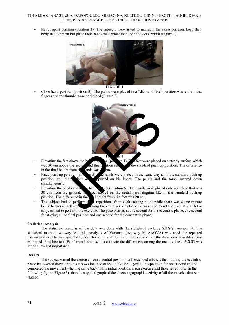
- Hands-apart position (position 2): The subjects were asked to maintain the same position, keep their
body in alignment but place their hands 50% wider than the shoulders’ width (Figure 1).
FIGURE 1
- Close hand position (position 3): The palms were placed in a “diamond-like” position where the index
fingers and the thumbs were conjoined (Figure 2).
FIGURE 2
- Elevating the feet above the hands position (position 4): The feet were placed on a steady surface which
was 30 cm above the ground and this position resembled the standard push-up position. The difference
in the final height from the hands was 20 cm.
- Knee push-up position (position 5): The hands were placed in the same way as in the standard push-up
position; yet, the subject was supported on his knees. The pelvis and the torso lowered down
simultaneously.
- Elevating the hands above the feet position (position 6): The hands were placed onto a surface that was
30 cm from the ground. The feet stayed on the metal parallelogram like in the standard push-up
position. The difference in the final height from the feet was 20 cm.
- The subject had to perform three repetitions from each starting point while there was a one-minute
break between each exercise. During the exercises a metronome was used to set the pace at which the
subjects had to perform the exercise. The pace was set at one second for the eccentric phase, one second
for staying at the final position and one second for the concentric phase.
Statistical Analysis
The statistical analysis of the data was done with the statistical package S.P.S.S. version 13. The
statistical method two-way Multiple Analysis of Variance (two-way M ANOVA) was used for repeated
measurements. The average, the typical deviation and the maximum value of all the dependent variables were
estimated. Post hoc test (Bonferroni) was used to estimate the differences among the mean values. P<0.05 was
set as a level of importance.
Results
The subject started the exercise from a neutral position with extended elbows; then, during the eccentric
phase he lowered down until his elbows inclined at about 90ο; he stayed at this position for one second and he
completed the movement when he came back to his initial position. Each exercise had three repetitions. In the
following figure (Figure 3), there is a typical graph of the electromyographic activity of all the muscles that were
studied.
JPES

TOPALIDOU ANASTASIA, DAFOPOULOU GEORGINA, KLEPKOU EIRINI - EROFILI, AGGELIGAKIS
JOHN, BEKRIS EVAGGELOS, SOTIROPOULOS ARISTOMENIS
JPES ® www.efsupit.ro
75
FIGURE 3
The following graph (Figure 4) shows the shift of the COP in the left and the right arm during a random attempt.
FIGURE 4
Results of Dynamic Analysis
There were no statistically important differences in the rates of force application for both the left and the
right upper extremity. However, there were some differences in the vertical forces of the left and right arm in
some positions. The following graphs show the mean values and the typical deviations of the vertical forces of
the two hands in all six positions (Figures 5-6).
FIGURE 5
JPES
TOPALIDOU ANASTASIA, DAFOPOULOU GEORGINA, KLEPKOU EIRINI - EROFILI, AGGELIGAKIS
JOHN, BEKRIS EVAGGELOS, SOTIROPOULOS ARISTOMENIS
JPES ® www.efsupit.ro
76
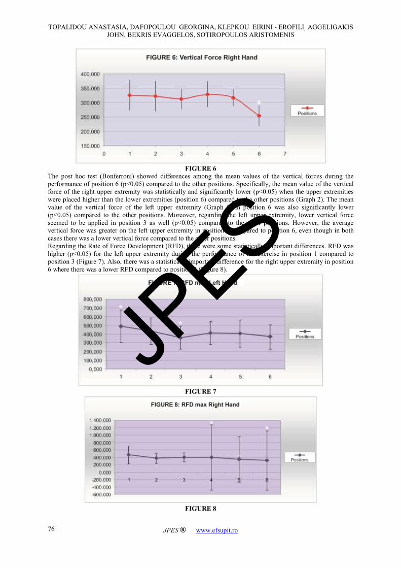
FIGURE 6
The post hoc test (Bonferroni) showed differences among the mean values of the vertical forces during the
performance of position 6 (p<0.05) compared to the other positions. Specifically, the mean value of the vertical
force of the right upper extremity was statistically and significantly lower (p<0.05) when the upper extremities
were placed higher than the lower extremities (position 6) compared to the other positions (Graph 2). The mean
value of the vertical force of the left upper extremity (Graph 1) in position 6 was also significantly lower
(p<0.05) compared to the other positions. Moreover, regarding the left upper extremity, lower vertical force
seemed to be applied in position 3 as well (p<0.05) compared to the other positions. However, the average
vertical force was greater on the left upper extremity in position 3 compared to position 6, even though in both
cases there was a lower vertical force compared to the other positions.
Regarding the Rate of Force Development (RFD), there were some statistically important differences. RFD was
higher (p<0.05) for the left upper extremity during the performance of the exercise in position 1 compared to
position 3 (Figure 7). Also, there was a statistically important difference for the right upper extremity in position
6 where there was a lower RFD compared to position 4 (Figure 8).
FIGURE 7
FIGURE 8
JPES
TOPALIDOU ANASTASIA, DAFOPOULOU GEORGINA, KLEPKOU EIRINI - EROFILI, AGGELIGAKIS
JOHN, BEKRIS EVAGGELOS, SOTIROPOULOS ARISTOMENIS
JPES ® www.efsupit.ro
77
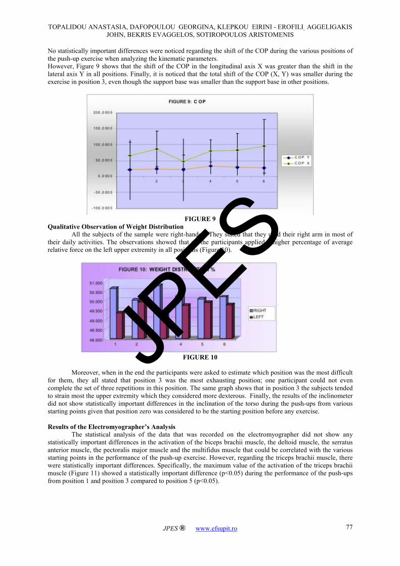
No statistically important differences were noticed regarding the shift of the COP during the various positions of
the push-up exercise when analyzing the kinematic parameters.
However, Figure 9 shows that the shift of the COP in the longitudinal axis X was greater than the shift in the
lateral axis Y in all positions. Finally, it is noticed that the total shift of the COP (X, Y) was smaller during the
exercise in position 3, even though the support base was smaller than the support base in other positions.
FIGURE 9
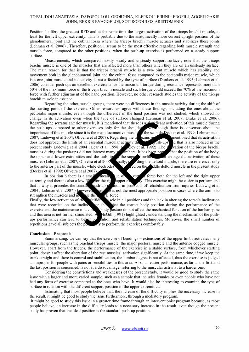
Qualitative Observation of Weight Distribution
All the subjects of the sample were right-handed. They stated that they used their right arm in most of
their daily activities. The observations showed that all the participants applied a higher percentage of average
relative force on the left upper extremity in all positions (Figure 10).
FIGURE 10
Moreover, when in the end the participants were asked to estimate which position was the most difficult
for them, they all stated that position 3 was the most exhausting position; one participant could not even
complete the set of three repetitions in this position. The same graph shows that in position 3 the subjects tended
to strain most the upper extremity which they considered more dexterous. Finally, the results of the inclinometer
did not show statistically important differences in the inclination of the torso during the push-ups from various
starting points given that position zero was considered to be the starting position before any exercise.
Results of the Electromyographer’s Analysis
The statistical analysis of the data that was recorded on the electromyographer did not show any
statistically important differences in the activation of the biceps brachii muscle, the deltoid muscle, the serratus
anterior muscle, the pectoralis major muscle and the multifidus muscle that could be correlated with the various
starting points in the performance of the push-up exercise. However, regarding the triceps brachii muscle, there
were statistically important differences. Specifically, the maximum value of the activation of the triceps brachii
muscle (Figure 11) showed a statistically important difference (p<0.05) during the performance of the push-ups
from position 1 and position 3 compared to position 5 (p<0.05).
JPES

TOPALIDOU ANASTASIA, DAFOPOULOU GEORGINA, KLEPKOU EIRINI - EROFILI, AGGELIGAKIS
JOHN, BEKRIS EVAGGELOS, SOTIROPOULOS ARISTOMENIS
JPES ® www.efsupit.ro
78
FIGURE 11
The mean value of the triceps brachii muscle shows statistically important differences in position 5 where it has
the lowest value (p<0.05) compared to the other positions (Figure 12).
FIGURE 12
Generally, in position 5 there was a smaller activation of the triceps brachii muscle compared to the
other positions. Finally, there was a smaller shift of the CPO both in the longitudinal axis X and the lateral axis
Y in position 1 and position 3 where there is the largest activation of the triceps brachii muscle.
Discussion
The study is restricted to a steady surface unlike other studies which compare steady and unsteady
support surfaces ( Freeman et al. 2006; Donkers et al. 1993; Lehman et al. 2007; Ludewig et al 2004; Lehman et
al. 2006). This is not a restriction since most research does not include all muscle groups and does not examine
the differences in the muscle activation in same surfaces with different support. Most research compares only
two surfaces. Also, it is important to observe whether the changes in the placement of the upper extremities
affect the result of muscle activation without any rotation and projection of the scapula, as other authors
distinctively mention (Freeman et al. 2006; Decker et al. 1999; Ludewig et al 2004; Lehman et al. 2006 ).
The present research showed that there are changes in muscle activation, RFD, the shift of the COP and
the vertical forces between the left and right upper extremity. This is probably due to which hand each subject
tends to use in order to maintain his balance. Even though there is not any research that mentions the use-
prevalence of one upper extremity in the push-up exercise, the highest distribution of weight on the right hand in
all positions remains a mystery.
Moreover, even though it was proven that in position 3 there is a smaller shift of the COP, both in the
longitudinal axis X and the lateral axis Y, and a maximum value of the triceps brachii muscle’s activation, yet it
is noticed a lower mean value of vertical force and a lower RFD for the left upper extremity (where there was a
smaller distribution of weight) in position 3 compared to the other positions. This is probably due to the
difficulty in performing the exercise as the participants stated. Based on that, we reach a conclusion that position
3 can activate the triceps brachii muscle to its maximum but this activation is slow. That means that the
performance of the exercise in this position is not recommended in cases where the aim is to improve the muscle
strength. However, it can be used to strengthen the triceps brachii muscle and it is likely to provide good results.
JPES
TOPALIDOU ANASTASIA, DAFOPOULOU GEORGINA, KLEPKOU EIRINI - EROFILI, AGGELIGAKIS
JOHN, BEKRIS EVAGGELOS, SOTIROPOULOS ARISTOMENIS
JPES ® www.efsupit.ro
79
Position 1 offers the greatest RFD and at the same time the largest activation of the triceps brachii muscle, at
least for the left upper extremity. This is probably due to the anatomically more correct upright position of the
glenohumeral joint and the cubital fossa where the triceps brachii muscle actuates and stabilizes these joints
(Lehman et al. 2006) . Therefore, position 1 seems to be the most effective regarding both muscle strength and
muscle force, compared to the other positions, when the push-up exercise is performed on a steady support
surface.
Measurements, which compared mostly steady and unsteady support surfaces, note that the triceps
brachii muscle is one of the muscles that are affected more than others when they are on an unsteady surface.
The main reason for that is that the triceps brachii muscle is a two-joint muscle which has stability and
movement both in the glenohumeral joint and the cubital fossa compared to the pectoralis major muscle, which
is a one-joint muscle and its activity is not affected by the type of surface (Donkers et al. 1993; Lehman et al.
2006) consider push-ups an excellent exercise since the maximum torque during resistance represents more than
50% of the maximum force of the triceps brachii muscle and such torque could exceed the 70% of the maximum
force with further adjustment of the hand position. However, no other research studies the activity of the triceps
brachii muscle in essence.
Regarding the other muscle groups, there were no differences in the muscle activity during the shift of
the starting point of the exercise. Other researchers agree with these findings, including the ones about the
pectoralis major muscle, even though the difference in the hand position was not studied, which showed no
change in its activation even when the type of surface changed (Lehman et al. 2007; Drake et al. 2006).
Regarding the serratus anterior muscle, it is mentioned that there is a significant activation of this muscle during
the push-ups compared to other exercises only for the shoulders, even though there is consensus about the
importance of this muscle since it is the main locomotive muscle of the scapula Decker et al. 1999; Lehman et al.
2007; Ludewig et al 2004; Oliveira et al 2007). Regarding the trapezius muscle, it is mentioned that its activation
does not approach the limits of an essential muscular activity during the push-ups and that is also noticed in the
present study Ludewig et al 2004 ; Lear et al. 1998; Moseley et al. 1992) .The activation of the biceps brachii
muscles during the push-ups did not interest many researchers. It has been proven that the position of the body,
the upper and lower extremities and the stability of the support surface do not change the activation of these
muscles (Lehman et al 2007; Oliveira et al 2007). Finally, regarding the deltoid muscle, there are references only
to the anterior part of the muscle, while electrodes were placed on the middle deltoid muscle in the present study
(Decker et al. 1999; Oliveira et al 2007) .
In position 6 there is a smaller mean value of the vertical force both for the left and the right upper
extremity and there is also a low RFD for the right upper extremity. This exercise might be easier to perform and
that is why it precedes the standard push-up position in protocols of rehabilitation from injuries Ludewig et al
2004 ; Lehman et al.2007 ) However, position 6 is not the most appropriate position in cases where the aim is to
strengthen the muscles and increase the muscle force.
Finally, the low activation of the multifidus muscle in all positions and the lack in altering the torso’s inclination
that were recorded on the inclinometer show that the correct body position during the performance of the
exercise and the maintenance of a correct body posture do not affect the mechanical function of the lumbar spine
and this area is not further stimulated. As McGill (1991) highlighted , understanding the mechanism of the push-
ups performance can lead to better prevention and rehabilitation techniques. Moreover, the small number of
repetitions gave all subjects the possibility to perform the exercises comfortably.
Conclusion – Proposals
Summarizing, we can say that the exercise of bendings – extensions of the upper limbs activates many
muscular groups, such as the brachial triceps muscle, the major pectoral muscle and the anterior cogged muscle.
However, apart from the triceps, the performance of the exercise in a stable surface, from whichever starting
point, doesn’t affect the alteration of the rest muscles’ activation significantly. At the same time, if we keep the
trunk straight and there is control and stabilization, the lumbar degree is not affected, thus the exercise is judged
as improper for people with pains or sensibilities in this area. Also, an easier performance, as far as the first and
the last position is concerned, is not at a disadvantage, referring to the muscular activity, to a harder one.
Considering the constrictions and weaknesses of the present study, it would be good to study the same
issue with a larger and more varied sample, such as a sample that includes females or even people who have not
had any form of exercise compared to the ones who have. It would also be interesting to examine the type of
surface in relation with the different support position of the upper extremities.
Estimating that most people believe that, the increase of the difficulty implies the necessary increase in
the result, it might be good to study the issue furthermore, through a mediatory program.
It might be good to study this issue in a greater time frame through an interventionist program because, as most
people believe, an increase in the difficulty leads to a necessary increase in the result, even though the present
study has proven that the ideal position is the standard push-up position.
JPES

TOPALIDOU ANASTASIA, DAFOPOULOU GEORGINA, KLEPKOU EIRINI - EROFILI, AGGELIGAKIS
JOHN, BEKRIS EVAGGELOS, SOTIROPOULOS ARISTOMENIS
JPES ® www.efsupit.ro
80
This study enabled us to apprehend that simple movements which are widely used have been remotely
examined and the field of further research is large.
The results of the present study could be useful in areas and recovery places so that the push-up exercise
would be included in their protocols.
References
Anderson D.S., Jacson M.F., Kropf D.S., Soderberg G.L (1984). Electromyographic analysis of selectedmuscles
during sitting push-ups. Effects of position and sex. Phys Ther 64, 24-32.
Bell N.S., Mangione T.W., Hemenway D., Amoroso P.J (2000). High iinjury rates among female army trainees.
A function of gender? American Journal of Preventive Medicine 18 (3S),141-146.
De Oliveira A.S., deMorais Carvalho M., de Brum D.P (2007). Activation of the shoulder and arm muscles
during axial load exercises on a stable base of support and on a medicine ball. Journal of Electromyographic and
Kinesiology 18(3), 472-479.
Decker M.J., Hintermeister R.A., Faber K.J., Hawkins R.J (1999). Serratus anterior muscle activity during
selected rehabilitation exercises. American Journal of Sports Medicine 27(6), 784-791.
Donkers M.J., An K.N., Chao E.Y.S., Morrey B.F (1993). Hand position affects elbow joint load during push-up
exercise. Journal of Biomechanics 26(6), 625-632.
Drake J.D.M., Fischer S.L., Brown S.H.M., Callaghan J.P (2006). Do exercise balls provide a training advantage
for trunk extensor exercises? A biomechanical evaluation. J Manipulative Physiol Ther 29(5), 354-362.
Ekstrom R.A., Donatelli R.A., Soderberg G.L (2003). Surface electromyographic analysis of exercises for the
trapezius and serratus anterior muscles. J Orthop Sports Phys Ther 33 (5), 247-258.
Freeman S., Karpowicz A., Gray J., McGill S. (2006). Quantifying muscle patterns and spine load during various
forms of the push-up. American College of Sports Medicine 570-577.
Gregg R.L., Banderet L.E., Reynolds K.L, Creedon J.F., Rice V.J. (2002). Psychological factors that influence
traumatic injury occurrence and physical performance. Work 18,133-139.
Hartmann J. & Tunnemann H. (1988) Fitness and Strength Training. Berlin , Sportverlag.
Kibler W.B. (1998). The role of the scapula in athletic shoulder function. American Journal of Sports Medicine
26(2), 325-337.
Knapik J.J., Canham-Chervak M., Hauret K., Laurin M.J., Hoedebecke E., Craig S., Montain S.J. (2001)
Seasonal variations in injury rates during US army basic combat training. Ann Occup Hyg 46(1), 15-23.
Kotani Y. & Tokuhiro A. (2002). Kinesiological study of the push-up motion in spinal cord injury patients:
involving measurement of hand pressure applied to a force plate. Acta Medica Okayama 56(2), 75-82.
Lear L.J. & Gross M.T. (1998) An electromyographical analysis of the scapular stabilizing synergists during a
push-up progression. J Orthop Sports Phys Ther 28(3), 146-157.
Lehman G.J., Gilas D., Patel U. (2007). An unstable support surface does not increase scapulothoracic
stabilizing muscle activity during push up and push ups plus exercises. Manual Therapy 13(6):500-506.
Lehman G.J., MacMillan B., MacIntyre I., Chivers M., Fluter M. (2006). Shoulder muscle EMG activity during
push up variations on and off a Swiss ball. Dynamic Medicine 5:7.
Lin H., Chie W., Lien H. (2006). Epidemiological analysis of factors influencing an episode of exertional
rhabdomyolysis in high school students. Am J Sports Med 34(3), 481-486.
Ludewig P.M., Hoff M.S., Osowski E.E., Meschke S.A., Rundquist P.J. (2004). Relative balance of serratus
anterior and upper trapezius muscle activity during push-up exercises. American Journal of Sports Medicine
32(2), 484-493.
McCann P.D., Wootten M.E., Kadaba M.P., Bigliani L.U. (1993). A kinematic and electromyographic study of
shoulder rehabilitation exercises. Clinical Orthopaedics and Related Research 288, 179-188.
McGill S.M. (1991). Electromyographic activity of the abdominal and low back musculature during the
generation of isometric and dynamic axial trunk torque: implications for lumbar mechanics. J Orthop Res 9, 91-
103.
Molonge T.S., Provencher M.T., Menzel K.A., Vachon T.A., Dewing C.B. (2007). Arthroscopic stabilization in
patients with an inverted pear glenoid: result in patients with bone loss of the anterior genoid. American Journal
of Sports Medicine 35 (8), 1276-1283.
Moseley J.B., Jobe F.B., Pink M., Perry J., Tibone J. (1992). EMG analysis of the scapular muscles during a
shoulder rehabilitation program. American Journal of Sports Medicine 20, 128-134.
White D.W., Wenke J.C., Mosely D.S., Mountcastle S.B., Basamania C.J. (2007). Incidence of major tendon
ruptures and anterior cruciate ligament tears in US Army Soldiers. American Journal of Sports Medicine 35(8),
1308-1314.
JPES