antibody-direct epifluorescent filter technique for …aem.asm.org/content/60/10/3553.full.pdf ·...
TRANSCRIPT

APPLIED AND ENVIRONMENTAL MICROBIOLOGY, OCt. 1994, p. 3553-35590099-2240/94/$04.00+0
Antibody-Direct Epifluorescent Filter Technique for Rapid,Direct Enumeration of Escherichia coli 0157:H7 in Beef
MARY LOU TORTORELLO* AND DIANA S. STEWART
National Center for Food Safety and Technology, U.S. Food and Drug Administration,Summit-Argo, Illinois 60501
Received 28 January 1994/Accepted 15 July 1994
Artificially inoculated Escherichia coli 0157:H7 was directly enumerated in ground beef and beef exudate,without enrichment or selection, by the antibody-direct epifluorescent filter technique (Ab-DEFT). The totalassay time of the Ab-DEFT was less than 1 h. The beef was homogenized, treated for 15 min with trypsin andTriton X-100, and passed through a 5-,Im-pore-size prefilter and then through a 0.2-,um-pore-size blackpolycarbonate filter. The final filter was stained directly with fluorescein-labeled anti-0157 polyclonalantibody, rinsed, and examined by epifluorescence microscopy. The sensitivity of the Ab-DEFT was comparedwith that of a standard enrichment culture technique. Both methods reliably determined the presence of thepathogen in beef at 16 CFU/g. The Ab-DEFT was also useful for quantifying the pathogen and monitoring itsgrowth in beef.
The direct epifluorescent filter technique (DEFT), an ex-tremely rapid microbiological analytical method, has been usedfor many years for direct quantitation of microbial load in avariety of applications (22), e.g., environmental testing (11),pharmaceutical manufacturing (9), and predicting the shelf lifeof food (29). The DEFT requires only 20 min of assay time fordetermination of microbial numbers in raw milk (5, 23);elimination of overnight incubation is a major advantage.Rapid microbiological testing is valuable in food manufactur-ing because the quality of materials coming into the productionline is often a critical control point, and assay speed is animportant economic consideration.
In the DEFT, microorganisms are entrapped on a mem-brane filter, which is stained with a fluorescent dye, e.g.,acridine orange, and examined by epifluorescence microscopy.The total microbial population is then rapidly enumerated. Forspecific rather than general staining of bacteria, fluorescentantibodies are used. Indirect fluorescent-antibody staining hasbeen adapted to the DEFT for specific enumeration of Salmo-nella (28) and Listeria (31) organisms in raw meats. A direct(one-step) fluorescent-antibody modification, the antibody-DEFT (Ab-DEFT), was developed to enumerate Eschenichiacoli 0157:H7 in milk and juice (34).The low sensitivity of traditional microscopic methods is
greatly improved in both the DEFT and Ab-DEFT by filterconcentration of the material to be analyzed and elimination ofirrelevant substances by passage through the filter. The pres-ence of these substances, which often interfere with theoutcome of the application of analytical techniques, is animportant reason for requiring enrichment and isolation stepsbefore the use of highly specific assays, such as the enzyme-linked immunosorbent assay and PCR.
Food-borne illnesses associated with E. coli 0157:H7 havebeen traced to consumption of undercooked ground beef (8).This report compares the sensitivity of the Ab-DEFT with thatof a conventional enrichment culture (EC) procedure foridentifying E. coli 0157:H7 in ground beef. The Ab-DEFT has
* Corresponding author. Mailing address: U.S. Food and DrugAdministration, 6502 S. Archer Rd., Summit-Argo, IL 60501. Phone:(708) 728-4146. Fax: (708) 728-4177.
also been useful in determining cell numbers of the microor-ganism in beef exudate and for monitoring growth and survivalin beef.
MATERIALS AND METHODS
Bacteria and media. E. coli 0157:H7 (ATCC 35150) as wellas 17 clinical isolates (University of Iowa Hjgienic Laboratory)were cultured at 37°C in Luria-Bertani broth supplementedwith 0.1% glucose. A dihydrostreptomycin-resistant variant ofthe ATCC 35150 strain, E. coli DHS-1, was isolated by suc-cessive culture in increasing concentrations of dihydrostrepto-mycin (Sigma Chemical Co., St. Louis, Mo.) by the gradientplate technique (7) and cultured at 37°C in Luria-Bertani brothsupplemented with 0.1% glucose and 1,000 ,ug of dihydrostrep-tomycin per ml. All microbiological culture media were pur-chased from Difco Laboratories (Detroit, Mich.).
Preparation of ground beef and beef serum for microbiolog-ical analysis. Ground beef, 80% lean, was purchased from a
local retail store. Log-phase bacterial cells, diluted appropri-ately in 0.01 M phosphate-buffered saline (PBS) (13), pH 7.2,were used in all artificial inoculations of beef and beef exudate.Before inoculation, the log-phase cells were plated onto Mac-Conkey sorbitol agar (MSA) (Difco) and incubated at 37°C for18 to 24 h to confirm the population size. Cells (1 to 10 ml)were mixed directly into the ground beef. The inoculated beefwas placed within the inner mesh lining of a plastic stomacherbag (model 400 filter bags; Seward Medical, London, UnitedKingdom), and diluent was added for homogenization. ForDEFT and Ab-DEFT analyses, 10 g of the inoculated beef wasadded to 90 ml of PBS; for the EC method, 25 g was added to225 ml of enrichment broth (17). The beef was then homoge-nized for 2 min in a stomacher model 400 (Tekmar Co.,Cincinnati, Ohio), and the slurry that passed through the meshlining was harvested. Beef exudate, i.e., the liquid that accu-mulates during shipment of unprocessed raw beef, was ob-tained from a local meat processor, and after inoculation withbacterial cells was treated for analysis.DEFT and Ab-DEFI. For analysis by DEFT and Ab-DEFT,
ground beef slurry and beef exudate were treated by modifi-cations of previously described protocols (6, 24, 34). Filtershoused in Swinnex-type filter holders, syringes, and reagents
3553
Vol. 60, No. 10
on June 13, 2018 by guesthttp://aem
.asm.org/
Dow
nloaded from
3554 TORTORELLO AND STEWART
were prewarmed by incubation at 50°C to facilitate filtration.To 4 ml of beef slurry or beef exudate, 4 ml of 0.5% (wt/vol)Triton X-100 and 1 ml of trypsin (Sigma) prepared in PBS at10 mg/ml were added. The ground beef mixture was incubatedat 50°C for 15 min before filtration. The beef exudate mixturewas incubated at 37°C for 15 min before filtration. Plastic 10-mlsyringes with Luer-Lok tips were used in the filtrations. Thehomogenized beef was passed through 5-,m-pore-size nylonprefilters (Micron Separations, Inc., Westboro, Mass.). Forbeef exudate, 5-,um-pore-size polycarbonate prefilters (Pore-tics Corp, Livermore, Calif.) were used. After prefiltration,portions (2 to 100 ml) of the beef or beef exudate filtrates werepassed through 0.2-,um-pore-size black polycarbonate filters(Poretics). If necessary, the mixture was diluted appropriatelyin PBS after prefiltration to obtain 10 to 100 cells per micro-scope field for ease in counting. The polycarbonate filters wererinsed by passing them through 5 ml of 0.1% (wt/vol) TritonX-100 and 5 ml of PBS before staining with acridine orange(DEFT) or fluorescent antibody (Ab-DEFT).The DEFT and Ab-DEFT were performed as described
previously (34), with the following modifications. P5 filterpaper discs (Fisher Scientific, Pittsburgh, Pa.) were placedunder the nylon prefilters and black polycarbonate filters toprovide support in Swinnex-type filter holders. For the DEFT,after acridine orange staining, the black polycarbonate filterswere washed with 5 ml of PBS, air dried, and mounted on glassslides in low-fluorescence immersion oil. For the Ab-DEFT,fluorescein-labeled, affinity-purified polyclonal antibody to E.coli 0157:H7 (Kirkegaard and Perry Laboratories, Inc., Gaith-ersburg, Md.) was diluted 1:2,000 in 1% (wt/vol) bovine serumalbumin in PBS and layered on top of the black polycarbonatefilters for 15 min; this was followed by rinses of 5 ml ofPBS-0.05% Tween 20 and 5 ml of PBS. The fluorescent-antibody-stained filters were mounted on glass slides in Vecta-Shield mounting medium (Vector Laboratories, Burlingame,Calif.).EC. EC was performed as recommended for the isolation
and identification of E. coli 0157:H7 in ground beef (18).Briefly, the EC procedure involved 24 h of enrichment inmodified EC-novobiocin broth (17) followed by selective iso-lation of E. coli colonies from the enrichment on MSAsupplemented with 5-bromo-4-chloro-3-indolyl-p-D-glucuronicacid (BCIG) (Biosynth International, Skokie, Ill.). A modifi-cation involved use of the sodium salt of BCIG instead of thecyclohexylammonium salt in MSA for selective isolation of E.coli colonies from the enrichment broth. The substitutionallowed the BCIG reagent to be solubilized directly in theaqueous MSA (MSA-BCIG) (15) instead of in alkaline etha-nol. The Minitek System (Becton Dickinson, Cockeysville,Md.) was used to characterize presumptively positive coloniesby indole production, H2S production, cellobiose utilization,Voges-Proskauer reaction, citrate utilization, lysine decarbox-ylase, and ornithine decarboxylase. Serological identificationwas performed with the Oxoid E. coli 0157 Test Kit (UnipathLtd., Hampshire, United Kingdom).
Bacterial cell counting. Bacterial cells were counted by theDEFT and Ab-DEFT as described previously (34). Membranefilter microscope factors (MFMF) were used to calculate cellconcentrations and depended on the microscope objective lensused: 1,670 for the 40x lens, 3,959 for the 63x lens, and 9,440for the 100x lens. The bacterial cell concentration was calcu-lated as follows: number of cells per milliliter = [(average cellcount/microscope field) (MFMF) (dilution factor)]/volumefiltered (milliliters).
For counting bacteria by viable plate counts (VPC), dilu-tions were made in PBS and then spread plated on agar media
TABLE 1. Sensitivities of Ab-DEFT and EC method for identifyingE. coli 0157:H7 artificially inoculated into ground beef
Ab-DEFT ECCFU inocu- Repli-lated' per caepl No. positive/g of beef cate Detection Count/g Detection 12 colonies
(%b)
240,000 1 + 320,000 + 12/12 (100)2 + 180,000 + 12/12 (100)3 + 500,000 + 12/12 (100)
820 1 + 770 + 12/12 (100)2 + 150 + 12/12 (100)3 + 740 + 12/12 (100)
210 1 + 62 + 11/12 (92)2 + 33 + 9/12 (75)3 + 50 + 8/12 (67)
16 1 + 5 + 12/12 (100)2 + 10 + 11/12 (92)3 + 1 + 10/12 (83)
2 1 - NDC + 1/12 (8)2 _ ND - 0/12 (0)3 - ND - 0/12 (0)
a Calculated from VPC of log-phase culture on MSA.b Percentage of colonies positively identified as E. coli 0157:H7 of 12 white
colonies from MSA-BCIG plates.c ND, not determined.
and incubated at 37°C for 18 to 24 h. For total aerobic platecounts, colonies cultured on standard plate count agar werecounted. For E. coli 0157:H7, white colonies cultured onMSA, indicative of sorbitol nonfermenters, were counted. ForE. coli DHS-1, white colonies cultured on MSA supplementedwith 1,000 ,ug of dihydrostreptomycin per ml were counted. AllVPC data were expressed in CFU.
Experimental design for comparison of the sensitivities ofAb-DEFT and EC. Ground beef was artificially inoculated withE. coli 0157:H7 at concentrations of approximately 105, 103,102, 10, and 1 CFU/g. For each inoculation level, log-phasecells were diluted appropriately in PBS, and portions (1 to 10ml) were mixed into 125 g of ground beef, which was thendivided into triplicate 10-g and 25-g portions. The 10-g por-tions were analyzed by the Ab-DEFT, and the 25-g portionswere analyzed by the EC method.
RESULTS
Comparison of the sensitivities of Ab-DEFI and EC. Theabilities of the Ab-DEFT and EC methods to determine thepresence of E. coli 0157:H7 artificially inoculated into groundbeef were compared (Table 1). Both methods identified allthree replicates of the inoculated beef at cell concentrations offrom 2.4 x 10⁵ to 16 CFU/g of beef as positive. However, at 2CFU/g of beef, neither method was reliable. The Ab-DEFTlacked sensitivity at this level: no positive identifications weremade for the three replicate beef portions. The EC method,which identified one of the three replicates as contaminated,was more sensitive than the Ab-DEFT. This result was sup-ported by a second trial of triplicate beef test portions inocu-lated at 2 CFU/g, in which the EC again identified one of threeinoculated beef replicates as positive.
In the description of the EC method, positive identificationswere reported for 11 of 12 beef test portions artificiallyinoculated at 0.6 to 0.7 CFU/g (18). Because our results did not
APPL. ENVIRON. MICROBIOL.
on June 13, 2018 by guesthttp://aem
.asm.org/
Dow
nloaded from
Ab-DEFT FOR E. COLI 0157:H7 IN BEEF 3555
TABLE 2. Detectability of artificially inoculatedE. coli 0157:H7 by Ab-DEFT
CFU inocu- Treated beet No. of No. oflated' per filtered Replicate fields' positive"g of beef (ml) examined fields
240,000 2 1 20 202 20 203 20 20
820 10 1 150 312 150 63 150 30
210 50 1 200 172 223 103 235 16
16 75 1 200 22 200 43 200 7
2 100 1 500 02 500 03 500 0
"Calculated from VPC of log-phase culture on MSA.^ Homogenized beef treated with trypsin and Triton X-100 for 15 min at 50°C
(see Materials and Methods).' Number of microscope fields examined with 40X objective lens.d Number of microscope fields that showed the presence of at least one
fluorescent cell.
agree with this reported sensitivity, we tested additional inoc-ulation levels of 0.6 and 0.1 CFU/g by EC. (The Ab-DEFT wasnot tested at these inoculation levels.) At 0.6 CFU/g, the ECmethod identified one of three inoculated beef replicates aspositive; at 0.1 CFU/g, none of the three replicates wasidentified as positive.The detectability of E. coli 0157:H7 by the Ab-DEFT may
be assessed for each inoculation level by considering thenumber of microscope fields examined, the number of positivemicroscope fields, and the volume of beef filtered (Table 2). At240,000 CFU/g, with only a 2-ml test portion filtered, everyfield examined contained fluorescent cells. At lower levels ofinoculation, larger test portions were necessary and more fieldshad to be examined for identification. At the lowest inocula-tion level of 2 CFU/g of beef and a filtration volume of 100 ml,the Ab-DEFT result was recorded as negative after 500 fieldsper filter were examined for each replicate. If it is assumed thatnone of the 500 fields overlapped, then the area examined ineach case represented approximately 30% of the total filterarea.
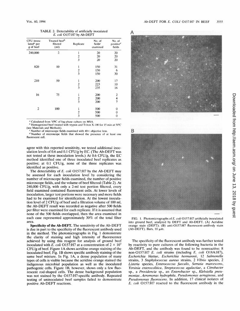
Specificity of the Ab-DEFI. The sensitivity of the Ab-DEFTis due in part to the specificity of the fluorescent antibody usedin the method. The photomicrographs in Fig. 1 demonstratethe clarity of staining and high intensity of fluorescenceachieved by using this reagent for analysis of ground beefinoculated with E. coli 0157:H7 at a concentration of 2 x 10⁵CFU/g of beef. Figure 1A shows acridine orange staining of theinoculated beef; Fig. 1B shows specific antibody staining of thesame beef mixture. In Fig. 1A, a dense population of manytypes of cells is visible because the acridine orange stained theindigenous microbial population as well as the inoculatedpathogenic cells. Figure 1B, however, shows only a few fluo-rescent rod-shaped cells. The dense background populationwas not stained by the 0157:H7-specific antibody. Repeatedtesting of uninoculated beef samples failed to demonstratepositive Ab-DEFT reactions.
A
R
FIG. 1. Photomicrographs of E. coli 0157:H7 artificially inoculatedinto ground beef, analyzed by DEFT and Ab-DEFT. (A) Acridineorange stain (DEFT); (B) anti-0157:H7 fluorescent-antibody stain(Ab-DEFT). Bars, 10 ,m.
The specificity of the fluorescent antibody was further testedby reactivity to pure cultures of the following bacteria in theAb-DEFT, and the antibody was found to be nonreactive: 8non-0157:H7 E. coli strains (including E. coli 018A:H7),Escherichia blattae, Escherichia hermannii, 12 Salmonellastrains, 3 Staphylococcus aureus strains, 2 Vibrio species, 2Listeria species, Enterococcus faecalis, Serratia marcescens,Yersinia enterocolitica, Streptococcus agalactiae, a Citrobactersp., a Providencia sp., an Enterobacter sp., Klebsiella pneu-moniae, Aeromonas hydrophila, Pseudomonas aeruginosa, andPseudoinonas fluorescens. In addition, 17 clinical isolates ofE. coli 0157:H7 reacted to the fluorescent antibody in the
VOL. 60, 1994
on June 13, 2018 by guesthttp://aem
.asm.org/
Dow
nloaded from

3556 TORTORELLO AND STEWART
7
a).-0
f! 6.-0
bCSV-5a)a)
00 4
a:CE1- 3
D-
2
0
0~~~
l0l I § I
I I I I I
1 2 3 4 5Log10 CFU/mI Filtered
6 7
FIG. 2. Recovery of E. coli 0157:H7 cells from artificially inocu-lated beef after filtration through 5-p.m-pore-size nylon prefilters. TheVPC on MSA was determined at various inoculation levels forhomogenized, trypsin-Triton X-100-treated beef mixtures before(loglo CFU/ml filtered) and after (log1(1 CFU/ml recovered) filtration.
Ab-DEFT. However, a false-positive reaction to Salmonellacholeraesuis serotype urbana was observed.
Enumeration of E. coli 0157:H7 in beef and beef exudate.Quantitation of contamination levels of E. coli 0157:H7 inground beef was possible by the Ab-DEFT but not by the ECmethod. With the Ab-DEFT, specific microbial cell concentra-tions were measured in the beef, generally with an accuracywithin an order of magnitude of the inoculation levels.
In previous studies there was inconsistent correlation be-tween inoculated cell counts and DEFT filter counts, depend-ing on the food analyzed (24). Poor correlations may be relatedto the prefiltration step. The 5-,um-pore-size nylon prefilter isnecessary for removal of large particulates and facilitatesfiltration of the food through the analytical 0.2-p.m-pore-sizefilter. However, the prefilter may be a barrier for microbialcells entrapped within the particulates and may cause lower-than-expected cell counts on the 0.2-plm-pore-size filter. Forground beef, virtually no cells were lost from the treatedmixture after prefiltration through 5-p.m-pore-size nylon filters(Fig. 2). This finding supports previous data obtained in similarstudies in which the DEFT was used for analysis of fresh meatand fish (24).
Bacterial cell enumeration by Ab-DEFT was also demon-strated for beef exudate, which accumulates in containers ofpacked beef during transport and is a convenient medium formicrobiological testing in meat processing plants. Figure 3shows the results of the Ab-DEFT analysis of beef exudateinoculated with E. coli 0157:H7 at levels varying from 107 to103 CFU/ml of beef exudate. The Ab-DEFT counts were linearand correlated well with numbers of cells inoculated, except atthe lowest concentrations (102 to 103 CFU/ml), for whichcounting accuracy declined. The use of 5-p.m-pore-size poly-carbonate (instead of nylon) prefilters was necessary to obtainthis correlation in the beef exudate experiments. In severaltrials using nylon prefilters, counts were linear but were
7
E 6enCD)a)0-
D) 50-JI-I
0 4
LL
03.3
22 3 4 5 6 7
Log10 CFU/mI InoculatedFIG. 3. Ab-DEFT count of E. coli 0157:H7 artificially inoculated
into beef exudate. The VPC on MSA was determined for log-phasecells inoculated at various levels into beef exudate (log,( CFU/mlinoculated) for analysis by the Ab-DEFT (log,(1 cells per ml).
approximately 10 times lower than the number of cells inocu-lated into the serum. This result may have been related toentrapment of cells on the nylon prefilter after the trypsin-Triton X-100 treatment at 50°C.
Validity of Ab-DEFI for measuring growth of E. coli0157:H7 in beef. The Ab-DEFT was compared with standardplate counting for its ability to monitor the increase in cellnumber of E. coli 0157:H7 during growth at 37°C in groundbeef. MSA has been recommended as a differential andselective medium for isolation and identification of E. coli0157:H7 (16). However, other microorganisms present inground beef can also grow and have a colony morphologyresembling that of E. coli 0157:H7 on this medium; i.e., MSAis not absolutely selective for this pathogen. Therefore, toperform this test with an unambiguously selectable microor-ganism, we isolated a dihydrostreptomycin-resistant variant ofE. coli 0157:H7, strain DHS-1, and incorporated the antibioticinto MSA (at 1,000 p.g/ml) to provide selective conditions. Themicroscopic counts in this growth experiment at 37°C werecompared with VPC (Fig. 4) and included total microbialcounts obtained by DEFT and specific E. coli DHS-1 countsobtained by Ab-DEFT. The VPC included total viable counts,obtained by plating on standard plate count agar, and specificE. coli DHS-1 counts, obtained by plating on MSA plus 1,000p.g of dihydrostreptomycin per ml.The Ab-DEFT counts were virtually identical to specific E.
coli DHS-1 VPC from the start time (0 h) through 6 h ofgrowth at 37°C (Fig. 4). The Ab-DEFT specifically enumeratedthe pathogen in the presence of approximately a 100-foldexcess of background indigenous microbial species, which weremeasured by DEFT and by total VPC. Enumeration by DEFTcompared with total VPC is also demonstrated in Fig. 4. TheDEFT counts and total VPC varied only at the starting point (0h), when the DEFT count was higher than the total VPC. Thisresult may be explained by the inclusion in the DEFT count of
I I I I
0
0
0
0~~~ ~ ~
APPL. ENVIRON. MICROBIOL.
on June 13, 2018 by guesthttp://aem
.asm.org/
Dow
nloaded from

Ab-DEFT FOR E. COLI 0157:H7 IN BEEF 3557
10 surements of original contamination levels in beef are notpossible by this method. For many years, total bacteria in foodshave been enumerated by using acridine orange stain in theDEFT. However, Pettipher (22) reported that accurate DEFT
9 counts of bacteria in milk were not possible at concentrations01n% 0 below approximately 103 cells per ml. At and below 820 cells
per g, detection is rare (Table 2) because the distribution of(D ' cells is not necessarily random or homogeneous. Nevertheless,B'8 enumeration by the Ab-DEFT may be considered approximate0 t/(i.e., accurate within an order of magnitude), and this level of
.JI'M 000000 0 accuracy may be sufficient for many applications (Table 1).D 71/_}Specific rapid and direct counting is a major advantage of theU. 4 Ab-DEFT._,§ The agreement we found between microscope counts and
VPC supports previous data on the applicability of filtrationo and epifluorescence microscopy to the microbiological analysis-_ of particulate foods such as meat (6, 24, 26, 30). The use of
6 / _ mesh-lined stomacher bags is particularly helpful for filtering9 | homogenized beef, as Qvist and Jakobsen (26) also noted in
their use of a gauze lining to contain large particulate matter_ _ _ ____ X within thebags.
0 1 2 3 4 5 6 Both the sensitivity and accuracy of enumeration by theTime (Hr) Ab-DEFT may be improved by the development of automatic
scanning and image analysis technologies. With automation,FIG. 4. Microbiological analysis of ground beef inoculated with E. the entire membrane surface and the multiple membranes
coli DHS-1 and incubated at 37°C for 6 h. Open circles, DEFT; closed prepared from a single test sample could be scanned, and thecircles, Ab-DEFT; open triangles, total VPC on standard plate count amount of sample examined could be increased without incur-agar; closed triangles, specific VPC on MSA supplemented with 1,000 ring operator fatigue, which is a limiting factor in the manual,ug of dihydrostreptomycin per ml. performance of the Ab-DEFT. An instrument which has been
described as providing full automation of the DEFT includesfiltration, staining, rinsing and drying of filters, and automated
both viable and nonviable cells, whereas the total VPC in- cell counting (25) and will most likely be applicable to thecluded only viable cells. The fact that the two counts converged Ab-DEFT as well.after this time point indicated the predominance of viable cells Unlike the EC and VPC techniques, which rely on microbialin the population as microbial growth occurred and as nonvi- cell growth, microscope counting techniques identify noncul-able cells diminished to negligible levels. turable microbial cells, e.g., those that have been injured or
killed, and chromogenic electron acceptors have been used toDISCUSSION distinguish nonviable cells microscopically (4). MSA, which
was used in VPC determinations, is not a good recoveryThe Ab-DEFT andEC methods were compared for analysis medium for resuscitation of stressed E. coli 0157:H7 cells (1).
of ground beef inoculated with E. coli 0157:H7. The sensitivity The Ab-DEFT should be able to identify these nonculturableof the Ab-DEFT compared favorably with that of the EC cells, regardless of their metabolic state, as long as the cellprocedure, if reliability is considered. Both methods identified surface antigens recognized by antibodies remain intact. Thus,the pathogen at a low concentration of 16 CFU/g of beef. The the Ab-DEFT may be able to detect nonviable cells when theEC method showed better sensitivity than the Ab-DEFT, source of disease outbreaks is being traced, since the pathogenpositively identifying one of the three replicates inoculated at is not always recoverable in culture from the epidemiologically2 CFU/g of beef. However, at this low inoculation level, two of implicated items (3). The data in Fig. 4 imply that the relevantthree test portions would have been incorrectly identified as cell surface antigens of E. coli 0157:H7 are expressed andpathogen free by the EC method. Therefore, considering the recognized by antibodies throughout at least nine cell divisionreliability of the method, the sensitivity of the Ab-DEFT (16 cycles in beef.CFU/g) was similar to that of the EC procedure. The specificity of the affinity-purified polyclonal antibody
In the initial description of the Ab-DEFT (34), a sensitivity was adequate to distinguish E. coli 0157:H7 from the indige-of 10 cells per ml was reported for a pure culture of E. coli nous microbial species in ground beef. Serological relatedness0157:H7 cells suspended in laboratory buffer. We report a between E. coli 0157:H7 and other microbial species, includ-similar sensitivity for cells inoculated into beef. Sensitivity ing E. hermannii, Salmonella 030 strains, Y enterocolitica 09,depends to some extent on the quality of the fluorescent and Brucella species, has been reported (2, 20, 27, 32). Theantibody. Use of a purified reagent that exhibited little non- fluorescent antibody used in this study did not cross-react withspecific binding resulted in very little background fluorescence, E. hermannii, Y enterocolitica, or E. coli 018A:H7. The lack ofso that bacterial cells were recognized when the 40x objective reactivity to the H7-expressing E. coli strain indicated thelens was used. Even with a complex substrate such as ground primary specificity of the antibody reagent to the 0157 antigen.beef, the background of the microscope field remained dark This finding was supported by the false-positive reaction of theand in sharp contrast to the specific fluorescent staining of the antibody to the urbana serotype of Salmonella choleraesuis,E. coli 0157:H7 cells (Fig. 1). which expresses an 0 antigen (030) with a structure identical
Specific enumeration of the pathogen is possible with the to that of 0157 (32).Ab-DEFT because of the direct nature of the analysis. In Reagent specificity will ultimately influence the usefulness ofcontrast, EC relies on a 24-h growth period; therefore, mea- the Ab-DEFT. Nevertheless, if certain strains cross-react with
VOL. 60, 1994
on June 13, 2018 by guesthttp://aem
.asm.org/
Dow
nloaded from

3558 TORTORELLO AND STEWART
the 0157 antibody, a more appropriate antibody can easily besubstituted. For example, monoclonal antibodies that recog-nize E. coli 0157:H7 (19, 21) may be fluorochrome labeled andadapted to the Ab-DEFT. A relevant antibody for use in theAb-DEFT might be one that is directed against the outermembrane protein which is the product of the eae gene. Thisgene product mediates the attaching and effacing mechanismof host cell invasion, which Gannon et al. (12) suggested maybe a good predictor of pathogenicity of enterohemorrhagic E.coli.Our results may not be directly extrapolated to practical
situations, because our experiments involved artificial inocula-tion of E. coli 0157:H7 cells in beef rather than naturallycontaminated beef. It is possible that the intense fluorescenceexhibited by laboratory-grown cells may not be matched bycells in a natural setting. This uncertainty ultimately dependson conditions governing expression of the 0157 and H7 cellsurface antigens recognized by fluorescent antibodies. Varia-tion in bacterial cell surface antigen expression is well docu-mented, even for this pathogen. For example, E. coli 0157:H7produced alterations in the 0 polysaccharide when the growthrate was varied in chemostat studies (10). Exopolysaccharideproduction by this strain has been reported and shown to bedependent upon culture conditions (14). It is likely that the0157 and H7 antigens exist in the natural state of theorganism. However, there is no direct proof of this, because noother methods attempt to identify these antigens directly in theenvironment; i.e., all methods for identification of this microberequire preliminary laboratory culture. Even in the unlikelycase that these antigens are uniquely associated with labora-tory culture, the technology of the Ab-DEFT would still beapplicable if an antibody that recognizes an expressed antigenis identified.Todd et al. (33) described a 24-h procedure that involved
specific antibody-based enumeration of E. coli 0157:H7 in ahydrophobic grid membrane filter format. The method in-volved no enrichment but only membrane filtration of foodsand overnight incubation of the membranes on selectivemedia. Membranes were reacted in a typical enzyme-labeledantibody procedure, and the technique specifically enumeratedthe pathogen at 10 to 1,000 cells per g in meats associated withfood-borne illness outbreaks. The Ab-DEFT is conceptuallysimilar to this technique, with apparently similar sensitivity, butsaves time because it eliminates the growth step and theenzymatic color development.The Ab-DEFT is a sensitive, specific, and direct method of
identifying and enumerating E. coli 0157:H7. The assay can becompleted in less than I h, making it feasible for use in HazardAnalysis Critical Control Point programs in the food industry.Although its use has been demonstrated for a particular food-borne pathogen, the method may be generalized to identifyother microbes or groups of microbes, provided that appropri-ate fluorescent-antibody probes are available.
ACKNOWLEDGMENTS
We thank C. P. Lattuada and the staff of the Food MicrobiologyBranch, Food Safety and Inspection Service, U.S. Department ofAgriculture, for technical help and information. We also thank MikeCirigliano, T.J. Lipton Co., for helpful comments and for providingbacterial cultures. Ravinder Reddy and Barbara VanTil, LaboratoryQuality Assurance Branch, U.S. Food and Drug Administration(FDA), generously donated bacterial cultures; clinical isolates of E.coli 0157:H7 were kindly provided by Pat Quinn, University of IowaHygienic Laboratory. Beef exudate was provided by Ray Frechette,OSI Industries, Inc. We thank Steven Gendel. Biotechnology StudiesBranch, FDA, for helpful review of the manuscript.
This publication was supported by a Cooperative Agreement, no.FD-000431, from the U.S. FDA and the National Center for FoodSafety anid Technology.
REFERENCES1. Abdul-Raouf, U. M., L. R. Beuchat, and M. S. Ammar. 1993.
Survival and growth of E.scherichia coli 0157:H7 in ground,roasted beef as affected by pH, acidulants, and temperature. Appl.Environ. Microbiol. 59:2364-2368.
2. Aleksic, S., H. Karch, and J. Bockemuhl. 1992. A biotyping schemefor shiga-like (vero) toxin-producing Eschcrichia coli 0157 and alist of serological cross-reactions between 0157 and other gramnegative bacteria. Zentralbl. Bakteriol. 276:221-230.
3. Anonymous. 1992. Waterborne E. coli 0157:H7 implicated in fourdeaths. Food Chem. News 33:71-72.
4. Baker, K. H., and A. L. Mills. 1982. Determination of the numberof respiring T/hiobacillus fc7nooxidans cells in water samples byusing combined fluorescenit antilbody-2-(v-iodophenyl)-3-()-nitro-phenyl)-5-phenyltetrazolium chloride staining. AppI. Environ. Mi-crobiol. 43:338-344.
5. Beck, C. G., and A. F. Hehir. 1982. The rapid fluorescence methodfor counting bacteria in milk. Aust. J. Dairy Technol. 37:66-67.
6. Boisen, F., N. Skovgaard, S. Ewald, G. Olsson, and G. Wirtanen.1992. Quantitation of microorganisms in raw minced meat usingthe direct epifluorescent filter technique: NMKL collaborativestudy. J. AOAC Int. 75:465-473.
7. Carlton, B. C., and B. J. Brown. 1981. Gene mutation, p. 222-242.In P. Gerhardt, R. G. E. Murray, R. N. Costilow, E. W. Nester,W. A. Wood, N. R. Krieg, and G. B. Phillips (ed.), Manual ofmethods for general bacteriology. American Society for Microbi-ology, Washington, D.C.
8. Centers for Disease Control. 1993. Update: multistate outbreak ofE.schreic/hia coli 0157:H7 infections from hamburgers westernUnited States, 1992-1993. Morbid. Mortal. Weekly Rep. 42:258-263.
9. Denyer, S. P., and K. H. Ward. 1983. A rapid method for thedetection of bacterial contaminants in intravenous fluids usingmembrane filtration and epifluorescence microscopy. J. Pareniter.Sci. Technol. 37:156-158.
10. Dodds, K. L., M. B. Perry, and I. J. McDonald. 1987. Alterationsin lipopolysaccharide produced by chemostat-grown LEsc/heiichiacoli 0157:H7 as a function of growth rate and growth-limitingnutrient. Can. J. Microbiol. 33:452-458.
11. Francisco, D. E., R. A. Mah, and A. C. Rabin. 1973. Acridineorange epifluorescence technique for counting bacteria in naturalwaters. Trans. Am. Microsc. Soc. 92:416-421.
12. Gannon, V. P. J., M. Rashed, R. K. King, and E. J. GolsteynThomas. 1993. Detection and characterization of the eae gene ofShiga-like toxin-producing Esclieicliia coli using polymerase chainreaction. J. Clin. Microbiol. 31:1268-1274.
13. Harlow, E., and D. Lane. 1988. Antibodies: a laboratory manual, p.684. Cold Spring Harbor Laboratory, Cold Spring Harbor, N.Y.
14. Junkins, A. D., and M. P. Doyle. 1992. Demonstration of exopoly-saccharide production by enterohemorrhagic Escherichia coli.Curr. Microbiol. 25:9-17.
15. Lattuada, C. P. (U.S. Department of Agriculture). 1993. Personalcommunication.
16. March, S. B., and S. Ratnam. 1986. Sorbitol-MacConkey mediumfor detection of Escherichia coli 0157:H7 associated with hemor-rhagic colitis. J. Clin. Microbiol. 23:869-872.
17. Okrend, A. J. G., B. E. Rose, and B. Bennett. 1990. A screeningmethod for the isolation of Fscheichia coli 0157:H7 from groundbeef. J. Food Prot. 53:249-252.
18. Okrend, A. J. G., B. E. Rose, and C. P. Lattuada. 1990. Use of5-bromo-4-chloro-3-indoxyl-l3-D-glucuronide in MacConkey sorbi-tol agar to aid in the isolation of Escherichia coli 0157:H7 fromground beef. J. Food Prot. 53:941-943.
19. Padhye, N. V., and M. P. Doyle. 1991. Production and character-ization of a monoclonal antibody specific for enterohemorrhagicEscherichia coli of serotypes 0157:H7 and 026:H11. J. Clin.Microbiol. 29:99-103.
20. Perry, M. B., and D. R. Bundle. 1990. Antigenic relationships ofthe lipopolysaccharides of Esce/icichia hemianinii strains with those
API'L. ENVIRON. MICROBIOL.
on June 13, 2018 by guesthttp://aem
.asm.org/
Dow
nloaded from

Ab-DEFT FOR E. COLI 0157:H7 IN BEEF 3559
of Escherichia coli 0157:H7, Brucella melitensis, and Brucellaabortus. Infect. Immun. 58:1391-1395.
21. Perry, M. B., D. R. Bundle, M. A. J. Gidney, and H. Lior. 1988.Identification of Eschenichia coli serotype 0157 strains by using amonoclonal antibody. J. Clin. Microbiol. 26:2391-2394.
22. Pettipher, G. L. 1986. Review: the direct epifluorescent filtertechnique. J. Food Technol. 21:535-546.
23. Pettipher, G. L., R. Mansell, C. H. McKinnon, and C. M. Cousins.1980. Rapid membrane filtration epifluorescent microscopy tech-nique for direct enumeration of bacteria in raw milk. Appl.Environ. Microbiol. 39:423-429.
24. Pettipher, G. L., and U. M. Rodrigues. 1982. Rapid enumerationof microorganisms in foods by the direct epifluorescent filtertechnique. Appl. Environ. Microbiol. 44:809-813.
25. Pettipher, G. L., Y. B. Watts, S. A. Langford, and R. G. Kroll. 1992.Preliminary evaluation of COBRA, an automated DEFT instru-ment, for the rapid enumeration of microorganisms in cultures,raw milk, meat and fish. Lett. Appl. Microbiol. 14:206-209.
26. Qvist, S. H., and M. Jakobsen. 1985. Application of the directepifluorescent filter technique as a rapid method in microbiolog-ical quality assurance in the meat industry. Int. J. Food Microbiol.2:139-144.
27. Rice, E. W., E. G. Sowers, C. H. Johnson, M. E. Dunnigan, N. A.Strockbine, and S. C. Edberg. 1992. Serological cross-reactionsbetween Escherichia coli 0157 and other species of the genusEscherichia. J. Clin. Microbiol. 30:1315-1316.
28. Rodrigues, U. M., and R. G. Kroll. 1990. Rapid detection ofsalmonellas in raw meats using a fluorescent antibody-microcolonytechnique. J. Appl. Bacteriol. 68:213-223.
29. Rodrigues, U. M., and G. L. Pettipher. 1984. Use of the directepifluorescent filter technique for predicting the keeping quality ofpasteurized milk within 24 hours. J. Appl. Bacteriol. 57:125-130.
30. Shaw, B. G., C. D. Harding, W. H. Hudson, and L. Farr. 1987.Rapid estimation of microbial numbers on meat and poultry by thedirect epifluorescent filter technique. J. Food Prot. 50:652-657.
31. Sheridan, J. J., I. Walls, J. McLauchlin, D. McDowell, and R.Welch. 1991. Use of a microcolony technique combined with anindirect immunofluorescence test for the rapid detection of List-eria in raw meat. Lett. Appl. Microbiol. 13:140-144.
32. Shimada, T., Y. Kosako, Y. Isshiki, and K. Hisatsune. 1992.Enterohemorrhagic Escherichia coli 0157:H7 possesses somatic(0) antigen identical with that of Salmonella 030. Curr. Micro-biol. 25:215-217.
33. Todd, E. C. D., R. A. Szabo, P. Peterkin, A. N. Sharpe, L.Parrington, D. Bundle, M. A. J. Gidney, and M. B. Perry. 1988.Rapid hydrophobic grid membrane filter-enzyme-labeled anti-body procedure for identification and enumeration of Escherichiacoli 0157 in foods. Appl. Environ. Microbiol. 54:2536-2540.
34. Tortorello, M. L., and S. M. Gendel. 1993. Fluorescent antibodiesapplied to direct epifluorescent filter technique for microscopicenumeration of Escherichia coli 0157:H7 in milk and juice. J.Food Prot. 56:672-677.
VOL. 60, 1994
on June 13, 2018 by guesthttp://aem
.asm.org/
Dow
nloaded from