angiopoietin-1 treated early endothelial outgrowth cells (eeocs)
TRANSCRIPT

Patschan et al. BMC Nephrology 2013, 14:227http://www.biomedcentral.com/1471-2369/14/227
RESEARCH ARTICLE Open Access
Angiopoietin-1 treated early endothelial outgrowthcells (eEOCs) are activated in vitro and reducerenal damage in murine acute ischemic kidneyinjury (iAKI)Daniel Patschan*, Jörg Rinneburger, Nazif Idrizi, Rico Backhaus, Katrin Schwarze, Elvira Henze, Susann Patschanand Gerhard A Müller
Abstract
Background: Acute kidney injury (AKI) severely worsens prognosis of hospitalized patients. Early EndothelialOutgrowth Cells act protective in murine acute ischemic renal failure and renoprotective actions of eEOCs havebeen documented to increase after cell pretreatment with 8-O-cAMP and Melatonin. Angiopoietin-1 is criticallyinvolved in maintaining vascular integrity and regeneration. Aim of the study was to analyze the consequences ofeEOC treatment with Ang-1 in murine AKI.
Methods: After 40 minutes of unilateral renal artery clamping with contralateral nephrectomy, male C57/Bl6N micewere injected with either untreated or pretreated (Ang-1) syngeneic murine eEOCs. Two days later serum creatininelevels and morphology were evaluated. Cultured, Ang-1 treated murine eEOCs were analyzed for production/releaseof proangiogenic and proinflammatory mediators, migratory activity, and cell survival, respectively.
Results: Angiopoietin-1 pretreatment of eEOCs significantly reduced serum creatinine in cell-injected mice. In vitroanalysis showed increased migration of Ang-1 treated eEOCs and supernatant from Ang-1 treated eEOCs stimulatedmigration of cultured mature endothelial cells. In addition, Ang-1 reduced percentages of Annexin V+/PI+ eEOCs.Intrarenal numbers of eEOCs remained unaffected by Ang-1 and eEOCs did not produce more or lessproangiogenic/proinflammatory mediators after being stimulated with Ang-1.
Conclusions: Angiopoietin-1 pretreatment of eEOCs increases the cells’ renoprotective competence in ischemic AKI.Thus, the armentarium of eEOC agonists in AKI is increasingly being expanded and the treatment of AKI with eEOCsbecomes a promising future option.
Keywords: Angiopoietin-1, eEOCs, AKI
BackgroundAcute renal failure (ARF) severely worsens prognosisof hospitalized patients [1]. Approximately 1-5% ofall patients treated in the hospital develop ARF duringthe course of the disease [2,3]. The most commoncause is prolonged hypoperfusion (acute ischemic kid-ney injury – iAKI) [4]. A new therapeutic approach ofiAKI was identified in 2006. Goligorsky’s group showed
* Correspondence: [email protected] of Nephrology and Rheumatology, University Hospital ofGöttingen, Robert-Koch-Straße 40, 37077 Göttingen, Germany
© 2013 Patschan et al.; licensee BioMed CentrCommons Attribution License (http://creativecreproduction in any medium, provided the or
that mice can be protected from iAKI by systemicallybeing injected with endothelial progenitor cells (EPCs) [5].EPCs have been described for the first time in 1997 [6].Since then, numerous studies evaluated the role of thesecells in vascular diseases [7-12]. Thus, it has widely beenaccepted that the cells can serve as therapeutic tool in is-chemic diseases such as ischemic heart, cerebrovascular,and renal disease [5,13-15].Since eEOCs (certain subpopulation of EPCs) have
been shown to protect the kidney from acute ischemic in-jury, our major interest focused on strategies to increasethe cells’ renoprotective capacity in iAKI.
al Ltd. This is an open access article distributed under the terms of the Creativeommons.org/licenses/by/2.0), which permits unrestricted use, distribution, andiginal work is properly cited.

Patschan et al. BMC Nephrology 2013, 14:227 Page 2 of 8http://www.biomedcentral.com/1471-2369/14/227
Angiopoietins are proteins with essential functions invascular homeostasis [16]. Targeted gene inactivation stud-ies showed that the early stages of vascular developmentrequire VEGF whereas Angiopoietin-1 mediates vascularremodeling during later stages [17]. Angiopoietin-2 in con-trast can disrupt in vivo angiogenesis [18]. Both proteinscompetetively interact with the endothelial-specific recep-tor tyrosine kinase Tie-2 [19]. One fundamental biologicalrole of Angiopoietin-1 is to stabilize interendothelial cellcontacts. This results from Angiopoietin-1 induced de-phosphorylation of PECAM-1 and VE-Cadherin, respect-ively [20]. Endothelial cell migration, proliferation, anddifferentiation on the other hand are also stimulated by theprotein [21]. Aim of the study was therefore to investigatethe effects of Angiopoietin-1 in the setting of an eEOC-based therapy of iAKI.
MethodsAnimal modelsThe animal study protocol was in accordance with theguidelines of the German Institute of Health Guide forthe Care and Use of Laboratory Animals and approvedby the Institutional Animal Care and Use Committee.C57BL/6 N mice were obtained from Jackson Labs (BarHarbor, ME, USA) and bred in the local animal facilityof the Göttingen University Hospital. All experimentswere performed with male 8–12 weeks old C57Bl/6 Nmice. All animals were caged separately with a 12:12-hlight–dark cycle and had free access to water and chowthroughout the study.
Surgical proceduresMice were anesthetized (300 μl of 6 mg/100 g ketaminehydrochloride plus 0.77 mg/100 g xylazine hydrochloride)and placed on a heated surgical pad. Rectal temperaturewas 37°C. After a 1.5-cm midlaparotomy, the left kidneywas exposed and clamping of the renal pedicle wasperformed with microserrefines (Fine Science Tools,Forster City, USA). Second, contralateral nephrectomywas performed in order to induce ischemic AKI. The su-ture placed around the right renale pedicle was held openuntil cell injection. A certain volume of eEOC-containingEBM-2 media (0.5 × 10⁶ cells in 50 μl) was injected intothe right renal vein (systemic circulation). Very shortlyafter cell injection, the suture was closed in order to pre-vent bleeding. The kidney was removed afterwards. Thus,the right kidney was removed before the contralateralclamps were released. Clamping was performed for 40 mi-nutes in all experiments. The vascular clamp was removedabout a minute before injecting eEOCs into the right renalvein. The abdominal incision was closed with a 4–0 sutureand surgical staples. Two days (48 hours) after this pro-cedure, animals were sacrificed and blood and kidney werecollected for further analysis. In each experimental group
8–10 animals were analyzed. Previous own studies showedthat creatinine levels peak at 48 hours after unilateral IRIpost uni-nephrectomy [5].
Culture of mouse-derived early endothelial outgrowthcells (eEOCs)In order to perform cell injection experiments eEOCswere isolated from C57Bl/6 N mice. Therefore, mousemononuclear cells (MNCs) were enriched by densitygradient centrifugation using Biocoll solution (Biochrom,Berlin, Germany) from peripheral blood and spleen cellextracts. The reason for pooling MNCs was to maximizethe total number of cells available for injection. Immedi-ately following isolation, mononuclear cells were mixedand 4 × 10⁶ cells were plated in individual wells of a24-well culture dish, coated with human fibronectin(Sigma, St Louis, MO) and maintained in endothelial cellmedium-2 (EGM-2 - Clonetics, Lonza, Walkersville, MD,USA) supplemented with endothelial growth medium(EGM) Single-Quots containing 5% FCS. After 4–5 days ofculture, eEOCs were identified by the uptake of DiI-labeledacetylated low density lipoprotein (acLDL) (Invitrogen,Carlsbad, CA, USA) and binding of FITC-labeled BS-1 lec-tin (BS-1) (Sigma Diagnostics, St. Louis, MO). For this pur-pose, cells were first incubated with 10 μg/ml DiI-ac-LDLat 37°C for 1 h and later fixed with 2% formaldehyde for10 min, followed by incubation with BS-1 lectin at 37°C for1 h. Cells that demonstrated double-positive immunofluor-escence in laser scanning microscopy were defined aseEOCs. Laser scanning microscopy was performed usingan inverted fluorescence microscope IX-71 (OlympusDeutschland GmbH, Hamburg, Germany) equipped withthe appropriate excitation and emission filters (AHFAnalysentechnik, Tuebingen, Germany). Images of respect-ive fluorescence channels were recorded as single highresolution 16bit b/w images using a F-View II ext. Camera(Olympus Deutschland GmbH, Hamburg, Germany). Theimages from every fluorescence channel were then auto-matically merged using the MFIP-module of the CELL-F®software. In some experiments cells that were used forinjection experiments were not labelled with acLDLand BS-1 lectin, but were incubated with CellTracker®(Molecular Probes, Eugene, OR, USA) according to themanufacturer’s protocol.
In vitro treatment of eEOCs before therapeuticadministrationEarly Endothelial Outgrowth Cells employed for sys-temic injections were detached by trypsinization afterthe first passage, and, after neutralization of trypsin, in-cubated with CellTracker®. After washing the cells oncewith PBS, they were resuspended in 50 μl of EGM-2 forsystemic injection or for further in vitro treatment. Forin vitro treatment eEOCs were incubated with Ang-1

Patschan et al. BMC Nephrology 2013, 14:227 Page 3 of 8http://www.biomedcentral.com/1471-2369/14/227
(250 ng/ml in EGM-2) (Celprogen stem cell research andtherapeutics, San Pedro, CA, USA) for 60 minutes at 37°C.After washing the cells once with EGM-2, they wereresuspended in 50 μl of EGM-2 for systemic injection.
Immunofluorescence microscopyTissue samples were fixed in a 4% formaldehyde solutionfor one hour, followed by incubation in 30% sucrose over-night at 4°C. Embedding was performed in an OCT com-pound (Tissue-Tek, Torrance, CA, USA), and embedded
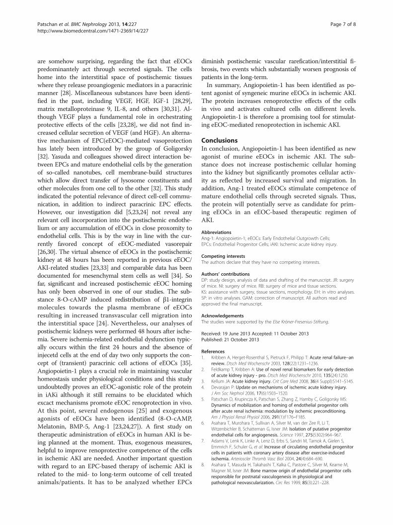
A
B
C.1
Figure 1 Cultured murine eEOCs and renal function after cell therapy. Ablue: nuclei, magnification × 100). Eighty-six ± 3.7% of all cultured cells showedAdministration of untreated eEOCs did not improve renal function but injectiocompared to mice without cell therapy and to mice with administration of unkidneys of cell-injected mice. Since previous investigations showed eEOC accumthe borderzone between medulla and papilla. Nevertheless, cell were de facells – C.2) (magnification in C × 40, IRI: ischemia reperfusion injury, Data as
samples were stored at −20°C. Frozen samples were cutinto 10 μm thick sections. Non-specific protein bindingwas blocked by 1 hour incubation with PBS-BSA (1%). Sec-tions were incubated with FITC-conjugated anti-mouseCD117 (c-Kit, 1:1000 in PBS-BSA 1%) (BD Biosciences,Rockville, MD, USA) or with the respective isotype controlfor 12 hours at 4°C. To visualize the nuclei, tissue sectionswere counterstained with DAPI (1:200 in PBS) (MolecularProbes, Eugene, OR, USA). Sections were examined as pre-viously described.
C.2
depicts BS 1+/acLDL+ cells (eEOCs) (green: BS 1 lectin-FITC, red: acLDL-Dil,double-positive staining. B shows renal function of cell injected animals.n of Ang-1 pretreated cells significantly improved renal function astreated eEOCs. C (C.1 and C.2) shows immunofluorescence images fromulation in the medullo-papillary area, our particular interest focused on
cto absent in both groups (untreated cells – C.1 and Ang-1 pretreatedmean ± SEM, *: p < 0.05).

Patschan et al. BMC Nephrology 2013, 14:227 Page 4 of 8http://www.biomedcentral.com/1471-2369/14/227
Serum creatinine analysisSerum creatinine concentration was measured using acommercially available kit (Creatinin PAP, Labor undTechnik - Eberhard Lehmann, Berlin, Germany) accordingto the manufacturer’s protocol.
Cell migration assaysThe eEOC cell migration assay was performed as pub-lished by Shi et al. [22]. Briefly, cells were grown on fibro-nectin coated 6 well plates. As soon as the well area wascompletely covered by cells (after approx. 5–6 days), anartificial wound was created, using the tip of a syringe.Cells remained in either Angiopoietin-1 free EBM-2 orin Ang-1 containing medium (250 ng/ml). Incubationtime was one hour in every series of experiments. Eachseries was performed three times. After cell washing withAng-1 free EBM-2, images of the respective wound areaswere taken at 0 and 24 hours. Commercially availablehuman umbilical vein endothelial cells (HUVECs – PCS-100-013, ATTC, Wesel, Germany) were cultured on fi-bronectin coated 6 well plates in EBM-2. An artificalwound was created as described. Cells were incubated
Figure 2 Effects of Ang-1 on migratory activity of cultured eEOCs (A) anmedium from Ang-1 treated eEOCs. Ang-1 induced faster direct and indirect wwound healing assay with C – Control; B: wound area reduction after 24 hoursreduction after 27 hours). Number of analysis in each group: 6, Data as mean ±
with supernatant EBM-2 from eEOCs that were incu-bated with Ang-1 for one hour. After incubation, super-natant was removed and cells remained in fresh EBM-2for one more hour. Supernatant was then used for treat-ment of HUVECs. Incubation time with supernatant was3 hours. Images of the respective wound areas were takenat 3 and 27 hours. The experiment was performed atleast three times.
ELISA studiesFor all in vitro cell experiments, a commercially availablemurine ’early outgrowth’ endothelial progenitor cell linewas purchased (66110–37 - Celprogen stem cell researchand therapeutics, San Pedro, CA, USA). Cells were cul-tured according to the manufacturer’s protocol. Celltreatment with Ang-1 was performed as described above.For measuring levels of Vascular Endothelial Growth Fac-tor, Insulin-like Growth Factor-1, Interleukin-6, andTransforming Growth Factor-β in the culture medium at24 hours after cell treatment, commercially availableELISA tests were performed according to the manufac-turer’s protocol (R&D Systems, Minneapolis, MN, USA).
d cultured mature endothelial cells (C). The latter were incubated withound closing, indicating proendothelial effects of the protein (A: direct; C: indirect wound healing assay with C – Control, D: wound areaSEM, *: p < 0.05).

Patschan et al. BMC Nephrology 2013, 14:227 Page 5 of 8http://www.biomedcentral.com/1471-2369/14/227
For measuring tissue levels of IL-6 and TGF-β, frozen kid-ney samples were thawed and frozen for 3 times in PBS.After the last cycle, tissue homogenates were centrifugedat 5.000 g and supernatants were employed for ELISAstudies.
Analysis of eEOC apoptosis and necrosisFor analyzing the effects of Ang-1 on eEOC apoptosisand necrosis, cultured murine eEOCs were incubatedwith either medium alone or with Ang-1 containingmedium (250 ng/ml). As reported earlier [23], apoptosis/necrosis was induced by treating the cells with TGF-β.Cells were first incubated with TGF-β (TGF-β 5 ng/ml), insome experiments Ang-1 (250 ng/ml) was added after aperiod of 2 hours, both mediators remained in the culturemedium for one more hour. Either untreated or pretreatedcells (TGF-β alone, TGF-β/Ang-1 combined) were washedand 24 hours later apoptosis and necrosis were evaluated.For analyzing apoptosis, the percentage of Annexin V +cells was quantified using a commercially available kit(BD, Heidelberg, Deutschland) according to the manufac-turer’s protocol. Necrosis was measured by cytometric ana-lysis. Briefly, cells with positive uptake of propidium iodidewere defined as necrotic.
Statistical analysisThe results were expressed as mean ± SEM. The meansof two populations were compared by Student’s t test
Figure 3 Analysis of eEOC release of VEGF (A), IGF-1 (B), and TGF-β (CeEOCs (Data as mean ± SEM).
(unpaired with equal variance). Differences between threeor more groups were compared by ANOVA one way ana-lysis of variance. Differences were considered significant atp < 0.05.
ResultsAngiopoietin-1 increases renoprotective competenceof eEOCs in iAKIAfter a 40 minutes period of ischemia mice were intra-venously injected with 0.5 × 10⁶ Ang-1 pretreated cells.Serum creatinine levels of cell injected animals signifi-cantly improved as compared to postischemic mice thatdid not receive any cells at all or that received un-treated cells (0.15 ± 0.01 mg/dl vs. 0.22 ± 0.03 mg/dl or vs.0.19 ± 0.004 mg/dl, p = 0.009 and p = 0.02 – Figure 1).
Angiopoietin-1 does neither increase nor decreasepostischemic eEOC homingIn order to analyze postischemic cell homing, some exper-iments required eEOC labelling with cell tracker® solution(CM-Dil). Cell tracker® incubation of eEOCs was exclu-sively performed for analyzing cell homing since prelimin-ary experiments using Ang-1 pretreated cells showed thatrenal function can deteriorate after labelling the cells (datanot shown). Immunofluorescence analysis showed de factono cell infiltrates in all injected animals as it has beenreported in previous studies [5,23,24]. Representative im-ages are shown in Figure 1.
). There were no differences between untreated and Ang-1 pretreated

02468
10121416
Ann
exin
V+
/PI+
cells
(%
)A
nnex
in V
+/P
I-ce
lls (
%)
0
20
40
60
80
100
*
A
B
Figure 4 Apoptosis and necrosis of Ang-1 treated eEOCs.Percentages of Annexin V+/PI+ (double-positive) cells were significantlylower in Ang-1 incubated cells as compared to untreated eEOCs (A).B shows percentages of Annexin V+/PI- cells. Percentages did not differbewteen the groups which indicated that differences in double-positivecells were the result of increased necrosis rather than apoptosis (Data asmean ± SEM, *: p < 0.05).
Patschan et al. BMC Nephrology 2013, 14:227 Page 6 of 8http://www.biomedcentral.com/1471-2369/14/227
Angiopoietin-1 stimulates migration of cultured murineeEOCsWe next aimed to determine the migratory activity ofcultured eEOCs in the presence of Ang-1. Angiopoietin-1 significantly accelerated wound closing as indicated bysmaller wound areas at 24 hours after treatment (69 ± 8%wound closing vs. 32 ± 22% wound closing, p = 0.03,Figure 2).
Angiopoietin-1 stimulates migration of cultured humanendothelial cells in an indirect mannerAfter artificial wound induction, human umbilical veinendothelial cells were incubated with medium fromAng-1 treated eEOCs. This measure significantly acceler-ated wound closing (92 ± 8% wound closing vs. 30 ± 6%wound closing, p = 0.0005, Figure 2), indicating thatAngiopoietin-1 treatment of eEOCs increases indirectproangiogenic effects of the cells.
Angiopoietin-1 does not increase eEOC release ofproangiogenic or inflammatory mediatorsWe next evaluated eEOC production/secretion of differentproangiogenic and proinflammatory mediators. Twenty-four hours after a one hour period of Ang-1 treatment,concentrations of vascular endothelial growth factor(VEGF), Insulin-like Growth Factor-1 (IGF-1), Interleukin-6 (IL-6), and Transforming Growth Factor-β (TGF-β) werenot different in the supernatants from untreated as com-pared to Ang-1 treated eEOCs (Figure 3). Results of IL-6measurements are not part of Figure 3 since the cytokinewas almost not detectable at all in supernatant fromuntreated/treated cells.
Angiopoietin-1 reduces apoptosis/necrosis of eEOCsFinally, we evaluated whether Angiopoietin-1 modu-lates TGF-β induced apoptosis/necrosis of cultured mur-ine eEOCs. Twenty-four hours after a one hour periodof Angiopoietin-1 treatment, eEOC were analyzed forAnnexin V expression and propidiumiodide uptake.Angiopoietin-1 significantly reduced percentages ofdouble-positive cells (6.8 ± 1.1% vs. 11.6 ± 1.9%,p = 0.0004 – Figure 4). In fact both processes of cell dam-age (apoptosis and necrosis) were reduced in the presenceof Ang-1 but necrosis, as represented by uptake of PI wasdecreased to an even greater extent. Thus, the AnnexinV/PI ratio was diminished as well.
DiscussionEarly Endothelial Outgrowth Cells significantly protectmice from acute ischemic renal failure. This has mean-while been proven in a number of different experimentalstudies [5,23-26]. In attempting to establish the cells asfuture therapeutic tool in ischemic AKI, different strategieshave been identified, helpful to increase renoprotective
effects of eEOCs. In 2010, 8-O-cAMP was identified aseEOC agonist, inducing redistribution of integrin mole-cules towards the plasma membrane of the cells therebyenhancing postischemic cell homing [24]. The hormonemelatonin improved antiischemic actions of eEOCs in AKIas well [23]. Recently, the protein BMP-5 was identified asanother eEOC agonist in this situation [27]. Thus, EarlyEndothelial Outgrowth Cells increasingly become a thera-peutic option in ischemic AKI.Aim of the current study was to analyze consequences
of Angiopoietin-1 (Ang-1) treatment of eEOCs in thecontext of eEOC-mediated postischemic kidney repair.Pretreatment of the cells resulted in significant lowerconcentrations of serum creatinine in cell-injected mice,which was accompanied by higher resistance of culturedeEOCs against TGF-β induced apoptosis/necrosis and byincreased cell migration. In addition, supernatant fromAng-1 incubated eEOCs markedly enhanced migratory ac-tivity of cultured mature endothelial cells. Postischemiccell homing or production/secretion of proangiogenic me-diators remained unaffected by Ang-1. The latter findings
Patschan et al. BMC Nephrology 2013, 14:227 Page 7 of 8http://www.biomedcentral.com/1471-2369/14/227
are somehow surprising, regarding the fact that eEOCspredominantely act through secreted signals. The cellshome into the interstitial space of postischemic tissueswhere they release proangiogenic mediators in a paracrinicmanner [28]. Miscellaneous substances have been identi-fied in the past, including VEGF, HGF, IGF-1 [28,29],matrix metalloproteinase 9, IL-8, and others [30,31]. Al-though VEGF plays a fundamental role in orchestratingprotective effects of the cells [23,28], we did not find in-creased cellular secretion of VEGF (and HGF). An alterna-tive mechanism of EPC(eEOC)-mediated vasoprotectionhas lately been introduced by the group of Goligorsky[32]. Yasuda and colleagues showed direct interaction be-tween EPCs and mature endothelial cells by the generationof so-called nanotubes, cell membrane-build structureswhich allow direct transfer of lysosome constituents andother molecules from one cell to the other [32]. This studyindicated the potential relevance of direct cell-cell commu-nication, in addition to indirect paracrinic EPC effects.However, our investigation did [5,23,24] not reveal anyrelevant cell incorporation into the postischemic endothe-lium or any accumulation of eEOCs in close proxomity toendothelial cells. This is by the way in line with the cur-rently favored concept of eEOC-mediated vasorepair[26,30]. The virtual absence of eEOCs in the postischemickidney at 48 hours has been reported in previous eEOC/AKI-related studies [23,33] and comparable data has beendocumented for mesenchymal stem cells as well [34]. Sofar, significant and increased postischemic eEOC hominghas only been observed in one of our studies. The sub-stance 8-O-cAMP induced redistribution of β1-integrinmolecules towards the plasma membrane of eEOCsresulting in increased transvascular cell migration intothe interstitial space [24]. Nevertheless, our analyses ofpostischemic kidneys were performed 48 hours after ische-mia. Severe ischemia-related endothelial dysfunction typic-ally occurs within the first 24 hours and the absence ofinjecetd cells at the end of day two only supports the con-cept of (transient) paracrinic cell actions of eEOCs [35].Angiopoietin-1 plays a crucial role in maintaining vascularhomeostasis under physiological conditions and this studyundoubtedly proves an eEOC-agonistic role of the proteinin iAKi although it still remains to be elucidated whichexcact mechanisms promote eEOC renoprotection in vivo.At this point, several endogenous [25] and exogenousagonists of eEOCs have been identified (8-O-cAMP,Melatonin, BMP-5, Ang-1 [23,24,27]). A first study ontherapeutic administration of eEOCs in human AKI is be-ing planned at the moment. Thus, exogenous measures,helpful to improve renoprotective competence of the cellsin ischemic AKI are needed. Another important questionwith regard to an EPC-based therapy of ischemic AKI isrelated to the mid- to long-term outcome of cell treatedanimals/patients. It has to be analyzed whether EPCs
diminish postischemic vascular rarefication/interstitial fi-brosis, two events which substantially worsen prognosis ofpatients in the long-term.In summary, Angiopoietin-1 has been identified as po-
tent agonist of syngeneic murine eEOCs in ischemic AKI.The protein increases renoprotective effects of the cellsin vivo and activates cultured cells on different levels.Angiopoietin-1 is therefore a promising tool for stimulat-ing eEOC-mediated renoprotection in ischemic AKI.
ConclusionsIn conclusion, Angiopoietin-1 has been identified as newagonist of murine eEOCs in ischemic AKI. The sub-stance does not increase postischemic cellular hominginto the kidney but significantly promotes cellular activ-ity as reflected by increased survival and migration. Inaddition, Ang-1 treated eEOCs stimulate competence ofmature endothelial cells through secreted signals. Thus,the protein will potentially serve as candidate for prim-ing eEOCs in an eEOC-based therapeutic regimen ofAKI.
AbbreviationsAng-1: Angiopoietin-1; eEOCs: Early Endothelial Outgrowth Cells;EPCs: Endothelial Progenitor Cells; iAKI: Ischemic acute kidney injury.
Competing interestsThe authors declare that they have no competing interests.
Authors’ contributionsDP: study design, analysis of data and drafting of the manuscript. JR: surgeryof mice. NI: surgery of mice. RB: surgery of mice and tissue sections.KS: assistance with surgery, tissue sections, morphology. EH: in vitro analyses.SP: in vitro analyses. GAM: correction of manuscript. All authors read andapproved the final manuscript.
AcknowledgementsThe studies were supported by the Else Kröner-Fresenius-Stiftung.
Received: 19 June 2013 Accepted: 11 October 2013Published: 21 October 2013
References1. Kribben A, Herget-Rosenthal S, Pietruck F, Philipp T: Acute renal failure--an
review. Dtsch Med Wochenschr 2003, 128(22):1231–1236.2. Feldkamp T, Kribben A: Use of novel renal biomarkers for early detection
of acute kidney injury - pro. Dtsch Med Wochenschr 2010, 135(24):1250.3. Kellum JA: Acute kidney injury. Crit Care Med 2008, 36(4 Suppl):S141–S145.4. Devarajan P: Update on mechanisms of ischemic acute kidney injury.
J Am Soc Nephrol 2006, 17(6):1503–1520.5. Patschan D, Krupincza K, Patschan S, Zhang Z, Hamby C, Goligorsky MS:
Dynamics of mobilization and homing of endothelial progenitor cellsafter acute renal ischemia: modulation by ischemic preconditioning.Am J Physiol Renal Physiol 2006, 291(1):F176–F185.
6. Asahara T, Murohara T, Sullivan A, Silver M, van der Zee R, Li T,Witzenbichler B, Schatteman G, Isner JM: Isolation of putative progenitorendothelial cells for angiogenesis. Science 1997, 275(5302):964–967.
7. Adams V, Lenk K, Linke A, Lenz D, Erbs S, Sandri M, Tarnok A, Gielen S,Emmrich F, Schuler G, et al: Increase of circulating endothelial progenitorcells in patients with coronary artery disease after exercise-inducedischemia. Arterioscler Thromb Vasc Biol 2004, 24(4):684–690.
8. Asahara T, Masuda H, Takahashi T, Kalka C, Pastore C, Silver M, Kearne M,Magner M, Isner JM: Bone marrow origin of endothelial progenitor cellsresponsible for postnatal vasculogenesis in physiological andpathological neovascularization. Circ Res 1999, 85(3):221–228.

Patschan et al. BMC Nephrology 2013, 14:227 Page 8 of 8http://www.biomedcentral.com/1471-2369/14/227
9. Khakoo AY, Finkel T: Endothelial progenitor cells. Annu Rev Med 2005,56:79–101.
10. Lambiase PD, Edwards RJ, Anthopoulos P, Rahman S, Meng YG, Bucknall CA,Redwood SR, Pearson JD, Marber MS: Circulating humoral factors andendothelial progenitor cells in patients with differing coronary collateralsupport. Circulation 2004, 109(24):2986–2992.
11. Rafii S, Lyden D: Therapeutic stem and progenitor cell transplantation fororgan vascularization and regeneration. Nat Med 2003, 9(6):702–712.
12. Urbich C, Dimmeler S: Endothelial progenitor cells: characterization androle in vascular biology. Circ Res 2004, 95(4):343–353.
13. Assmus B, Fischer-Rasokat U, Honold J, Seeger FH, Fichtlscherer S, Tonn T,Seifried E, Schächinger V, Dimmeler S, Zeiher AM: Transcoronarytransplantation of functionally competent BMCs is associated with adecrease in natriuretic peptide serum levels and improved survival ofpatients with chronic postinfarction heart failure: results of theTOPCARE-CHD Registry. Circ Res 2007, 100(8):1234–1241.
14. Assmus B, Honold J, Schächinger V, Britten MB, Fischer-Rasokat U, Lehmann R,Teupe C, Pistorius K, Martin H, Abolmaali ND, et al: Transcoronarytransplantation of progenitor cells after myocardial infarction. N Engl JMed 2006, 355(12):1222–1232.
15. Shaffer RG, Greene S, Arshi A, Supple G, Bantly A, Moore JS, Parmacek MS,Emile RM: Effect of acute exercise on endothelial progenitor cells inpatients with peripheral arterial disease. Vasc Med 2006, 11(4):219–226.
16. Thurston G: Role of Angiopoietins and Tie receptor tyrosine kinases inangiogenesis and lymphangiogenesis. Cell Tissue Res 2003, 314(1):61–68.
17. Suri C, McClain J, Thurston G, McDonald DM, Zhou H, Oldmixon EH, Sato TN,Yancopoulos GD: Increased vascularization in mice overexpressingangiopoietin-1. Science 1998, 282(5388):468–471.
18. Maisonpierre PC, Suri C, Jones PF, Bartunkova S, Wiegand SJ, Radziejewski C,Compton D, McClain J, Aldrich TH, Papadopoulos N, et al: Angiopoietin-2,a natural antagonist for Tie2 that disrupts in vivo angiogenesis. Science 1997,277(5322):55–60.
19. Hansen TM, Singh H, Tahir TA, Brindle NP: Effects of angiopoietins-1 and −2on the receptor tyrosine kinase Tie2 are differentially regulated at theendothelial cell surface. Cell Signalling 2010, 22(3):527–532.
20. Gamble JR, Drew J, Trezise L, Underwood A, Parsons M, Kasminkas L, Rudge J,Yancopoulos G, Vadas MA: Angiopoietin-1 is an antipermeability andanti-inflammatory agent in vitro and targets cell junctions. Circ Res 2000,87(7):603–607.
21. Abdel-Malak NA, Mofarrahi M, Mayaki D, Khachigian LM, Hussain SNA: Earlygrowth response-1 regulates angiopoietin-1-induced endothelial cellproliferation, migration, and differentiation. Arterioscler Thromb Vasc Biol2009, 29(2):209–216.
22. Shi H, Patschan D, Dietz GPH, Bähr M, Plotkin M, Goligorsky MS: Glial cellline-derived neurotrophic growth factor increases motility and survivalof cultured mesenchymal stem cells and ameliorates acute kidney injury.Am J Physiol Renal Physiol 2008, 294(1):F229–F235.
23. Patschan D, Hildebrandt A, Rinneburger J, Wessels JT, Patschan SA, Becker JU,Henze E, Kruger A, Muller GA: The hormone Melatonin stimulatesrenoprotective effects of early outgrowth endothelial progenitor cells inacute ischemic kidney injury. Am J Physiol Renal Physiol 2012, 302:F1305–F1312.
24. Patschan D, Patschan S, Wessels JT, Becker JU, David S, Henze E, Goligorsky MS,Müller GA: Epac-1 activator 8-O-cAMP augments renoprotective effects ofsyngeneic [corrected] murine EPCs in acute ischemic kidney injury. Am JPhysiol Renal Physiol 2010, 298(1):F78–F85.
25. Patschan D, Patschan S, Gobe GG, Chintala S, Goligorsky MS: Uric acidheralds ischemic tissue injury to mobilize endothelial progenitor cells.J Am Soc Nephrol 2007, 18(5):1516–1524.
26. Patschan D, Plotkin M, Goligorsky MS: Therapeutic use of stem andendothelial progenitor cells in acute renal injury: ça ira. Curr OpinPharmacol 2006, 6(2):176–183.
27. Patschan D, Schwarze K, Lange A, Meise N, Henze E, Becker JU, Patschan S,Muller GA: Bone morphogenetic protein-5 and early endothelial outgrowthcells (eEOCs) in acute ischemic kidney injury (AKI) and 5/6-chronic kidneydisease. Am J Physiol Renal Physiol 2013, 305(3):F314–F322.
28. Patschan D, Patschan S, Muller GA: Endothelial progenitor cells in acuteischemic kidney injury: strategies for increasing the cells’ renoprotectivecompetence. Int J Nephrol 2011, 2011:828369.
29. Rehman J, Li J, Orschell CM, March KL: Peripheral blood “endothelialprogenitor cells” are derived from monocyte/macrophages and secreteangiogenic growth factors. Circulation 2003, 107(8):1164–1169.
30. Goligorsky MS, Yasuda K, Ratliff B: Dysfunctional endothelial progenitorcells in chronic kidney disease. J Am Soc Nephrol 2010, 21(6):911–919.
31. Pula G, Mayr U, Evans C, Prokopi M, Vara DS, Yin X, Astroulakis Z, Xiao Q, Hill J,Xu Q, et al: Proteomics identifies thymidine phosphorylase as a key regulatorof the angiogenic potential of colony-forming units and endothelialprogenitor cell cultures. Circ Res 2009, 104(1):32–40.
32. Yasuda K, Khandare A, Burianovskyy L, Maruyama S, Zhang F, Nasjletti A,Goligorsky MS: Tunneling nanotubes mediate rescue of prematurelysenescent endothelial cells by endothelial progenitors: exchange oflysosomal pool. Aging 2011, 3(6):597–608.
33. Patschan D, Backhaus R, Elle HJ, Schwarze K, Henze E, Becker JU, Patschan S,Muller GA: Angiopoietin-2 modulates eEOC-mediated renoprotection inAKI in a dose-dependent manner. J Nephrol 2013, 26(4):667–674.
34. Togel F, Yang Y, Zhang P, Hu Z, Westenfelder C: Bioluminescence imagingto monitor the in vivo distribution of administered mesenchymal stem cellsin acute kidney injury. Am J Physiol Renal Physiol 2008, 295(1):F315–F321.
35. Yamamoto T, Tada T, Brodsky SV, Tanaka H, Noiri E, Kajiya F, Goligorsky MS:Intravital videomicroscopy of peritubular capillaries in renal ischemia.Am J Physiol Renal Physiol 2002, 282(6):F1150–F1155.
doi:10.1186/1471-2369-14-227Cite this article as: Patschan et al.: Angiopoietin-1 treated early endothelialoutgrowth cells (eEOCs) are activated in vitro and reduce renal damage inmurine acute ischemic kidney injury (iAKI). BMC Nephrology 2013 14:227.
Submit your next manuscript to BioMed Centraland take full advantage of:
• Convenient online submission
• Thorough peer review
• No space constraints or color figure charges
• Immediate publication on acceptance
• Inclusion in PubMed, CAS, Scopus and Google Scholar
• Research which is freely available for redistribution
Submit your manuscript at www.biomedcentral.com/submit