truncated trkb receptor-induced outgrowth of dendritic
TRANSCRIPT

IntroductionThe protein family of mammalian neurotrophins – consistingof NGF, BDNF, NT-3 and NT-4/5 – is known to supportsurvival, differentiation and several forms of synaptic plasticityin CNS neurons (Poo, 2001; Lu, 2003).
The secreted neurotrophins mediate their effects viaactivation of two different classes of plasma membranereceptors (Patapoutian and Reichardt, 2001). First, eachneurotrophin can bind as a homodimer to its specific Trktyrosine kinase receptor (NGF binds to TrkA, BDNF and NT-4/5 to TrkB and NT-3 preferentially binds to TrkC). Secondly,all neurotrophins are known to bind with equal specificity to thep75 neurotrophin receptor (p75NTR). Upon binding ofneurotrophins, Trks activate a tyrosine kinase signalling cascadethat accounts for numerous neurotrophin-dependent biologicaleffects in neurons (Patapoutian and Reichardt, 2001). Thep75NTR modulates Trk signalling and can even initiate cellularresponses independently from Trk signalling (see Kaplan andMiller, 2000). This is accomplished via protein-proteininteractions with a set of intracellular binding partners includinge.g., RhoA (a small G-protein involved in neurite outgrowth),NRAGE, NRIF and TRAFs (Roux and Barker, 2002). In
addition, p75NTR can initiate the production of ceramide viaintermediate activation of neutral sphingomyelinase(Dobrowsky and Carter, 1998). This pathway has recently beenimplicated in p75-mediated growth of neuronal processes inhippocampal neurons (Brann et al., 1999; Brann et al., 2002).
The rat TrkB tyrosine kinase receptor for BDNF and NT-4/5(TrkB.FL) can also exist as alternatively spliced truncatedreceptor isoforms TrkB.T1 and TrkB.T2, respectively, whichlack the intracellular kinase domain (Klein et al., 1990;Middlemas, 1991). Although it is recognised that the TrkB.T1receptor is the dominant TrkB isoform expressed in the adultrodent CNS (Cabelli, 1996), the physiological function of thesetruncated receptors are still not well established andintracellular downstream effectors have long remained elusive(see Baxter et al., 1997; Kryl and Barker, 2000; Rose et al.,2003). Most evidence suggests a role of TrkB.T1 and T2 inligand presentation or in ligand scavenging for TrkB.FL(Bothwell, 1995; Biffo et al., 1995), or as a dominant negativeinhibitor of TrkB.FL signalling via formation of heterodimers(Eide et al., 1996; Ninkina et al., 1996).
Since one of the classical actions of neurotrophins is topromote neuronal fibre outgrowth (Levi-Montalcini and
5803
The Trk family of receptor tyrosine kinases and the p75receptor (p75NTR) mediate the effects of neurotrophins onneuronal survival, differentiation and synaptic plasticity.The neurotrophin BDNF and its cognate receptor tyrosinekinase, TrkB.FL, are highly expressed in neurons of thecentral nervous system. At later stages in postnataldevelopment the truncated TrkB splice variants (TrkB.T1,TrkB.T2) become abundant. However, the signalling andfunction of these truncated receptors remained largelyelusive.
We show that overexpression of TrkB.T1 in hippocampalneurons induces the formation of dendritic filopodia, whichare known precursors of synaptic spines. The induction offilopodia by TrkB.T1 occurs independently of neurotrophinbinding and of kinase activity of endogenous TrkB.FL.Coexpression of a p75NTR lacking an intracellular domain
inhibits the TrkB.T1-induced effect in a dominant negativemanner. Steric hindrance of extracellular p75NTR
interactions with a specific antibody, or absence of p75NTR
with an intact extracellular domain also inhibit thisTrkB.T1-induced effect.
We thus propose a novel signalling pathway initiated byneurotrophin-independent extracellular or intramembraneinteraction of TrkB.T1 with the p75NTR receptor, whichmodulates dendritic growth via p75NTR signalling cascades.
Supplementary material available online athttp://jcs.biologists.org/cgi/content/full/117/24/5803/DC1
Key words: Neurotrophins, Trk, p75 receptor, Filopodia, BDNF,Spines
Summary
Truncated TrkB receptor-induced outgrowth ofdendritic filopodia involves the p75 neurotrophinreceptorMatthias Hartmann1,2, Tanja Brigadski1, Kai S. Erdmann2, Bettina Holtmann3, Michael Sendtner3, Frank Narz2
and Volkmar Leßmann1,*1Institute of Physiology and Pathophysiology, Johannes Gutenberg-University Mainz, Duesbergweg 6, 55128 Mainz, Germany2Department of Molecular Neurobiochemistry, Ruhr-University Bochum, Universitätsstraße 150, 44801 Bochum, Germany3Institute for Clinical Neurobiology, Julius-Maximilians University Würzburg, Sanderring 2 - 97070 Würzburg, Germany*Author for correspondence (e-mail: [email protected])
Accepted 24 August 2004Journal of Cell Science 117, 5803-5814 Published by The Company of Biologists 2004doi:10.1242/jcs.01511
Research Article

5804
Hamburger, 1953), it was an intriguing recent finding, thatoverexpression of TrkB.T1 receptors in cortical slice culturescan initiate dendritic branching, which is negatively regulatedby phosphorylated TrkB.FL receptors coexpressed in the samecells (Yacoubian and Lo, 2000). Comparable effects werereported for filopodial growth induced by ectopic expressionof TrkB.T1 in a neuroblastoma cell line (Haapasalo et al.,1999). However, both studies did not delineate the mechanismof these TrkB.T1-induced effects.
In order to investigate the underlying signalling mechanismof TrkB.T1-induced process outgrowth in postnatal rathippocampal neurons, we overexpressed GFP-tagged versionsof the different TrkB receptor isoforms in neuronmicrocultures. We found that overexpression of TrkB.T1 andTrkB.T2 induced a two- to threefold increase in the numberof dendritic filopodia. This effect was independent ofneurotrophin binding and was also seen with a mutant TrkB.T1receptor that completely lacked the cytoplasmic tail.Coexpression of TrkB.FL had a dominant negative effect onTrkB.T1-induced filopodial growth. As revealed by applicationof an antibody that binds to the extracellular epitope of thep75NTR and that inhibits filopodia formation, this TrkB.T1-induced growth of filopodia involves p75NTR signalling.Accordingly, the TrkB.T1-induced effect is inhibited bycoexpression of a dominant negative p75NTR, which lacks thecytoplasmic domain, and is significantly reduced in neuronsderived from exon III p75NTR knockout mice.
These data add a new facet to the functions of NT receptorsignalling, suggesting an activation of certain aspects ofp75NTR signalling by interaction with non-liganded truncatedTrkB receptors. This interaction could play a role in promotingsynapse formation via dendritic filopodia.
Materials and MethodsConstruction of TrkB expression vectors To facilitate detection of protein expression in living cells, greenfluorescent protein (GFP) was fused C-terminally, i.e. to the intracel-lular domain, of the three known rat TrkB isoforms TrkB.FL, TrkB.T1and TrkB.T2 (Middlemas et al., 1991). TrkB cDNAs used as templatesfor PCR amplification were kindly provided by D. Middlemas.Oligonucleotides were synthesised for the respective 3′ and 5′ terminito allow restriction digestion after the PCR reaction and deletion ofstop codons in case of GFP fusion proteins (e.g. 3′ primers: TrkB.FL-GFP, GATGGATCCCGGCCTAGGATGTCCAGGTAG; TrkB.T1-GFP, GATGGATCCCGCCCATCCAGGGGGATCTTATG; TrkB.T2-GFP, GATGGATCCCGAGAAGCAAAATAAGCACACTTCTG;TrkB.T1∆ICD-GFP, GATGGATCCGTCGCCAACTTGAGCAGA-AGC. 5′ primer for all four constructs: ACGAATTCGCCACCAT-GTCGCCCTGGCCGAGGTG). Amplified fragments were digestedwith EcoRI and BamHI and ligated into the pEGFP-N1 or pDsRed2-N1 vector (CLONTECH Inc.). Unfused controls (TrkB.T1, wtTrkB.FL) were constructed accordingly without removal of the stopcodon. In order to examine the role of p75NTR signalling, a p75NTR-GFP fusion protein and the deletion construct p75∆ICD (fused toDsRed) were cloned using a similar strategy. All constructs wereverified by sequencing. Expression of all constructs used in this studywas driven by CMV promoters.
Cell cultureCOS7 and PC12 cells were grown in Dulbecco’s Modified Eagle’sMedium (DMEM; Gibco) containing either 10% fetal calf serum
(COS7) or 10% horse serum and 5% fetal calf serum (PC12) andsupplemented with penicillin and streptomycin (37°C in 10% CO2).Upon reaching confluence, cells were passaged 1:3 (PC12) or 1:7(COS7). For transfection and fluorescence imaging, cells were platedon uncoated glass coverslips with a density of 100,000 cells/cm2.Transfection was performed 1 day after plating, fluorescence imaging1-2 days after transfection.
Hippocampal microculturesDissociated postnatal rat hippocampal microcultures were prepared asdescribed previously (Lessmann and Heumann, 1998), with minormodifications. Primary postnatal (P0-P2) neocortical astrocytes wereisolated and cultured on glass cover slips at low density in DMEMmedium containing 10% FCS, to yield astrocyte islands of 100-300µm in diameter after 7-14 days in vitro (DIV). Postnatal rathippocampal neurons were plated at a density of 5-20 neurons perastrocyte island onto the astrocyte coverslips, and neurons werecultured in serum-free medium (Neurobasal with 2% B27; Gibco).After 4 days, 4 µM ARAC was added to inhibit glial proliferation.Cultures from p75NTR knockout mice were prepared accordingly fromthe offspring of homozygous male and female p75NTR knockout mice(Lee et al., 1992), obtained from a local breeding colony.
TransfectionRat hippocampal microcultures were transfected with the expressionplasmids at 8-9 DIV, using the calcium phosphate precipitationmethod as described previously (Haubensak et al., 1998). Aftertransfection, 4% fetal calf serum (FCS) was added. Cells were usedfor experiments 2-3 days after transfection (10-11 DIV). Culturesfrom p75NTR knockout mice and from C57/BL6 wild-type mice weretransfected at 7-8 DIV with TrkB.T1-GFP and GFP, respectively (1µg), and cotransfected with DsRed2 plasmid (2 µg). No FCS wasadded. Quantification of filopodia in these experiments was performedby a person blind to the identity of the cultures.
PC12 and COS7 cells were transfected with the polyethylenimine(PEI) method as described by others (Lambert et al., 1996). Briefly,5 µg DNA per 35 mm dish were diluted in 125 µl of 0.15 M NaCl,mixed with 15 µl PEI solution (900 µg/ml 50% PEI 800 kDa, pH 6.5)and 110 µl 0.15 M NaCl, incubated for 10 minutes at roomtemperature and added to 1 ml serum-free DMEM. After incubatingthe cells with this solution for 3 hours (37°C, 10% CO2), the mediumwas changed to normal cell culture medium (see above). For the fibreoutgrowth assay, 20-30 green fluorescent cells were evaluated percoverslip.
Fluorescence imagingCoverslips with transfected cells were transferred into Petripermdishes (Vivascience) with folio bottoms and observation offluorescence signals was performed as described previously(Hartmann et al., 2001).
Cells were inspected through high aperture oil immersionobjectives (40×, NA: 1.0; 100×, NA: 1.35) of an invertedepifluorescence microscope (Olympus IX 70). GFP fluorescence wasdetected with narrow excitation (450-490 nm) and emission (dichroicmirror: 495 nm; 500-550 nm) band pass filters. Red fluorescence wasdetected with a custom built filter set (excitation: 530-550 nm;dichroic mirror: 570 nm; emission: 590-650 nm). Illumination of theprobe was restricted to image capture and controlled with anelectronic shutter device (Uni-Blitz). Pictures were captured with adigital CCD camera (Sensys 1401E, Photometrics). Data acquisitionand analysis was performed with MetaView software (UniversalImaging). Brightness and contrast of images were adjusted to makeuse of the dynamic range of colour representation. All images of agiven figure were treated equally.
Journal of Cell Science 117 (24)

5805TrkB.T1 induces dendritic filopodia via p75NTR
ImmunocytochemistryCoverslips of transfected cells were fixed (4% formaldehyde in PBS)and permeabilized (0.1% Triton X-100) using standard procedures(see Haubensak et al., 1998). Tau antibody (mouse, 1:200; BoehringerMannheim), TrkB.T1 antibody (TrkB[TK-], rabbit, 1:200; Santa CruzBiotechnology) and MAP2 antibody (mouse, 1:200; Sigma) wereincubated for 1 hour at room temperature (RT) followed by incubation(RT, 1 hour) with Alexa Green-coupled anti-rabbit secondary antibodyor Alexa Red-coupled anti-mouse secondary antibody (both 1:1000;Molecular Probes). Where required, polymerized filamentous actinwas stained by incubating with Phalloidin-TRITC (1:100, Sigma) for20 minutes (at RT).
Sholl analysisTo measure the complexity of the dendritic tree of cultured neurons,Sholl analysis was performed on single transfected neurons onmicroislands 2-3 days after transfection (10-11 DIV). Imaginarycircles with radii in increments of 5 µm up to 30 µm were drawnaround the cell body, and the number of intersections with fluorescentneurites of the same neuron were counted.
Quantification of the density of filopodiaTo determine the effect of TrkB.T1 expression on filopodia formation,the filopodic density of single transfected neurons on microislandswas determined 2 days after transfection (10 DIV). Of every neuronunder examination a 40 µm section of a typical dendrite that was inthe plane of focus was chosen for counting. For these sections thenumber of protrusions between 1 and 10 µm in length weredetermined. Inhibitors, ligands etc. were applied on the day oftransfection at concentrations as indicated. All results are given asmean values ± s.e.m. Statistical significance was determined using thetwo-tailed Student’s t-test. The absolute numbers of filopodia weredependent on experimental conditions. The batch of FCS used forplating, the person responsible for counting and the necessity to addFCS after transfection to rat but not to mouse neurons were the mostsensitive factors for the count of filopodia. Therefore negative (GFPtransfected or TrkB.FL transfected) and positive controls (TrkB.T1-GFP) were included in all experiments. Only data from experimentswith identical conditions were pooled.
ReagentsK252a and neurotrophins were obtained from Alomone Labs, TrkB-Fc receptor bodies from R&D Systems and MC192 monoclonalantibody was purchased from Chemicon. Intact biological activity ofneurotrophins, of TrkB-Fc receptor bodies, and of k252a was testedat regular intervals in a PC12 cell fibre outgrowth assay (seesupplementary material).
Intact biological function of neurotrophinsThe intact biological function of the exogenously addedneurotrophins in our experiments (i.e. NGF, BDNF, NT-4/5, 100ng/ml each), of the Trk kinase inhibitor k252a (200 nM) and of theTrkB receptor bodies (TrkB-Fc; 1 µg/ml) was tested at regularintervals using the above mentioned PC12 cell assay (see Fig. S1 insupplementary material). GFP-transfected PC12 cells (endogenouslyexpressing TrkA) were used to test NGF and k252a. TrkB.FL-GFP-expressing PC12 cells were employed to test BDNF and NT-4/5.TrkB.FL-expressing PC12 cells cotransfected with GFP were used toexamine the intact function of TrkB-Fc. Only neurotrophin batchesyielding maximal stimulation (i.e. >50%, compare Fig. S1 insupplementary material) of fibre outgrowth at a concentration of 100ng/ml were used for experiments with neurons. The Trk kinaseinhibitor k252a (200 nM) significantly inhibited NGF-induced fibre
outgrowth and proliferation of NGF-treated (100 ng/ml) PC12 cells(data not shown).
ResultsIn order to visualize the localization and assess the biologicalfunction of truncated TrkB receptors in hippocampal neuronswe constructed GFP-tagged versions of rat TrkB.FL, TrkB.T1and TrkB.T2 receptors. The GFP tag was fused via a sevenamino acid flexible spacer to the C terminus of the differentTrkB receptor isoforms (Fig. 1). These GFP-labeled constructsallowed us to directly identify transfected cells and to ascertainintact cell membrane targeting of the overexpressed constructsin each cell investigated (Klau et al., 2001). The intactbiological function of the GFP-tagged TrkB.FL receptor(TrkB.FL-GFP) was investigated by testing ligand-inducedprocess formation in TrkB.FL-GFP-transfected PC12 cells.This cell line is known to show fibre outgrowth whenectopically expressed TrkB receptors are activated by BDNF(Ip et al., 1993b; Fanger et al., 1995). One day after transienttransfection with our TrkB-GFP construct, BDNF or NT-4/5(100 ng/ml; 3 days) induced fibre outgrowth in these PC12cells to an extent, which was indistinguishable from, (i)the respective process formation obtained followingoverexpression of untagged TrkB.FL, and, (ii) from NGF-induced fibre outgrowth in the same cells (Fig. S1 insupplementary material). This is in line with the intact ligand-induced tyrosine autophosphorylation of TrkB-GFP observedin COS7 cells (data not shown) (see also Watson et al., 1999).
Upon transfection (at 8 DIV) of postnatal rat hippocampalneurons with the TrkB.T1-GFP and TrkB.FL-GFP constructs,we observed a correct plasma membrane localization of bothreceptor isoforms in the soma and in the processes of living
GFP
DsRed
TrkB.FL-GFP
TrkB.FL-DsRed
expression vector topology
extracellular intracellular
TrkB.FL
TrkB.FL
GFPTrkB.T2-GFP TrkB.T2
GFP
GFP
TrkB.T1-GFP
TrkB.T1
T1 ICD-GFP TrkB.T1 ICD
TrkB.T1
TrkB.T1
GFP
DsRed
p75-GFP
p75 ICD-DsRed p75 ICD
p75
p
p
p
p
p
p
p
p
Fig. 1. Topology of cloned TrkB and p75 fusion proteins. Greenfluorescent protein (GFP) and red fluorescent DsRed was fused C-terminally to full length rat TrkB (TrkB.FL), yielding TrkB.FL-GFPand TrkB.FL-DsRed. Similarly, the truncated TrkB isoforms wereconstructed to yield TrkB.T1-GFP and TrkB.T2-GFP. As a control,unfused TrkB.T1 was cloned into the same vector. The intracellulardomain of TrkB.T1 (black bar) was deleted in the construct T1∆ICD-GFP. C-terminal fusion of GFP to rat p75NTR yielded p75-GFP. Adominant negative p75NTR (lowermost construct) was generated byfusing DsRed to the transmembrane domain of p75NTR. The greyvertical bar shows the location of the plasma membrane; theextracellular space is to the left. The p indicates the pre sequencedirecting the receptor mRNAs to the rough ER.

5806
neurons (Fig. 2A). As expected, a substantial fraction of GFP-labeled TrkB.FL and of TrkB.T1 receptors was also found inintracellular compartments (Du et al., 2000; Haapasalo et al.,2002), the relative abundance of which was not furtheranalyzed here. As indicated by colocalization with thedendritic marker MAP2 (Fig. 2B), both receptors were targetedto dendritic compartments (see also Kryl et al., 1999). Inaddition, axonal targeting was also evident in all neuronsinvestigated, as indicated by the GFP fluorescence in MAP2-negative thin processes of transfected neurons (see arrows inFig. 2B), and from colocalization with the axonal markerprotein Tau (see Fig. S3 in supplementary material). Similarresults were obtained when TrkB.T2 receptors wereoverexpressed (data not shown). These results suggest generalneuronal (non-polarized) expression of the different TrkBreceptor variants in our hippocampal neurons.
Interestingly, the expression of TrkB.T1-GFP in postnatal rathippocampal neurons induced a highly significant (P<10–5)roughly twofold increase in the number of dendritic filopodia,as compared to TrkB.FL-GFP and wt-GFP-overexpressingcontrols (see Fig. 3). For these and all further experiments onlycells with a clear plasma membrane localization of theoverexpressed receptors were selected, however, owing toimage processing procedures necessary to visualize thinprocesses, membrane localization of the receptors is notevident in these pictures. Staining with TRITC-labeledphalloidin revealed the presence of filamentous actin in thesemembrane protrusions, which is a characteristic marker fordendritic filopodia. These filopodia typically give rise to the
formation of synaptic spines at later stages ofdevelopment (see Harris, 1999). As revealedby Sholl analysis, the number of primarydendrites and of proximal dendritic brancheswas not significantly affected in theseexperiments (see Fig. 3C), corroborating theselective effect on filopodia. In spite of theunpolarized expression of TrkB.T1 in ourneurons, we did not observe a similar effectof TrkB.T1 on the growth of axonal filopodia.
Given the membrane targeting of bothreceptors (see Fig. 2), the selective inductionof filopodia in TrkB.T1-GFP-overexpressingversus TrkB.FL-GFP-overexpressing cells
clearly indicates true induction of processes rather than justhighlighting of processes by membrane targeted GFP.
This interpretation is supported by control experimentsin which filopodia formation was analyzed by the redfluorescence of coexpressed DsRed protein in neuronstransfected with either TrkB.T1-GFP or wt-GFP: mousehippocampal neurons were cotransfected at 8 DIV with eitherTrkB.T1-GFP and DsRed (3:1) or with GFP and DsRed (3:1).Two days posttransfection, dendritic filopodia were counted inthe red fluorescent channel (DsRed) for both conditions. Therewas a roughly twofold visible increase in TrkB.T1-GFP-expressing neurons (significantly different with P<0.0001; seeFig. S2 in supplementary material). These results indicate thatthe TrkB.T1-induced induction of filopodia is also visible whencytosolic DsRed fluorescence is evaluated in both conditions.This is also evident from the largely overlapping fluorescenceof TrkB-T1-GFP and DsRed fluorescence in the same cells.Thus, either GFP- or TrkB.FL-GFP-expressing cells could beused as negative control in all subsequent experiments.
Further control experiments employing overexpression ofuntagged TrkB.T1 receptors and analysis of filopodia by thered fluorescence of coexpressed DsRed demonstrated a similarinduction of filopodia also for untagged TrkB.T1 receptors: rathippocampal neurons were cotransfected at 8 DIV withTrkB.T1 receptors (either TrkB.T1-GFP or wt TrkB.T1) and aDsRed expression plasmid (DNA ratio TrkB.T1:DsRed, 3:1).As a negative control, sister cultures were cotransfected withGFP and DsRed (DNA ratio, 3:1). Two days after transfection,dendritic filopodia were counted in the three groups in the red
Journal of Cell Science 117 (24)
Fig. 2. TrkB.FL-GFP and TrkB.T1-GFP arelocalized in the plasma membrane of hippocampalneurons and are targeted to dendrites and axons.(A) Hippocampal neurons, transfected withTrkB.FL-GFP (left) and TrkB.T1-GFP (right)expression plasmids at 8 days in vitro (DIV),showed a clear accumulation of GFP fluorescenceat the plasma membrane (2 days aftertransfection). (B) Immunocytochemistry oftransfected neurons with an antibody against thedendritic marker MAP2 (at 16 DIV, 8 days aftertransfection). TrkB.FL-GFP and TrkB.T1-GFP inhippocampal neurons (green), were targeted toboth, dendrites (MAP2-positive, red) and axons(MAP2-negative, arrows). Filopodia (smallprotrusions) were restricted to dendrites, whereasthe appearance of axons was generally smooth.Scale bars: 5 µm (A); 20 µm (B).

5807TrkB.T1 induces dendritic filopodia via p75NTR
fluorescent channel (DsRed). GFP-tagged and wt TrkB.T1 receptor bothshowed a 1.6-fold increased number ofdendritic filopodia compared to thenegative control (see Fig. S4 insupplementary material; both TrkB.T1groups significantly different from GFPwith P<0.001). These results indicatethat induction of filopodia by TrkB.T1is not an artefact of the GFP tag. Thisrules out that the GFP tag of TrkB.T1was responsible for the indcution offilopodia.
A similar increase in the growth ofdendritic filopodia was observed whenTrkB.T2-GFP was overexpressed inhippocampal neurons (data not shown).However, since TrkB.T1 is more highlyconserved between species and is theonly truncated receptor in humans(Biffo et al., 1995; Shelton et al., 1995),all subsequent experiments wereperformed with the TrkB.T1-GFPconstruct.
To assess the signaling mechanism ofthe TrkB.T1-induced outgrowth ofdendritic filopodia, we examined theeffects of endogenously released and ofexogenously added TrkB receptorligands, respectively (Fig. 4). In order toscavenge endogenously released BDNFand/or NT-4/5 in our cultures, 0.4 µg/mlTrkB-Fc receptor bodies (Shelton et al.,1995) were added at the day oftransfection (8 DIV). This treatment hadno effect on the magnitude of filopodialgrowth in either TrkB.T1-GFP- orTrkB.FL-GFP-overexpressing neurons.Likewise, the TrkB.T1-GFP-inducedgrowth of filopodia and the basal levelsof filopodia formation in TrkB.FL-GFP-expressing and wt-GFP-expressingneurons was not affected by theexogenous application of BDNF or NT-4/5 (100 ng/ml). These results indicatethat the TrkB.T1-induced growth offilopodia occurs independently fromneurotrophin binding. In addition, these data demonstrate thatthe basal formation of dendritic filopodia in our cultures isindependent of ligand-induced signalling via endogenouslyexpressed or overexpressed full length TrkB receptors.
The independence of the TrkB.T1-induced formation ofdendritic filopodia from full length TrkB signalling was furthercorroborated by experiments wherein the signalling of Trkreceptors was completely inhibited by application ofsupramaximal concentrations of the Trk kinase inhibitor K252a(200 nM, Fig. 4B): again, this treatment did not affect eitherthe TrkB.T1-induced formation of filopodia or the basal levelsof filopodia in control cultures expressing endogenous levelsof TrkB.FL. Thus, the TrkB.T1-induced formation of filopodiacannot be explained by an interference of the overexpressed
TrkB.T1 with the full-length TrkB tyrosine kinase signalling.Rather, our results suggest a novel independent signalling ofnon-liganded TrkB.T1 receptors in hippocampal neurons.
To examine whether the short intracellular tail of the TrkB.T1receptor, which is highly conserved among species, is necessaryto observe the effect on filopodial growth, we generated amutant lacking all of the intracellular domain of TrkB.T1(TrkB.T1∆ICD-GFP; see Fig. 1). Again, the GFP tag allowedus to directly ascertain that this mutant receptor was targeted tothe membrane (data not shown). To our surprise, overexpressionof this construct yielded induction of dendritic filopodia inhippocampal neurons to an extent that was indistinguishablefrom the TrkB.T1-GFP-induced effect (not significantlydifferent with P>0.14; Fig. 4C). The most likely explanation of
Fig. 3. TrkB.T1 expression in hippocampal neurons increases the number of dendriticfilopodia. (A) Typical examples of the morphology of TrkB.T1-GFP-, TrkB.FL-GFP- and GFP-expressing hippocampal neurons (at 11 DIV, 3 days after transfection). The increased numberof filopodia in TrkB.T1-GFP expressing neurons lead to a ruffled appearance (left) compared tothe much smoother TrkB.FL-GFP and GFP controls. (B) The increase in density of filopodiawas highly significant (n=26-91 cells, 5-13 independent experiments, P<10–5). (C) Shollanalysis of all neurons from B. The expression of TrkB.T1-GFP, had no significant effect on thecomplexity of the dendritic tree. (D) Typical dendrite of a TrkB.T1-GFP expressinghippocampal neuron (green, at 10 DIV). Phalloidin-TRITC staining (red) of TrkB.T1-GFP-induced filopodia revealed their filamentous actin content, which is characteristic for filopodia.Scale bars: 10 µm (A); 5 µm (D).

5808
this result is, that an extracellular or intramembrane interactionof the TrkB.T1 receptor with an unidentified membrane proteinis sufficient for the induction of filopodia.
Since the truncated TrkB receptors are known to formheterodimers with TrkB.FL (Eide et al., 1996; Ninkina et al.,1996) we reasoned that unliganded TrkB.FL could be theinteracting membrane protein for TrkB.T1 in our cells. Thus, wecoexpressed TrkB.FL-DsRed together with TrkB.T1-GFP (ratioTrkB.FL:TrkB.T1 DNA: 3:1) and compared the formation ofdendritic filopodia in these coexpressers to neurons expressingonly TrkB.T1-GFP in the same culture dishes (Fig. 5). In orderto definitely rule out endogenous Trk kinase signalling, 200 nMK252a was added at the day of transfection. Instead of disclosing
a role for a signalling competentTrkB.FL/TrkB.T1 heterodimer, theseexperiments revealed a dominant negativeeffect of coexpressed TrkB.FL on theTrkB.T1-induced formation of filopodia.These results clearly indicate that the full-length TrkB receptor can not be the transducerof TrkB.T1-dependent formation of dendritic
Journal of Cell Science 117 (24)
mµ04/
aidopoliffo.on
B10
8
6
4
2
0
TrkB.T1- + - + - +
TrkB.FL GFPK252a
*
C
mµ04/
aidopoliff o.on
8
6
4
2
0TrkB.T1
-GFPT1 ICD
-GFPTrkB.FL
-GFP
p > 0.14
*
mµ04/
aidop ol iff o.o n
A10
8
6
4
2
0 - - -
TrkB.FL-GFPTrkB.T1-GFP
FN
DB
+
FN
DB
+
FN
DB
+
cF-
BkrT
+
cF-
BkrT
+
5/4-
TN
+
GFP
* *
Fig. 4. The induction of filopodia by TrkB.T1 is independent fromligand binding, TrkB kinase activity, and from the intracellulardomain of TrkB.T1. (A) Hippocampal neurons were transfected withTrkB.T1-GFP, TrkB.FL-GFP or GFP expression plasmids. On theday of transfection, either TrkB ligand (BDNF or NT-4/5) was added(100 ng/ml), or endogenously released ligand was scavenged withTrkB-Fc receptor bodies (0.4 µg/ml). The density of filopodia wasdetermined 2 days later. Neither exogenous application of ligand norscavenging of endogenously released ligands with TrkB-Fc receptorbodies influenced the density of dendritic filopodia (n=10-50 cells;two to five independent experiments, *P<10–4, compared withTrkB.T1-GFP positive control). (B) K252a (200 nM), did not affectthe growth of filopodia, indicating the lack of an effect of Trk kinasesignalling. *Significantly different from TrkB.T1-GFP with P<0.05.(C) The induction of filopodia by a deletion mutant of TrkB.T1without intracellular domain (T1∆ICD-GFP) was not significantlydifferent (P>0.14) from the TrkB.T1-GFP-induced effect. Theincrease in filopodia caused by T1∆ICD-GFP is statisticallysignificant compared to TrkB.FL-GFP transfected controls (*P<0.05,n=8-26 cells).
Fig. 5. Expression of TrkB.FL inhibits theTrkB.T1-induced growth of filopodia in adominant negative fashion. Hippocampalmicrocultures were cotransfected with TrkB.T1-GFP and TrkB.FL-DsRed (relative amounts ofDNA: 1:3). (A) A typical neuron, expressing bothTrkB.T1-GFP and TrkB.FL-DsRed. Note thesmooth dendritic surface, compared to B, aneuron expressing TrkB.T1-GFP only. (Insets inA an B show boxed areas at highermagnification.) (C) The reduction in the densityof filopodia by coexpression of TrkB.FL is highlysignificant compared to controls expressingTrkB.T1 only (P<10–5 n=16-22 cells; threeindependent experiments). Both groups ofneurons from a given experiment were located onthe same glass cover slip. All experiments wereperformed in the presence of 200 nM K252a.Therefore, TrkB receptor tyrosine kinase activityis not required for the dominant negative action ofTrkB.FL. Scale bars: 20 µm (A); 5 µm (inset).
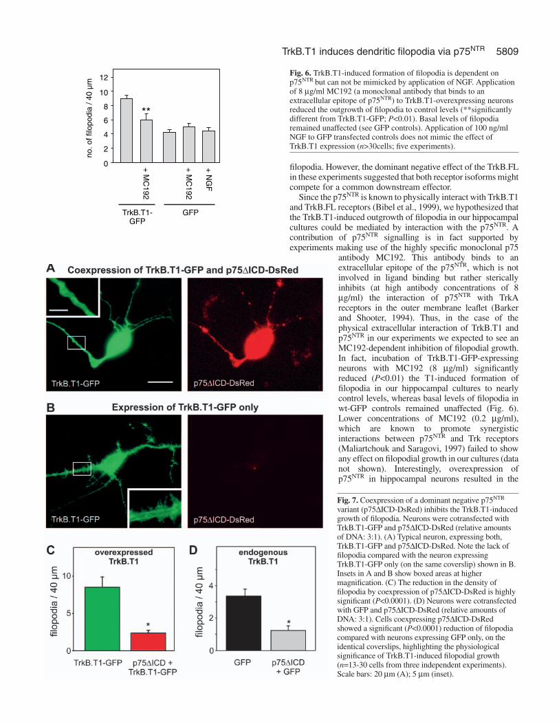
5809TrkB.T1 induces dendritic filopodia via p75NTR
filopodia. However, the dominant negative effect of the TrkB.FLin these experiments suggested that both receptor isoforms mightcompete for a common downstream effector.
Since the p75NTR is known to physically interact with TrkB.T1and TrkB.FL receptors (Bibel et al., 1999), we hypothesized thatthe TrkB.T1-induced outgrowth of filopodia in our hippocampalcultures could be mediated by interaction with the p75NTR. Acontribution of p75NTR signalling is in fact supported byexperiments making use of the highly specific monoclonal p75
antibody MC192. This antibody binds to anextracellular epitope of the p75NTR, which is notinvolved in ligand binding but rather stericallyinhibits (at high antibody concentrations of 8µg/ml) the interaction of p75NTR with TrkAreceptors in the outer membrane leaflet (Barkerand Shooter, 1994). Thus, in the case of thephysical extracellular interaction of TrkB.T1 andp75NTR in our experiments we expected to see anMC192-dependent inhibition of filopodial growth.In fact, incubation of TrkB.T1-GFP-expressingneurons with MC192 (8 µg/ml) significantlyreduced (P<0.01) the T1-induced formation offilopodia in our hippocampal cultures to nearlycontrol levels, whereas basal levels of filopodia inwt-GFP controls remained unaffected (Fig. 6).Lower concentrations of MC192 (0.2 µg/ml),which are known to promote synergisticinteractions between p75NTR and Trk receptors(Maliartchouk and Saragovi, 1997) failed to showany effect on filopodial growth in our cultures (datanot shown). Interestingly, overexpression ofp75NTR in hippocampal neurons resulted in the
10
12
8
6
4
2
0
TrkB.T1-GFP
291
CM
+
291
CM
+
FG
N+
GFP
mµ04/
aidop oliffo.on
**
Fig. 6. TrkB.T1-induced formation of filopodia is dependent onp75NTR but can not be mimicked by application of NGF. Applicationof 8 µg/ml MC192 (a monoclonal antibody that binds to anextracellular epitope of p75NTR) to TrkB.T1-overexpressing neuronsreduced the outgrowth of filopodia to control levels (**significantlydifferent from TrkB.T1-GFP; P<0.01). Basal levels of filopodiaremained unaffected (see GFP controls). Application of 100 ng/mlNGF to GFP transfected controls does not mimic the effect ofTrkB.T1 expression (n>30cells; five experiments).
Fig. 7. Coexpression of a dominant negative p75NTR
variant (p75∆ICD-DsRed) inhibits the TrkB.T1-inducedgrowth of filopodia. Neurons were cotransfected withTrkB.T1-GFP and p75∆ICD-DsRed (relative amountsof DNA: 3:1). (A) Typical neuron, expressing both,TrkB.T1-GFP and p75∆ICD-DsRed. Note the lack offilopodia compared with the neuron expressingTrkB.T1-GFP only (on the same coverslip) shown in B.Insets in A and B show boxed areas at highermagnification. (C) The reduction in the density offilopodia by coexpression of p75∆ICD-DsRed is highlysignificant (P<0.0001). (D) Neurons were cotransfectedwith GFP and p75∆ICD-DsRed (relative amounts ofDNA: 3:1). Cells coexpressing p75∆ICD-DsRedshowed a significant (P<0.0001) reduction of filopodiacompared with neurons expressing GFP only, on theidentical coverslips, highlighting the physiologicalsignificance of TrkB.T1-induced filopodial growth(n=13-30 cells from three independent experiments).Scale bars: 20 µm (A); 5 µm (inset).

5810
induction of filopodial growth on its own (p75: 5.5±0.5filopodia per 40 µm dendrite; TrkB.T1: 6.3±0.6;TrkB.FL: 2.8±1.1, significantly different with P<0.01).However, in many cases the overexpression of p75NTR
also induced neuronal death, which is in line withneurotrophin-independent induction of neuronal deathvia the intracellular domain of p75NTR in vivo (Majdanet al., 1997).
Since TrkA receptors are absent from hippocampalneurons, application of NGF can be used to selectivelyactivate p75NTR signalling in these cells (Ip et al.,1993a; Brann et al., 1999; Friedman, 2000). Toexamine whether NGF-induced p75NTR activation issufficient to induce filopodia, we stimulated GFP-transfected neurons with NGF (100 ng/ml).Interestingly, NGF failed to reproduce the TrkB.T1-induced growth of filopodia (Fig. 6), suggesting thatpartially different signalling mechanisms are at workdownstream of the p75NTR, depending on whetherNGF or TrkB.T1 is interacting.
Altogether, these data suggested that the TrkB.T1-induced formation of filopodia is transduced vianeurotrophin-independent interaction of theextracellular or transmembrane domains of p75NTR andTrkB.T1 receptors, initiating downstream activation ofcertain aspects of p75NTR signalling.
Intriguingly, coexpression of TrkB.T1-GFP with amutant p75NTR, which lacks the intracellular domain(p75∆ICD-DsRed), indeed inhibits the TrkB.T1-induced effect in a dominant negative manner: theformation of dendritic filopodia in these coexpresserswas reduced fourfold, when compared to neuronsexpressing only TrkB.T1-GFP, in the same culture(Fig. 7). It is of note, that in these experiments TrkB.T1-GFP-expressing cells were collected for both groups from identicalcoverslips. Thus, the observed differences in filopodial growthare only related to the presence/absence of p75∆ICD-DsRed inthe cells, ruling out any bias in our analysis. This experimentprovides strong evidence for the model of endogenous p75NTR
being a downstream mediator of TrkB.T1-induced filopodiaformation. Importantly, p75∆ICD-DsRed also significantlyreduced basal levels of filopodia formation in wt-GFP-expressing controls (Fig. 7D), thus suggesting that endogenouslyexpressed TrkB.T1 contributes to basal filopodia formation. Inorder to test for endogenous expression of TrkB.T1 in ourneurons we performed immunocytochemistry with an antibodythat reacts selectively with truncated TrkB receptors (anti-TrkB[TK–]). The antibody recognized truncated TrkB receptorsin astrocytes (see also Rose et al., 2003) and in neurons of ourhippocampal microcultures (Fig. 8). This supports the view thatendogenous TrkB.T1 could account for basal levels of filopodialgrowth in our cultures (Fig. 7D).
In order to further establish TrkB.T1-induced signallingvia p75NTR we performed experiments in hippocampalmicrocultures from exon III p75NTR knockout mice (Lee et al.,1992), which show a strong reduction, albeit not a completedeletion of all p75NTR isoforms (von Schack et al., 2001).Importantly, the TrkB.T1-induced formation of filopodia wassignificantly reduced in these cultures compared to matchedwild-type controls (TrkB.T1 in p75 knockout: 14.5±0.9 filopodiaper 40 µm dendrite; TrkB.T1 in wt: 20.0±1.1; significantly
different with P<0.0005, see Fig. 9). Nevertheless, the TrkB.T1-induced formation of filopodia was not abolished in thesep75NTR knockout cultures. This suggests that the remainingextracellularly truncated p75NTR in these mice (von Schack etal., 2001) can partially substitute for the full length p75NTR.
Journal of Cell Science 117 (24)
Fig. 8. TrkB.T1 is endogenously expressed in hippocampal neurons and glia.(A) untransfected rat hippocampal microcultures were stained at 10 DIV withan antibody that selectively reacts with truncated TrkB receptors (TrkB[TK–]).(B) As a positive control, rat hippocampal neurons were transfected at 9 DIVwith wt TrkB.T1 receptor and also processed for TrkB [TK–]immunocytochemistry. Neurons were identified by co-staining with a MAP2antibody. Truncated TrkB receptors can be immunostained in untransfectedneurons (arrows) and glia (asterisk). Specificity of the TrkB[TK–] antibody wasevident from the intense signal in TrkB.T1-overexpressing neurons in B. Owingto the tenfold shorter exposure time for the overexpressed truncated receptorstaining in the glia is not visible in B (lower panel). Scale bar: 10 µm.
p < 0,0005
p75 k.o. micewildtype
20
15
10
5
0TrkB.T1
*
TrkB.T1 GFP GFP
*
no
. of
filo
po
dia
/ 40
µm
Fig. 9. Reduction of TrkB.T1-induced filopodia formation inhippocampal neurons from p75 knockout mice. Neurons from wt orp75NTR knockout mice were transfected with either TrkB.T1-GFP orGFP. The reduction in the density of TrkB.T1-induced filopodia inp75NTR knockout compared with wild-type mice is highly significant(P<0.0005). Both TrkB.T1-transfected groups were also significantlydifferent from the respective GFP-transfected negative controls(*P<0.0001). The number of filopodia in GFP-expressing cells fromp75NTR knockout mice was not significantly different from GFP-expressing wild-type cultures (P>0.15) (n=28-52 cells from two tothree independent experiments).

5811TrkB.T1 induces dendritic filopodia via p75NTR
In trying to rescue the inhibited filopodial growth in the exonIII p75NTR knockout mice, we cotransfected one group of theknockout-derived cells with TrkB.T1-GFP and the wt-p75NTR
receptor, respectively. However, as previously observed in ratneurons (see above) overexpression of wt-p75NTR also inducedthe death of mice neurons, thus impairing the observation of arescue effect.
DiscussionOur results indicate a novel signalling mechanism of truncatedTrkB receptors in regulating dendritic growth of hippocampalneurons. Overexpression of truncated TrkB receptors (TrkB.T1and TrkB.T2) induced a twofold increase in the number ofdendritic filopodia. This effect was independent of the presenceof neurotrophins and of tyrosine kinase signalling of endogenousTrkB.FL. Our data suggest an extracellular or intramembraneinteraction between TrkB.T1 and p75NTR receptors, initiatingfilopodial growth via p75NTR intracellular signalling. This modelis corroborated by the dominant negative regulation of theTrkB.T1-induced filopodia by coexpression of a p75NTR lackingthe intracellular domain (p75∆ICD) and by the significantreduction of the TrkB.T1 effect in p75NTR knockout mice.Coexpressing surplus TrkB.FL receptors antagonized theTrkB.T1-induced filopodial growth independently of Trk kinaseactivity, suggesting a competitive interference of TrkB.FL in theformation of putative p75NTR-TrkB.T1 heterodimers.
Since overexpression of the dominant negative p75∆ICD wasable to also reduce basal levels of filopodia in our hippocampalcultures (Fig. 7D), these data suggest a role of endogenousTrkB.T1-p75NTR signalling in dendritic growth. AccordinglyTrkB.T1 was observed to be expressed endogeneously in ourhippocampal neurons (Fig. 8).
The absence of a similar inhibition of basal levels of filopodiaupon overexpression of TrkB.FL (Fig. 4), might reflect thedifferent potencies of these two dominant negative inhibitors. Inthis context it is of note that p75∆ICD was able to inhibitTrkB.T1-GFP-induced filopodia when coexpressed at a DNAratio of only 1:3 (Fig. 7) whereas TrkB.FL needed to beoverexpressed at a DNA ratio of 3:1 (Fig. 5) in order to have asimilar dominant negative effect.
The unperturbed presence of filopodia in GFP-expressingcontrols from exon III p75NTR knockout mice might be accountedfor by the expression of the p75NTR short in these neurons, whichcan partially substitute for the wt p75NTR. In addition, it seemsconceivable that TrkB.T1-p75NTR-mediated induction offilopodia is not the sole mechanism inducing dendritic growth inhippocampal neurons, thus leaving space for induction offilopodia in the p75 knockout mice via other pathways.
Previous studies showed that cell surface targeting of TrkB.FLin hippocampal neurons and in retinal ganglion cells is promotedby electrical activity and decreased by coexpression of TrkB.T1(Meyer-Franke et al., 1998; Du et al., 2000; Haapasalo et al.,2002). Since we observed cell surface targeting of TrkB.FL andof all other TrkB receptors tested, and given the comparablesurface targeting of GFP-tagged and untagged TrkB.FL (Klau etal., 2001), these data suggest that sufficient levels of electricalactivity are present in our neurons to support this targetingprocess, both in the absence and presence of C-terminal GFP.Interestingly, membrane delimited fluorescence of TrkB.FL-GFP was decreased upon action potential blockade with TTX
(M.H. and V.L., unpublished results), further supporting the viewthat activity-dependent targeting of TrkB.FL-GFP is intact in ourneurons.
We can not definitely exclude that overexpression of TrkB.T1decreases surface expression of endogenous TrkB.FL in ourneurons (Haapasalo et al., 2002). However, since induction offilopodia by TrkB.T1 occurs independently from ligand binding,it would remain unclear how reduced endogenous TrkB.FLlevels could increase the number of filopodia, if not by themechanism suggested in Fig. 10: given the dominant negativeeffect of coexpressed TrkB.FL on TrkB.T1-induced filopodia(Fig. 5), reduced surface levels of endogenous TrkB.FL shouldfacilitate the growth of filopodia, thus being in accordance withour model (see Fig. 10).
TrkB.T1-induced morphological changesA previous study by Yacoubian and (Lo Yacoubian and Lo,2000) in ferret cortical slices suggested a role of overexpressedTrkB.T1 receptors in promoting the growth of distal dendrites,whereas TrkB.FL induced the growth of proximal dendrites.Exogenous TrkB ligands reversed the effect of TrkB.T1 in akinase-dependent manner, whereas TrkB.FL effects werepotentiated. Although this was an intriguing finding, possibledownstream effectors of this TrkB.T1-induced growth remainedelusive. Their results were most compatible with a mutualregulation of dendritic growth by both receptor isoforms: highlocal expression ratios of TrkB.FL:TrkB.T1 would favour aligand-dependent growth of dendrites, whereas the relativeabundance of TrkB.T1 would promote dendritic growth in theabsence of ligands.
In our hippocampal cultures, the basic finding of TrkB.T1-induced growth of dendritic filopodia is comparable with theTrkB.T1-induced dendritic branching observed by Yacoubian
p75TrkB.FL TrkB.FL
TrkB.T1
BDNF, NGF, TrkB IgG: without effect on T1 induced filopodia
k252a:no change indominant negativeeffect of TrkB.FL
MC192
p75 ICD
dendritic filopodia
p75 k.o.
Fig. 10. Proposed model of TrkB.T1 action in the induction offilopodia. Interaction of TrkB.T1 with the p75NTR, leads to theformation of filopodia (grey arrow). This effect can be blocked byeither the MC192 antibody or the dominant negative p75∆ICD(inhibitory agents in red). The dominant negative action of theTrkB.FL receptor can be explained by its competition with TrkB.T1for binding to either p75NTR (right margin) or by scavenging TrkB.T1(left margin). NGF, BDNF, TrkB-IgG or K252a were without effecton this signalling mechanism. The short p75NTR (in blue) indicates apossible (albeit weaker) such signalling via a p75NTR lacking anintact extracellular domain (see p75NTR knockout mice experiments)

5812
and Lo (Yacoubian and Lo, 2000). However, two differences areevident. First, in our study, the proximal dendrites were the siteof filopodia formation. Secondly, all TrkB.T1-induced effects onthe formation of filopodia in our hippocampal cultures werecompletely independent of endogenous and exogenous TrkBligands, and of TrkB.FL kinase signalling (Fig. 4). It remains anattractive hypothesis, that subcellular differences in endogenousexpression levels of TrkB.FL versus TrkB.T1 (e.g. inhippocampus versus neocortex) could account for this dissimilarligand dependence in TrkB-dependent dendritic growth(Yacoubian and Lo, 2000).
We directly addressed this issue by coexpressing both receptorisoforms in the same cells (Fig. 5). The observed dominantnegative effect of TrkB.FL on TrkB.T1-induced formation ofdendritic filopodia is the first direct demonstration of full lengthTrkB-mediated inhibition of TrkB.T1-induced signalling inneurons. Interestingly, a comparable dominant negativeregulation of TrkB.T1-induced morphological changes byTrkB.FL receptors has recently been shown in a neuroblastomacell line (Haapasalo et al., 1999). Taken together, these dataprovide evidence for a new function of coordinated TrkB.T1 andTrkB.FL signalling in regulating dendritic growth.
In contrast to the effects on filopodial growth observed here,most previous studies, addressing TrkB-dependent modulationof dendritic growth, focussed on BDNF-dependent modulationof dendritic branching and dendritic length (McAllister et al.,1997; Lom and Cohen-Cory, 1999; Tolwani et al., 2002).However, Horch and colleagues (Horch et al., 1999) reported aBDNF overexpression-induced sprouting of dendriticprotrusions in cortical neurons, similar to the TrkB.T1-induced(albeit ligand independent) filopodial growth in our hippocampalneurons. The relation between these two effects remains to bedetermined.
Given the delayed expression of TrkB.T1 receptors duringdevelopment compared with TrkB.FL (Fryer et al., 1996), itseems to be an attractive hypothesis that BDNF-dependentsignalling via full length receptors supports the outgrowth ofprimary dendrites early in the development, whereas the delayedexpression of TrkB.T1 allows for the ligand-independentsprouting of dendritic filopodia during synapse formation(Yacoubian and Lo, 2000). In a preliminary set of experimentswe determined whether the increased number of dendriticfilopodia is accompanied by an increase in synaptic contacts assuggested previously (Ziv and Smith, 1996). However, activity-dependent staining of presynaptic terminals using FM 4-64did not indicate a rise in functional synapse numbers in ourTrkB.T1-overexpressing neurons (M.H. and V.L., unpublishedobservation) (see also Klau et al., 2001). It remains to beestablished whether such an effect on synaptic differentiationwill be detected in TrkB.T1-GFP-overexpressing neurons inhippocampal slice cultures that show a larger number of coherentafferent fibres innervating the dendritic tree of a given neuron.
Signalling mechanism of TrkB.T1-induced growth ofdendritic filopodiaOur data provide three independent lines of evidence for aninvolvement of the p75NTR in the observed TrkB.T1-inducedformation of filopodia: (1) inhibition in the presence of the MC192 monoclonal p75NTR antibody; (2) dominant negativeinhibition by a p75NTR lacking the intracellular domain; and, (3)
significant reduction of the effect in exon III p75NTR knockoutmice.
Furthermore, growth of filopodia is intact uponoverexpression of the mutant TrkB∆ICD receptor variant lackingthe TrkB.T1 intracellular domain. Coexpression of TrkB.FLreceptors inhibits TrkB.T1-induced filopodia in a dominantnegative fashion.
Taken together, these results lead us to propose that ligand-independent interaction of the extracellular or theintramembrane domains of TrkB.T1 and p75NTR provokes theformation of dendritic filopodia (Fig. 10). This pathway can beblocked by sterical hindrance of the TrkB.T1-P75NTR interactionby extracellular binding of the MC192 antibody to the p75NTR
receptor (Barker and Shooter, 1994; Maliartchouk and Saragovi,1997; Kimpinski et al., 1999). The inability of a different p75NTR
antibody to block a similar TrkB.T1-induced growth of filopodiain N2a cells (Haapasalo et al., 1999) might reflect the differentspecificities of these antibodies to interfere with the interactionof p75NTR and TrkB.T1.
In accordance with the model, the filopodial growth is blockedwhen the TrkB.T1-p75NTR interaction is inhibited by a dominantnegative p75NTR (Fig. 7). Furthermore we suggest that TrkB.FLinhibits the binding of TrkB.T1 to the p75NTR receptor by eitherforming heterodimers with TrkB.T1 or with p75NTR,respectively, thereby inhibiting the specific downstreamsignalling of the putative TrkB.T1-p75NTR heterodimers. Thus,the tendency of TrkB.T1 to induce growth of filopodia in a givendendritic segment would critically depend on the relativeabundance of these three receptors at the specified subcellularlocation (see also Yacoubian and Lo, 2000).
Interestingly, the TrkB.T1 receptor, which lacks anintracellular domain (TrkB∆ICD), still induced filopodia (Fig.4C), indicating that the required motifs for interaction with thep75NTR are localized in the extracellular or intramembranedomain of TrkB.T1. Although these domains are identical inTrkB.FL, this receptor lacked an effect on filopodia. These datasuggest that the intracellular domain of TrkB.FL prevents theappropriate interaction of the extracellular or intramembranedomain of the TrkB receptor with the p75NTR, or hinders thecontact of a putative TrkB.FL-p75NTR complex with theintracellular filopodia inducing machinery. Similarly, thedominant negative effect of coexpressed TrkB.FL on TrkB.T1-induced filopodia (Fig. 5) could be accounted for by thismechanism. Consequently, future studies should aim atidentifying the TrkB.FL intracellular domains that preventTrkB.FL from inducing filopodia.
The significant reduction of the TrkB.T1-induced filopodia inp75 knockout cultures supports the model proposed in Fig. 10.However, the exon III p75 knockout mouse used in this studyhas been reported previously to give rise to expression of a shortp75NTR lacking most of the extracellular domain, which canpartially substitute for full length p75NTR receptor signalling(von Schack et al., 2001). Thus, the remaining effect on filopodiaformation in our p75 knockout experiments (see Fig. 9) couldbe explained by intact signalling of this short p75NTR. In factrecent evidence directly suggests signalling of p75NTR lackingan extracellular domain, which results from intramembraneproteolytic processing of the p75NTR (Yamashita et al., 1999;Jung et al., 2003; Kanning et al., 2003; Yamashita and Tohyama,2003).
In accordance with the model in Fig. 10 recent evidence in
Journal of Cell Science 117 (24)
5813TrkB.T1 induces dendritic filopodia via p75NTR
fact indicates a ligand-independent molecular interaction of bothTrkB.FL and intracellularly truncated TrkB.FL (resemblingTrkB.T1) with the p75NTR receptor in HEK A293 cells, and alsoshows a role of these heteromeric receptor complexes in liganddiscrimination (Bibel et al., 1999). Similar results have beendescribed with respect to a molecular interaction of TrkA withp75NTR (Ross et al., 1996; Gargano et al., 1997) and a functionalinteraction of TrkC receptors with p75NTR (Hapner et al., 1998)in non-neuronal cells. Our data now provide the first evidencefor a possible physiological function of a TrkB-p75NTR
heteromeric receptor in neurons (see above).NGF, which is known to selectively activate p75NTR and to
induce fibre outgrowth via ceramide production in hippocampalneurons (Brann et al., 1999; Friedman, 2000), did not mimicthe effect of overexpressed TrkB.T1 in our cultures. Theinvolvement of different p75NTR-dependent signalling cascadesin response to TrkB.T1 versus NGF could account for thedissimilar effects of these two unlike ligands in stimulatingp75NTR-induced dendritic growth. The p75NTR-mediateddendritic growth in hippocampal neurons following stimulationwith NGF was restricted to very young embryonic cultures(Brann et al., 1999; Brann et al., 2002). In more mature neurons,NGF-induced effects on dendritic branching were absentalthough the expression level of p75 receptors was not decreased(Brann, 2002). It thus seems plausible to assume that p75NTR
signalling is experiencing subtle changes during neuronaldevelopment in culture, which can not be explained by, forexample, reduced expression levels of p75NTR protein. Theongoing expression of p75NTR as observed by Brann et al. (Brannet al., 2002) is in line with stable expression of p75 mRNA inour hippocampal microcultures until at least 14 DIV (M.Knipper and V. L., unpublished). In support of a role of p75NTR
in the formation of neuronal processes, Yamashita andcolleagues (Yamashita et al., 1999) identified the small G proteinRhoA as interacting with the intracellular domain of p75NTR.This interaction leads to a neurotrophin-independent activationof RhoA, indicating ligand-independent signalling of the p75receptors. Binding of neurotrophins to p75NTR reduces theactivity of RhoA, resulting in axonal outgrowth in PNS neurons(Yamashita et al., 1999). Interestingly, Rho has recently alsobeen demonstrated to regulate the growth of filopodia and spineformation in hippocampal dendrites (Nakayama et al., 2000). Itis thus tempting to speculate that RhoA signalling is involved inthe TrkB.T1-p75NTR-mediated growth of filopodia observed inour study.
Myelin-associated glycoprotein (MAG) was recently found tobind to a complex consisting of p75NTR and ganglioside GT1b,thus retarding neurite outgrowth via intermediate activation ofRho (Yamashita et al., 2002), whereas neurotrophin-stimulatedp75NTR promotes neurite outgrowth. Likewise, two recentstudies implicated the p75NTR in the signal transduction of theNoGo receptor in complex with its ligands Nogo66, nMAG andoMPG (Wang et al., 2002; Wong et al., 2002). These findingsare particularly relevant to our results, stressing the bindingof p75NTR receptors to ligands other than neurotrophins.Furthermore, in the study by Yamashita and colleagues(Yamashita et al., 2002) MAG was shown to stimulate Rhoactivity downstream of p75NTR, whereas activation vianeurotrophins elicits an opposite p75NTR signalling mechanism.This suggests that p75NTR-induced RhoA signalling could alsobe involved in the TrkB.T1-induced outgrowth of filopodia in
our hippocampal neurons. Since manipulation of Rho activityinduces marked changes in dendritic morphology on its own(Nakayama et al., 2000), a role of Rho in TrkB.T1-inducedgrowth of filopodia can not be easily determined by inhibitionof Rho function. Thus, future studies employing mutant p75receptors lacking the Rho interacting mastoparan binding sitecould help to demonstrate a causal connection between Rhobinding to p75 and TrkB.T1-induced formation of filopodia inhippocampal neurons.
The authors want to thank David Middlemas for providing rat TrkBconstructs, Sabine Rickheim-Lowack and Sabine Laerbusch forexcellent technical assistance and Heiko Luhmann and Rolf Heumannfor their support. This work was supported by grants from the DFG toV.L. (SFB 509 and SFB 553), M.S. (SFB 487) and K.S.E. (SFB 452).
ReferencesBarker, P. A. and Shooter, E. M. (1994). Disruption of NGF binding to the low
affinity neurotrophin receptor p75LNTR reduces NGF binding to TrkA onPC12 cells. Neuron 13, 203-215.
Baxter, G. T., Radeke, M. J., Kuo, R. C., Makrides, V., Hinkle, B., Hoang,R., Medina-Selby, A., Coit, D., Valenzuela, P. and Feinstein, S. C. (1997).Signal transduction mediated by the truncated trkB receptor isoforms, trkB.T1and trkB.T2. J. Neurosci. 17, 2683-2690.
Bibel, M., Hoppe, E. and Barde, Y. A. (1999). Biochemical and functionalinteractions between the neurotrophin receptors trk and p75NTR. EMBO J.18, 616-622.
Biffo, S., Offenhauser, N., Carter, B. D. and Barde, Y. A. (1995). Selectivebinding and internalisation by truncated receptors restrict the availability ofBDNF during development. Development 121, 2461-2470.
Bothwell, M. (1995). Functional interactions of neurotrophins and neurotrophinreceptors. Annu. Rev. Neurosci. 18, 223-253.
Brann, A. B., Scott, R., Neuberger, Y., Abulafia, D., Boldin, S., Fainzilber,M. and Futerman, A. H. (1999). Ceramide signaling downstream of the p75neurotrophin receptor mediates the effects of nerve growth factor onoutgrowth of cultured hippocampal neurons. J. Neurosci. 19, 8199-8206.
Brann, A. B., Tcherpakov, M., Williams, I. M., Futerman, A. H. andFainzilber, M. (2002). Nerve growth factor-induced p75-mediated death ofcultured hippocampal neurons is age-dependent and transduced throughceramide generated by neutral sphingomyelinase. J. Biol. Chem. 277, 9812-9818.
Cabelli, R. J., Allendoerfer, K. L., Radeke, M. J., Welcher, A. A., Feinstein,S. C. and Shatz, C. J. (1996). Changing patterns of expression and subcellularlocalization of TrkB in the developing visual system. J. Neurosci. 16, 7965-7980.
Dobrowsky, R. T. and Carter, B. D. (1998). Coupling of the p75 neurotrophinreceptor to sphingolipid signaling. Ann. N. Y. Acad. Sci. 845, 32-45.
Du, J., Feng, L., Yang, F. and Lu, B. (2000). Activity- and Ca(2+)-dependentmodulation of surface expression of brain-derived neurotrophic factorreceptors in hippocampal neurons. J. Cell Biol. 150, 1423-1434.
Eide, F. F., Vining, E. R., Eide, B. L., Zang, K., Wang, X. Y. and Reichardt,L. F. (1996). Naturally occurring truncated trkB receptors have dominantinhibitory effects on brain-derived neurotrophic factor signaling. J. Neurosci.16, 3123-3129.
Fanger, G. R., Jones, J. R. and Maue, R. A. (1995). Differential regulation ofneuronal sodium channel expression by endogenous and exogenous tyrosinekinase receptors expressed in rat pheochromocytoma cells. J. Neurosci. 15,202-213.
Friedman, W. J. (2000). Neurotrophins induce death of hippocampal neuronsvia the p75 receptor. J. Neurosci. 20, 6340-6346.
Fryer, R. H., Kaplan, D. R., Feinstein, S. C., Radeke, M. J., Grayson, D. R.and Kromer, L. F. (1996). Developmental and mature expression of full-lengthand truncated TrkB receptors in the rat forebrain. J. Comp Neurol. 374, 21-40.
Gargano, N., Levi, A. and Alema, S. (1997). Modulation of nerve growth factorinternalization by direct interaction between p75 and TrkA receptors. J.Neurosci. Res. 50, 1-12.
Haapasalo, A., Saarelainen, T., Moshnyakov, M., Arumae, U., Kiema,T. R., Saarma, M., Wong, G. and Castren, E. (1999). Expression ofthe naturally occurring truncated trkB neurotrophin receptor inducesoutgrowth of filopodia and processes in neuroblastoma cells. Oncogene 18,1285-1296.

5814
Haapasalo, A., Sipola, I., Larsson, K., Akerman, K. E., Stoilov, P., Stamm,S., Wong, G. and Castren, E. (2002). Regulation of TRKB surfaceexpression by brain-derived neurotrophic factor and truncated TRKBisoforms. J. Biol Chem. 277, 43160-43167.
Hapner, S. J., Boeshore, K. L., Large, T. H. and Lefcort, F. (1998). Neuraldifferentiation promoted by truncated trkC receptors in collaboration withp75(NTR). Dev. Biol. 201, 90-100.
Harris, K. M. (1999). Structure, development, and plasticity of dendritic spines.Curr. Opin. Neurobiol. 9, 343-348.
Hartmann, M., Heumann, R. and Lessmann, V. (2001). Synaptic secretion ofBDNF after high-frequency stimulation of glutamatergic synapses. EMBO J.20, 5887-5897.
Haubensak, W., Narz, F., Heumann, R. and Lessmann, V. (1998). BDNF-GFP containing secretory granules are localized in the vicinity of synapticjunctions of cultured cortical neurons. J. Cell Sci. 111, 1483-1493.
Horch, H. W., Kruttgen, A., Portbury, S. D. and Katz, L. C. (1999).Destabilization of cortical dendrites and spines by BDNF. Neuron 23, 353-364.
Ip, N. Y., Li, Y., Yancopoulos, G. D. and Lindsay, R. M. (1993a). Culturedhippocampal neurons show responses to BDNF, NT-3, and NT-4, but not NGF.J. Neurosci. 13, 3394-3405.
Ip, N. Y., Stitt, T. N., Tapley, P., Klein, R., Glass, D. J., Fandl, J., Greene, L.A., Barbacid, M. and Yancopoulos, G. D. (1993b). Similarities anddifferences in the way neurotrophins interact with the Trk receptors inneuronal and nonneuronal cells. Neuron 10, 137-149.
Jung, K. M., Tan, S., Landman, N., Petrova, K., Murray, S., Lewis, R., Kim,P. K., Kim, D. S., Ryu, S. H., Chao, M. V. and Kim, T. W. (2003). Regulatedintramembrane proteolysis of the p75 neurotrophin receptor modulates itsassociation with the TrkA receptor. J. Biol Chem. 278, 42161-42169.
Kanning, K. C., Hudson, M., Amieux, P. S., Wiley, J. C., Bothwell, M. andSchecterson, L. C. (2003). Proteolytic processing of the p75 neurotrophinreceptor and two homologs generates C-terminal fragments with signalingcapability. J. Neurosci. 23, 5425-5436.
Kaplan, D. R. and Miller, F. D. (2000). Neurotrophin signal transduction in thenervous system. Curr. Opin. Neurobiol. 10, 381-391.
Kimpinski, K., Jelinski, S. and Mearow, K. (1999). The anti-p75 antibody,MC192, and brain-derived neurotrophic factor inhibit nerve growth factor-dependent neurite growth from adult sensory neurons. Neuroscience 93, 253-263.
Klau, M., Hartmann, M., Erdmann, K. S., Heumann, R. and Lessmann, V.(2001). Reduced number of functional glutamatergic synapses in hippocampalneurons overexpressing full-length TrkB receptors. J. Neurosci. Res. 66, 327-336.
Klein, R., Conway, D., Parada, L. F. and Barbacid, M. (1990). The trkBtyrosine protein kinase gene codes for a second neurogenic receptor that lacksthe catalytic kinase domain. Cell 61, 647-656.
Kryl, D. and Barker, P. A. (2000). TTIP is a novel protein that interacts withthe truncated T1 TrkB neurotrophin receptor. Biochem. Biophys. Res.Commun. 279, 925-930.
Kryl, D., Yacoubian, T., Haapasalo, A., Castren, E., Lo, D. and Barker, P.A. (1999). Subcellular localization of full-length and truncated Trk receptorisoforms in polarized neurons and epithelial cells. J. Neurosci. 19, 5823-5833.
Lambert, R. C., Maulet, Y., Dupont, J. L., Mykita, S., Craig, P., Volsen, S.and Feltz, A. (1996). Polyethylenimine-mediated DNA transfection ofperipheral and central neurons in primary culture: probing Ca2+ channelstructure and function with antisense oligonucleotides. Mol. Cell Neurosci. 7,239-246.
Lee, K. F., Li, E., Huber, L. J., Landis, S. C., Sharpe, A. H., Chao, M. V.and Jaenisch, R. (1992). Targeted mutation of the gene encoding the lowaffinity NGF receptor p75 leads to deficits in the peripheral sensory nervoussystem. Cell 69, 737-749.
Lessmann, V. and Heumann, R. (1998). Modulation of unitary glutamatergicsynapses by neurotrophin-4/5 or brain-derived neurotrophic factor inhippocampal microcultures: presynaptic enhancement depends on pre-established paired-pulse facilitation. Neuroscience 86, 399-413.
Levi-Montalcini, R. and Hamburger, V. (1953). A diffusible agent ofmouse sarcoma, producing hyperplasia of sympathetic-ganglia andhyperneurotization of viscera in the chick embryo. J. Exp. Zool. 123, 233-287.
Lom, B. and Cohen-Cory, S. (1999). Brain-derived neurotrophic factordifferentially regulates retinal ganglion cell dendritic and axonal arborizationin vivo. J. Neurosci. 19, 9928-9938.
Lu, B. (2003). BDNF and activity-dependent synaptic modulation. Learn. Mem.10, 86-98.
Majdan, M., Lachance, C., Gloster, A., Aloyz, R., Zeindler, C., Bamji, S.,Bhakar, A., Belliveau, D., Fawcett, J., Miller, F. D. and Barker, P. A.(1997). Transgenic mice expressing the intracellular domain of the p75neurotrophin receptor undergo neuronal apoptosis. J. Neurosci. 17, 6988-6998.
Maliartchouk, S. and Saragovi, H. U. (1997). Optimal nerve growth factortrophic signals mediated by synergy of TrkA and p75 receptor-specificligands. J. Neurosci. 17, 6031-6037.
McAllister, A. K., Katz, L. C. and Lo, D. C. (1997). Opposing roles forendogenous BDNF and NT-3 in regulating cortical dendritic growth. Neuron18, 767-778.
Meyer-Franke, A., Wilkinson, G. A., Kruttgen, A., Hu, M., Munro, E.,Hanson, M. G., Jr, Reichardt, L. F. and Barres, B. A. (1998).Depolarization and cAMP elevation rapidly recruit TrkB to the plasmamembrane of CNS neurons. Neuron 21, 681-693.
Middlemas, D. S., Lindberg, R. A. and Hunter, T. (1991). trkB, a neuralreceptor protein-tyrosine kinase: evidence for a full-length and two truncatedreceptors. Mol. Cell. Biol. 11, 143-153.
Nakayama, A. Y., Harms, M. B. and Luo, L. (2000). Small GTPases Rac andRho in the maintenance of dendritic spines and branches in hippocampalpyramidal neurons. J. Neurosci. 20, 5329-5338.
Ninkina, N., Adu, J., Fischer, A., Pinon, L. G., Buchman, V. L. and Davies,A. M. (1996). Expression and function of TrkB variants in developing sensoryneurons. EMBO J. 15, 6385-6393.
Patapoutian, A. and Reichardt, L. F. (2001). Trk receptors: mediators ofneurotrophin action. Curr. Opin. Neurobiol. 11, 272-280.
Poo, M. M. (2001). Neurotrophins as synaptic modulators. Nat. Rev. Neurosci.2, 24-32.
Rose, C. R., Blum, R., Pichler, B., Lepier, A., Kafitz, K. W. and Konnerth,A. (2003). Truncated TrkB-T1 mediates neurotrophin-evoked calciumsignalling in glia cells. Nature 426, 74-78.
Ross, A. H., Daou, M. C., McKinnon, C. A., Condon, P. J., Lachyankar, M.B., Stephens, R. M., Kaplan, D. R. and Wolf, D. E. (1996). The neurotrophinreceptor, gp75, forms a complex with the receptor tyrosine kinase TrkA. J.Cell Biol. 132, 945-953.
Roux, P. and Barker, P. (2002). Neurotrophin signaling through the p75neurotrophin receptor. Prog. Neurobiol. 67, 203.
Shelton, D. L., Sutherland, J., Gripp, J., Camerato, T., Armanini, M. P.,Phillips, H. S., Carroll, K., Spencer, S. D. and Levinson, A. D. (1995).Human trks: molecular cloning, tissue distribution, and expression ofextracellular domain immunoadhesins. J. Neurosci. 15, 477-491.
Tolwani, R., Buckmaster, P., Varma, S., Cosgaya, J., Wu, Y., Suri, C. andShooter, E. (2002). BDNF overexpression increases dendrite complexity inhippocampal dentate gyrus. Neuroscience 114, 795.
Von Schack, D., Casademunt, E., Schweigreiter, R., Meyer, M., Bibel, M.and Dechant, G. (2001). Complete ablation of the neurotrophin receptorp75NTR causes defects both in the nervous and the vascular system. Nat.Neurosci. 4, 977-978.
Wang, K. C., Kim, J. A., Sivasankaran, R., Segal, R. and He, Z. (2002). p75interacts with the Nogo receptor as a co-receptor for Nogo, MAG and OMgp.Nature 420, 74-78.
Watson, F. L., Heerssen, H. M., Moheban, D. B., Lin, M. Z., Sauvageot, C.M., Bhattacharyya, A., Pomeroy, S. L. and Segal, R. A. (1999). Rapidnuclear responses to target-derived neurotrophins require retrograde transportof ligand-receptor complex. J. Neurosci. 19, 7889-7900.
Wong, S. T., Henley, J. R., Kanning, K. C., Huang, K. H., Bothwell, M. andPoo, M. M. (2002). A p75(NTR) and Nogo receptor complex mediatesrepulsive signaling by myelin-associated glycoprotein. Nat. Neurosci. 5, 1302-1308.
Yacoubian, T. A. and Lo, D. C. (2000). Truncated and full-length TrkBreceptors regulate distinct modes of dendritic growth. Nat. Neurosci. 3, 342-349.
Yamashita, T. and Tohyama, M. (2003). The p75 receptor acts as adisplacement factor that releases Rho from Rho-GDI. Nat. Neurosci. 6, 461-467.
Yamashita, T., Tucker, K. L. and Barde, Y. A. (1999). Neurotrophin bindingto the p75 receptor modulates Rho activity and axonal outgrowth. Neuron 24,585-593.
Yamashita, T., Higuchi, H. and Tohyama, M. (2002). The p75 receptortransduces the signal from myelin-associated glycoprotein to Rho. J. Cell Biol.157, 565-570.
Ziv, N. E. and Smith, S. J. (1996). Evidence for a role of dendritic filopodia insynaptogenesis and spine formation. Neuron 17, 91-102.
Journal of Cell Science 117 (24)