andrew melnykovych - missouri public service … cases/2012-00428/public comments...1 melnykovych,...
TRANSCRIPT

1
Melnykovych, Andrew (PSC)
From: Melnykovych, Andrew (PSC)Sent: Monday, February 24, 2014 1:18 PMTo: 'song bird'Subject: your comments in case 2012-00428
Ms. Holloway‐ These comments and documents, along with those below, will be placed into the case file in the above‐referenced matter.
Andrew Melnykovych Director of Communications Kentucky Public Service Commission 211 Sower Boulevard Frankfort, KY 40601 502‐782‐2564 cell:502‐330‐5981 From: song bird Sent: Sunday, February 23, 2014 3:34 PM Subject: SM @900MHz Causes Brain Damage Dear Mr. Melnykovych, Please add this to Case 2012-00428 Here is another study showing brain damage caused by 900MHz radiation exposure. Smart Meters transmit every 2 seconds at this frequency. This is what my daughter and her pets started experiencing during her exposure to the smart meters! I myself as well as my daughter have noticed me having symptoms since these smart meters were installed in my neighborhood without my permission. Please read and share this with the PSC! (And remember...you are liable.) These are deadly devices! Also notice that it is killing the pollenators. What would you suggest we eat when there is no food left because you approved the radiation of every living thing? My daughter tells me that the meters work just like a nuclear fallout except its quiet and less noticable!
1
Melnykovych, Andrew (PSC)
From: song bird Sent: Saturday, February 22, 2014 6:22 PMSubject: Smart Meters cause Thyroid Damage and MoreAttachments: SM 900MHz Thyroid Damage OMJ-D-09-00089.pdf
Dear Mr. Melnykovych, Please read and then add this to Case 2012-00428 I am begging you to take this seriously and consider the health of your friends and loved ones! These meters are deadly! Ruby Holloway
1
Melnykovych, Andrew (PSC)
From: song bird Sent: Sunday, February 23, 2014 1:59 PMSubject: Smart Meter Case StudiesAttachments: SM 900MHz Thyroid Damage OMJ-D-09-00089.pdf; SM AAEM -wireless-smart-meter-
case-studies.pdf
Dear Mr. Melnykovych, Please read and add this AAEM Notice to Case File 2012-00428. I am asking you to make sure the entire PSC reads and understands the Smart Meter and EMF studies that I have submitted to Case File 2012-00428. Please make sure you are all aware of the liability involved if you keep allowing the installation of these meters and you decide to go forward with the smart grid. Just remember, your family and friends as well as yourself are going to become sick just as my family has! No one or any living thing is free from the damage these meters cause! Sincerely, Ruby Holloway
1
Melnykovych, Andrew (PSC)
From: song bird Sent: Sunday, February 23, 2014 2:32 PMSubject: Smart Meters are Hackable by Terrorists and othersAttachments: Smart Grid Technology 100% Hackable.doc
Dear Mr. Melnykovych, Please add this information to Case File 2012-00428. Also, please read and then watch the interview with Cyber Expert David Chalk. Remember, not only are these meters causing health problems, deaths, and fires, but it is also possible to shut down all of them not only through the government, but through our enemies! You, your family and friends will be a victim just like me if you don't stop this! Please be a part of the solution and stop the installation of these "death meters." They are a threat to life and liberty. Sincerely, Ruby Holloway
http://www.businesswire.com/news/home/20120412005992/en/Hacking-Expert-David-
Chalk-Joins-Urgent-Call
Hacking Expert David Chalk Joins
Urgent Call to Halt Smart Grid "100% certainty of catastrophic failure of energy grid within 3 years"
April 12, 2012 10:26 AM Eastern Daylight Time
VANCOUVER, British Columbia--(BUSINESS WIRE)--The vulnerability of the energy
industry's new wireless smart grid will inevitably lead to lights out for everyone,
according to leading cyber expert David Chalk. In an online interview for an upcoming
documentary film entitled 'Take Back Your Power' (www.ThePowerFilm.org), Chalk
says the entire power grid will be at risk to being taken down by cyber attack, and if
installations continue it's only a matter of time.
“The so-called 'smart grid' that is as vulnerable as what we've got now is not
smart at all”
“We're in a state of crisis,” said Chalk. “The front door is open and there is no lock to be
had. There is not a power meter or device on the grid that is protected from hacking - if
not already infected - with some sort of trojan horse that can cause the grid to be shut
down or completely annihilated.”
“One of the most amazing things that has happened to mankind in the last 100 years is
the Internet. It's given us possibility beyond our wildest imagination. But we also know
the vulnerabilities that exist inside of it. And then we have the backbone, the power grid
that powers our nations. Those two are coming together. And it's the smart meter on your
home or business that's now allowing that connectivity.”
Chalk also issued a challenge to governments, media and technology producers to show
him one piece of digital technology that is hack-proof.
“The computer companies that are involved, the manufacturers that are involved, bring
forward a technology and I will show you that it's penetrable,” said Chalk. “I'll do it on
national TV, I'll do it anywhere. But I can guarantee you 100% that there is nothing out
there today – nothing – that can't be penetrated.”
Chalk's strong words come amidst increasing reports of the smart grid's fatal insecurities,
even from the governments and energy companies who are forcing their hand with the
smart program. “Every endpoint [meter] is a new potential threat vector,” according to
Doug Powell, manager, SMI Security, Privacy & Safety, for Canadian utility BC Hydro.
And in an interview with EnergyNow.com, former CIA Director James Woolsey was also
highly critical of energy policy makers, whose plans received multi-billion dollar funding
as part of the Economic Stimulus Act of 2008. “The so-called 'smart grid' that is as
vulnerable as what we've got now is not smart at all,” said Woolsey. “It's a really, really
stupid grid.”
But there's more. In an audit released in January, the US Inspector General Gregory
Friedman was also highly critical. “Without a formal risk assessment and associated
mitigation strategy, threats and weaknesses may go unidentified and expose the ...
systems to an unacceptable level of risk,” Friedman wrote.
Energy officials knew of these weaknesses but approved plans for the projects anyway,
auditors said. “The initial weaknesses had not always been fully addressed, and did not
include a number of security practices commonly recommended for federal government
and industry systems.”
And security is not the only technologically-based obstacle faced by smart grid
proponents. In March, alarm bells were rung following current CIA Director David
Patraeus' confirmation that governments will use wireless smart appliances to spy on
citizens. “Items of interest will be located, identified, monitored, and remotely controlled
through technologies such as radio-frequency identification, sensor networks, tiny
embedded servers, and energy harvesters,” Patraeus said at a meeting of In-Q-Tel, the
CIA's venture capital firm. He added that this will prompt a rethink of “our notions of
identity and secrecy.”
With strong criticism to the smart grid now coming from many directions, energy
corporations and governments now have the challenge to explain to an increasingly
unapproving public why they continue to fast-track smart grid installations.
Citizen groups and organizations throughout the US, Canada and Europe have launched
legal actions to stop the installation of smart meters. They cite issues such as cost
increases, health risks, privacy concerns, grid vulnerability and the lack of democratic
process. In Chalk's home province of British Columbia, Citizens for Safe Technology
(www.citizensforsafetechnology.org) and the BC Coalition to Stop Smart Meters are
leading a growing challenge.
Options for opting out of the smart metering program have been announced in markets
including California, Maine, Vermont, Louisiana, Michigan, Connecticut, Quebec, the
UK and the Netherlands. In the US, several regions including the counties of Santa Cruz
and Marin are enforcing outright moratoriums.
“Unless we wake up and realize what we're doing, there is 100% certainty of total
catastrophic failure of the entire power infrastructure within 3 years,” said Chalk. “This
could actually be worse than a nuclear war, because it would happen everywhere. How
governments and utilities are blindly merging the power grid with the Internet, and
effectively without any protection, is insanity at its finest.”
The full video interview with David Chalk can be seen on www.thepowerfilm.org. The
feature film documentary 'Take Back Your Power', which critically examines the smart
grid program, will be released online this spring.
Contacts Josh del Sol, 604-629-7945
Producer, Take Back Your Power
or
Lori Patrick, 778-384-1601
Executive Assistant to David Chalk
http://tmoney777.empowernetwork.com/blog/smart-meters-and-the-end-of-bees/
Smart Meters and the End of Bees Posted by Brian Thiesen on November 14, 2012
0 Comments
Smart Meters and the End of Bees Smart Meters WILL be the nail in the coffin for bees, for heirloom seeds/plants and all
cells within creation that they touch if we don’t stop them. Many of the people looking
into CCD have a very closed mind. “It is only this,” “it is only that,” “there is no other
factor but X.” Therefor, even posing that smart meters (click kill bees, and mutate seeds)
causes a stir for some.
It is as if somehow all the laws that show death and disease in humans and animals noted
HERE HERE and HERE do not apply to bees or plants. All things in creation obey laws
and are really the same:
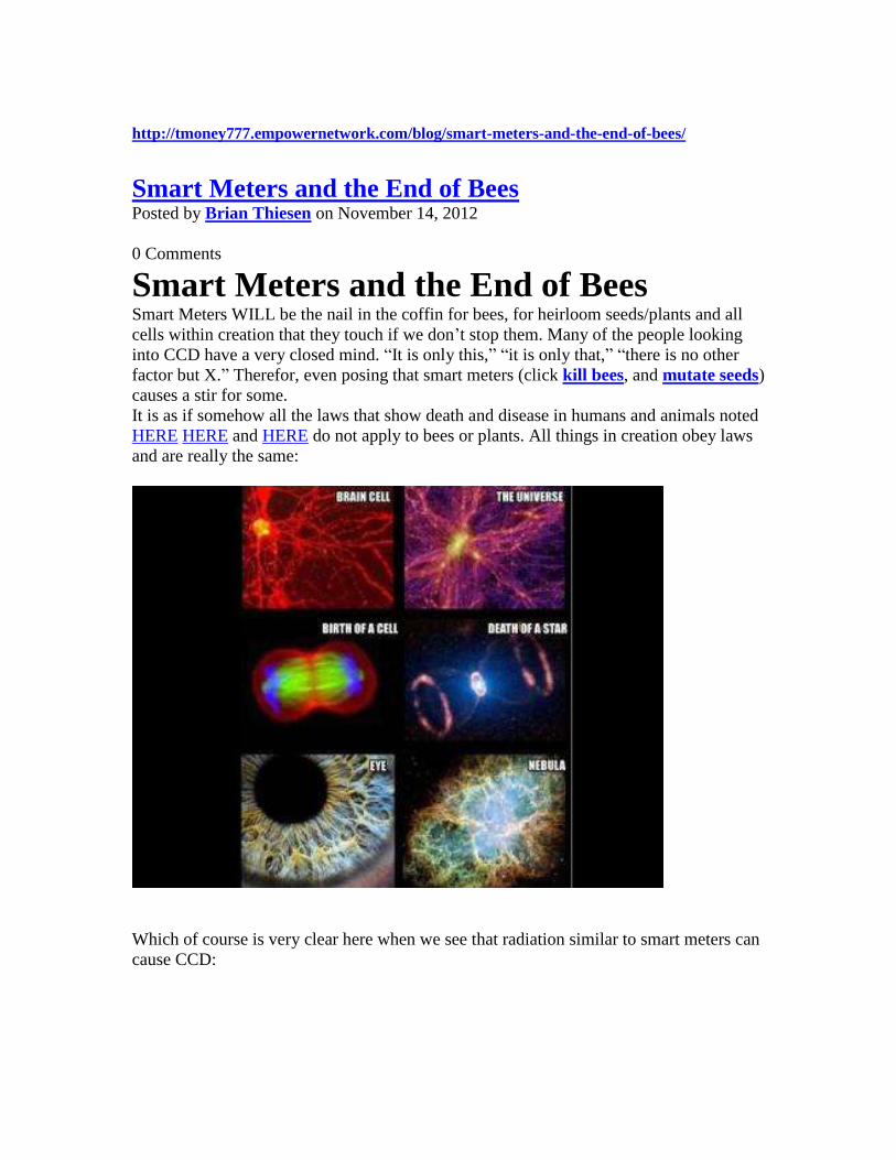
Which of course is very clear here when we see that radiation similar to smart meters can
cause CCD:
Note that now we use phones over 6 X more powerful (928 Million hz vs 6 Billion Hz)
than the ones used in the study above. Smart Meters operate at this frequency and at
least one other one (2.4 Ghz) Constantly, for life.
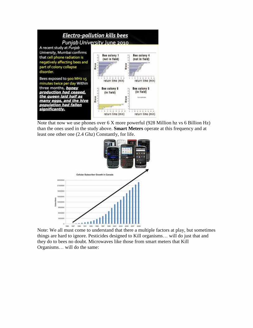
Note: We all must come to understand that there a multiple factors at play, but sometimes
things are hard to ignore. Pesticides designed to Kill organisms… will do just that and
they do to bees no doubt. Microwaves like those from smart meters that Kill
Organisms… will do the same:
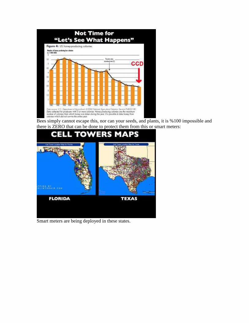
Bees simply cannot escape this, nor can your seeds, and plants, it is %100 impossible and
there is ZERO that can be done to protect them from this or smart meters:
Smart meters are being deployed in these states.
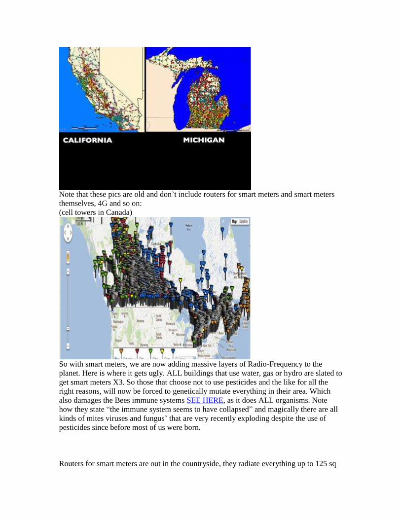
Note that these pics are old and don’t include routers for smart meters and smart meters
themselves, 4G and so on:
(cell towers in Canada)
So with smart meters, we are now adding massive layers of Radio-Frequency to the
planet. Here is where it gets ugly. ALL buildings that use water, gas or hydro are slated to
get smart meters X3. So those that choose not to use pesticides and the like for all the
right reasons, will now be forced to genetically mutate everything in their area. Which
also damages the Bees immune systems SEE HERE, as it does ALL organisms. Note
how they state “the immune system seems to have collapsed” and magically there are all
kinds of mites viruses and fungus’ that are very recently exploding despite the use of
pesticides since before most of us were born.
Routers for smart meters are out in the countryside, they radiate everything up to 125 sq
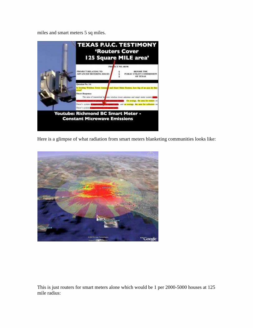
miles and smart meters 5 sq miles.
Here is a glimpse of what radiation from smart meters blanketing communities looks like:
This is just routers for smart meters alone which would be 1 per 2000-5000 houses at 125
mile radius:
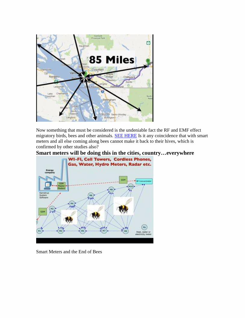
Now something that must be considered is the undeniable fact the RF and EMF effect
migratory birds, bees and other animals. SEE HERE Is it any coincidence that with smart
meters and all else coming along bees cannot make it back to their hives, which is
confirmed by other studies also?
Smart meters will be doing this in the cities, country…everywhere
Smart Meters and the End of Bees
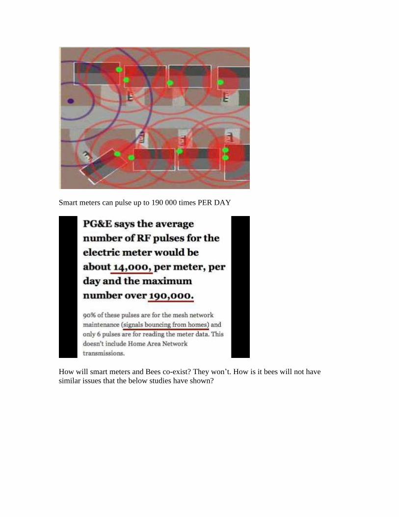
Smart meters can pulse up to 190 000 times PER DAY
How will smart meters and Bees co-exist? They won’t. How is it bees will not have
similar issues that the below studies have shown?
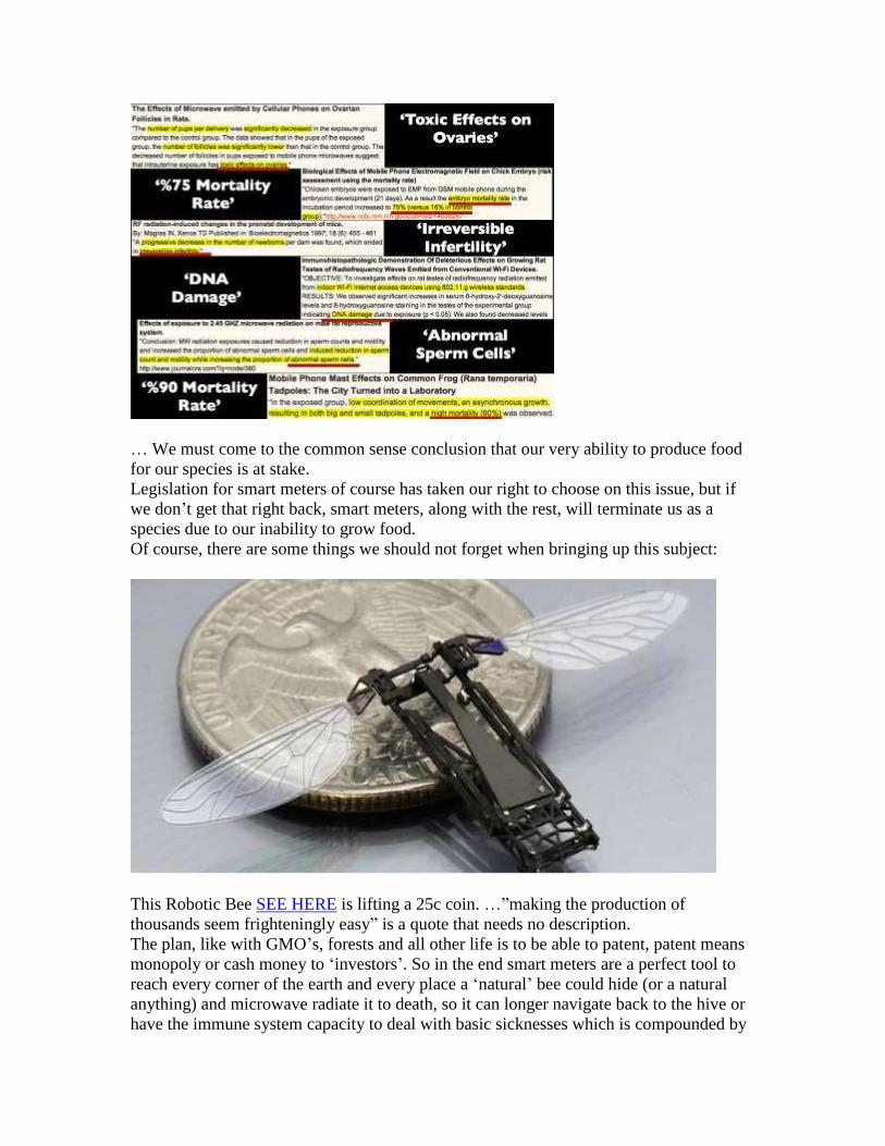
… We must come to the common sense conclusion that our very ability to produce food
for our species is at stake.
Legislation for smart meters of course has taken our right to choose on this issue, but if
we don’t get that right back, smart meters, along with the rest, will terminate us as a
species due to our inability to grow food.
Of course, there are some things we should not forget when bringing up this subject:
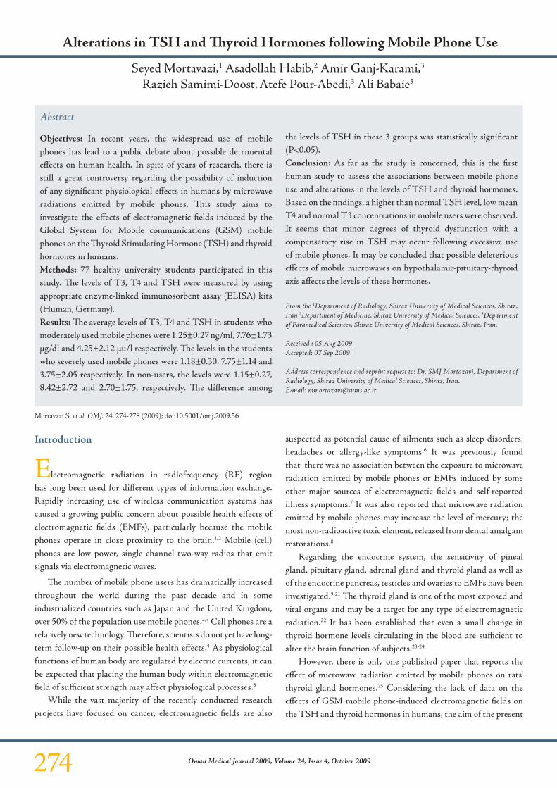
This Robotic Bee SEE HERE is lifting a 25c coin. …”making the production of
thousands seem frighteningly easy” is a quote that needs no description.
The plan, like with GMO’s, forests and all other life is to be able to patent, patent means
monopoly or cash money to ‘investors’. So in the end smart meters are a perfect tool to
reach every corner of the earth and every place a ‘natural’ bee could hide (or a natural
anything) and microwave radiate it to death, so it can longer navigate back to the hive or
have the immune system capacity to deal with basic sicknesses which is compounded by

pesticides. Those are top issues with CCD. Thus creating a new revenue stream for Bio-
Tech.
While there are unimaginable consequences to smart meters being deployed, the
jeopardizing of 1/3 +/- of the Entire Food Supply of the world seems to be quite an issue
wouldn’t you say?
Instead of smart meters, we could just keep our analogues. We, knowing the above must
fight to get smart meters removed, and for now, discontinue the use of the items we have
a choice to not use like cell phones cordless phones etc.
Thanks for reading:
Smart Meters and the End of Bees

Executive Committee
President Amy L. Dean, D.O., FAAEM
1955 Pauline Blvd Ste 100D Ann Arbor, MI 48103
President-Elect
Janette Hope, M.D., FAAEM 304 W Los Olivos
Santa Barbara, CA 93105
Secretary
Jennifer Armstrong, M.D., FAAEM 3364 Carling Ave.
Ottawa, Ontario, Canada
Treasurer Richard G. Jaeckle, M.D., FAAEM
8220 Walnut Hill Ln Ste 404 Dallas, TX 75231
Immediate Past President A.L. Barrier, M.D., FAAO-HNS
Advisor
William J. Rea, M.D.,FAAEM Gary R. Oberg, M.D., FAAEM
Board of Directors
Craig Bass, M.D.
Robin Bernhoft, M.D., FAAEM Martha Grout, M.D., MD(H)
W. Alan Ingram, M.D. Derek Lang, D.O.
Allan D. Lieberman, M.D., FAAEM Lisa Nagy, M.D.
Kalpana D. Patel, M.D., FAAEM Continuing Medical Education
Chair James W. Willoughby, II, D.O.
24 Main St. Liberty, MO 64068
Assistant-Chair
Wm. Alan Ingram, M.D. 18015 Oak St Ste B Omaha, NE 68130
American Academy of Environmental Medicine 6505 E Central • Ste 296 • Wichita, KS 67206
Tel: (316) 684-5500 • Fax: (316) 684-5709
www.aaemonline.org
Wireless Smart Meter Case Studies
Founded in 1965 as a non-profit medical association, the American Academy of Environmental Medicine (AAEM) is an international organization of physician and scientists interested in the complex relationship between the environment and health.
AAEM physicians and physicians world-wide are treating patients who report adverse, debilitating health effects following the installation of smart meters, which emit electromagnetic frequencies (EMF) and radiofrequencies (RF).
The peer reviewed, scientific literature demonstrates the correlation between EMF/RF exposure and neurological, cardiac, and pulmonary disease as well as reproductive disorders, immune dysfunction, cancer and other health conditions. The evidence is irrefutable. Despite this research, claims have been made that studies correlating smart meter emissions with adverse health effects do not exist.
The AAEM has received a case series submitted by Dr. Federica Lamech, MBBS, Self-Reporting of Symptom Development from Exposure to Wireless Smart Meters’ Radiofrequency Fields in Victoria. AAEM supports this research. It is a well documented 92 case series that is scientifically valid. It clearly demonstrates adverse health effects in the human population from smart meter emissions.
The symptoms reported in this case series closely correlate not only with the clinical findings of environmental physicians, but also with the scientific literature. Many of the symptoms reported including fatigue, headaches, heart palpitations, dizziness and other symptoms have been shown to be triggered by electromagnetic field exposure under double blind, placebo controlled conditions. Symptoms in this case series also correlate with the Austrian Medical Association’s Guidelines for the Diagnosis and Treatment of EMF Related Health Problems.
It is critically important to note that the data in this case series indicates that the “vast majority of cases” were not electromagnetically hypersensitive until after installation of smart meters. Dr. Lamech concludes that smart meters “may have unique characteristics that lower people’s threshold for symptom development”.
This research is the first of its kind, clearly demonstrating the correlation between smart meters and adverse health effects.
Based on the findings of this case series, AAEM calls for:
Further research regarding smart meter health effects
Accommodation for health considerations regarding smart meters.
Avoidance of smart meter EMF/RF emissions based on health considerations, including the option to maintain analog meters.
A moratorium on smart meters and implementation of safer technology
Physicians and health care providers to consider the role of EMF and RF in the disease process, diagnosis and treatment of patients.
Passed by the Board of Directors of the American Academy of Environmental Medicine October 23, 2013 Please note: Smart Meter case series research to be released upon publication
274 Oman Medical Journal 2009, Volume 24, Issue 4, October 2009
Alterations in TSH and Thyroid Hormones following Mobile Phone Use
Seyed Mortavazi,1 Asadollah Habib,2 Amir Ganj-Karami,3 Razieh Samimi-Doost, Atefe Pour-Abedi,3 Ali Babaie3
Introduction
Electromagnetic radiation in radiofrequency (RF) region has long been used for different types of information exchange. Rapidly increasing use of wireless communication systems has caused a growing public concern about possible health effects of electromagnetic fields (EMFs), particularly because the mobile phones operate in close proximity to the brain.1,2 Mobile (cell) phones are low power, single channel two-way radios that emit signals via electromagnetic waves.
The number of mobile phone users has dramatically increased throughout the world during the past decade and in some industrialized countries such as Japan and the United Kingdom, over 50% of the population use mobile phones.2,3 Cell phones are a relatively new technology. Therefore, scientists do not yet have long-term follow-up on their possible health effects.4 As physiological functions of human body are regulated by electric currents, it can be expected that placing the human body within electromagnetic field of sufficient strength may affect physiological processes.5
While the vast majority of the recently conducted research projects have focused on cancer, electromagnetic fields are also
suspected as potential cause of ailments such as sleep disorders, headaches or allergy-like symptoms.6 It was previously found that there was no association between the exposure to microwave radiation emitted by mobile phones or EMFs induced by some other major sources of electromagnetic fields and self-reported illness symptoms.7 It was also reported that microwave radiation emitted by mobile phones may increase the level of mercury; the most non-radioactive toxic element, released from dental amalgam restorations.8
Regarding the endocrine system, the sensitivity of pineal gland, pituitary gland, adrenal gland and thyroid gland as well as of the endocrine pancreas, testicles and ovaries to EMFs have been investigated.9-21 The thyroid gland is one of the most exposed and vital organs and may be a target for any type of electromagnetic radiation.22 It has been established that even a small change in thyroid hormone levels circulating in the blood are sufficient to alter the brain function of subjects.23-24
However, there is only one published paper that reports the effect of microwave radiation emitted by mobile phones on rats' thyroid gland hormones.25 Considering the lack of data on the effects of GSM mobile phone-induced electromagnetic fields on the TSH and thyroid hormones in humans, the aim of the present
Abstract
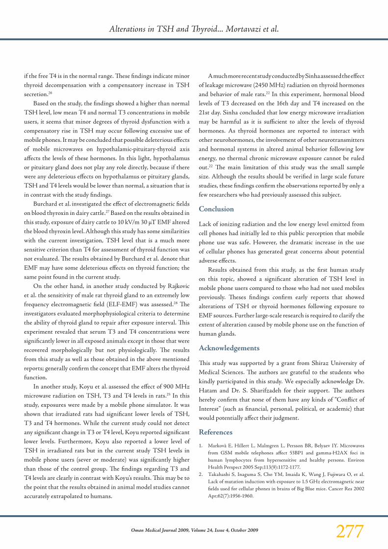
Objectives: In recent years, the widespread use of mobile phones has lead to a public debate about possible detrimental effects on human health. In spite of years of research, there is still a great controversy regarding the possibility of induction of any significant physiological effects in humans by microwave radiations emitted by mobile phones. This study aims to investigate the effects of electromagnetic fields induced by the Global System for Mobile communications (GSM) mobile phones on the Thyroid Stimulating Hormone (TSH) and thyroid hormones in humans. Methods: 77 healthy university students participated in this study. The levels of T3, T4 and TSH were measured by using appropriate enzyme-linked immunosorbent assay (ELISA) kits (Human, Germany). Results: The average levels of T3, T4 and TSH in students who moderately used mobile phones were 1.25±0.27 ng/ml, 7.76±1.73 µg/dl and 4.25±2.12 µu/l respectively. The levels in the students who severely used mobile phones were 1.18±0.30, 7.75±1.14 and 3.75±2.05 respectively. In non-users, the levels were 1.15±0.27, 8.42±2.72 and 2.70±1.75, respectively. The difference among
the levels of TSH in these 3 groups was statistically significant (P<0.05). Conclusion: As far as the study is concerned, this is the first human study to assess the associations between mobile phone use and alterations in the levels of TSH and thyroid hormones. Based on the findings, a higher than normal TSH level, low mean T4 and normal T3 concentrations in mobile users were observed. It seems that minor degrees of thyroid dysfunction with a compensatory rise in TSH may occur following excessive use of mobile phones. It may be concluded that possible deleterious effects of mobile microwaves on hypothalamic-pituitary-thyroid axis affects the levels of these hormones.
From the 1Department of Radiology, Shiraz University of Medical Sciences, Shiraz, Iran 2Department of Medicine, Shiraz University of Medical Sciences, 3Department of Paramedical Sciences, Shiraz University of Medical Sciences, Shiraz, Iran.
Received : 05 Aug 2009Accepted: 07 Sep 2009
Address correspondence and reprint request to: Dr. SMJ Mortazavi, Department of Radiology, Shiraz University of Medical Sciences, Shiraz, Iran.E-mail: [email protected]
Mortavazi S, et al. OMJ. 24, 274-278 (2009); doi:10.5001/omj.2009.56

277Oman Medical Journal 2009, Volume 24, Issue 4, October 2009
if the free T4 is in the normal range. These findings indicate minor thyroid decompensation with a compensatory increase in TSH secretion.26
Based on the study, the findings showed a higher than normal TSH level, low mean T4 and normal T3 concentrations in mobile users, it seems that minor degrees of thyroid dysfunction with a compensatory rise in TSH may occur following excessive use of mobile phones. It may be concluded that possible deleterious effects of mobile microwaves on hypothalamic-pituitary-thyroid axis affects the levels of these hormones. In this light, hypothalamus or pituitary gland does not play any role directly, because if there were any deleterious effects on hypothalamus or pituitary glands, TSH and T4 levels would be lower than normal, a situation that is in contrast with the study findings.
Burchard et al. investigated the effect of electromagnetic fields on blood thyroxin in dairy cattle.27 Based on the results obtained in this study, exposure of dairy cattle to 10 kV/m 30 µT EMF altered the blood thyroxin level. Although this study has some similarities with the current investigation, TSH level that is a much more sensitive criterion than T4 for assessment of thyroid function was not evaluated. The results obtained by Burchard et al. denote that EMF may have some deleterious effects on thyroid function; the same point found in the current study.
On the other hand, in another study conducted by Rajkovic et al. the sensitivity of male rat thyroid gland to an extremely low frequency electromagnetic field (ELF-EMF) was assessed.28 The investigators evaluated morphophysiological criteria to determine the ability of thyroid gland to repair after exposure interval. This experiment revealed that serum T3 and T4 concentrations were significantly lower in all exposed animals except in those that were recovered morphologically but not physiologically. The results from this study as well as those obtained in the above mentioned reports; generally confirm the concept that EMF alters the thyroid function.
In another study, Koyu et al. assessed the effect of 900 MHz microwave radiation on TSH, T3 and T4 levels in rats.25 In this study, exposures were made by a mobile phone simulator. It was shown that irradiated rats had significant lower levels of TSH, T3 and T4 hormones. While the current study could not detect any significant change in T3 or T4 level, Koyu reported significant lower levels. Furthermore, Koyu also reported a lower level of TSH in irradiated rats but in the current study TSH levels in mobile phone users (sever or moderate) was significantly higher than those of the control group. The findings regarding T3 and T4 levels are clearly in contrast with Koyu’s results. This may be to the point that the results obtained in animal model studies cannot accurately extrapolated to humans.
A much more recent study conducted by Sinha assessed the effect of leakage microwave (2450 MHz) radiation on thyroid hormones and behavior of male rats.22 In this experiment, hormonal blood levels of T3 decreased on the 16th day and T4 increased on the 21st day. Sinha concluded that low energy microwave irradiation may be harmful as it is sufficient to alter the levels of thyroid hormones. As thyroid hormones are reported to interact with other neurohormones, the involvement of other neurotransmitters and hormonal systems in altered animal behavior following low energy, no thermal chronic microwave exposure cannot be ruled out.22 The main limitation of this study was the small sample size. Although the results should be verified in large scale future studies, these findings confirm the observations reported by only a few researchers who had previously assessed this subject.
Conclusion
Lack of ionizing radiation and the low energy level emitted from cell phones had initially led to this public perception that mobile phone use was safe. However, the dramatic increase in the use of cellular phones has generated great concerns about potential adverse effects.
Results obtained from this study, as the first human study on this topic, showed a significant alteration of TSH level in mobile phone users compared to those who had not used mobiles previously. Theses findings confirm early reports that showed alterations of TSH or thyroid hormones following exposure to EMF sources. Further large-scale research is required to clarify the extent of alteration caused by mobile phone use on the function of human glands.
Acknowledgements
This study was supported by a grant from Shiraz University of Medical Sciences. The authors are grateful to the students who kindly participated in this study. We especially acknowledge Dr. Hatam and Dr. S. Sharifzadeh for their support. The authors hereby confirm that none of them have any kinds of “Conflict of Interest” (such as financial, personal, political, or academic) that would potentially affect their judgment.
References
1. Markovà E, Hillert L, Malmgren L, Persson BR, Belyaev IY. Microwaves from GSM mobile telephones affect 53BP1 and gamma-H2AX foci in human lymphocytes from hypersensitive and healthy persons. Environ Health Perspect 2005 Sep;113(9):1172-1177.
2. Takahashi S, Inaguma S, Cho YM, Imaida K, Wang J, Fujiwara O, et al. Lack of mutation induction with exposure to 1.5 GHz electromagnetic near fields used for cellular phones in brains of Big Blue mice. Cancer Res 2002 Apr;62(7):1956-1960.
Alterations in TSH and Thyroid... Mortavazi et al.
278 Oman Medical Journal 2009, Volume 24, Issue 4, October 2009
3. Myerson SG, Mitchell AR. Mobile phones in hospitals. BMJ 2003 Mar;326(7387):460-461.
4. Frumkin H, Jacobson A, Gansler T, Thun MJ. Cellular phones and risk of brain tumors. CA Cancer J Clin 2001 Mar-Apr;51(2):137-141.
5. Karinen A, Heinävaara S, Nylund R, Leszczynski D. Mobile phone radiation might alter protein expression in human skin. BMC Genomics 2008;9:1-5 .
6. Seitz H, Stinner D, Eikmann T, Herr C, Röösli M. Electromagnetic hypersensitivity (EHS) and subjective health complaints associated with electromagnetic fields of mobile phone communication–a literature review published between 2000 and 2004. Sci Total Environ 2005 Oct;349(1-3):45-55.
7. Mortazavi SM, Ahmadi J, Shariati M. Prevalence of subjective poor health symptoms associated with exposure to electromagnetic fields among university students. Bioelectromagnetics 2007 May;28(4):326-330.
8. Mortazavi SM, Daiee E, Yazdi A, Khiabani K, Kavousi A, Vazirinejad R, et al. Mercury release from dental amalgam restorations after magnetic resonance imaging and following mobile phone use. Pak J Biol Sci 2008 Apr;11(8):1142-1146.
9. Stevens RG. Electric power use and breast cancer: a hypothesis. Am J Epidemiol 1987 Apr;125(4):556-561.
10. Löscher W, Mevissen M. Animal studies on the role of 50/60-Hertz magnetic fields in carcinogenesis. Life Sci 1994;54(21):1531-1543.
11. Brainard GC, Kavet R, Kheifets LI. The relationship between electromagnetic field and light exposures to melatonin and breast cancer risk: a review of the relevant literature. J Pineal Res 1999 Mar;26(2):65-100.
12. Graham C, Cook MR, Gerkovich MM, Sastre A. Examination of the melatonin hypothesis in women exposed at night to EMF or bright light. Environ Health Perspect 2001 May;109(5):501-507.
13. Ossenkopp KP, Koltek WT, Persinger MA. Prenatal exposure to an extremely low frequency-low intensity rotating magnetic field and increases in thyroid and testicle weight in rats. Dev Psychobiol 1972;5(3):275-285.
14. Zagorskaya EA. Endocrine responses to low frequency electromagnetic fields of continuous and intermittent generation. Kosm Biol Aviakosm Med 1989;23:4-14.
15. Picazo ML, Miguel MP, Leyton V, Franco P, Varela L, Paniagua R, et al. Long-term effects of ELF magnetic fields on the mouse testis and serum testosterone levels. Electro-Magnetobiol 1995;14:127-134.
16. Zagorskaya EA, Klimovitsky VY, Melnichenko VP, Rodina GP, Semyonov SN. The effect of low frequency electromagnetic fields on physiological systems: a review. Kosm Biol Aviakosm Med 1990;24:3-11.
17. Forgács Z, Thuróczy G, Paksy K, Szabó LD. Effect of sinusoidal 50 Hz magnetic field on the testosterone production of mouse primary Leydig cell culture. Bioelectromagnetics 1998;19(7):429-431.
18. Burchard JF, Nguyen DH, Block E. Progesterone concentrations during estrous cycle of dairy cows exposed to electric and magnetic fields. Bioelectromagnetics 1998;19(7):438-443.
19. Feria-Velasco A, Castillo-Medina S, Verdugo-Díaz L, Castellanos E, Orozco-Suárez S, Sánchez-Gómez C, et al. Neuronal differentiation of chromaffin cells in vitro, induced by extremely low frequency magnetic fields or nerve growth factor: a histological and ultrastructural comparative study. J Neurosci Res 1998 Sep;53(5):569-582.
20. Uscebrka G, Zikic D, Matavulj M, Rajkovic V, Gledic D. Electromagnetic field effects on the morphometrical characteristics of rat adrenal glands. In: Bersani, F. (Ed.), Electricity and Magnetism in Biology and Medicine. Kluwer Academic/Plenum Publishers, New York, pp. 485–488, 1999.
21. Matavulj M, Rajkovic V, Uscebrka G, Lukac T, Stevanovic D, Lazetic B. Studies on the possible endocrinological effects of an 50 Hz electromagnetic field. Centr. Europ. J. Occup. Environ. Med 2000;6:183-188.
22. Sinha RK. Chronic non-thermal exposure of modulated 2450 MHz microwave radiation alters thyroid hormones and behavior of male rats. Int J Radiat Biol 2008 Jun;84(6):505-513.
23. Bauer M, Goetz T, Glenn T, Whybrow PC. The thyroid-brain interaction in thyroid disorders and mood disorders. J Neuroendocrinol 2008 Oct;20(10):1101-1114.
24. Bernal J. Thyroid hormone receptors in brain development and function. Nat Clin Pract Endocrinol Metab 2007 Mar;3(3):249-259.
25. Koyu A, Cesur G, Ozguner F, Akdogan M, Mollaoglu H, Ozen S. Effects of 900 MHz electromagnetic field on TSH and thyroid hormones in rats. Toxicol Lett 2005 Jul;157(3):257-262.
26. Larsen RP, Davies TF, Schlumberger MJ, Hay ID. Thyroid physiology and diagnostic evaluation of patient with thyroid disorders. In: Williams textbook of endocrinology. 11th edition. Saunders, pp: 319-320, 2008.
27. Burchard JF, Nguyen DH, Rodriguez M. Plasma concentrations of thyroxine in dairy cows exposed to 60 Hz electric and magnetic fields. Bioelectromagnetics 2006 Oct;27(7):553-559.
28. Rajkovic V, Matavulj M, Gledic D, Lazetic B. Evaluation of rat thyroid gland morphophysiological status after three months exposure to 50 Hz electromagnetic field. Tissue Cell 2003 Jun;35(3):223-231.
Alterations in TSH and Thyroid... Mortavazi et al.
Brain proteome response following whole bodyexposure of mice to mobile phone or wireless DECTbase radiation
Adamantia F. Fragopoulou1, Athina Samara2, Marianna H. Antonelou1,Anta Xanthopoulou3, Aggeliki Papadopoulou3, Konstantinos Vougas3,Eugenia Koutsogiannopoulou2, Ema Anastasiadou2,Dimitrios J. Stravopodis1, George Th. Tsangaris3 & Lukas H. Margaritis1
1Department of Cell Biology and Biophysics, Athens University, Athens, Greece, 2Geneticsand Gene Therapy Division, Center of Basic Research II, Biomedical Research Foundation ofthe Academy of Athens, Athens, Greece, and 3Proteomics Research Unit, Center of BasicResearch II, Biomedical Research Foundation of the Academy of Athens, Athens, Greece
The objective of this study was to investigate the effects of two sources of electromagnetic fields(EMFs) on the proteome of cerebellum, hippocampus, and frontal lobe in Balb/c mice followinglong-term whole body irradiation. Three equally divided groups of animals (6 animals/group)were used; the first group was exposed to a typical mobile phone, at a SAR level range of 0.17–0.37W/kg for 3 h daily for 8 months, the second group was exposed to a wireless DECT base(Digital Enhanced Cordless Telecommunications/Telephone) at a SAR level range of 0.012–0.028W/kg for 8 h/day also for 8 months and the third group comprised the sham-exposedanimals. Comparative proteomics analysis revealed that long-term irradiation from both EMFsources altered significantly (p , 0.05) the expression of 143 proteins in total (as low as 0.003fold downregulation up to 114 fold overexpression). Several neural function related proteins (i.e.,Glial Fibrillary Acidic Protein (GFAP), Alpha-synuclein, Glia Maturation Factor beta (GMF), andapolipoprotein E (apoE)), heat shock proteins, and cytoskeletal proteins (i.e., Neurofilaments andtropomodulin) are included in this list as well as proteins of the brain metabolism (i.e., Aspartateaminotransferase, Glutamate dehydrogenase) to nearly all brain regions studied. Western blotanalysis on selected proteins confirmed the proteomics data. The observed protein expressionchanges may be related to brain plasticity alterations, indicative of oxidative stress in the nervous
Authors’ contributions: AFF and LHM conceived the concept and design of the experiments, madethe literature survey and the final biologically valid interpretation of the EMF impact upon the brain,wrote and finalized the manuscript. AFF carried out all animal handling, welfare, EMF exposure,part of brain dissection and immunoassays. AS performed the brain dissection and brain regions’separation, contributed to the non EMF writing of the manuscript and together with MHA, EK andEA carried out a part of the immunoassays and contributed to the data evaluation related toneuroproteomics. AX, AP and KV were involved in 2 DE experiments, Maldi ToF/MS, proteinidentification and statistical analysis. DJS participated in the conception of the design andcontributed to the interpretation and evaluation of the overall data. GThT participated in theexperimental design and experimental protocols optimization, coordinated the proteomics study,carried out the overall differential proteomics analysis and data evaluation and contributed to theproteomics writing of the manuscript. All authors read and approved the final manuscript.
Address correspondence to Lukas H. Margaritis, Adamantia F. Fragopoulou, Department of CellBiology and Biophysics, Faculty of Biology, Athens University, Panepistimiopolis, 15784 Athens,Greece. E mails: [email protected], [email protected]
Electromagnetic Biology and Medicine, Early Online: 1 25, 2012Copyright Q Informa Healthcare USA, Inc.ISSN: 1536 8378 print / 1536 8386 onlineDOI: 10.3109/15368378.2011.631068
1
Ele
ctro
mag
n B
iol M
ed D
ownl
oade
d fr
om in
form
ahea
lthca
re.c
om b
y U
nive
rsity
of
Ath
ens
on 0
1/20
/12
For
pers
onal
use
onl
y.
system or involved in apoptosis and might potentially explain human health hazards reported sofar, such as headaches, sleep disturbance, fatigue, memory deficits, and brain tumor long-terminduction under similar exposure conditions.
Keywords Microwaves, Radiofrequencies, Wireless phones, Proteomics, Brain plasticity,Hippocampus, Frontal lobe, Cerebellum
INTRODUCTION
Wireless technology emitting electromagnetic radiation (EMR) is spread worldwideaffecting directly or indirectly all social levels, all countries, and all ages since itincludes mobile phones, cordless DECT telephones, Wi-Fi, wi-max, baby monitors,local TV, and FM broadcast stations. The concern about possible health hazards hasled to extensive research, concerning exclusively the effects of mobile phonetechnology (devices and mast stations) at the cellular, lab animal, andepidemiological level, using a variety of model systems and approaches but not ina coordinated manner (Chavdoula et al., 2010; Fragopoulou et al., 2010a,b,c;Fragopoulou and Margaritis, 2010; Hardell and Carlberg, 2009; Hillert et al., 2008;Khurana et al., 2009, 2010), although there have been international efforts (i.e.,interphone study; Cardis et al., 2011) to reveal the truth about the possible EMFhealth risks. The importance of mobile phone (MP) radiation research lies in thefact that there are currently 5 billion users on the planet and the vast majority is usingthe MP in contact with the brain (Frey, 1998).
A number of reports have dealt with possible changes on gene/protein expression,either at an individual gene/protein level or using the “omics” approaches. Theindividual approach has focused mainly on heat shock proteins and their mRNAs(French et al., 2001; McNamee and Chauhan, 2009), but other proteins and geneshave also been studied with conflicting, so far, results (Fritze et al., 1997; Cleary et al.,1997; Nikolova et al., 2005; Zhao et al., 2007). In order to assess large numbers ofgenes and proteins, high throughput approaches have been applied in the lastdecade. These “omics” approaches, also used in the present work, have gainedground in the study of EMF effects mainly on cell cultures. Belyaev et al. (2006),analyzing by Affymetrix U34 Gene Chips cerebellum of brain samples after wholebody 2 h exposure of rats at 915 GSM in TEM cells, revealed overexpression of12 genes and downregulation of 1 gene. The same (Salford’s) research group 2 yearslater applied Microarray hybridizations on Affymetrix rat2302 chips of RNA extractsfrom cortex and hippocampus of GSM 1800 exposed rats for just 6 h within TEM cells(Nittby et al., 2008). Using four exposed and four control animals they found that alarge number of genes were altered at hippocampus and cortex. The vast majoritywere downregulated. In a series of publications by Leszczynski’s research group,consistently using human endothelial cell lines EA.hy926 and EA.hy926v1, proteinexpression changes after exposure to 900 MHz were shown (Leszczynski et al., 2002,2004; Nylund and Leszczynski, 2004, 2006; Remondini et al., 2006). These effectshave been recently confirmed by the same group in the two types of mobile phoneexposure protocols: GSM 900 and 1800 MHz (Nylund et al., 2009). Another “omics”group exposing human lens epithelial cells has detected heat-shock protein (HSP) 70and heterogeneous nuclear ribonucleoprotein K (hnRNP K) to be upregulatedfollowing exposure to GSM 1800 MHz for 2 h (Li et al., 2007), whereas a third researchgroup exposed human breast cancer cells MCF-7 to an RF generator simulating GSM1800 MHz signal at various SAR values and duration of exposures (Zeng et al., 2006a).They analyzed the transcriptome and the proteome of the cells after continuous orintermittent exposure and concluded that EMF exposure caused distinct effects ongene and protein expression. The same authors suggested that the protein
2 A. F. Fragopoulou et al.
Electromagnetic Biology and Medicine
Ele
ctro
mag
n B
iol M
ed D
ownl
oade
d fr
om in
form
ahea
lthca
re.c
om b
y U
nive
rsity
of
Ath
ens
on 0
1/20
/12
For
pers
onal
use
onl
y.
expression changes might depend on duration and mode of exposure andtherefore a number of biological processes might be affected (Zeng et al., 2006b).Since the above in vitro effects cannot be easily translated into humans, in 2008,Leszczynski’s group performed a pilot study on volunteers (Karinen et al., 2008)and showed that mobile phone radiation might alter protein expression inhuman skin cells. Gene expression changes as revealed using transcriptomicshad not effects on C3H 10T(1/2) mouse cells (Whitehead et al., 2006). However,and as previously mentioned, such a limited and non systematic number ofpublications using “omics” approaches does not allow for any conclusions to bedrawn concerning the impact of mobile phone emitted radiation upon cellproteome, physiology and function (Nylund et al., 2009), as also pointed out byVanderstraeten and Verschaeve (2008).
Concerning research on wireless DECT base and handset radiation exposurewhich is potentially harmful to millions of people, no actual experiments have beenconducted, besides the clinical studies reported by Soderqvist et al. (2009a,b),Havas et al. (2010) and the epidemiological studies showing increased risk for braintumors (Hardell and Carlberg, 2009; Khurana et al., 2009). A recently publishedarticle highlighted the importance of mobile phone epidemiology studies inproperly addressing DECT phone use as a strong and likely confounder (Redmayneet al., 2010).
Given the limited available data on animal models, our objective was toinvestigate the effects of two sources of EMFs on the proteome of the cerebellum,hippocampus and frontal lobe in Balb/c mice.
These three brain regions were chosen since they are related to mainfunctions of the brain, such as memory, attention, reward, planning, equilibrium,and motor control. Their common role is the correlation with cognitive functions(Okano et al., 2000), which have been reported in a number of studies to bealtered after EMF exposure (for a review see Fragopoulou and Margaritis, 2010).The hippocampus mainly controls spatial memory, the cerebellum is responsiblefor motor learning, and the frontal lobe plays an important role in retaininglonger term memories associated with emotions. The frontal lobe does not seemto be involved in any particular discrete perceptual sensory or so called motorfunction, but in spite of that, it seems to have a very critical role on how weuse the kind of information that other parts of the brain are dedicated todetermine.
Our high-throughput approach challenges the gaps in the literatureinvestigating whether EMFs can provoke changes on the mouse brain proteome;changes that could be correlated with EMF memory impairments reported so faror with neurological diseases, such as Alzheimer’s and even with brain tumorinduction.
Three groups of 18 animals were used in the present study (6 animals/group): thefirst group was exposed to a commercially available mobile phone, operating at GSM900 MHz configuration and frequency and at normal speaking emission mode at aSAR level range of 0.17 –0.37 W/kg for 3 h daily for 8 months. The second group wasexposed to a wireless DECT base at a SAR level range of 0.012 –0.028 W/kg for 8 h/dayduring the lights-off period also for 8 months. The third group comprised the sham-exposed animals.
The novelty of this work lies in the fact that no brain proteome studies have beenreported so far following EMF exposure and, in particular, of isolated brain regions inany animal model. In addition, to our knowledge this is the first experimental reportof wireless DECT exposure effects on any biological model system and in particularfollowing proteome analysis.
EMFs affect mouse brain proteome 3
Copyright Q Informa Healthcare USA, Inc.
Ele
ctro
mag
n B
iol M
ed D
ownl
oade
d fr
om in
form
ahea
lthca
re.c
om b
y U
nive
rsity
of
Ath
ens
on 0
1/20
/12
For
pers
onal
use
onl
y.
MATERIALS AND METHODS
AnimalsA total of 18 healthy adult male mice Mus musculus, strain Balb/c, were obtainedfrom the Hellenic Pasteur Institute Animal Facility and then transferred to ouranimal facility in the Department of Cell Biology and Biophysics of Athens Universitywhere they were left for two weeks to get acclimatized. Animals were housed equallydivided into 3 groups in Techniplast, USA Plexiglas cages, 1290D Eurostandard TypeIII, 425 £ 266 £ 155 mm - floor area 820 cm2. The first and the second studygroup were exposed to a commercially available dual band mobile phone and awireless DECT base, respectively. Free moving mice were exposed within their cages,as reported previously (Fragopoulou et al., 2010b). The third group comprised thesham-exposed group. All animals were kept under standard laboratory conditions:(22 ^ 2)oC, (40– 60)% relative humidity, 12 h:12 h light/dark cycle (lights on at7:00 am) and received food (pellets) and water ad libitum. Taking into considerationthe welfare of the animals, enrichment material was used within their home cages,i.e., paper and plastic tubes. All experimental procedures were carried out inagreement with the ethical recommendations of the European Communities CouncilDirective of 24 November 1986 (86/609/EEC) and with the ethical rules of theBioethics Committee of the Faculty of Biology of Athens University. The 3R’s conceptof Russell and Burch (Refinement, Reduction and Replacement) was seriously takeninto consideration (Russell and Burch, 1959).
EMF Exposure Conditions and Field MeasurementsSince the objective of this work is the exploration of any changes in the brainproteome, special attention was given to ensure that the only factor affecting theanimals would be the radiation emitted from mobile phone or the base of the DECTwireless device. Therefore, other fields or noise (i.e., magnetic field, other RFs ofvarious frequencies and noise levels) were measured and negligible and in any casethey were the same quantitatively and qualitatively with the sham-exposed group.
Mobile Phone ExposureThe animals of this group (n 6) were exposed to radiation within their home cagethree hours per day for 8 months. The exposure protocol of “3 h/day £ 8 months”has been chosen in order to mimic a daily typical mobile phone operation by anactive person. The mobile phone was placed underneath the cage. A semi-Faradaycage was specially constructed having one open surface to allow mobile phonecommunication and at the same time to prevent radiation leakage towards sham-exposed animals. The GSM 900 MHz electrical field intensity of the radiation emittedby the mobile phone was measured using the Smartfieldmeter, EMC Test Design,LLC, Newton, MA, USA placing the dual band omni directional probe (900,1800 MHz) inside a similar cage housing the animals positioned at the same placeeither at the end or in the beginning of exposure. The obtained measurements werereproducible on a daily basis (minimum-maximum value depending on the soundintensity). In order to simulate the conditions of human voice and activate mobilephone ELF modulated EMF emission, radio station was playing as a source ofauditory stimulation throughout the exposure time. The measured electrical fieldintensity was below ICNIRP’s recommendations (ICNIRP, 1998) within the range of15–22 V/m in the various areas within the cage with the animals followingalso the typical GSM power modulation by the sound intensity. The SAR value(SAR s*E^2/r) calculated as previously described (Fragopoulou et al., 2010a,b)was between 0.17 and 0.37 W/kg. This is a rough estimation of the whole body
4 A. F. Fragopoulou et al.
Electromagnetic Biology and Medicine
Ele
ctro
mag
n B
iol M
ed D
ownl
oade
d fr
om in
form
ahea
lthca
re.c
om b
y U
nive
rsity
of
Ath
ens
on 0
1/20
/12
For
pers
onal
use
onl
y.
average SAR of individual animals. The aim was to achieve similar exposureconditions occurring to a human user when holding the mobile phone next tohis/her ear with the only difference that the mice were receiving whole body and nothead-only exposure.
Wireless DECT Base ExposureThe animals of this group were exposed to a commercially available wireless DECTbase, which constantly emits radiation at a bandwidth of 1880–1900 MHz, very closeto the GSM1800 band, scanning all 10 allocated RF channels without any handsetcommunicating with the base. The DECT base was placed close to the mouse cageand was programmed to operate for 8 h per day during the lights-off period for8 months. This exposure protocol of 8 h/day has been chosen to correspond tohuman occupational or home DECT base exposure. A semi-Faraday cage wasspecially constructed to prevent radiation leakage towards sham-exposed animals.Electrical field levels were measured with Smartfieldmeter as described aboveand the values recorded were from 4–6 V/m depending on the position within thecage. No voice modulation is required for DECT operation, but the same radiostation was playing for comparative purposes to the mobile phone exposure.Therefore, SAR value calculated, as described above, ranged from 0.012– 0.028 W/Kg.
Sham-exposed GroupMice were kept in a similar room as the exposed groups, under the same conditions ofliving. The cages of the animals were inside a Faraday cage to prevent radiation entryfrom the mobile phone and DECT base when in operation. A radio was playing atthe same station and the same volume as the one in the rooms of the exposed animals.Non significant levels of Radio-frequency (RF) field deriving from the exposure sourceswas detected inside the cage with the animals, as measured by the Smartfieldmeter.
BRAIN TISSUE REMOVAL AND HOMOGENIZATION
At the end of the experiment, mice were euthanized according to the bioethical rulesof the European Committee for animal protection, with cervical dislocation followedby rapid brain tissue removal between 8 and 10 am. Parts of the brain (frontallobe, hippocampus, and cerebellum) were quickly separated, immediately frozenin liquid nitrogen, and then stored at 80oC until sample processing for furthermanipulation.
TWO-DIMENSIONAL ELECTROPHORESIS
The tissue was homogenized in a glass Wheaton (tight) homogenizer in a bufferconsisting of 8 M urea, 40 mM Tris-HCL (pH 8.5), 2 M thiourea, 4% CHAPS, 1%dithioerythritol (DTE), 0.2% IPG buffer pH 3– 10 (Amersham Biosciences) and1 mg/mL of a mixture of protease inhibitors (1 mM PMSF and 1 tablet (RocheDiagnostics) per 50 mL of wash buffer and phosphatase inhibitors (0.2 mM Na3VO3
and 1 mM NaF)). The homogenate was left at room temperature for 1 h andcentrifuged at 13,000 rpm for 30 min. The protein content of the supernatant wasdetermined using the Bradford quantification method.
Two-dimensional gel electrophoresis was performed as previously reported(Anagnostopoulos et al., 2010). Samples of 1 mg total protein were applied on 18 cmIPG strips with pI 3–10 NL or 4–7 L (Bio-Rad Lab, Hercules, CA), at their basic andacidic ends, using sample cups. IPG strips had been prepared for IEF by 20 hrehydration in a buffer of 8 M urea, 4% CHAPS and 1% DTE.
EMFs affect mouse brain proteome 5
Copyright Q Informa Healthcare USA, Inc.
Ele
ctro
mag
n B
iol M
ed D
ownl
oade
d fr
om in
form
ahea
lthca
re.c
om b
y U
nive
rsity
of
Ath
ens
on 0
1/20
/12
For
pers
onal
use
onl
y.
First dimension focusing, for separation by two-dimensional gel electrophoresis,started at 250 V and voltage was gradually increased to 8000 V, with 3 V/min, keptconstant for 25 h (approximately 150,000 Vh totally). IEF was conducted in aPROTEAN IEF Cell, Bio-Rad apparatus. After focusing, IPG strips were equilibratedfirst in 6 M urea, 50 mM Tris-HCL (pH 8.8), 2% (w/v) SDS, 30% (v/v) glycerol, and0.5% (w/v) DTE for 15 min then in the same buffer containing 4% (w/v)iodoacetamide instead of DTE, for 15 more min. Second dimensional electrophoresiswas performed on 12% SDS-polyacrylamide gels (180 £ 200 £ 1.5 mm) with a run of40 mA/ gel, in PROTEIN-II multi-cell apparatuses (Bio-Rad, Hercules, CA).
PROTEIN VISUALIZATION AND IMAGE ANALYSIS
After vertical electrophoresis, gels were fixed in 50% methanol containing 5%phosphoric acid for 2 h. The fixative solution was washed off by agitation in distilledwater for 45 min. Protein spots were visualized by application of Coomassie BlueG-250 staining solution (Novex, San Diego, CA) on 2-DE gels for 12 h. Gel imageswere scanned in a GS-800 Calibrated Densitometer (Bio-Rad Laboratories, Hercules,CA) using the scanning application/tool of the PD-Quest v8.0 software (Bio-Rad,Hercules, CA). Protein spots of all gels contained in the analysis, were detected,aligned, matched, and quantified using the PD-Quest v8.0 image processingsoftware, according to the manufacturer’s instructions. Manual inspection of thespots was used to verify the accuracy of matching. Spot volume was used as theanalysis parameter to quantify protein expression. Normalization of each individualspot was performed according to the total quantity of the valid spots in each gel, aftersubtraction of the background values. Optical Density (O.D.) level (%) of eachprotein from the sham-exposed or exposed groups was determined separately andcalculated as the sum of the volume % of all spots from all gels containing the sameprotein. Selection of protein spots or entire gel regions for MS analysis was basedupon O.D. alteration between the two groups analysed. A minimum of 1.25 foldchange in the expression level was used as the selection criterion.
PEPTIDE MASS FINGERPRINTING AND IDENTIFICATION OF PROTEINS
Peptide mass fingerprinting analysis was essentially performed as describedpreviously (Mavrou et al., 2008). Briefly, all spots on the gels were annotated semi-automatically using the Melanie 4.02 software, excised with a Proteiner SPII robot(Bruker Daltonics, Bremen, Germany) and placed into 96-well microtiter plates. Theexcised spots were destained using 180ml of 100 mM ammonium bicarbonate in 30%ACN and the gel piece was dried in a speed vacuum concentrator (MaxiDry Plus,Heto, Denmark). The dried gel piece was rehydrated with 5mL of 20mg/mLrecombinant trypsin (proteomics grade, Roche diagnostics, Basel, Swiss) solution.After 16 h at room temperature, 10mL of 50% acetonitrile containing 0.3%trifluroacetic acid were added, and the gel pieces were incubated for 15 min withgentle shaking. Sample application to a target plate and analysis as well as peptidematching and protein searching were carried out as described previously (Mavrouet al., 2008). Briefly, tryptic peptide mixtures (1mL) were applied on an anchorchip MALDI plate with 1mL of matrix solution, consisting of 0.08% CHCA (Sigma),the internal standard peptides des-Arg-bradykinin (Sigma, 904.4681 Da), andadrenocorticotropic hormone fragment 18–39 (Sigma, 2465.1989 Da) in 65% ethanol,50% CAN, and 0.1% TFA. Peptide mixtures were analysed in a MALDI-ToF massspectrometer (Ultraflex II, Bruker Daltonics). Laser shots (n 1000) of intensitybetween 40% and 60% were collected and summarized and the peak list was created
6 A. F. Fragopoulou et al.
Electromagnetic Biology and Medicine
Ele
ctro
mag
n B
iol M
ed D
ownl
oade
d fr
om in
form
ahea
lthca
re.c
om b
y U
nive
rsity
of
Ath
ens
on 0
1/20
/12
For
pers
onal
use
onl
y.
using the FlexAnalysis v2.2 software (Bruker). Peptide matching and proteinsearches were performed automatically with MASCOT Server 2 (Matrix Science).Peptide masses were compared with the theoretical peptide masses of allavailable proteins of Mus musculus in the SWISS-PROT database. Stringent criteriawere used for protein identification with a maximum allowed mass error of10 ppm and a minimum of four matching peptides. Probability score withp , 0.05 was used as the criterion for affirmative protein identification.Monoisotopic masses were used, and one missed trypsin cleavage site wascalculated for proteolytic products.
WESTERN BLOT ANALYSIS
Frozen tissues were sonicated in RIPA (radioimmunoprecipitation) lysis buffer(50 mM Tris, pH 8.0, 150 mM NaCl, 1% NP-40, 5 mM EDTA, 0.5% sodiumdeoxycholate, and 0.1% SDS), in the presence of protease inhibitors on ice.The homogenate was centrifuged at 20,000 rpm for 20 min at 48C. The proteinconcentration of each brain extract was determined by Bradford assay and 50mg wasloaded onto 12% SDS-PAGE (sodium dodecyl sulphate polyacrylamide gelelectrophoresis) after boiling in SDS sample buffer, and electroblotted ontonitrocellulose membrane (Bio-Rad). The membrane was blocked in 5% dried non fatmilk diluted in PBS-T (0.1%) for 60 min at room temperature and probed withprimary antibodies, mouse monoclonal anti-GMF (diluted at 1:100), goat polyclonalanti-ApoE (sc-6384, diluted at 1:1000), and rabbit polyclonal anti-GFAP (ab7260,diluted at 1:4000) using standard immunoblotting techniques. After the 1 h RTapplication of species-specific HRP- (horseradish peroxidase) conjugated secondaryantibodies (anti-rabbit, Amersham-Pharmacia Biotechnology, Piscataway, NJ, USA,at 1:8.000, anti-mouse, Dako, Denmark at 1:10.000 and anti-goat, Sigma, Germanyat 1:14000) appropriately diluted in blocking solution, the immunoblots weredeveloped using an enhanced chemiluminescence (ECL) reagent kit (AmershamBiosciences, Piscataway, NJ, USA) or ECL Plus (GE Healthcare, AmershamBiosciences) western blotting detection reagent. Unspecific protein bands wereused as internal loading controls. The molecular weight (MW) definition of unknownbands was identified against a lane of MW protein standards (Fermentas, Hanover,MD, USA).
Following exposure and development the negatives were scanned and processedthrough image analysis “Gel analyzer” software (v.1.0, Biosure, Ltd, Greece) toquantitatively estimate band densities. The immunoblots shown are derived fromdifferent animals randomly selected.
NETWORK ANALYSIS
All protein identifications, both the ones solely expressed in exposed regions, andthose differentially expressed among exposed and sham-exposed regions, were usedfor Pathway Analysis. For this purpose, the Swiss-Prot accession numbers wereinserted into the Ingenuity Pathway Analysis (IPA) software (Ingenuity Systems,Mountain View, CA). This software categorizes gene products based on the locationof the protein within cellular components and suggests possible biochemical,biological, and molecular functions. Furthermore, proteins were mapped to geneticnetworks available in the Ingenuity database and ranked by score. These geneticnetworks describe functional relationships between gene products based on knowninteractions in literature. Through the IPA software, the newly formed networks areassociated with known biological pathways.
EMFs affect mouse brain proteome 7
Copyright Q Informa Healthcare USA, Inc.
Ele
ctro
mag
n B
iol M
ed D
ownl
oade
d fr
om in
form
ahea
lthca
re.c
om b
y U
nive
rsity
of
Ath
ens
on 0
1/20
/12
For
pers
onal
use
onl
y.
STATISTICAL ANALYSIS
To ensure confidence in our experimental approach we employed a design whichinvolved duplicate 2-DE gels per sample (i.e., to determine analytical variation) andseparate preparations for each replicate sample per experiment (i.e., to determinebiological variation), summing up to 36 2-DE gels in total.
Mean densitometry values of all spots corresponding to a specific protein fromeach group were first checked for normal distribution using the Kolmogorov-Smirnov/Lilliefor test (StatPlus 2007 software, AnalystSoft, Vancouver, Canada).Data with normally distributed densitometric values were exported to MicrosoftExcel 2007 software and compared with the two pair t-test assuming unequalvariances. Means of spot intensities for proteins with not normally distributed valueswere compared for statistical significance with the Mann-Whitney non parametrictest (GraphPad Instat 3 software, GraphPad software Inc, La Jolla, CA). Statisticalsignificance (a-level) was defined as p , 0.05. In order to control the False DiscoveryRate (FDR), individual a-levels for each spot were adjusted following the FDRcorrection procedure (Benjamini and Hochberg, 1995).
The above analysis was performed in order to increase the sensitivity withoutcompromising the accuracy of the statistical output. As such, all the normallydistributed populations were tested using a t-test. If these had been tested usingMann-Whitney some statistically significant differentiations would have beenmissed. FDR was used to correct for multiple comparisons.
RESULTS
In this study we examined the protein expression levels in different mouse brainregions after whole body exposure of Balb/c mice, separately to mobile phone andwireless DECT base electromagnetic radiation.
Protein expression was estimated by proteomics analysis using 2-DE with broad(3–10 NL) and narrow (4–7 L) IPG strips. All brain tissue samples were analyzed induplicate. Hippocampi were pooled in order to assure the protein quantity (1 mgtotal protein per 2-DE gel) needed for the analysis. In total, 36 gels were performed inthis study. Coomassie blue staining revealed a mean number of 843 ^ 73 and587 ^ 45 protein spots within the pH range 3–10 and the pH range 4–7, respectively.Areas of interest with reproducible spot intensity and/or pattern differencesobserved in pI 3–10 2-DE gels, were mainly monitored in the acidic regions. Furtherexamination therefore, using 4–7 IPG strips guaranteed greater detail of spot analysisin the specific areas.
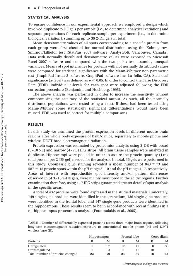
A total of 432 proteins were found expressed in the studied materials. Concretely,149 single gene products were identified in the cerebellum, 136 single gene productswere identified in the frontal lobe, and 147 single gene products were identified inthe hippocampus. These results seem to be in accordance with recent findings in arat hippocampus proteomics analysis (Fountoulakis et al., 2005).
TABLE 1 Number of differentially expressed proteins across three major brain regions, followinglong term electromagnetic radiation exposure to conventional mobile phone (M) and DECTwireless base (B).
Hippocampus Frontal lobe Cerebellum
Proteins B M B M B M
Upregulated 11 37 12 19 8 36Downregulated 11 33 11 18 10 18Total number of proteins changed 22 70 23 37 18 54
8 A. F. Fragopoulou et al.
Electromagnetic Biology and Medicine
Ele
ctro
mag
n B
iol M
ed D
ownl
oade
d fr
om in
form
ahea
lthca
re.c
om b
y U
nive
rsity
of
Ath
ens
on 0
1/20
/12
For
pers
onal
use
onl
y.

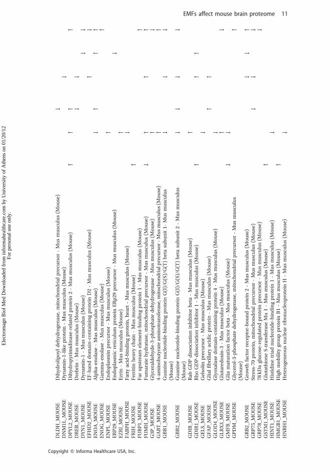
Statistical analysis under the criteria described above, revealed that 143 singlegene products were found differentially expressed among the studied brain tissuesamples, as shown in Suppl. Table 1. This table summarizes the identified proteins,gives the spot numbers under which the proteins appeared on the 2-DE gels, theiridentity, SwissProt accession numbers, theoretical pI, molecular weight, MASCOTscore, the number of peptides used per identification, protein coverage, and theexpression level, as calculated with the PD Quest 8.0 software. Proteins withdifference in expression at a level of 1.25 were considered upregulated, while a 0.75difference designated downregulated proteins.
FIGURE 1 Representative 2 DE gel of mouse hippocampus. Arrows indicate the proteinsdownregulated after the exposure of the mice to mobile (black arrows) and to base (white arrows)compared to the sham exposed animals.
EMFs affect mouse brain proteome 9
Copyright Q Informa Healthcare USA, Inc.
Ele
ctro
mag
n B
iol M
ed D
ownl
oade
d fr
om in
form
ahea
lthca
re.c
om b
y U
nive
rsity
of
Ath
ens
on 0
1/20
/12
For
pers
onal
use
onl
y.

TA
BL
E2
Dif
fere
nti
all
yex
pre
ssed
pro
tein
sin
the
mo
use
bra
ina
fter
exp
osu
reto
ba
sea
nd
mo
bil
ep
ho
ne
rad
iati
on
.
Hip
po
cam
pu
sF
ron
tal
Lo
be
Cer
ebel
lum
Acc
essi
on
Na
me
Pro
tein
Na
me
BM
BM
BM
14
33E
_MO
US
E1
4-3
-3p
rote
inep
silo
n-
Mu
sm
usc
ulu
s(M
ou
se)
#
AA
TC
_MO
US
EA
spa
rta
tea
min
otr
an
sfer
ase
,cy
top
lasm
ic-
Mu
sm
usc
ulu
s(M
ou
se)
""
""
AC
TY
_MO
US
EB
eta
-cen
tra
ctin
-M
us
mu
scu
lus
(Mo
use
)"
AH
SA
1_M
OU
SE
Act
iva
tor
of
90
kD
ah
eat
sho
ckp
rote
inA
TP
ase
ho
mo
log
1-
Mu
sm
usc
ulu
s(M
ou
se)
"
AIN
X_M
OU
SE
Alp
ha
-in
tern
exin
-M
us
mu
scu
lus
(Mo
use
)"
#
AL
DH
2_M
OU
SE
Ald
ehyd
ed
ehyd
roge
na
se,
mit
och
on
dri
al
pre
curs
or
-M
us
mu
scu
lus
(Mo
use
)#
AL
DO
A_M
OU
SE
Fru
cto
se-b
isp
ho
sph
ate
ald
ola
seA
-M
us
mu
scu
lus
(Mo
use
)"
AL
DO
C_M
OU
SE
Fru
cto
se-b
isp
ho
sph
ate
ald
ola
seC
-M
us
mu
scu
lus
(Mo
use
)#
""
AL
DR
_MO
US
EA
ldo
sere
du
cta
se-
Mu
sm
usc
ulu
s(M
ou
se)
""
AN
XA
5_M
OU
SE
An
nex
inA
5-
Mu
sm
usc
ulu
s(M
ou
se)
#
AP
OA
1_M
OU
SE
Ap
oli
po
pro
tein
A-I
pre
curs
or
-M
us
mu
scu
lus
(Mo
use
)"
AP
OE
_MO
US
EA
po
lip
op
rote
inE
pre
curs
or
-M
us
mu
scu
lus
(Mo
use
)"
"
AT
P5
H_M
OU
SE
AT
Psy
nth
ase
sub
un
itd
,m
ito
cho
nd
ria
l-
Mu
sm
usc
ulu
s(M
ou
se)
"#
AT
PA
_MO
US
EA
TP
syn
tha
sesu
bu
nit
alp
ha
,m
ito
cho
nd
ria
lp
recu
rso
r-
Mu
sm
usc
ulu
s(M
ou
se)
""
"
AT
PB
_MO
US
EA
TP
syn
tha
sesu
bu
nit
bet
a,
mit
och
on
dri
al
pre
curs
or
-Mu
sm
usc
ulu
s(M
ou
se)
#"
AT
PG
_MO
US
EA
TP
syn
tha
sesu
bu
nit
gam
ma
,m
ito
cho
nd
ria
lp
recu
rso
r-
Mu
sm
usc
ulu
s(M
ou
se)
#
BA
CH
_MO
US
EC
yto
soli
ca
cyl
coen
zym
eA
thio
este
rh
ydro
lase
-M
us
mu
scu
lus
(Mo
use
)#
BL
VR
B_M
OU
SE
Fla
vin
red
uct
ase
-M
us
mu
scu
lus
(Mo
use
)#
C1
QB
P_M
OU
SE
Co
mp
lem
ent
com
po
nen
t1
Qsu
bco
mp
on
ent-
bin
din
gp
rote
in,
mit
och
on
dri
al
-M
us
mu
scu
lus
(Mo
use
)#
CA
H2
_MO
US
EC
arb
on
ica
nh
ydra
se2
-M
us
mu
scu
lus
(Mo
use
)"
#"
#
CA
LR
_MO
US
EC
alr
etic
uli
np
recu
rso
r-
Mu
sm
usc
ulu
s(M
ou
se)
#"
CH
60
_MO
US
E6
0k
Da
hea
tsh
ock
pro
tein
,m
ito
cho
nd
ria
lp
recu
rso
r-
Mu
sm
usc
ulu
s(M
ou
se)
#
CIS
Y_M
OU
SE
Cit
rate
syn
tha
se,
mit
och
on
dri
al
pre
curs
or
-M
us
mu
scu
lus
(Mo
use
)"
CL
PP
_MO
US
EP
uta
tive
AT
P-d
epen
den
tC
lpp
rote
ase
pro
teo
lyti
csu
bu
nit
,m
ito
cho
nd
ria
l-
Mu
sm
usc
ulu
s(M
ou
se)
"
CN
TN
2_M
OU
SE
Co
nta
ctin
-2p
recu
rso
r-
Mu
sm
usc
ulu
s(M
ou
se)
"
CP
NE
6_M
OU
SE
Co
pin
e-6
-M
us
mu
scu
lus
(Mo
use
)"
CR
YM
_MO
US
EM
u-c
ryst
all
inh
om
olo
g-
Mu
sm
usc
ulu
s(M
ou
se)
"
CS
N4
_MO
US
EC
OP
9si
gna
loso
me
com
ple
xsu
bu
nit
4-
Mu
sm
usc
ulu
s(M
ou
se)
"
DC
TN
2_M
OU
SE
Dyn
act
insu
bu
nit
2-
Mu
sm
usc
ulu
s(M
ou
se)
#
DD
AH
1_M
OU
SE
N(G
),N
(G)-
dim
eth
yla
rgin
ine
dim
eth
yla
min
oh
ydro
lase
1-
Mu
sm
usc
ulu
s(M
ou
se)
#
DH
E3
_MO
US
EG
luta
mat
ed
ehyd
roge
na
se1
,m
ito
cho
nd
ria
lp
recu
rso
r-
Mu
sm
usc
ulu
s(M
ou
se)
""
#
DH
PR
_MO
US
ED
ihyd
rop
teri
din
ere
du
cta
se-
Mu
sm
usc
ulu
s(M
ou
se)
"
10 A. F. Fragopoulou et al.
Electromagnetic Biology and Medicine
Ele
ctro
mag
n B
iol M
ed D
ownl
oade
d fr
om in
form
ahea
lthca
re.c
om b
y U
nive
rsity
of
Ath
ens
on 0
1/20
/12
For
pers
onal
use
onl
y.
DL
DH
_MO
US
ED
ihyd
roli
po
yld
ehyd
roge
na
se,
mit
och
on
dri
al
pre
curs
or
-M
us
mu
scu
lus
(Mo
use
)#
DN
M1
L_M
OU
SE
Dyn
amin
-1-l
ike
pro
tein
-M
us
mu
scu
lus
(Mo
use
)#
DP
YL
2_M
OU
SE
Dih
ydro
pyr
imid
ina
se-r
ela
ted
pro
tein
2-
Mu
sm
usc
ulu
s(M
ou
se)
""
""
DR
EB
_MO
US
ED
reb
rin
-M
us
mu
scu
lus
(Mo
use
)#
#
DY
N1
_MO
US
ED
ynam
in-1
-M
us
mu
scu
lus
(Mo
use
)#
#
EF
HD
2_M
OU
SE
EF
-han
dd
om
ain
-co
nta
inin
gp
rote
inD
2-
Mu
sm
usc
ulu
s(M
ou
se)
"#
EN
OA
_MO
US
EA
lph
a-e
no
lase
-M
us
mu
scu
lus
(Mo
use
)#
""
"
EN
OG
_MO
US
EG
am
ma-
eno
lase
-M
us
mu
scu
lus
(Mo
use
)"
EN
PL
_MO
US
EE
nd
op
lasm
inp
recu
rso
r-
Mu
sm
usc
ulu
s(M
ou
se)
"
ER
P2
9_M
OU
SE
En
do
pla
smic
reti
culu
mp
rote
inE
Rp
29
pre
curs
or
-M
us
mu
scu
lus
(Mo
use
)#
EZ
RI_
MO
US
EE
zrin
-M
us
mu
scu
lus
(Mo
use
)"
FA
BP
H_M
OU
SE
Fa
tty
aci
d-b
ind
ing
pro
tein
,h
eart
-M
us
mu
scu
lus
(Mo
use
)#
FR
IH_M
OU
SE
Fer
riti
nh
eavy
cha
in-
Mu
sm
usc
ulu
s(M
ou
se)
"
FU
BP
1_M
OU
SE
Fa
ru
pst
rea
mel
emen
t-b
ind
ing
pro
tein
1-
Mu
sm
usc
ulu
s(M
ou
se)
"
FU
MH
_MO
US
EF
um
ara
teh
ydra
tase
,m
ito
cho
nd
ria
lp
recu
rso
r-
Mu
sm
usc
ulu
s(M
ou
se)
#"
""
G3
P_M
OU
SE
Gly
cera
ldeh
yde-
3-p
ho
sph
ate
deh
ydro
gen
ase
-M
us
mu
scu
lus
(Mo
use
)"
#
GA
BT
_MO
US
E4
-am
ino
bu
tyra
tea
min
otr
an
sfer
ase
,mit
och
on
dri
alp
recu
rso
r-
Mu
sm
usc
ulu
s(M
ou
se)
""
GB
B1
_MO
US
EG
ua
nin
en
ucl
eoti
de-
bin
din
gp
rote
inG
(I)/
G(S
)/G
(T)
bet
asu
bu
nit
1-
Mu
sm
usc
ulu
s(M
ou
se)
##
#
GB
B2
_MO
US
EG
ua
nin
en
ucl
eoti
de-
bin
din
gp
rote
inG
(I)/
G(S
)/G
(T)
bet
asu
bu
nit
2-
Mu
sm
usc
ulu
s(M
ou
se)
##
##
GD
IB_M
OU
SE
Ra
bG
DP
dis
soci
ati
on
inh
ibit
or
bet
a-
Mu
sm
usc
ulu
s(M
ou
se)
"
GD
IR1_
MO
US
ER
ho
GD
P-d
isso
cia
tio
nin
hib
ito
r1
-M
us
mu
scu
lus
(Mo
use
)"
""
GE
LS
_MO
US
EG
elso
lin
pre
curs
or
-M
us
mu
scu
lus
(Mo
use
)#
GF
AP
_MO
US
EG
lia
lfi
bri
lla
rya
cid
icp
rote
in-
Mu
sm
usc
ulu
s(M
ou
se)
""
GL
OD
4_M
OU
SE
Gly
oxa
lase
do
ma
in-c
on
tain
ing
pro
tein
4-
Mu
sm
usc
ulu
s(M
ou
se)
#
GL
RX
3_M
OU
SE
Glu
tare
do
xin
-3-
Mu
sm
usc
ulu
s(M
ou
se)
"#
GM
FB
_MO
US
EG
lia
ma
tura
tio
nfa
cto
rb
eta
-M
us
mu
scu
lus
(Mo
use
)#
#
GP
DM
_MO
US
EG
lyce
rol-
3-p
ho
sph
ate
deh
ydro
gen
ase
,m
ito
cho
nd
ria
lp
recu
rso
r-
Mu
sm
usc
ulu
s(M
ou
se)
"
GR
B2
_MO
US
EG
row
thfa
cto
rre
cep
tor-
bo
un
dp
rote
in2
-M
us
mu
scu
lus
(Mo
use
)#
#"
GR
P7
5_M
OU
SE
Str
ess-
70
pro
tein
,m
ito
cho
nd
ria
lp
recu
rso
r-
Mu
sm
usc
ulu
s(M
ou
se)
##
#
GR
P7
8_M
OU
SE
78
kD
agl
uco
se-r
egu
late
dp
rote
inp
recu
rso
r-
Mu
sm
usc
ulu
s(M
ou
se)
#
GS
TM
1_M
OU
SE
Glu
tath
ion
eS
-tra
nsf
era
seM
u1
-M
us
mu
scu
lus
(Mo
use
)"
HIN
T1_
MO
US
EH
isti
din
etr
iad
nu
cleo
tid
e-b
ind
ing
pro
tein
1-
Mu
sm
usc
ulu
s(M
ou
se)
#
HM
GB
1_M
OU
SE
Hig
hm
ob
ilit
ygr
ou
pp
rote
inB
1-
Mu
sm
usc
ulu
s(M
ou
se)
"
HN
RH
1_M
OU
SE
Het
ero
gen
eou
sn
ucl
ear
rib
on
ucl
eop
rote
inH
-M
us
mu
scu
lus
(Mo
use
)#
EMFs affect mouse brain proteome 11
Copyright Q Informa Healthcare USA, Inc.
Ele
ctro
mag
n B
iol M
ed D
ownl
oade
d fr
om in
form
ahea
lthca
re.c
om b
y U
nive
rsity
of
Ath
ens
on 0
1/20
/12
For
pers
onal
use
onl
y.

TA
BL
E2
(continued
)
Hip
po
cam
pu
sF
ron
tal
Lo
be
Cer
ebel
lum
Acc
essi
on
Na
me
Pro
tein
Na
me
BM
BM
BM
HN
RP
K_M
OU
SE
Het
ero
gen
eou
sn
ucl
ear
rib
on
ucl
eop
rote
inK
-M
us
mu
scu
lus
(Mo
use
)"
#
HS
90A
_MO
US
EH
eat
sho
ckp
rote
inH
SP
90
-alp
ha
-M
us
mu
scu
lus
(Mo
use
)"
HS
90B
_MO
US
EH
eat
sho
ckp
rote
inH
SP
90
-bet
a-
Mu
sm
usc
ulu
s(M
ou
se)
"
HS
P7
C_M
OU
SE
Hea
tsh
ock
cogn
ate
71
kD
ap
rote
in-
Mu
sm
usc
ulu
s(M
ou
se)
#
IMM
T_M
OU
SE
Mit
och
on
dri
al
inn
erm
emb
ran
ep
rote
in-
Mu
sm
usc
ulu
s(M
ou
se)
#"
KA
D1
_MO
US
EA
den
yla
tek
ina
seis
oen
zym
e1
-M
us
mu
scu
lus
(Mo
use
)"
"
KC
RB
_MO
US
EC
rea
tin
ek
ina
seB
-typ
e-
Mu
sm
usc
ulu
s(M
ou
se)
"#
KC
RU
_MO
US
EC
rea
tin
ek
ina
se,
ub
iqu
ito
us
mit
och
on
dri
al
pre
curs
or
-M
us
mu
scu
lus
(Mo
use
)"
KC
Y_M
OU
SE
UM
P-C
MP
kin
ase
-M
us
mu
scu
lus
(Mo
use
)#
#
KP
YM
_MO
US
EP
yru
vate
kin
ase
iso
zym
esM
1/M
2-
Mu
sm
usc
ulu
s(M
ou
se)
#"
""
LA
SP
1_M
OU
SE
LIM
an
dS
H3
do
ma
inp
rote
in1
-M
us
mu
scu
lus
(Mo
use
)"
LD
HA
_MO
US
EL
-la
cta
ted
ehyd
roge
na
seA
cha
in-
Mu
sm
usc
ulu
s(M
ou
se)
"
LG
UL
_MO
US
EL
act
oyl
glu
tath
ion
ely
ase
-M
us
mu
scu
lus
(Mo
use
)"
LIS
1_M
OU
SE
Pla
tele
t-a
ctiv
ati
ng
fact
or
ace
tylh
ydro
lase
IBsu
bu
nit
alp
ha
-M
us
mu
scu
lus
(Mo
use
)"
MD
HC
_MO
US
EM
ala
ted
ehyd
roge
na
se,
cyto
pla
smic
-M
us
mu
scu
lus
(Mo
use
)"
MD
HM
_MO
US
EM
ala
ted
ehyd
roge
na
se,
mit
och
on
dri
al
pre
curs
or
-M
us
mu
scu
lus
(Mo
use
)"
ML
RB
_MO
US
EM
yosi
nre
gula
tory
ligh
tch
ain
2-B
,sm
oo
thm
usc
leis
ofo
rm-
Mu
sm
usc
ulu
s(M
ou
se)
"
ND
KB
_MO
US
EN
ucl
eosi
de
dip
ho
sph
ate
kin
ase
B-
Mu
sm
usc
ulu
s(M
ou
se)
#
ND
UA
A_M
OU
SE
NA
DH
deh
ydro
gen
ase
[ub
iqu
ino
ne]
1a
lph
asu
bco
mp
lex
sub
un
it1
-M
us
mu
scu
lus
(Mo
use
)#
ND
UB
A_M
OU
SE
NA
DH
deh
ydro
gen
ase
[ub
iqu
ino
ne]
1b
eta
sub
com
ple
xsu
bu
nit
10
-M
us
mu
scu
lus
(Mo
use
)"
ND
US
1_M
OU
SE
NA
DH
-ub
iqu
ino
ne
oxi
do
red
uct
ase
75
kD
asu
bu
nit
,m
ito
cho
nd
ria
l-
Mu
sm
usc
ulu
s(M
ou
se)
#
ND
US
3_M
OU
SE
NA
DH
deh
ydro
gen
ase
[ub
iqu
ino
ne]
iro
n-s
ulf
ur
pro
tein
3,
mit
och
on
dri
al
-M
us
mu
scu
lus
(Mo
use
)"
ND
US
8_M
OU
SE
NA
DH
deh
ydro
gen
ase
[ub
iqu
ino
ne]
iro
n-s
ulf
ur
pro
tein
8,
mit
och
on
dri
al
-M
us
mu
scu
lus
(Mo
use
)#
ND
UV
1_M
OU
SE
NA
DH
deh
ydro
gen
ase
[ub
iqu
ino
ne]
fla
vop
rote
in1
,m
ito
cho
nd
ria
l-
Mu
sm
usc
ulu
s(M
ou
se)
""
"
NF
L_M
OU
SE
Neu
rofi
lam
ent
ligh
tp
oly
pep
tid
e-
Mu
sm
usc
ulu
s(M
ou
se)
##
""
NF
M_M
OU
SE
Neu
rofi
lam
ent
med
ium
po
lyp
epti
de
-M
us
mu
scu
lus
(Mo
use
)"
OA
T_M
OU
SE
Orn
ith
ine
am
ino
tra
nsf
era
se,
mit
och
on
dri
al
pre
curs
or
-M
us
mu
scu
lus
(Mo
use
)"
12 A. F. Fragopoulou et al.
Electromagnetic Biology and Medicine
Ele
ctro
mag
n B
iol M
ed D
ownl
oade
d fr
om in
form
ahea
lthca
re.c
om b
y U
nive
rsity
of
Ath
ens
on 0
1/20
/12
For
pers
onal
use
onl
y.

OD
O1_
MO
US
E2
-oxo
glu
tara
ted
ehyd
roge
na
seE
1co
mp
on
ent,
mit
och
on
dri
al
pre
curs
or
-M
us
mu
scu
lus
(Mo
use
)"
OD
P2
_MO
US
ED
ihyd
roli
po
ylly
sin
e-re
sid
ue
ace
tylt
ran
sfer
ase
com
po
nen
to
fp
yru
vate
deh
ydro
gen
ase
com
ple
x,m
ito
cho
nd
ria
l-M
us
mu
scu
lus
(Mo
use
)"
"
OD
PB
_MO
US
EP
yru
vate
deh
ydro
gen
ase
E1
com
po
nen
tsu
bu
nit
bet
a,
mit
och
on
dri
al
-M
us
mu
scu
lus
(Mo
use
)#
##
PD
C6I
_MO
US
EP
rogr
am
med
cell
dea
th6
-in
tera
ctin
gp
rote
in-
Mu
sm
usc
ulu
s(M
ou
se)
"
PD
IA3
_MO
US
EP
rote
ind
isu
lfid
e-is
om
era
seA
3p
recu
rso
r-
Mu
sm
usc
ulu
s(M
ou
se)
"
PE
BP
1_M
OU
SE
Ph
osp
ha
tid
ylet
ha
no
lam
ine-
bin
din
gp
rote
in1
-M
us
mu
scu
lus
(Mo
use
)"
"
PG
AM
1_M
OU
SE
Ph
osp
ho
glyc
era
tem
uta
se1
-M
us
mu
scu
lus
(Mo
use
)#
PG
K1_
MO
US
EP
ho
sph
ogl
ycer
ate
kin
as
e1
-M
us
mu
scu
lus
(Mo
use
)"
PIM
T_M
OU
SE
Pro
tein
-L-i
soa
spa
rta
te(D
-asp
art
ate
)O
-met
hyl
tra
nsf
era
se-
Mu
sm
usc
ulu
s(M
ou
se)
#
PP
CK
M_M
OU
SE
Ph
osp
ho
eno
lpyr
uva
teca
rbo
xyk
ina
se[G
TP
],m
ito
cho
nd
ria
l-
Mu
sm
usc
ulu
s(M
ou
se)
"
PR
DX
1_M
OU
SE
Per
oxi
red
oxi
n-1
-M
us
mu
scu
lus
(Mo
use
)"
PR
DX
3_M
OU
SE
Th
iore
do
xin
-dep
end
ent
per
oxi
de
red
uct
ase
,mit
och
on
dri
al
pre
curs
or
-M
us
mu
scu
lus
(Mo
use
)#
"
PR
DX
6_M
OU
SE
Per
oxi
red
oxi
n-6
-M
us
mu
scu
lus
(Mo
use
)"
"
PR
S7
_MO
US
E2
6S
pro
tea
sere
gula
tory
sub
un
it7
-M
us
mu
scu
lus
(Mo
use
)#
"
PS
A6
_MO
US
EP
rote
aso
me
sub
un
ita
lph
aty
pe-
6-
Mu
sm
usc
ulu
s(M
ou
se)
"
PS
B4
_MO
US
EP
rote
aso
me
sub
un
itb
eta
typ
e-4
pre
curs
or
-M
us
mu
scu
lus
(Mo
use
)"
PU
RA
_MO
US
ET
ran
scri
pti
on
al
act
iva
tor
pro
tein
Pu
r-a
lph
a-
Mu
sm
usc
ulu
s(M
ou
se)
#
PY
C_M
OU
SE
Pyr
uva
teca
rbo
xyla
se,
mit
och
on
dri
al
pre
curs
or
-M
us
mu
scu
lus
(Mo
use
)"
QC
R1
_MO
US
EC
yto
chro
me
b-c
1co
mp
lex
sub
un
it1
,m
ito
cho
nd
ria
lp
recu
rso
r-
Mu
sm
usc
ulu
s(M
ou
se)
""
#
QC
R2
_MO
US
EC
yto
chro
me
b-c
1co
mp
lex
sub
un
it2
,m
ito
cho
nd
ria
lp
recu
rso
r-
Mu
sm
usc
ulu
s(M
ou
se)
"
RO
A2
_MO
US
EH
eter
oge
neo
us
nu
clea
rri
bo
nu
cleo
pro
tein
sA
2/B
1-
Mu
sm
usc
ulu
s(M
ou
se)
#
SA
M5
0_M
OU
SE
So
rtin
ga
nd
ass
emb
lym
ach
iner
yco
mp
on
ent
50
ho
mo
log
-M
us
mu
scu
lus
(Mo
use
)"
SC
OT
_MO
US
ES
ucc
inyl
-Co
A3
-ket
oa
cid
-co
enzy
me
Atr
an
sfer
ase
1,
mit
och
on
dri
al
-M
us
mu
scu
lus
(Mo
use
)#
SE
P11
_MO
US
ES
epti
n-1
1-
Mu
sm
usc
ulu
s(M
ou
se)
#
SE
PT
2_M
OU
SE
Sep
tin
-2-
Mu
sm
usc
ulu
s(M
ou
se)
#
SE
PT
5_M
OU
SE
Sep
tin
-5-
Mu
sm
usc
ulu
s(M
ou
se)
#
SE
PT
8_M
OU
SE
Sep
tin
-8-
Mu
sm
usc
ulu
s(M
ou
se)
#
SH
3G
2_M
OU
SE
En
do
ph
ilin
-A1
-M
us
mu
scu
lus
(Mo
use
)#
#
SN
AB
_MO
US
EB
eta
-so
lub
leN
SF
att
ach
men
tp
rote
in-
Mu
sm
usc
ulu
s(M
ou
se)
#
SN
AG
_MO
US
EG
am
ma
-so
lub
leN
SF
att
ach
men
tp
rote
in-
Mu
sm
usc
ulu
s(M
ou
se)
#
EMFs affect mouse brain proteome 13
Copyright Q Informa Healthcare USA, Inc.
Ele
ctro
mag
n B
iol M
ed D
ownl
oade
d fr
om in
form
ahea
lthca
re.c
om b
y U
nive
rsity
of
Ath
ens
on 0
1/20
/12
For
pers
onal
use
onl
y.
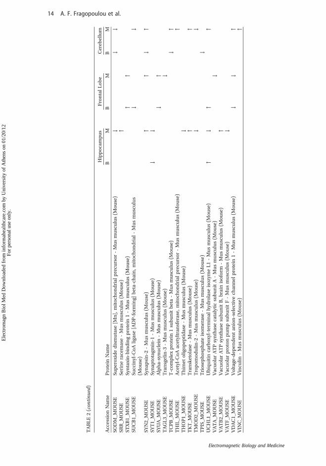
TA
BL
E2
(continued
)
Hip
po
cam
pu
sF
ron
tal
Lo
be
Cer
ebel
lum
Acc
essi
on
Na
me
Pro
tein
Na
me
BM
BM
BM
SO
DM
_MO
US
ES
up
ero
xid
ed
ism
uta
se[M
n],
mit
och
on
dri
al
pre
curs
or
-M
us
mu
scu
lus
(Mo
use
)#
##
SR
R_M
OU
SE
Ser
ine
race
mas
e-
Mu
sm
usc
ulu
s(M
ou
se)
"
ST
XB
1_M
OU
SE
Syn
taxi
n-b
ind
ing
pro
tein
1-
Mu
sm
usc
ulu
s(M
ou
se)
""
SU
CB
1_M
OU
SE
Su
ccin
yl-C
oA
liga
se[A
DP
-fo
rmin
g]b
eta
-ch
ain
,m
ito
cho
nd
ria
l-
Mu
sm
usc
ulu
s(M
ou
se)
##
SY
N2
_MO
US
ES
yna
psi
n-2
-M
us
mu
scu
lus
(Mo
use
)"
"#
"
SY
T1
_MO
US
ES
yna
pto
tagm
in-1
-M
us
mu
scu
lus
(Mo
use
)#
#
SY
UA
_MO
US
EA
lph
a-s
ynu
clei
n-
Mu
sm
usc
ulu
s(M
ou
se)
#"
TA
GL
3_M
OU
SE
Tra
nsg
elin
-3-
Mu
sm
usc
ulu
s(M
ou
se)
#
TC
PB
_MO
US
ET
-co
mp
lex
pro
tein
1su
bu
nit
bet
a-
Mu
sm
usc
ulu
s(M
ou
se)
#"
TH
IL_M
OU
SE
Ace
tyl-
Co
Aa
cety
ltra
nsf
era
se,
mit
och
on
dri
al
pre
curs
or
-M
us
mu
scu
lus
(Mo
use
)"
TH
OP
1_M
OU
SE
Th
imet
oli
gop
epti
da
se-
Mu
sm
usc
ulu
s(M
ou
se)
#
TK
T_M
OU
SE
Tra
nsk
eto
lase
-M
us
mu
scu
lus
(Mo
use
)"
"
TM
OD
2_M
OU
SE
Tro
po
mo
du
lin
-2-
Mu
sm
usc
ulu
s(M
ou
se)
##
TP
IS_M
OU
SE
Tri
ose
ph
osp
ha
teis
om
eras
e-
Mu
sm
usc
ulu
s(M
ou
se)
#
UC
HL
1_M
OU
SE
Ub
iqu
itin
carb
oxy
l-te
rmin
al
hyd
rola
seis
ozy
me
L1
-M
us
mu
scu
lus
(Mo
use
)"
#"
"
VA
TA
_MO
US
EV
acu
ola
rA
TP
syn
tha
seca
taly
tic
sub
un
itA
-M
us
mu
scu
lus
(Mo
use
)#
VA
TB
2_M
OU
SE
Va
cuo
lar
AT
Psy
nth
ase
sub
un
itB
,b
rain
iso
form
-M
us
mu
scu
lus
(Mo
use
)"
VA
TF
_MO
US
EV
acu
ola
rp
roto
np
um
psu
bu
nit
F-
Mu
sm
usc
ulu
s(M
ou
se)
#
VD
AC
1_M
OU
SE
Vo
lta
ge-d
epen
den
ta
nio
n-s
elec
tive
cha
nn
elp
rote
in1
-M
us
mu
scu
lus
(Mo
use
)#
#"
VIN
C_M
OU
SE
Vin
culi
n-
Mu
sm
usc
ulu
s(M
ou
se)
"
14 A. F. Fragopoulou et al.
Electromagnetic Biology and Medicine
Ele
ctro
mag
n B
iol M
ed D
ownl
oade
d fr
om in
form
ahea
lthca
re.c
om b
y U
nive
rsity
of
Ath
ens
on 0
1/20
/12
For
pers
onal
use
onl
y.
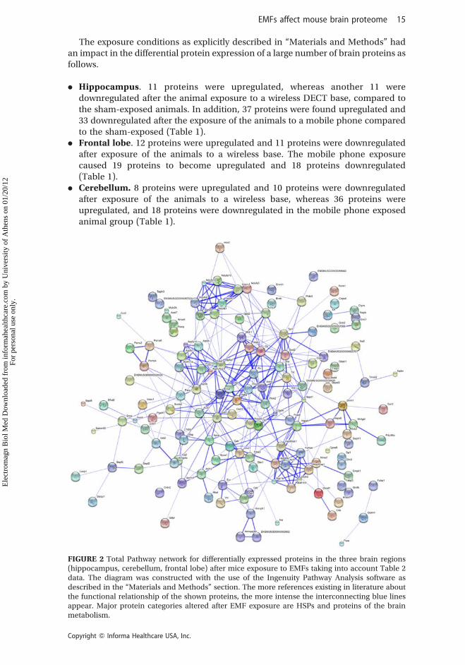
The exposure conditions as explicitly described in “Materials and Methods” hadan impact in the differential protein expression of a large number of brain proteins asfollows.
. Hippocampus. 11 proteins were upregulated, whereas another 11 weredownregulated after the animal exposure to a wireless DECT base, compared tothe sham-exposed animals. In addition, 37 proteins were found upregulated and33 downregulated after the exposure of the animals to a mobile phone comparedto the sham-exposed (Table 1).
. Frontal lobe. 12 proteins were upregulated and 11 proteins were downregulatedafter exposure of the animals to a wireless base. The mobile phone exposurecaused 19 proteins to become upregulated and 18 proteins downregulated(Table 1).
. Cerebellum. 8 proteins were upregulated and 10 proteins were downregulatedafter exposure of the animals to a wireless base, whereas 36 proteins wereupregulated, and 18 proteins were downregulated in the mobile phone exposedanimal group (Table 1).
FIGURE 2 Total Pathway network for differentially expressed proteins in the three brain regions(hippocampus, cerebellum, frontal lobe) after mice exposure to EMFs taking into account Table 2data. The diagram was constructed with the use of the Ingenuity Pathway Analysis software asdescribed in the “Materials and Methods” section. The more references existing in literature aboutthe functional relationship of the shown proteins, the more intense the interconnecting blue linesappear. Major protein categories altered after EMF exposure are HSPs and proteins of the brainmetabolism.
EMFs affect mouse brain proteome 15
Copyright Q Informa Healthcare USA, Inc.
Ele
ctro
mag
n B
iol M
ed D
ownl
oade
d fr
om in
form
ahea
lthca
re.c
om b
y U
nive
rsity
of
Ath
ens
on 0
1/20
/12
For
pers
onal
use
onl
y.
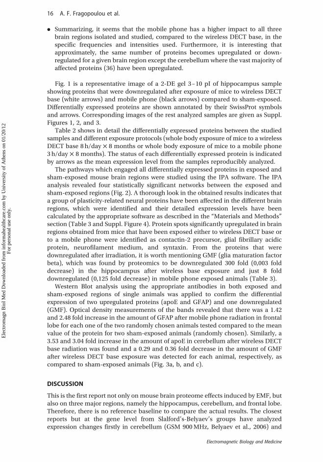
. Summarizing, it seems that the mobile phone has a higher impact to all threebrain regions isolated and studied, compared to the wireless DECT base, in thespecific frequencies and intensities used. Furthermore, it is interesting thatapproximately, the same number of proteins becomes upregulated or down-regulated for a given brain region except the cerebellum where the vast majority ofaffected proteins (36) have been upregulated.
Fig. 1 is a representative image of a 2-DE gel 3–10 pI of hippocampus sampleshowing proteins that were downregulated after exposure of mice to wireless DECTbase (white arrows) and mobile phone (black arrows) compared to sham-exposed.Differentially expressed proteins are shown annotated by their SwissProt symbolsand arrows. Corresponding images of the rest analyzed samples are given as Suppl.Figures 1, 2, and 3.
Table 2 shows in detail the differentially expressed proteins between the studiedsamples and different exposure protocols (whole body exposure of mice to a wirelessDECT base 8 h/day £ 8 months or whole body exposure of mice to a mobile phone3 h/day £ 8 months). The status of each differentially expressed protein is indicatedby arrows as the mean expression level from the samples reproducibly analyzed.
The pathways which engaged all differentially expressed proteins in exposed andsham-exposed mouse brain regions were studied using the IPA software. The IPAanalysis revealed four statistically significant networks between the exposed andsham-exposed regions (Fig. 2). A thorough look in the obtained results indicates thata group of plasticity-related neural proteins have been affected in the different brainregions, which were identified and their detailed expression levels have beencalculated by the appropriate software as described in the “Materials and Methods”section (Table 3 and Suppl. Figure 4). Protein spots significantly upregulated in brainregions obtained from mice that have been exposed either to wireless DECT base orto a mobile phone were identified as contactin-2 precursor, glial fibrillary acidicprotein, neurofilament medium, and syntaxin. From the proteins that weredownregulated after irradiation, it is worth mentioning GMF (glia maturation factorbeta), which was found by proteomics to be downregulated 300 fold (0,003 folddecrease) in the hippocampus after wireless base exposure and just 8 folddownregulated (0,125 fold decrease) in mobile phone exposed animals (Table 3).
Western Blot analysis using the appropriate antibodies in both exposed andsham-exposed regions of single animals was applied to confirm the differentialexpression of two upregulated proteins (apoE and GFAP) and one downregulated(GMF). Optical density measurements of the bands revealed that there was a 1.42and 2.48 fold increase in the amount of GFAP after mobile phone radiation in frontallobe for each one of the two randomly chosen animals tested compared to the meanvalue of the protein for two sham-exposed animals (randomly chosen). Similarly, a3.53 and 3.04 fold increase in the amount of apoE in cerebellum after wireless DECTbase radiation was found and a 0.29 and 0.36 fold decrease in the amount of GMFafter wireless DECT base exposure was detected for each animal, respectively, ascompared to sham-exposed animals (Fig. 3a, b, and c).
DISCUSSION
This is the first report not only on mouse brain proteome effects induced by EMF, butalso on three major regions, namely the hippocampus, cerebellum, and frontal lobe.Therefore, there is no reference baseline to compare the actual results. The closestreports but at the gene level from Slalford’s-Belyaev’s groups have analyzedexpression changes firstly in cerebellum (GSM 900 MHz, Belyaev et al., 2006) and
16 A. F. Fragopoulou et al.
Electromagnetic Biology and Medicine
Ele
ctro
mag
n B
iol M
ed D
ownl
oade
d fr
om in
form
ahea
lthca
re.c
om b
y U
nive
rsity
of
Ath
ens
on 0
1/20
/12
For
pers
onal
use
onl
y.
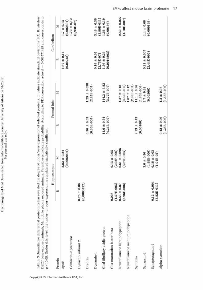
TA
BL
E3
Qu
an
tita
tive
dif
fere
nti
alp
rote
om
ics
ha
sre
vea
led
the
deg
ree
of
un
der
/ove
r-ex
pre
ssio
no
fse
lect
edp
rote
ins.^
valu
esin
dic
ate
sta
nd
ard
dev
iati
on
s(S
D).
B:w
irel
ess
DE
CT
ba
seex
po
sed
an
ima
ls,M
:mo
bil
ep
ho
ne
exp
ose
da
nim
als
.Pa
ren
thes
esin
dic
ate
pro
ba
bil
ity.
Acc
ord
ing
toF
DR
corr
ecti
on
,a-l
evel
¼0
.00
35
71
42
9a
nd
corr
esp
on
ds
top,
0.0
5.
Un
der
this
leve
l,th
eu
nd
er-
or
ove
r-ex
pre
ssio
nis
con
sid
ered
sta
tist
ica
lly
sign
ific
an
t.
Hip
po
cam
pu
sF
ron
tal
lob
eC
ereb
ellu
m
Pro
tein
BM
BM
BM
Ap
oE
2.826
0.24
(0.0002004)
1.676
0.14
(0.00248)
1.7
60.13
(0.002801)
Co
nta
ctin
-2p
recu
rso
r1.726
0,12
(6,93E-07)
Dyn
act
insu
bu
nit
20.756
0.06
(0,0003472)
Dre
bri
n0.566
0.03
(6.30E-005)
1.256
0.098
(2.85E-005)
Dyn
am
in-1
0.186
0.07
(2,75E-07)
3.466
0.36
(3.99E-011)
Gli
al
fib
rill
ary
aci
dic
pro
tein
11.6
60.54
(4.24E-007)
114.2
63.82
(3.77E-007)
1.566
0.26
(0,0016803)
1.686
0.19
(0,00198)
Gli
am
atu
rati
on
fact
or
bet
a0.003
(1.57E-005)
0.136
0.05
(2.02E-006)
Neu
rofi
lam
ent
ligh
tp
oly
pep
tid
e0.656
0.87
(3.96E-006)
0.616
0.096
(6,87E-005)
1.376
0.18
(4.62E-006)
2.626
0.027
(4.10E-007)
Neu
rofi
lam
ent
med
ium
po
lyp
epti
de
1.876
0.12
(2.03E-005)
Syn
taxi
n2.136
0.43
(0,00180)
11.1
60.96
(1.40E-006)
Syn
ap
sin
-23.6
60.56
(4.89E-006)
3.7
60.62
(0.00266)
0.216
0.007
(2,44E-007)
1.4
60.08
(0.000169)
Syn
ap
tota
gmin
-10.126
0.004
(3.82E-011)
0.166
0.03
(1.01E-05)
Alp
ha
-syn
ucl
ein
0.426
0.06
(1.28E-006)
1.2
60.08
(1.68E-006)
EMFs affect mouse brain proteome 17
Copyright Q Informa Healthcare USA, Inc.
Ele
ctro
mag
n B
iol M
ed D
ownl
oade
d fr
om in
form
ahea
lthca
re.c
om b
y U
nive
rsity
of
Ath
ens
on 0
1/20
/12
For
pers
onal
use
onl
y.
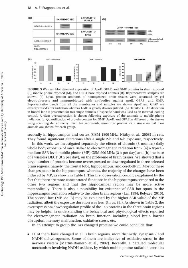
secondly in hippocampus and cortex (GSM 1800 MHz, Nittby et al., 2008) in rats.They found significant alterations after a single 2-h and 6-h exposure, respectively.
In this work, we investigated separately the effects of chronic (8 months) dailywhole body exposure of mice Balb/c to electromagnetic radiation from: (a) a typical-medium SAR level mobile phone (MP) GSM 900 MHz (3 h per day) and (b) the baseof a wireless DECT (8 h per day), on the proteome of brain tissues. We showed that alarge number of proteins become overexpressed or downregulated in three selectedbrain regions, namely, the frontal lobe, hippocampus, and cerebellum. Most of thesechanges occur in the hippocampus, whereas, the majority of the changes have beeninduced by MP, as shown in Table 1. This first observation could be explained by thefact that there are more concentrated functions in the hippocampus compared to theother two regions and that the hippocampal region may be more activemetabolically. There is also a possibility for existence of SAR hot spots in thehippocampus formation relative to the other brain regions (Lai, 1994; Belyaev, 2010).The second fact (MP .. B) may be explained by the higher SAR value of the MPradiation, albeit the exposure duration was less (3 h vs. 8 h). As shown in Table 2, theoverexpression/downregulation profile of the 143 proteins in the three brain regionsmay be helpful in understanding the behavioral and physiological effects reportedfor electromagnetic radiation on brain function including blood brain barrierdisruption, memory malfunction, oxidative stress, etc.
In an attempt to group the 143 changed proteins we could conclude that:
. 11 of them have changed in all 3 brain regions, more distinctly, synapsin-2 andNADH dehydrogenase. Some of them are indicative of oxidative stress in thenervous system (Martin-Romero et al., 2002). Recently, a detailed molecularmechanism involving NADH oxidase, by which mobile phone radiation exerts its
FIGURE 3 Western blot detected expression of ApoE, GFAP, and GMF proteins in sham exposed(S), mobile phone exposed (M), and DECT base exposed animals (B). Representative samples areshown. (a) Equal protein amounts of homogenized brain tissues were separated by gelelectrophoresis and immunoblotted with antibodies against apoE, GFAP, and GMF.Representative bands from all the membranes and samples are shown. ApoE and GFAP areoverexpressed after radiation whereas GMF is greatly downregulated. (b) Detailed GFAP detectionin frontal lobe is presented for two single animals. Unspecific band was used as an internal loadingcontrol. A clear overexpression is shown following exposure of the animals to mobile phoneradiation. (c) Quantification of protein content for GMF, ApoE, and GFAP in different brain tissuesusing scanning densitometry. Each bar represents amount of protein for a single animal. Twoanimals are shown for each group.
18 A. F. Fragopoulou et al.
Electromagnetic Biology and Medicine
Ele
ctro
mag
n B
iol M
ed D
ownl
oade
d fr
om in
form
ahea
lthca
re.c
om b
y U
nive
rsity
of
Ath
ens
on 0
1/20
/12
For
pers
onal
use
onl
y.
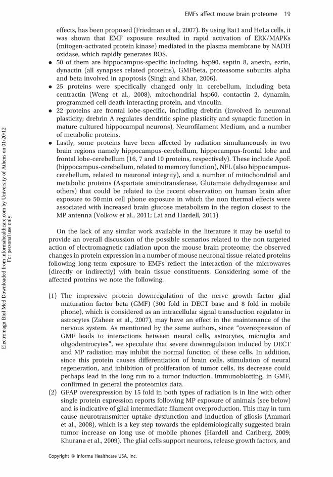
effects, has been proposed (Friedman et al., 2007). By using Rat1 and HeLa cells, itwas shown that EMF exposure resulted in rapid activation of ERK/MAPKs(mitogen-activated protein kinase) mediated in the plasma membrane by NADHoxidase, which rapidly generates ROS.
. 50 of them are hippocampus-specific including, hsp90, septin 8, anexin, ezrin,dynactin (all synapses related proteins), GMFbeta, proteasome subunits alphaand beta involved in apoptosis (Singh and Khar, 2006).
. 25 proteins were specifically changed only in cerebellum, including betacentractin (Weng et al., 2008), mitochondrial hsp60, contactin 2, dynamin,programmed cell death interacting protein, and vinculin.
. 22 proteins are frontal lobe-specific, including drebrin (involved in neuronalplasticity; drebrin A regulates dendritic spine plasticity and synaptic function inmature cultured hippocampal neurons), Neurofilament Medium, and a numberof metabolic proteins.
. Lastly, some proteins have been affected by radiation simultaneously in twobrain regions namely hippocampus-cerebellum, hippocampus-frontal lobe andfrontal lobe-cerebellum (16, 7 and 10 proteins, respectively). These include ApoE(hippocampus-cerebellum, related to memory function), NFL (also hippocampus-cerebellum, related to neuronal integrity), and a number of mitochondrial andmetabolic proteins (Aspartate aminotransferase, Glutamate dehydrogenase andothers) that could be related to the recent observation on human brain afterexposure to 50 min cell phone exposure in which the non thermal effects wereassociated with increased brain glucose metabolism in the region closest to theMP antenna (Volkow et al., 2011; Lai and Hardell, 2011).
On the lack of any similar work available in the literature it may be useful toprovide an overall discussion of the possible scenarios related to the non targetedaction of electromagnetic radiation upon the mouse brain proteome; the observedchanges in protein expression in a number of mouse neuronal tissue-related proteinsfollowing long-term exposure to EMFs reflect the interaction of the microwaves(directly or indirectly) with brain tissue constituents. Considering some of theaffected proteins we note the following.
(1) The impressive protein downregulation of the nerve growth factor glialmaturation factor beta (GMF) (300 fold in DECT base and 8 fold in mobilephone), which is considered as an intracellular signal transduction regulator inastrocytes (Zaheer et al., 2007), may have an effect in the maintenance of thenervous system. As mentioned by the same authors, since “overexpression ofGMF leads to interactions between neural cells, astrocytes, microglia andoligodentrocytes”, we speculate that severe downregulation induced by DECTand MP radiation may inhibit the normal function of these cells. In addition,since this protein causes differentiation of brain cells, stimulation of neuralregeneration, and inhibition of proliferation of tumor cells, its decrease couldperhaps lead in the long run to a tumor induction. Immunoblotting, in GMF,confirmed in general the proteomics data.
(2) GFAP overexpression by 15 fold in both types of radiation is in line with othersingle protein expression reports following MP exposure of animals (see below)and is indicative of glial intermediate filament overproduction. This may in turncause neurotransmitter uptake dysfunction and induction of gliosis (Ammariet al., 2008), which is a key step towards the epidemiologically suggested braintumor increase on long use of mobile phones (Hardell and Carlberg, 2009;Khurana et al., 2009). The glial cells support neurons, release growth factors, and
EMFs affect mouse brain proteome 19
Copyright Q Informa Healthcare USA, Inc.
Ele
ctro
mag
n B
iol M
ed D
ownl
oade
d fr
om in
form
ahea
lthca
re.c
om b
y U
nive
rsity
of
Ath
ens
on 0
1/20
/12
For
pers
onal
use
onl
y.

remove debris after injury or neuronal death. Astrocytes help form the bloodbrain barrier that prevents toxic substances circulating in the blood fromentering the brain. It was proposed many years ago that overexpression of GFAPis the response of astrocytes to oxidative stress (Morgan et al., 1997), which isbeing reported to take place in brain tissues after exposure of guinea pigs tomobile phone radiation (Meral et al., 2007). Since GFAP is a sensitive biomarkerfor neurotoxicity, our findings may indicate neuronal tissue injury caused byelectromagnetic radiation or a probable injury of the blood brain barrier,reported to be an effect of exposure (Nittby et al., 2009; Sirav and Seyhan, 2009).Immunoblotting with anti-GFAP confirmed in general the proteomics data.
(3) ApoE is a class of apolipoprotein found in the chylomicron and LDLs that bind toa specific receptor on liver cells and peripheral cells. It has been studied for itsrole in several biological processes not directly related to lipoprotein transport,including Alzheimer’s disease (AD), immunoregulation, and cognition. So, theoverexpression in the cerebellum and hippocampus after mobile exposuremight be related to the memory deficits reported by our group (Fragopoulouet al., 2010b; Fragopoulou and Margaritis, 2010; Ntzouni et al., 2011). This is inagreement with the observation that ApoE4 knock-in mice exhibit an age-dependent decrease in hilar GABAergic interneurons correlated with the extentof learning and memory deficits as found by the Morris water maze task(Andrews-Zwilling et al., 2010).
(4) Synapsin-2 and syntaxin-1 overexpression by both radiation types (MP andDECT) in hippocampus may indicate a compensatory neuronal response toradiation by making more synapses.
(5) Synaptotagmin levels in the hippocampus are in line with the above-mentionedGMF dramatic downregulation. This protein species is known to function as acalcium sensor in the regulation of neurotransmitter release and hormonesecretion.
The significance of the present results may be noticeable in relation to theepidemiological, clinical, and other experimental data reported so far concerningbehavioral deficits and brain structural/functional alterations induced by EMF inrodents. Although at the epidemiological level Schuz et al. (2006) found as anoutcome of the Interphone study no overall increased risk of glioma or meningiomaobserved among cellular phone users, however, for long-term cellular phone users,the same authors suggested that the results need to be reconsidered before firmconclusions can be drawn. In fact, recent data by Hardell’s group have providedsolid evidence for a long term effect on brain tumors (Hardell and Carlberg, 2009;Khurana et al., 2009) which might be supported by the protein expression changesfound in our results. Along the same lines, reports dealing with EMF-induced brainnetworking dysfunction can be explained. For instance, in a clinical study with 41volunteers participating, it was reported that 890 MHz mobile phone-like signalalters the integrity of the human blood–brain and blood–cerebrospinal fluid barriers(Soderqvist et al., 2009). There is also a relationship of MP radiation with behavioralproblems in prenatally exposed children (Divan et al., 2008).
Our data using the cordless DECT base as a source of EMF may appear surprisingdue to the low SAR level, as deduced by measuring the field within the animal cage,approximately 20 mW/Kg, but one explanation could be the intensity windows effect(Blackman, 2009; Belyaev, 2010). Interestingly, Salford’s work with rats, applyingsimilar low SAR value (0.6 and 60 mW/Kg), but using mobile phone radiation for just2 h per week for 55 week, demonstrated significantly altered performance during anepisodic-like memory test (Nittby et al., 2008).
20 A. F. Fragopoulou et al.
Electromagnetic Biology and Medicine
Ele
ctro
mag
n B
iol M
ed D
ownl
oade
d fr
om in
form
ahea
lthca
re.c
om b
y U
nive
rsity
of
Ath
ens
on 0
1/20
/12
For
pers
onal
use
onl
y.
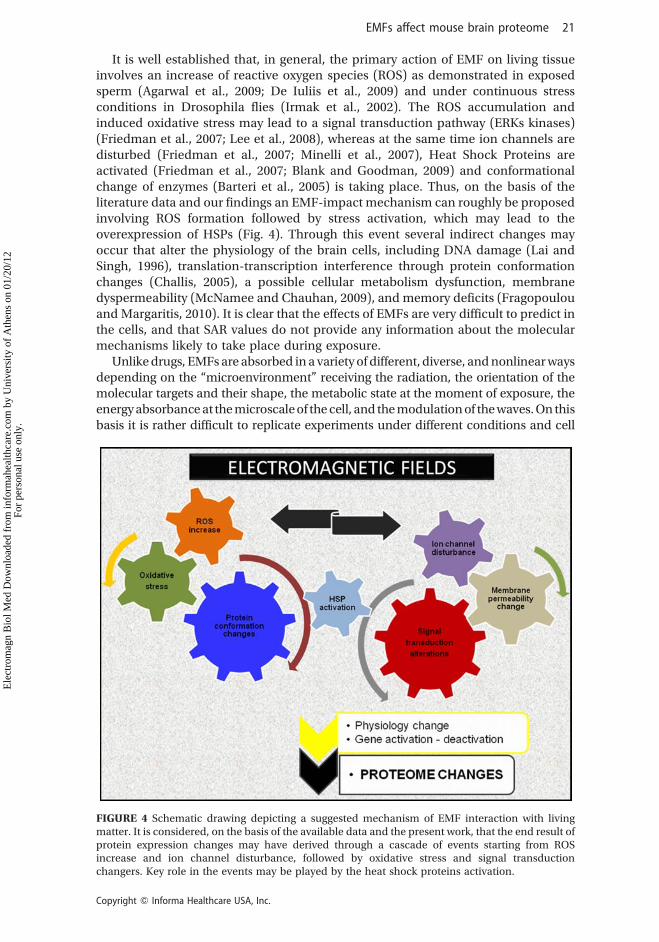
It is well established that, in general, the primary action of EMF on living tissueinvolves an increase of reactive oxygen species (ROS) as demonstrated in exposedsperm (Agarwal et al., 2009; De Iuliis et al., 2009) and under continuous stressconditions in Drosophila flies (Irmak et al., 2002). The ROS accumulation andinduced oxidative stress may lead to a signal transduction pathway (ERKs kinases)(Friedman et al., 2007; Lee et al., 2008), whereas at the same time ion channels aredisturbed (Friedman et al., 2007; Minelli et al., 2007), Heat Shock Proteins areactivated (Friedman et al., 2007; Blank and Goodman, 2009) and conformationalchange of enzymes (Barteri et al., 2005) is taking place. Thus, on the basis of theliterature data and our findings an EMF-impact mechanism can roughly be proposedinvolving ROS formation followed by stress activation, which may lead to theoverexpression of HSPs (Fig. 4). Through this event several indirect changes mayoccur that alter the physiology of the brain cells, including DNA damage (Lai andSingh, 1996), translation-transcription interference through protein conformationchanges (Challis, 2005), a possible cellular metabolism dysfunction, membranedyspermeability (McNamee and Chauhan, 2009), and memory deficits (Fragopoulouand Margaritis, 2010). It is clear that the effects of EMFs are very difficult to predict inthe cells, and that SAR values do not provide any information about the molecularmechanisms likely to take place during exposure.
Unlike drugs, EMFs are absorbed in a variety of different, diverse, and nonlinear waysdepending on the “microenvironment” receiving the radiation, the orientation of themolecular targets and their shape, the metabolic state at the moment of exposure, theenergy absorbance at the microscale of the cell, and the modulation of the waves. On thisbasis it is rather difficult to replicate experiments under different conditions and cell
FIGURE 4 Schematic drawing depicting a suggested mechanism of EMF interaction with livingmatter. It is considered, on the basis of the available data and the present work, that the end result ofprotein expression changes may have derived through a cascade of events starting from ROSincrease and ion channel disturbance, followed by oxidative stress and signal transductionchangers. Key role in the events may be played by the heat shock proteins activation.
EMFs affect mouse brain proteome 21
Copyright Q Informa Healthcare USA, Inc.
Ele
ctro
mag
n B
iol M
ed D
ownl
oade
d fr
om in
form
ahea
lthca
re.c
om b
y U
nive
rsity
of
Ath
ens
on 0
1/20
/12
For
pers
onal
use
onl
y.
systems, which may explain the discrepancy of the results among research groups.Especially in the case of proteomics analysis used to approach the effects of EMFsin this article, it is even more difficult, since 2D electrophoresis is inherently variablefrom one run to the next, especially when being performed in different labs. However,the 2-DE approach is still largely refractory to high-throughput methods due to anumber of reasons and can be judiciously coupled to several types of biologicalexperiments to provide meaningful data. Indeed, efforts to improve reproducibilityhave largely centered on robotics, improved visualization methods as better spot-detection algorithms (Ong and Pandey, 2001; Rogers and Graham, 2007) and asclearly stated by Ong and Pandey (2001), 2-DE-based approaches can still be effectivelyused when applied with a clear under-standing of its strengths and limitations.
CONCLUSIONS
Our differential proteomic analysis results suggest that conventional MP and DECTbase EMFs affect the proteome of hippocampus, cerebellum, and frontal lobefollowing whole body exposure of Balb/c mice. Since this is the first report showingmouse brain proteome changes induced by EMFs, there is no reference baseline tocompare the actual results. However, it is more likely that the observed proteomechanges reflect EMF impact and not variability between individual mice, since ithas been found just recently that genetic background in both out-bred mousestocks and inbred mouse strains has a negligible effect on the brain proteomeprofile (Foldi et al., 2011). Based on the currently available literature it is assumedthat EMF may function as a stress factor creating ROS and inducing oxidative stress,whereas at the same time ion channels are disturbed and Heat Shock Proteins areactivated. This, in turn, may affect gene over/under-expression, possibly throughtranscription factor activation/deactivation (Kar et al., 2011) in a random manner,since EMF impact is non targeted although stress-related events within the cell aremost likely affected. As an end result, functions related to stress response may betriggered. The altered protein expression in this report may reflect such a cascade ofevents, in which some proteins are related to neural plasticity whereas othersbelong to the general metabolic processes. The reported herein effects can beconsidered non thermal since the actual SAR values calculated are well belowICNIRP’s (1998) guidelines. In any case, it is seriously considered by pioneers in thetopic of EMFs that the relatively low field strengths capable to affect biochemicalreactions is a further indication that cells are in a position to sense potential dangerlong before there is an increase in temperature (Blank and Goodman, 2009).
Further work is underway to reveal the onset of the proteome changes after shortterm exposure conditions (data under analysis). Also, it is necessary to usemultidisciplinary and multilevel approaches in order to delineate the mechanismsof EMF interaction with living organisms.
ACKNOWLEDGEMENTS
The authors thank Professors Ramon Lim and Asgar Zaheer from the Department ofNeurology of Iowa University for generously providing them with the affinity purifiedmonoclonal antibody against GMF and also the Reviewers and the Editor for theirconstructive criticism.
Declaration of interestThis study was supported by the Special Account for Research Grants of theUniversity of Athens to the Research Group of Professor L.H. Margaritis. AFF is a
22 A. F. Fragopoulou et al.
Electromagnetic Biology and Medicine
Ele
ctro
mag
n B
iol M
ed D
ownl
oade
d fr
om in
form
ahea
lthca
re.c
om b
y U
nive
rsity
of
Ath
ens
on 0
1/20
/12
For
pers
onal
use
onl
y.
scholarship recipient by the Hellenic State Scholarship Foundation – “N.D.Xrysovergis” Bequest (PhD fellowship). The authors report no conflicts of interest.The authors alone are responsible for the content and writing of the article.
REFERENCES
Agarwal, A., Desai, N. R., Makker, K., et al. (2009). Effects of radiofrequency electromagnetic waves
(RF EMW) from cellular phones on human ejaculated semen: an in vitro pilot study. Fertil. Steril. 92(4):
1318 1325.
Ammari, M., Brillaud, E., Gamez, C., et al. (2008). Effect of a chronic GSM 900 MHz exposure on glia in the
rat brain. Biomed. Pharmacother. 62(4):273 281.
Anagnostopoulos, A. K., Kolialexi, A., Mavrou, A., et al. (2010). Proteomic analysis of amniotic fluid in
pregnancies with Klinefelter syndrome foetuses. J. Proteomics 5:943 950.
Andrews Zwilling, Y., Bien Ly, N., Xu, Q., et al. (2010). Apolipoprotein E4 causes age and Tau dependent
impairment of GABAergic interneurons, leading to learning and memory deficits in mice. J. Neurosci.
30(41):13707 13717.
Benjamini, Y., Hochberg, Y. (1995). Controlling the false discovery rate: a practical and powerful approach
to multiple testing. J. Roy. Statist. Soc. (Ser. B) 57(1):289 300.
Barteri, M., Pala, A., Rotella, S. (2005). Structural and kinetic effects of mobile phone microwaves on
acetylcholinesterase activity. Biophys. Chem. 113(3):245 253.
Belyaev, I. (2010). Dependence of non thermal biological effects of microwaves on physical and biological
variables: implications for reproducibility and safety standards. In: Giuliani, L., Soffritti, M. European
Journal of Oncology Library, vol. 5 Non Thermal Effects And Mechanisms Of Interaction Between
Electromagnetic Fields And Living Matter. An ICEMS Monograph, (pp. 187 218) Bologna, Italy:
Ramazzini Institute , http://www.icems.eu/papers.htm?f ¼ /c/a/2009/12/15/MNHJ1B49KH.DTL . .
Belyaev, I. Y., Koch, C. B., Terenius, O., et al. (2006). Exposure of rat brain to 915 MHz GSM microwaves
induces changes in gene expression but not double stranded DNA breaks or effects on chromatin
conformation. Bioelectromagnetics 27(4):295 306.
Blackman, C. (2009). Cell phone radiation: Evidence from ELF and RF studies supporting more inclusive
risk identification and assessment. Pathophysiology 16(2 3):205 216.
Blank, M., Goodman, R. (2009). Electromagnetic fields stress living cells. Pathophysiology 16(2 3):71 78.
Cardis, E., Armstrong, B. K., Bowman, J. D., et al. (2011). Risk of brain tumours in relation to estimated RF
dose from mobile phones: results from five Interphone countries. Occup. Environ. Med. 68(9):631 640.
Challis, L. J. (2005). Mechanisms for interaction between RF fields and biological tissue.
Bioelectromagnetics 26(Suppl 7):98 106.
Chavdoula, E. D., Panagopoulos, D. J., Margaritis, L. H. (2010). Comparison of biological effects between
continuous and intermittent exposure to GSM 900 MHz mobile phone radiation: Detection of
apoptotic cell death features. Mutat. Res. 700(1 2):51 61.
Cleary, S. F., Cao, G., Liu, L. M., et al. (1997). Stress proteins are not induced in mammalian cells exposed
to radiofrequency or microwave radiation. Bioelectromagnetics 18(7):499 505.
De Iuliis, G. N., Newey, R. J., King, B. V., Aitken, R. J. (2009). Mobile phone radiation induces
reactive oxygen species production and DNA damage in human spermatozoa in vitro. PLoS One 4(7):
e6446.
Divan, H. A., Kheifets, L., Obel, C., Olsen, J. (2008). Prenatal and postnatal exposure to cell phone use and
behavioral problems in children. Epidemiology 19(4):523 529.
Fragopoulou, A. F., Koussoulakos, S. L., Margaritis, L. H. (2010a). Cranial and postcranial skeletal
variations induced in mouse embryos by mobile phone radiation. Pathophysiology 17(3):169 177.
Fragopoulou, A. F., Miltiadous, P., Stamatakis, A., et al. (2010b). Whole body exposure with GSM 900 MHz
affects spatial memory in mice. Pathophysiology 17(3):179 187.
Fragopoulou, A., Grigoriev, Y., Johansson, O., et al. (2010c). Scientific panel on electromagnetic field health
risks consensus points, recommendations and rationales. Rev. Environ. Health 25(4):307 317.
Fragopoulou, A. F., Margaritis, L. H. (2010). Is cognitive function affected by mobile phone radiation
exposure? In: Giuliani, L., Soffritti, M. European J. Oncology Library, vol. 5 Non Thermal Effects And
Mechanisms Of Interaction Between Electromagnetic Fields And Living Matter. An ICEMS Monograph,
(pp. 261 272) Bologna, Italy: Ramazzini Institute.
French, P. W., Penny, R., Laurence, J. A., McKenzie, D. R. (2001). Mobile phones, heat shock protein and
cancer. Differentiation 67:93 97.
Frey, A. H. (1998). Headaches from cellular telephones: Are they real and what are the implications?
Environ. Health Perspect. 106(3):101 103.
Friedman, J., Kraus, S., Hauptman, Y., et al. (2007). Mechanism of short term ERK activation by
electromagnetic fields at mobile phone frequencies. Biochem. J. 405(3):559 568.
EMFs affect mouse brain proteome 23
Copyright Q Informa Healthcare USA, Inc.
Ele
ctro
mag
n B
iol M
ed D
ownl
oade
d fr
om in
form
ahea
lthca
re.c
om b
y U
nive
rsity
of
Ath
ens
on 0
1/20
/12
For
pers
onal
use
onl
y.

Fritze, K., Wiessner, C., Kuster, N., et al. (1997). Effect of global system for mobile communication
microwave exposure on the genomic response of the rat brain. Neuroscience 81(3):627 639.
Foldi, I., Muller, G., Penke, B., Janaky, T. (2011). Characterisation of the variation of mouse brain proteome
by two dimensional electrophoresis. J. Proteomics 74(6):894 901.
Fountoulakis, M., Tsangaris, G. T., Maris, A., Lubec, G. (2005). The rat brain hippocampus. J. Chromatogr.
B 819(1):115 129.
Hardell, L., Carlberg, M. (2009). Mobile phones, cordless phones and the risk for brain tumours. Int.
J. Oncol. 35(1):5 17.
Havas, M., Marrongelle, J., Pollner, B., et al. (2010). Provocation study using heart rate variability shows
microwave radiation from DECT phone affects autonomic nervous system. In: Giuliani, L., Soffritti, M.
European J. Oncology Library, vol. 5 NON THERMAL EFFECTS AND MECHANISMS OF INTERAC
TION BETWEEN ELECTROMAGNETIC FIELDS AND LIVING MATTER. An ICEMS Monograph,
(pp. 273 300) Bologna, Italy: Ramazzini Institute.
Hillert, L., Akerstedt, T., Lowden, A., et al. (2008). The effects of 884 MHz GSM wireless communication
signals on headache and other symptoms: an experimental provocation study. Bioelectromagnetics
29(3):185 196.
ICNIRP(1998). Guidelines for limiting exposure to time varying electric, magnetic and electromagnetic
fields (up to 300 GHz). Health Phys. 74:494 522.
Irmak, M. K., Fadillioglu, E., Gulec, M., et al. (2002). Effects of electromagnetic radiation from
a cellular telephone on the oxidant and antioxidant levels in rabbits.Cell Biochem. Funct. 20(4):279 283.
Kar, P., Nelson, C., Parekh, A. B. (2011). Selective activation of the transcription factor NFAT1 by calcium
microdomains near Ca2 þ release activated Ca2 þ (CRAC) channels. J. Biol. Chem. 286(17):
14795 14803.
Karinen, A., Heinavaara, S., Nylund, R., Leszczynski, D. (2008). Mobile phone radiation might alter protein
expression in human skin. BMC Genom. 9:77.
Khurana, V. G., Teo, C., Kundi, M., et al. (2009). Cell phones and brain tumors: a review including the long
term epidemiologic data. Surg. Neurol. 72(3):205 214.
Khurana, V. G., Hardell, L., Everaert, J., et al. (2010). Epidemiological evidence for a health risk from mobile
phone base stations. Int. J. Occup. Environ. Health 16(3):263 267.
Lai, H. (1994). Neurological effects of radiofrequency electromagnetic radiation. In: Lin, J. C. Advances in
Electromagnetic Fields in Living Systems (pp. 17 88) New York: Plenum Press vol. 1.
Lai, H., Hardell, L. (2011). Cell phone radiofrequency radiation exposure and brain glucose metabolism.
JAMA (editorial) 305(8).
Lai, H., Singh, N. P. (1996). Single and double strand DNA breaks in rat brain cells after acute exposure to
radiofrequency electromagnetic radiation. Int. J. Radiat. Biol. 69(4):513 521.
Lee, K. S., Choi, J. S., Hong, S. Y., et al. (2008). Mobile phone electromagnetic radiation activates MAPK
signaling and regulates viability in Drosophila. Bioelectromagnetics 29(5):371 379.
Leszczynski, D., Joenvaara, S., Reivinen, J., Kuokka, R. (2002). Non thermal activation of the hsp27/
p38MAPK stress pathway by mobile phone radiation in human endothelial cells: Molecular
mechanism for cancer and blood brain barrier related effects. Differentiation 70:120 129.
Leszczynski, D., Nylund, R., Joenvaara, S., Reivinen, J. (2004). Applicability of discovery science approach
to determine biological effects of mobile phone radiation. Proteomics 4:426 431.
Li, H. W., Yao, K., Jin, H. Y., et al. (2007). Proteomic analysis of human lens epithelial cells exposed to
microwaves. Jpn. J. Ophthalmol. 51(6):412 416.
Martin Romero, F. J., Gutierrez Martın, Y., Henao, F., Gutierrez Merino, C. (2002). The NADH oxidase
activity of the plasma membrane of synaptosomes is a major source of superoxide anion and is
inhibited by peroxynitrite. J. Neurochem. 82(3):604 614.
Mavrou, A., Anagnostopoulos, A. K., Kolialexi, A., et al. (2008). Proteomic analysis of amniotic fluid in
pregnancies with Turner syndrome fetuses. J. Proteome Res. 5:1862 1866.
McNamee, J. P., Chauhan, V. (2009). Radiofrequency radiation and gene/protein expression: a review.
Radiat. Res. 172(3):265 287 Review.
Meral, I., Mert, H., Mert, N., et al. (2007). Effects of 900 MHz electromagnetic field emitted from cellular
phone on brain oxidative stress and some vitamin levels of guinea pigs. Brain Res. 1169:120 124.
Minelli, T. A., Balduzzo, M., Milone, F. F., Nofrate, V. (2007). Modeling cell dynamics under mobile phone
radiation. Nonlin. Dyn. Psychol. Life Sci. 11(2):197 218.
Morgan, T. E., Rozovsky, I., Goldsmith, S. K., et al. (1997). Increased transcription of the astrocyte gene
GFAP during middle age is attenuated by food restriction: implications for the role of oxidative stress.
Free Radic. Biol. Med. 23(3):524 528.
Nikolova, T., Czyz, J., Rolletschek, A., et al. (2005). Electromagnetic fields affect transcript levels of apoptosis
related genes in embryonic stem cell derived neural progenitor cells. FASEB J. 19(12):1686 1688.
Nittby, H., Brun, A., Eberhardt, J., et al. (2009). Increased blood brain barrier permeability in mammalian brain
7 days after exposure to the radiation from a GSM 900 mobile phone. Pathophysiology 16(2 3):103 112.
24 A. F. Fragopoulou et al.
Electromagnetic Biology and Medicine
Ele
ctro
mag
n B
iol M
ed D
ownl
oade
d fr
om in
form
ahea
lthca
re.c
om b
y U
nive
rsity
of
Ath
ens
on 0
1/20
/12
For
pers
onal
use
onl
y.
Nittby, H., Grafstrom, G., Tian, D. P., et al. (2008). Cognitive impairment in rats after long term exposure to
GSM 900 mobile phone radiation. Bioelectromagnetics 29(3):219 232.
Nittby, H., Widegren, B., Krogh, M., et al. (2008). Exposure to radiation from global system for mobile
communications at 1,800 MHz significantly changes gene expression in rat hippocampus and cortex.
Environmentalist 28(4):458 465.
Ntzouni, M. P., Stamatakis, A., Stylianopoulou, F., Margaritis, L. H. (2011). Short term memory in mice is
affected by mobile phone radiation. Pathophysiology 18(3):193 199.
Nylund, R., Leszczynski, D. (2004). Proteomics analysis of human endothelial cell line EA.hy926 after
exposure to GSM 900 radiation. Proteomics 4:1359 1365.
Nylund, R., Leszczynski, D. (2006). Mobile phone radiation causes changes in gene and protein expression
in human endothelial cell lines and the response seems to be genome and proteome dependent.
Proteomics 6:4769 4780.
Nylund, R., Tammio, H., Kuster, N., Leszczynski, D. (2009). Proteomic Analysis of the Response of Human
Endothelial Cell Line EA.hy926 to 1800 GSM Mobile Phone Radiation. J. Proteom. Bioinform. 2(10):
455 462.
Okano, H., Hirano, T., Balaban, E. (2000). Learning and memory. PNAS 97(23):12403 12404.
Ong, S. E., Pandey, A. (2001). Review: An evaluation of the use of two dimensional gel electrophoresis in
proteomics. Biomolec. Eng. 18:195 205.
Redmayne, M., Inyang, I., Dimitriadis, C., et al. (2010). Cordless telephone use: implications for mobile
phone research. J. Environ. Monit. 12(4):809 812.
Remondini, D., Nylund, R., Reivinen, J., et al. (2006). Gene expression changes in human cells after
exposure to mobile phone microwaves. Proteomics 6(17):4745 4754.
Rogers, M., Graham, J. (2007). Robust and accurate registration of 2 D electrophoresis gels using point
matching. IEEE Trans. Image Proc. 16(3):624 635.
Russell, W. M. S., Burch, R. L. (1959). The Principles of Humane Experimental Technique London: Methuen.
Schuz, J., Bohler, E., Berg, G., et al. (2006). Cellular phones, cordless phones, and the risks of glioma and
meningioma (Interphone Study Group, Germany). Amer. J. Epidemiol. 163(6):512 520.
Singh, S., Khar, A. (2006). Activation of NFkB and Ub proteasome pathway during apoptosis induced by a
serum factor is mediated through the upregulation of the 26S proteasome subunits. Apoptosis 11(5):
845 859.
Sirav, B., Seyhan, N. (2009). Blood brain barrier disruption by continuous wave radio frequency radiation.
Electromagn. Biol. Med. 28(2):215 222.
Soderqvist, F., Carlberg, M., Hansson, M. K., Hardell, L. (2009). Exposure to an 890 MHz mobile phone like
signal and serum levels of S100B and transthyretin in volunteers. Toxicol. Lett. 189(1):63 66.
Soderqvist, F., Carlberg, M., Hardell, L. (2009). Mobile and cordless telephones, serum transthyretin and
the blood cerebrospinal fluid barrier: a cross sectional study. Environ. Health 8:19.
Vanderstraeten, J., Verschaeve, L. (2008). Gene and Protein Expression following Exposure to
Radiofrequency Fields from Mobile Phones. Environ. Health Perspect. 116(9):1131 1135 Commentary.
Volkow, N. D., Tomasi, D., Wang, G. J., et al. (2011). Effects of cell phone radiofrequency signal exposure on
brain glucose metabolism. JAMA 305(8):808 813.
Weng, Y. Q., Qiu, S. J., Liu, Y. K., et al. (2008). Down regulation of beta centractin might be involved in
dendritic cells dysfunction and subsequent hepatocellular carcinoma immune escape: a proteomic
study. J. Cancer Res. Clin. Oncol. 134(2):179 186.
Whitehead, T. D., Moros, E. G., Brownstein, B. H., Roti Roti, J. L. (2006). Gene expression does not change
significantly in C3H 10T(1/2) cells after exposure to 847.74 CDMA or 835.62 FDMA radiofrequency
radiation. Radiat. Res. 165(6):626 635.
Zaheer, A., Zaheer, S., Sahu, S. K., et al. (2007). A novel role of glia maturation factor: induction of
granulocyte macrophage colony stimulating factor and pro inflammatory cytokines. J. Neurochem.
101(2):364 376.
Zeng, Q., Chen, G., Weng, Y., et al. (2006a). Effects of global system for mobile communications 1800 MHz
radiofrequency electromagnetic fields on gene and protein expression in MCF 7 cells. Proteomics 6:
4732 4738.
Zeng, Q. L., Weng, Y., Chen, G. D., et al. (2006b). Effects of GSM 1800 MHz radiofrequency electromagnetic
fields on protein expression profile of human breast cancer cell MCF 7. Zhonghua Yu Fang Yi Xue Za
Zhi 40(3):153 158.
Zhao, T. Y., Zou, S. P., Knapp, P. E. (2007). Exposure to cell phone radiation up regulates apoptosis genes
in primary cultures of neurons and astrocytes. Neurosci. Lett. 412(1):34 38.
Supplementary table and figures available online.
EMFs affect mouse brain proteome 25
Copyright Q Informa Healthcare USA, Inc.
Ele
ctro
mag
n B
iol M
ed D
ownl
oade
d fr
om in
form
ahea
lthca
re.c
om b
y U
nive
rsity
of
Ath
ens
on 0
1/20
/12
For
pers
onal
use
onl
y.