anchorage-independent phosphorylation of p130cas protects lung adenocarcinoma cells from anoikis
TRANSCRIPT

Journal of Cellular Biochemistry 87:439–449 (2002)
Anchorage-Independent Phosphorylation of p130Cas
Protects Lung Adenocarcinoma Cells From Anoikis
Lin Wei, Yu Yang, Xin Zhang, and Qiang Yu*
Pulmonary Center, Department of Medicine, and Department of Biochemistry,Boston University Medical Center, Boston, Massachusetts 02118
Abstract The regulation and functionof the signaling adaptor proteinp130Cas in tumor cell anchorage-independentsurvival, or anoikis resistance, were investigated in human lung adenocarcinoma cells. The tyrosine phosphorylation andfunction of p130Cas during cell detachment were analyzed in tumor cells and compared with that of normal epithelialcells. Cell detachment trigged rapid dephosphorylation of p130Cas in the nontumorigenic and anoikis-sensitive normalepithelial cells, but had no effect on the tyrosine phosphorylation of p130Cas in the anoikis-resistant lung adenocarcinomacells. Further analysis revealed that the total tyrosine kinase activities associated with p130Cas in the lung tumor cells areanchorage-independent andare significantly higher than that in the normal cells, inwhich the p130Cas-associated tyrosinekinase activities are anchorage-dependent. Analysis of two known p130Cas-associated tyrosine kinases FAK and Srcindicated that the regulation of tyrosine phosphorylation of FAK and Src are altered in the tumor cells. Inhibition of Srcspecifically abolished phosphorylation of p130Cas and induced anoikis. Furthermore, overexpression of dominant-negative forms of p130Cas also induced apoptosis. Taken together, these data suggest that p130Cas mediates a cell survivalsignal from cell–matrix interaction. Alterations in tumor cells that lead to constitutive phosphorylation of p130Cas canprevent cells from anoikis, hence contribute to tumor cell anchorage independence and metastasis. J. Cell. Biochem. 87:439–449, 2002. � 2002 Wiley-Liss, Inc.
Key words: p130Cas; Src; FAK; anoikis; lung adenocarcinoma cells
The interaction of cells with the extracellularmatrix (ECM) proteins plays a fundamentalrole in regulating their proliferation, differen-tiation, migration, and survival. Prohibition ofcell–ECM interaction causes apoptosis of manytypes of cells, particularly epithelial and endo-thelial cells [Meredith et al., 1993; Frisch andFrancis, 1994; Frisch and Screaton, 2001]. Thistype of apoptosis was termed anoikis [Frischand Francis, 1994]. Anoikis is physiologicallyimportant for maintaining homeostasis andarchitecture of epithelia of various organs. Epi-
thelial cells accidentally detached from theirappropriate ECM environment are eliminatedby anoikis to prevent them from growing ininappropriate sites.
Malignant tumor cells, more than 80% ofwhich are derived from epithelial cells, are char-acterized by their ability to survive and growindependent of anchorage. They are anoikisresistant [Wei et al., 2001]. This property oftumor cells is critical for tumor cell metastasissince metastatic cells have to survive in bloodcirculation before they reattach and spread atdistant sites. Although certain cytoplasmic on-cogenes, such as Src and Ras, have been shownto be capable of preventing normal epithelialcells from anoikis [Frisch and Francis, 1994;Khwaja et al., 1997], the mechanism of anoikisresistance of many human tumor cells is essen-tially unknown.
The molecular mechanism of anoikis isnot fully understood. Accumulating evidencessuggest that cell–ECM interaction is one ofthe essential cell survival signals to suppressotherwise a default cell death pathway. Cell
� 2002 Wiley-Liss, Inc.
Grant sponsor: National Institute of Health, Institute ofGeneral Medical Sciences; Grant number: R01 GM59678(NIH Grant).
*Correspondence to: Qiang Yu, Pulmonary Center, BostonUniversity Medical Center, 715 Albany Street, Boston, MA02118. E-mail: [email protected]
Received 8 July 2002; Accepted 12 August 2002
DOI 10.1002/jcb.10322

detachment from their ECM activates thisdefault endogenous cell death pathway as aresult of de-inhibition. The cell survival signalsgenerated from ECM are mainly mediated bycell surface ECM receptor integrins [Frisch andRuoslahti, 1997]. Ligation of integrins withECM proteins leads to phosphorylation andactivation of the focal adhesion tyrosine kinaseFAK. FAK interacts with a number of intracel-lular signaling proteins, including Src, Grb2,Shc, PI 3K, paxillin, and p130Cas [Schaller andParsons, 1994, 1995; Schlaepfer et al., 1994;Polte and Hanks, 1995; Richardson andParsons, 1995; Schlaepfer et al., 1999]. Activa-tion of FAK leads to increased tyrosine phos-phorylation and complex formation of thesesignaling mediators, which subsequently acti-vate a set of kinases, such as PI 3K and Akt thatare crucial for protecting cells from death[Khwaja et al., 1997]. Disruption of integrin–EMC interaction inactivates FAK, leading toloss of FAK-mediated cell survival signals andcell death. Activated FAK has been shown toprevent epithelial cells from anoikis [Frischet al., 1996]. Therefore, FAK and its down-stream signaling partners play important rolesin regulating anoikis.
One of the important downstream signalingcomponents of FAK pathway is p130Cas (Crk-associated substrate), a member of a family ofstructurally related signaling adaptor pro-teins. p130Cas was originally identified as amajor tyrosine-phosphorylated protein in v-Crk[Mayer and Hanafusa, 1990; Matsuda et al.,1990; Birge et al., 1992] or v-Src [Reynolds et al.,1989; Kanner et al., 1990, 1991] transformedcells. It contains multiple structural motifs ofadapter proteins, which include a SH3 domain,several proline-rich regions, and a cluster of 15tyrosine phosphorylation sites that are SH2binding motifs [Sakai et al., 1994]. AlthoughFAK physically binds to p130Cas and have a rolein mediating p130Cas phosphorylation [Polteand Hanks, 1995; Vuori et al., 1996; Cary et al.,1998], the direct tyrosine kinases for p130Cas
are Src family kinases [Hamasaki et al., 1996;Sakai et al., 1997]. p130Cas binds to FAK via itsSH3 domain [Polte and Hanks, 1995; Burnhamet al., 1996; Harte et al., 1996; Astier et al.,1997], and to Src and Lyn via its C-terminalproline-rich sequence and the phosphorylatedtyrosine 668 [Manie et al., 1997; Nakamotoet al., 1997]. Tyrosine-phosphorylated p130Cas
binds to a number of SH2-domain-containing
proteins, which include Crk, Src, PI-3-kinase,Nck, PLCg, via its distinct SH2 binding motifs[Sakai et al., 1994; Burnham et al., 1996; Vuoriet al., 1996]. These multiple interactions allowp130Cas function as a signal assembly protein tointegrate and coordinate a variety of signalsto control cellular activities. Multiple factorswere found which can stimulate tyrosine phos-phorylation of p130Cas, ranging from ECM pro-teins such as fibronectin [Nojima et al., 1995;Petch et al., 1995; Vuori and Ruoslahti, 1995;Schlaepfer et al., 1997], peptide growth factorsNGF [Ribon and Saltiel, 1996], EGF [Ojaniemiand Vuori, 1997], and PDGF [Casamassimaand Rozengurt, 1997], neuropeptides bombasin,vasopressin, endothelin, and bradykinin, tobioactive lipids LPA and SPC [Seufferlein andRozengurt, 1994; Casamassima and Rozengurt,1997]. Functionally, p130Cas has been shown tobe involved in cell transformation [Burnhamet al., 1996; Nojima et al., 1996; Salgia et al.,1996; Honda et al., 1998], cell adhesion [Nojimaet al., 1996; Vuori et al., 1996; Nakamoto et al.,1997], actin organization [Nakamura et al.,1998], and cell migration [Cary et al., 1998;Klemke et al., 1998].
Recently, a number of experiments suggestthat p130Cas is also involved in regulation ofcell death. Dephosphorylation and cleavage ofp130Cas have been found closely associated withapoptosis induced by a variety of agents. [Chanet al., 1999; Weng et al., 1999; Kook et al., 2000;Weyant et al., 2000; Harrington et al., 2001;Lesay et al., 2001; Shim et al., 2001; Wang et al.,2001]. Overexpression of wild-type p130Cas pro-tected cells from apoptosis [Weng et al., 1999;Cho and Klemke, 2000], whereas overexpres-sion of dominant-negative forms of p130Cas
induces cell death [Chan et al., 1999; Almeidaet al., 2000]. The exact role of p130Cas inregulation of apoptosis, however, has not beenclearly defined. In this report, we analyzedthe regulation and function of p130Cas in anoikisand anoikis resistance. We found that tyrosinephosphorylation of p130Cas was anchorage-dependent in normal anoikis-sensitive nor-mal epithelial cells but became anchorage-independent in anoikis-resistant human lungadenocarcinoma cells. The constitutive phos-phorylation of p130Cas protects tumor cells fromanoikis. Our data provide the first direct evi-dence to demonstrate how alteration of focaladhesion components may contribute to onco-genic transformation of tumor cells.
440 Wei et al.

MATERIALS AND METHODS
Cell Culture and Anoikis Assay
Human bronchial epithelial cell line HBE4-E6/E7, Madin–Darby canine kidney epithelialcell line (MDCK), 293, and lung adenocarci-noma cell lines A549, H1792, and SK-LU-1 werepurchased from the American Type CultureCollection (Manassas, VA). The cells, exceptHBE4-E6/E7, were maintained in DMEM (LifeTechnologies, Inc., Grand Island, NY) supple-mented with 10% heat-inactivated fetal bovineserum. HBE4-E6/E7 was cultured in Keratino-cyte-SFM supplemented with brain pituitaryextracts and human recombinant EGF assuggested by the manufacturer (GIBCO-BRL,Grand Island, NY).
The anoikis assay was essentially performedas described by Frisch and Francis [1994].Briefly, cells were grown to confluence in100-mm tissue culture dishes. Cells were thentrypsinized, counted, and 106 cells were platedinto 60-mm polyHEMA-coated Petri dishes. ThepolyHEMA-coated dishes were prepared byapplying 2-ml polyHEMA solution (10 mg/mlpolyhydroxyethylmethacrylate, Aldrich Chemi-cal Co., Milwaukee, WI, in ethanol) onto thedish, drying in tissue culture hood, and re-peating once, followed by extensive wash withPBS (>3 times). Cells cultured in the poly-HEMA dishes were collected by pipetting;cells cultured in regular tissue culture disheswere collected by scraping. Cytosolic nucleicacids, which contain both fragmented genomicDNA and RNA, were extracted with a 0.6-mlsolution of 0.5% Triton X-100, 10 mM EDTA,and 10 mM Tris (pH 7.4), pheno-chloroformextracted three times, and ethanol precipitat-ed and analyzed on a 1.5% agarose gel. The gelwas incubated in RNase A-containing solution(5 mg/ml) to digest away the RNAs in the gelbefore photographing.
Antibodies, Immunoblotting, andImmunoprecipitation
A mouse monoclonal antibody to phosphotyr-osine (4G10) was purchased from UpstateBiotechnology (Lake Placid, NY). Mouse mono-clonal antibodiesagainstp130Cas, phospho-FAK,and FAK were purchased from TransductionLaboratory (San Diego, CA). A rabbit poly-clonal antibody to phospho-Src at amino acidresidue 418 was purchased from BioSource, Inc.(Camarillo, CA). The mouse monoclonal Anti-
body to Src was purchased from Santa CruzeBiotech, Inc. (Santa Cruze, CA). Rabbit poly-clonal antibodies to phospho-Akt, Akt, phospho-MAPK, and MAPK were purchased from NewEngland BioLabs (Beverly, MA). Protein A-Agarose beads were purchased from SantaCruze Biotech, Inc. All commercially purchasedantibodies were used as recommended by themanufacturers.
For immunoblotting, cells were lysed in amodified RIPA buffer [50 mM Tris-HCl (pH 7.4),150 mM NaCl, 1% NP40, 0.5% (w/v) sodiumdeoxycholate, 0.1% SDS, 0.2 mM phenylme-thylsulfonyl fluoride, leupeptin (5 mg/ml),aprotinin (5 mg/ml), and 1 mM Na3VO4]. Cellnuclei were removed from lysates by centrifuga-tion for 10 min. Protein concentration wasdetermined with the Bradford reagent (Bio-Rad, Hercules, CA). Proteins were resolved bySDS–PAGE (7.5%) and transferred to a nitro-cellulose membrane. For immunoblotting withmonoclonal antibody 4G10, the membraneswere preincubated for 20 min at room tem-perature with 3% (w/v) nonfat dry milk in PBS;for immunoblotting with other antibodies, themembrane was incubated for 1 h at room tem-perature in a solution containing 5% nonfat drymilk and 10 mM Tris-HCl (pH 8.0), 150 mMNaCl, and 0.1% (v/v) Tween 20. After preincu-bation, the membranes were incubated withprimary antibodies at 48C overnight. The mem-branes were then washed three times with theblocking solution with no milk and incubatedfor 1 h at room temperature with horseradishperoxidase- conjugated secondary antibodies(Promega, Madison, WI), followed by ECLdetection (Amersham Pharmacia Biotech, Pis-cataway, NJ).
For immunoprecipitation, cells were lysedin either the modified RIPA buffer or in alow stringency buffer [20 mM Tris-HCl(pH 7.4), 1% Triton X-100, 5 mM EGTA (pH8.0), 0.2 mM phenylmethylsulfonyl fluoride,leupeptin (5 g/ml), aprotinin (5 g/ml), and1 mM Na3VO4] and the cell lysates containing500–1,000 mg of protein in a volume of 500 mlwere used for experiments. Immunoprecipita-tion was carried out at 48C. After pre-clearing,the cell lysates were incubated with variousantibodies for 2 h, followed by 2 h incubationwith protein A-Agarose beads with rotation.After washing three times with the cell lysisbuffer, the protein A-Agarose beads were boiledin 50 ml of Laemmli sample buffer.
p130Cas and Tumor Cell Anoikis Resistance 441

Plasmid Constructs
A full-length and four deletion mutant con-structs of p130Cas (Cas-DSD: deletion of sub-strate domain from amino acid residue 119 to445; Cas-DSDP: deletion of part of the substratedomain from amino acid residue 249 to 445; Cas-DSrcBD: deletion of the Src binding domainfrom amino acid residue 446 to 945; Cas–FAK:FAK binding domain from amino acid residue1 to 118) were subcloned in frame into a EGFPc2 expression vector (Clontech, CA) to generategreen fluorescent protein (GFP) fusion pro-teins. All the constructs were verified by re-striction enzyme digestion mapping. The GFP–Cas fusion proteins produced from these con-structs were analyzed by transfection andimmunoblotting.
Transfection and Apoptosis Assay
Cells (293, MDCK, or A549) were plated onto30-mm cell culture dishes at 60–75% confluencein DMEM supplemented with 10% FBS. Cellswere transfected using the Lipofectamine Plustransfection kit (GIBCO BRL). One microgramof plasmid DNA was used per transfection.Protein expression of the transfected constructswas analyzed by visual inspection of the greenfluorescence produced by the GFP fusion pro-teins and by immunoblotting of lysates of thetransfected cells 24 h after transfection. Forapoptosis analysis, three separate transfectionswere performed for each construct. Cells wereanalyzed under a Nikon inverted fluorescencemicroscope equipped with dual FITC/DAPI fil-ters. Total GFP positive and GFP positive withfragmented nuclei apoptotic cells were counted.The apoptotic index represents the percentageof GFP-positive cells that were scored as apop-totic after DAPI staining.
Treatment of Cells With Src Inhibitor
The specific Src inhibitor, PP2 was purchasedfrom Calbiochem (La Jolla, CA) and dissolved inDMSO. In experiments that cells were treatedwith the Src inhibitor, the same volumes ofDMSO were added to the controls.
In Vitro Kinase Assay
For assessing the p130Cas-associated tyrosinekinase activities, cells were lysed in NP40 buf-fer containing 0.5% NP-40, 1% Triton X-100,150 mM NaCl, 10 mM Tris-HCl (pH 7.4), 1 mMEDTA, 1 mM EGTA, 0.2 mM phenylmethylsul-
fonyl fluoride, leupeptin (5 mg/ml), aprotinin(5 mg/ml), and 1 mM Na3VO4. p130Cas was im-munoprecipited with a rabbit polyclonal anti-body to p130Cas from Santa Cruze Biotech, Inc.The bacterial-expressed p130Cas–GST fusionprotein was used as the substrate. After wash-ing, the above p130Cas immunoprecipitateswere suspended in 60 ml kinase buffer contain-ing 10 mg p130Cas–GST fusion protein, 200 mMATP, 25 mM Tris-HCl (pH 7.5), 5 mM b-glycerophosphate, 2 mM DTT, 0.1 mM Na3VO4,10 mM MgCl2, and incubated at 308C for 30 min.After kinase reaction, the proteinA-Agarosebeads of the p130Cas immunoprecipitates wereremoved from the samples by centrifugationand the supernatants were added with equalvolume of 2� Laemmli sample buffer andresolved by 7.5% SDS–PAGE, transferred toa nitrocellulose membrane, and analyzed byimmunoblotting with the anti-phosphotyrosineantibody 4G10.
RESULTS
Tyrosine Phosphorylation of p130Cas
Is Anchorage-Dependent in Anoikis-SensitiveNormal Epithelial Cells, but Anchorage-
Independent in Anoikis-Resistant Tumor Cells
We and others previously reported that nor-mal epithelial cells, such as MDCK cells, under-go rapid apoptosis when they were detached andcultured in suspension, termed anoikis [Frischand Francis, 1994], whereas many tumor cellsremained alive under the same culturing con-dition [Wei et al., 2001]. We also observed thatprotein tyrosine kinases are critical in transdu-cing cell adhesion-generated cell survival sig-nals and in protecting tumor cells from anoikis[Wei et al., 2001]. In a separate study, we notic-ed that signaling adaptor protein p130Cas andits tyrosine phosphorylation play an impor-tant role in protecting cells from activation oftyrosinephosphatase-induced apoptosis [Wenget al., 1999]. To explore a potential role ofp130Cas in anoikis and anoikis resistance, weanalyzed the regulation of tyrosine-phos-phorylation of p130Cas upon cell detachment inanoikis-sensitive non-transformed epithelialcells (MDCK and HBE4-E6/E7) and comparedto that of anoikis-resistant human lung adeno-carcinoma cells (A549, NCI-H1792, and NCI-H23). All three lung adenocarcinoma cellsare resistant to anoikis [Wei et al., 2001].Detachment induced dephosphorylation of
442 Wei et al.

p130Cas in the two non-transformed epithelialcells (Fig. 1A). On the contrary, the tyrosinephosphorylation of p130Cas in the three anoikis-resistant lung tumor cells remained largelyunaffected by cell detachment (Fig. 1A). Thedephosphorylation of p130Cas in the non-trans-formed cells was rapid, within 2 h after celldetachment (Fig. 1B and data not shown). At5 h, most of the p130Cas protein was depho-
sphorylated in MDCK cells. Cell death wasdetectable by 5 h after cell detachment andbecame evident at 7 h (Fig. 1B). The phosphor-ylation of p130Cas in the anoikis-resistant A549cells remained unchanged after detachment(Fig. 1B). Therefore, the regulation of p130Cas
tyrosine phosphorylation is anchorage-depen-dent in anoikis-sensitive normal epithelial cellsbut anchorage-independent in the anoikis re-sistant lung tumor cells. The distinct differencein the requirement of anchorage for p130Cas
phosphorylation in anoikis sensitive and resis-tant cells suggests a possible functional role ofp130Cas phosphorylation in regulation of anoi-kis sensitivity.
p130Cas-Associated Tyrosine Kinase ActivitiesAre Anchorage Dependent in Normal Epithelial
Cells but Independent in Lung Tumor Cells
The constitutively phosphorylated p130Cas inthe lung tumor cells could be due to alteredkinase and/or phosphatase activities. Since ourprevious studies suggest that altered tyrosinekinase activities are responsible for anoikisresistance of the lung tumor cells [Wei et al.,2001], we analyzed tyrosine kinase activitiesassociated with p130Cas in an in vitro kinaseassay using immunoprecipitated p130Cas fromnormal MDCK and lung tumor A549 and H1792cells. A bacteria-expressed p130Cas–GST fusionprotein was used as a substrate. p130Cas-associated tyrosine kinase activities were de-tectable in the p130Cas immunoprecipitatesfrom both attached normal and tumor cells(Fig. 2). The p130Cas-associated tyrosine kinaseactivity is higher in the two tumor cell lines thanthat in the MDCK cells. After detachment ofcells, the p130Cas-associated kinase activity inthe MDCK cells was diminished, whereas thelevels of p130Cas-associated kinase activity inthe two detached tumor cells were essentiallyunaffected (Fig. 2). These data suggest that ap130Cas-associated tyrosine kinase(s) is regu-lated by cell adhesion in non-tumor cells. Theregulation of this kinase activity is likely alter-ed in the tumor cells and may be responsible forthe anchorage-independent phosphorylation ofp130Cas in the tumor cells.
Src Is Responsible for the Anchorage-IndependentPhosphorylation of p130Cas in the Lung
Tumor Cells
Both FAK and Src physically interact withp130Cas and regulate tyrosine phosphorylation
Fig. 1. Effect of cell detaching on tyrosine-phosphorylation ofp130Cas and cell death. A: Confluent normal (MDCK and HBE)and human lung adenocarcinoma (A549, H1792, and SK-LU-1)cells from regular tissue culture dishes were trypsinized andcultured in suspensiononpoly-HEMA-coateddishes.A: attachedcells. S6: cells cultured in suspension for 6 h. S24: cells culturedin suspension for 24 h. For analyzing tyrosine-phosphorylation ofp130Cas, p130Cas protein was immunoprecipitated by anti-Casantibody and analyzed by immunoblotting using anti-phos-phorylated tyrosine (aP-Tyr) and anti-p130Cas (aCas) antibodiessequentially. B: Time course of p130Cas dephosphorylation andcell death. Confluent MDCK and A549 cells from regular tissueculture dishes were trypsinized and cultured in suspension onpoly-HEMA-coated dishes for indicated time periods. Half of thecell lysates were used for protein phosphorylation analysis andthe other half were used for DNA analysis. For analyzing DNA,cytosolic DNA were extracted as described in Methods andMaterials and analyzed by agarose gel electrophoresis.
p130Cas and Tumor Cell Anoikis Resistance 443

of p130Cas. Src is a key kinase involved inmediating cell adhesion-dependent phosphory-lation of p130Cas in normal cells [Hamasakiet al., 1996]. To determine whether FAK or Srcis responsible for the anchorage-independentphosphorylation of p130Cas in the tumor cells,we examined the phosphorylation of FAK andSrc in response to cell detachment in these celllines. The phosphorylation of both FAK Tyr-397and Src Tyr-418, which are the indicatives of theactivated kinases, are regulated by cell adhe-sion in normal cells. Cell detachment decreasedphosphorylation of FAK significantly in MDCKcells, but had much less or no effect on thephosphorylation of FAK in A549 and H1792cells (Fig. 3 and data not shown). Phosphoryla-tion of Src Tyr-418 was decreased in detachedMDCK cells, and interestingly, dramaticallyincreased in the two tumor cells after cell de-tachment (Fig. 3). These data suggest that thephosphorylation of both FAK and Src, and,therefore, their activities, are altered in thetumor cells and may contribute to the ancho-rage-independent phosphorylation of p130Cas inthe tumor cells.
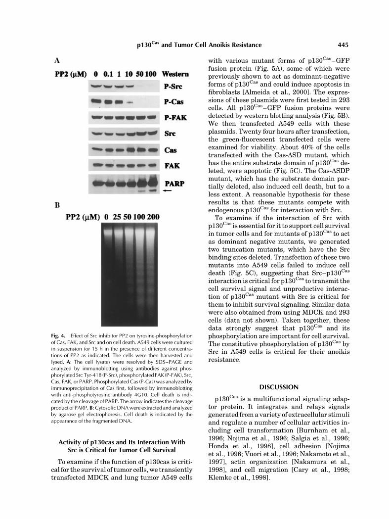
To further understand the role of Src andFAK in the constitutive phosphorylation ofp130Cas in the tumor cells, we inhibited Srcactivity by using a Src-specific inhibitor PP2and analyzed its effect on phosphorylation ofp130Cas and FAK. Treatment of A549 cells with
PP2 specifically abolished phosphorylation ofSrc and p130Cas, without affecting the phos-phorylation of FAK (Fig. 4A). The treatment ofSrc inhibitor also completely abolished thep130Cas-associated tyrosine kinase activity inthe in vitro kinase assay (data not shown).Taken together, these data strongly suggestthat Src is the key kinase that is responsible forthe constitutive phosphorylation of p130Cas inthe tumor cells.
Inhibition of Src and p130Cas PhosphorylationInduces Anoikis in A549 Tumor Cells
To test whether constitutive phosphoryla-tion of p130Cas is critical to the anoikis re-sistance of the lung tumor cells, we altered itsactivity by two different methods. We firstinhibited phosphorylation of p130Cas by theSrc inhibitor and analyzed its effect on celldeath of the lung tumor cells. We observed adose-dependent inhibition of p130Cas phos-phorylation and the induction of cell death bythe Src inhibitor, as indicated by PARP clea-vage and by DNA fragmentation analysis(Fig. 4). At 50 mM concentration of PP2, thephosphorylation of p130Cas was completelyinhibited and the cell death began to occur,suggesting that phosphorylation of p130Cas bySrc may be essential for transmitting the cellsurvival signal.
Fig. 2. In vitro kinase assay of p130Cas-associated tyrosinekinase activity. MDCK, A549, and H1792 cells were culturedas attached (A) or as suspension (S) cultures for 15 h. Cellswere then lysed and p130Cas were immunoprecipitated withanti-Cas antibody. The immunocomplexes were mixed withequal amounts of purified bacteria-expressed Cas–GST fusionprotein in kinase assay buffer and incubated for 30 min. Afterkinase reaction, the proteins were separated by SDS–PAGEand analyzed by immunoblotting using anti-phosphorylatedtyrosine antibody (aP-Tyr) and anti-GST antibodies (aCas-GST)sequentially. (C) is a control without addition of p130Cas
immunocomplex.
Fig. 3. Anchorage dependence of Src and FAK phosphoryla-tion/activation. MDCK, A549, and H1792 cells were cultured asattached (A) or as suspension (S) cultures for 15 h. Cellswere thenlysed in protein lysis buffer. Cell lysates were resolved by SDS–PAGE and analyzed by immunoblotting using the antibodiesagainst phosphorylated Src Tyr-418 (P-Src), phosphorylated FAKTyr-397 (P-FAK), Src, or FAK as indicated.
444 Wei et al.
Activity of p130cas and Its Interaction WithSrc is Critical for Tumor Cell Survival
To examine if the function of p130cas is criti-cal for the survival of tumor cells, we transientlytransfected MDCK and lung tumor A549 cells
with various mutant forms of p130Cas–GFPfusion protein (Fig. 5A), some of which werepreviously shown to act as dominant-negativeforms of p130Cas and could induce apoptosis infibroblasts [Almeida et al., 2000]. The expres-sions of these plasmids were first tested in 293cells. All p130Cas–GFP fusion proteins weredetected by western blotting analysis (Fig. 5B).We then transfected A549 cells with theseplasmids. Twenty four hours after transfection,the green-fluorescent transfected cells wereexamined for viability. About 40% of the cellstransfected with the Cas-DSD mutant, whichhas the entire substrate domain of p130Cas de-leted, were apoptotic (Fig. 5C). The Cas-DSDPmutant, which has the substrate domain par-tially deleted, also induced cell death, but to aless extent. A reasonable hypothesis for theseresults is that these mutants compete withendogenous p130Cas for interaction with Src.
To examine if the interaction of Src withp130Cas is essential for it to support cell survivalin tumor cells and for mutants of p130Cas to actas dominant negative mutants, we generatedtwo truncation mutants, which have the Srcbinding sites deleted. Transfection of these twomutants into A549 cells failed to induce celldeath (Fig. 5C), suggesting that Src–p130Cas
interaction is critical for p130Cas to transmit thecell survival signal and unproductive interac-tion of p130Cas mutant with Src is critical forthem to inhibit survival signaling. Similar datawere also obtained from using MDCK and 293cells (data not shown). Taken together, thesedata strongly suggest that p130Cas and itsphosphorylation are important for cell survival.The constitutive phosphorylation of p130Cas bySrc in A549 cells is critical for their anoikisresistance.
DISCUSSION
p130Cas is a multifunctional signaling adap-tor protein. It integrates and relays signalsgenerated from a variety of extracellular stimuliand regulate a number of cellular activities in-cluding cell transformation [Burnham et al.,1996; Nojima et al., 1996; Salgia et al., 1996;Honda et al., 1998], cell adhesion [Nojimaet al., 1996; Vuori et al., 1996; Nakamoto et al.,1997], actin organization [Nakamura et al.,1998], and cell migration [Cary et al., 1998;Klemke et al., 1998].
Fig. 4. Effect of Src inhibitor PP2 on tyrosine-phosphorylationof Cas, FAK, and Src and on cell death. A549 cells were culturedin suspension for 15 h in the presence of different concentra-tions of PP2 as indicated. The cells were then harvested andlysed. A: The cell lysates were resolved by SDS–PAGE andanalyzed by immunoblotting using antibodies against phos-phorylatedSrc Tyr-418 (P-Src), phosphorylated FAK (P-FAK), Src,Cas, FAK, or PARP. Phosphorylated Cas (P-Cas) was analyzed byimmunoprecipitation of Cas first, followed by immunoblottingwith anti-phosphotyrosine antibody 4G10. Cell death is indi-cated by the cleavage of PARP. The arrow indicates the cleavageproduct of PARP.B: CytosolicDNAwere extracted and analyzedby agarose gel electrophoresis. Cell death is indicated by theappearance of the fragmented DNA.
p130Cas and Tumor Cell Anoikis Resistance 445

Evidences linking p130Cas to regulation of celldeath/survival have been emerging recently. Anumber of experiments have shown thatp130Cas undergo dephosphorylation or cleavageduring apoptosis induced by various agents
ranging from anti-cancer drugs [Kook et al.,2000; Weyant et al., 2000; Shim et al., 2001],collagen gel overlay [Wang et al., 2001], adeno-sine/homocysteine [Harrington et al., 2001], celldetachment [Lesay et al., 2001], UV irradiation[Chan et al., 1999], tyrosine phosphatase [Wenget al., 1999], to serum withdraw [Almeida et al.,2000]. More direct evidences supporting anactive role of p130Cas in apoptosis came fromoverexpression experiments, in which overex-pression of wild-type p130Cas protects cells fromtyrosine phosphatase-induced apoptosis [Wenget al., 1999] and increases survival of migratorycells [Cho and Klemke, 2000], whereas over-expression of dominant-negative forms ofp130Cas blocks FAK-mediated cell survival[Chan et al., 1999; Almeida et al., 2000].
In this report, we analyzed the regulation andfunction of p130Cas in anoikis of normal epithe-lial cells and compared with that of anoikis-resistant human lung adenocarcinoma cells. Wefound that the phosphorylation of p130Cas isanchorage-dependent in the normal anoikis-sensitive epithelial cells, but anchorage-inde-pendent in the anoikis-resistant lung tumorcells. Further, we found that inhibition of thephosphorylation of p130Cas or overexpression ofdominant-negative forms of p130Cas inducedapoptosis in the tumor cells. Our data, togetherwith others, strongly support a critical role ofp130Cas in mediating cell survival signals gene-rated from cell adhesion. Inactivation of p130Cas
is an important step to fully execute a celldeath program. Alterations in the regulation ofp130Cas may contribute to resistance to apopto-sis of tumor cells.
Fig. 5. Cell death induced by transfection of p130Cas mutants.A: Schematic representation of GFP–Cas fusion protein and itsmutant forms. The GFP portion and the various functionaldomains of p130Cas are indicated. GFP–Cas: fusion protein ofGFP and wild type p130Cas; GFP–Cas-DSD: fusion protein ofGFP and substrate domain-deletion mutant of p130Cas; GFP–Cas-DSDP: fusion protein of GFP and partial substrate domain-deletion mutant of p130Cas; GFP–Cas-DSrc–BD: fusion proteinof GFP and Src binding domain-deletion mutant of p130Cas;GFP–Cas–FAK–BD: fusion protein of GFP and FAK bindingdomain of p130Cas; GFP:GFP alone.B: Immunoblot showing theprotein expression of theGFP–Cas fusion constructs in 293 cells.C: Cell death induced by transfection of GFP–Cas fusion con-structs. A549 cells cultured in regular tissue culture dishes weretransfectedwith the indicated constructs. Twenty four hours aftertransfection, cells were stained with DABI. Total green fluor-escent cells and green fluorescent dead cell were scored andcalculated. The numbers were presented as the percentage ofgreen fluorescent dead cells.
446 Wei et al.

The mechanism responsible for the constitu-tive phosphorylation of p130Cas in the tumorcells has yet to be fully understood. Eitheraltered tyrosine kinases or phosphatases ofp130Cas could contribute to the constitutivephosphorylation of p130Cas. Our data suggestthat it is a p130Cas-associated tyrosine kinasethat may be altered in the tumor cells and isresponsible for the anchorage-independentphosphorylation of p130Cas. Both Src and FAKare physically associated with p130Cas. Thefunctional importance of Src in the constitutivephosphorylation of p130Cas in the tumor cellswas analyzed by using its specific inhibitor.Inactivation of Src by its inhibitor specificallyand completely abolished the phosphorylationof p130Cas, confirming that Src is the key tyro-sine kinase in the constitutive phosphorylationof p130Cas in the tumor cells.
Elevated levels of Src activity have been re-ported in a number of human cancers, includinglung cancer [Biscardi et al., 1999]. In certainbreast cancer cells, Src is activated by increasedlevels of a Src tyrosine-530-specific phosphatase[Egan et al., 1999]. We did not observe de-creased phosphorylation at the tyr-530 of Srcin the lung tumor cells (data not shown). We,however, observed an increased phosphoryla-tion at the tyrosine-418 of Src upon cell detach-ment in the tumor cells. Therefore, increasedSrc tyrosine phosphatase activity may not bethe mechanism for the activation of Src uponcell detachment in the lung tumor cells. Themechanism of Src activation in the lung tumorcells remains to be determined.
FAK is also constitutively phosphorylated insome of the lung tumor cells. The mechanism ofconstitutive phosphorylation of FAK and its rolein phosphorylation of p130Cas are not clear atpresent. FAK apparently does not phosphory-late p130Cas directly since inhibition of Src issufficient to abolish phosphorylation of p130Cas
completely. This, however, does not exclude therole of the constitutively phosphorylated FAK inphysically linking Src and p130Cas together tofacilitate the phosphorylation of p130Cas by Src.
p130Cas interacts with a number of signalingproteins that are involved in regulation of phos-phorylation/activation of JNK, Erk, and PI 3K,all of which are important regulators of cellsurvival/death [Vuori et al., 1996; Schlaepferet al., 1997; Zhu et al., 1998; Blaukat et al., 1999;Oktay et al., 1999; Almeida et al., 2000; Cho andKlemke, 2000; Xing et al., 2000; Yoshizumi
et al., 2000]. We previously reported thatphosphorylation of Erk and Akt were decreasedin MDCK cells but increased in A549 cells uponcell detachment. However, inhibition of Erk andAkt phosphorylation/activation did not inducecell death [Wei et al., 2001], suggesting thatsignaling molecules other than Erk and Aktmay mediate the cell survival signals fromp130Cas. We recently also analyzed the regula-tion of phosphorylation of JNK and p38 MAPKby cell adhesion in A549 cells. No phosphory-lated JNK or p38 was detected in this cell line(unpublished data). Since functional p130Cas
is essential for the anoikis resistance of A549cell, we propose that p130Cas transmits a cellsurvival signal through a novel signaling mole-cule, which remains to be identified.
ACKNOWLEDGMENTS
We thank Dr. Jun-Lin Guan, Dr. Steven K.Hanks, and Amy H. Bouton for providing plas-mids containing p130Cas.
REFERENCES
Almeida EA, Ilic D, Han Q, Hauck CR, Jin F, Kawakatsu H,Schlaepfer DD, Damsky CH. 2000. Matrix survivalsignaling: From fibronectin via focal adhesion kinase toc-Jun NH(2)-terminal kinase. J Cell Biol 149:741–754.
Astier A, Avraham H, Manie SN, Groopman J, Canty T,Avraham S, Freedman AS. 1997. The related adhesionfocal tyrosine kinase is tyrosine-phosphorylated afterbeta1-integrin stimulation in B cells and binds top130cas. J Biol Chem 272:228–232.
Birge RB, Fajardo JE, Mayer BJ, Hanafusa H. 1992.Tyrosine-phosphorylated epidermal growth factor recep-tor and cellular p130 provide high affinity bindingsubstrates to analyze Crk-phosphotyrosine-dependentinteractions in vitro. J Biol Chem 267:10588–10595.
Biscardi JS, Tice DA, Parsons SJ. 1999. c-Src, receptortyrosine kinases, and human cancer. Adv Cancer Res 76:61–119.
Blaukat A, Ivankovic-Dikic I, Gronroos E, Dolfi F, TokiwaG, Vuori K, Dikic I. 1999. Adaptor proteins Grb2 and Crkcouple Pyk2 with activation of specific mitogen-activatedprotein kinase cascades. J Biol Chem 274:14893–14901.
Burnham MR, Harte MT, Richardson A, Parsons JT,Bouton AH. 1996. The identification of p130cas-bindingproteins and their role in cellular transformation. Onco-gene 12:2467–2472.
Cary LA, Han DC, Polte TR, Hanks SK, Guan JL. 1998.Identification of p130Cas as a mediator of focal adhesionkinase-promoted cell migration. J Cell Biol 140:211–221.
Casamassima A, Rozengurt E. 1997. Tyrosine phosphor-ylation of p130(cas) by bombesin, lysophosphatidicacid, phorbol esters, and platelet-derived growth factor.Signaling pathways and formation of a p130(cas)–Crkcomplex. J Biol Chem 272:9363–9370.
p130Cas and Tumor Cell Anoikis Resistance 447

Chan PC, Lai JF, Cheng CH, Tang MJ, Chiu CC, ChenHC. 1999. Suppression of ultraviolet irradiation-inducedapoptosis by overexpression of focal adhesion kinase inMadin–Darby canine kidney cells. J Biol Chem 274:26901–26906.
Cho SY, Klemke RL. 2000. Extracellular-regulated kinaseactivation and CAS/Crk coupling regulate cell migrationand suppress apoptosis during invasion of the extra-cellular matrix. J Cell Biol 149:223–236.
Egan C, Pang A, Durda D, Cheng HC, Wang JH, Fujita DJ.1999. Activation of Src in human breast tumor cell lines:Elevated levels of phosphotyrosine phosphatase activitythat preferentially recognizes the Src carboxy terminalnegative regulatory tyrosine 530. Oncogene 18:1227–1237.
Frisch SM, Francis H. 1994. Disruption of epithelial cell–matrix interactions induces apoptosis. J Cell Biol 124:619–626.
Frisch SM, Ruoslahti E. 1997. Integrins and anoikis. CurrOpin Cell Biol 9:701–706.
Frisch SM, Screaton RA. 2001. Anoikis mechanisms. CurrOpin Cell Biol 13:555–562.
Frisch SM, Vuori K, Ruoslahti E, Chan-Hui PY. 1996.Control of adhesion-dependent cell survival by focaladhesion kinase. J Cell Biol 134:793–799.
Hamasaki K, Mimura T, Morino N, Furuya H, NakamotoT, Aizawa S, Morimoto C, Yazaki Y, Hirai H, Nojima Y.1996. Src kinase plays an essential role in integrin-mediated tyrosine phosphorylation of Crk-associatedsubstrate p130Cas. Biochem Biophys Res Commun 222:338–343.
Harrington EO, Smeglin A, Newton J, Ballard G, Rounds S.2001. Protein tyrosine phosphatase-dependent proteoly-sis of focal adhesion complexes in endothelial cell apop-tosis. Am J Physiol Lung Cell Mol Physiol 280:L342–L353.
Harte MT, Hildebrand JD, Burnham MR, Bouton AH,Parsons JT. 1996. p130Cas, a substrate associated withv-Src and v-Crk, localizes to focal adhesions andbinds to focal adhesion kinase. J Biol Chem 271:13649–13655.
Honda H, Oda H, Nakamoto T, Honda Z, Sakai R, Suzuki T,Saito T, Nakamura K, Nakao K, Ishikawa T, Katsuki M,Yazaki Y, Hirai H. 1998. Cardiovascular anomaly, im-paired actin bundling and resistance to Src-inducedtransformation in mice lacking p130Cas [see comments].Nat Genet 19:361–365.
Kanner SB, Reynolds AB, Vines RR, Parsons JT. 1990.Monoclonal antibodies to individual tyrosine-phosphory-lated protein substrates of oncogene-encoded tyrosinekinases. Proc Natl Acad Sci USA 87:3328–3332.
Kanner SB, Reynolds AB, Wang HC, Vines RR, Parsons JT.1991. The SH2 and SH3 domains of pp60src direct stableassociation with tyrosine phosphorylated proteins p130and p110. Embo J 10:1689–1698.
Khwaja A, Rodriguez-Viciana P, Wennstrom S, Warne PH,Downward J. 1997. Matrix adhesion and Ras transfor-mation both activate a phosphoinositide 3-OH kinase andprotein kinase B/Akt cellular survival pathway. EmboJ 16:2783–2793.
Klemke RL, Leng J, Molander R, Brooks PC, Vuori K,Cheresh DA. 1998. CAS/Crk coupling serves as a ‘‘mole-cular switch’’ for induction of cell migration. J Cell Biol140:961–972.
Kook S, Shim SR, Choi SJ, Ahnn J, Kim JI, Eom SH, JungYK, Paik SG, Song WK. 2000. Caspase-mediated clea-vage of p130cas in etoposide-induced apoptotic Rat-1cells. Mol Biol Cell 11:929–939.
Lesay A, Hickman JA, Gibson RM. 2001. Disruption of focaladhesions mediates detachment during neuronal apop-tosis. Neuroreport 12:2111–2115.
Manie SN, Astier A, Haghayeghi N, Canty T, Druker BJ,Hirai H, Freedman AS. 1997. Regulation of integrin-mediated p130(Cas) tyrosine phosphorylation in humanB cells. A role for p59(Fyn) and SHP2. J Biol Chem 272:15636–15641.
Matsuda M, Mayer BJ, Fukui Y, Hanafusa H. 1990. Bind-ing of transforming protein, P47gag-crk, to a broad rangeof phosphotyrosine-containing proteins. Science 248:1537–1539.
Mayer BJ, Hanafusa H. 1990. Association of the v-crkoncogene product with phosphotyrosine-containing pro-teins and protein kinase activity. Proc Natl Acad Sci USA87:2638–2642.
Meredith JE, Jr., Fazeli B, Schwartz MA. 1993. The extra-cellular matrix as a cell survival factor. Mol Biol Cell 4:953–961.
Nakamoto T, Sakai R, Honda H, Ogawa S, Ueno H, SuzukiT, Aizawa S, Yazaki Y, Hirai H. 1997. Requirements forlocalization of p130cas to focal adhesions. Mol Cell Biol17:3884–3897.
Nakamura I, Jimi E, Duong LT, Sasaki T, Takahashi N,Rodan GA, Suda T. 1998. Tyrosine phosphorylation ofp130Cas is involved in actin organization in osteoclasts.J Biol Chem 273:11144–11149.
Nojima Y, Morino N, Mimura T, Hamasaki K, Furuya H,Sakai R, Sato T, Tachibana K, Morimoto C, Yazaki Y,et al. 1995. Integrin-mediated cell adhesion promotestyrosine phosphorylation of p130Cas, a Src homology3-containing molecule having multiple Src homology2-binding motifs. J Biol Chem 270:15398–15402.
Nojima Y, Mimura T, Morino N, Hamasaki K, Furuya H,Sakai R, Nakamoto T, Yazaki Y, Hirai H. 1996. Tyrosinephosphorylation of p130Cas in cell adhesion and trans-formation. Hum Cell 9:169–174.
Ojaniemi M, Vuori K. 1997. Epidermal growth factor modu-lates tyrosine phosphorylation of p130Cas. Involvementof phosphatidylinositol 30-kinase and actin cytoskeleton.J Biol Chem 272:25993–25998.
Oktay M, Wary KK, Dans M, Birge RB, Giancotti FG. 1999.Integrin-mediated activation of focal adhesion kinase isrequired for signaling to Jun NH2-terminal kinase andprogression through the G1 phase of the cell cycle. J CellBiol 145:1461–1469.
Petch L, Bockholt S, Bouton A, Parsons J, Burridge K.1995. Adhesion-induced tyrosine phosphorylation of thep130 SRC substrate. J Cell Sci 108:1371–1379.
Polte TR, Hanks SK. 1995. Interaction between focal adhe-sion kinase and Crk-associated tyrosine kinase substratep130Cas. Proc Natl Acad Sci USA 92:10678–10682.
Reynolds AB, Kanner SB, Wang HC, Parsons JT. 1989.Stable association of activated pp60src with two tyrosine-phosphorylated cellular proteins. Mol Cell Biol 9:3951–3958.
Ribon V, Saltiel AR. 1996. Nerve growth factor stimulatesthe tyrosine phosphorylation of endogenous Crk-II andaugments its association with p130Cas in PC-12 cells.J Biol Chem 271:7375–7380.
448 Wei et al.

Richardson A, Parsons JT. 1995. Signal transductionthrough integrins: A central role for focal adhesionkinase? Bioessays 17:229–236.
Sakai R, Iwamatsu A, Hirano N, Ogawa S, Tanaka T, ManoH, Yazaki Y, Hirai H. 1994. A novel signaling molecule,p130, forms stable complexes in vivo with v-Crk andv-Src in a tyrosine phosphorylation-dependent manner.Embo J 13:3748–3756.
Sakai R, Nakamoto T, Ozawa K, Aizawa S, Hirai H. 1997.Characterization of the kinase activity essential for tyro-sine phosphorylation of p130Cas in fibroblasts. Oncogene14:1419–1426.
Salgia R, Pisick E, Sattler M, Li JL, Uemura N, WongWK, Burky SA, Hirai H, Chen LB, Griffin JD. 1996.p130CAS forms a signaling complex with the adapterprotein CRKL in hematopoietic cells transformed by theBCR/ABL oncogene. J Biol Chem 271:25198–25203.
Schaller MD, Parsons JT. 1994. Focal adhesion kinase andassociated proteins. Curr Opin Cell Biol 6:705–710.
Schaller MD, Parsons JT. 1995. pp125FAK-dependenttyrosine phosphorylation of paxillin creates a high-affinity binding site for Crk. Mol Cell Biol 15:2635–2645.
Schlaepfer DD, Hanks SK, Hunter T, van der Geer P. 1994.Integrin-mediated signal transduction linked to Raspathway by GRB2 binding to focal adhesion kinase.Nature 372:786–791.
Schlaepfer DD, Broome MA, Hunter T. 1997. Fibronectin-stimulated signaling from a focal adhesion kinase-c–Srccomplex: Involvement of the Grb2, p130cas, and Nckadaptor proteins. Mol Cell Biol 17:1702–1713.
Schlaepfer DD, Hauck CR, Sieg DJ. 1999. Signalingthrough focal adhesion kinase. Prog Biophys Mol Biol71:435–478.
Seufferlein T, Rozengurt E. 1994. Lysophosphatidic acidstimulates tyrosine phosphorylation of focal adhesionkinase, paxillin, and p130. Signaling pathways and cross-talk with platelet-derived growth factor. J Biol Chem269:9345–9351.
Shim SR, Kook S, Kim JI, Song WK. 2001. Degradation offocal adhesion proteins paxillin and p130cas by caspases
or calpains in apoptotic rat-1 and L929 cells. BiochemBiophys Res Commun 286:601–608.
Vuori K, Ruoslahti E. 1995. Tyrosine phosphorylation ofp130Cas and cortactin accompanies integrin-mediatedcell adhesion to extracellular matrix. J Biol Chem 270:22259–22262.
Vuori K, Hirai H, Aizawa S, Ruoslahti E. 1996. Intro-duction of p130cas signaling complex formation uponintegrin-mediated cell adhesion: A role for Src familykinases. Mol Cell Biol 16:2606–2613.
Wang YK, Lin HH, Tang MJ. 2001. Collagen gel overlayinduces two phases of apoptosis in MDCK cells. AmJ Physiol Cell Physiol 280:C1440–C1448.
Wei L, Yang Y, Yu Q. 2001. Tyrosine kinase-dependent,phosphatidylinositol 30-kinase, and mitogen-activatedprotein kinase-independent signaling pathways preventlung adenocarcinoma cells from anoikis. Cancer Res 61:2439–2444.
Weng LP, Wang X, Yu Q. 1999. Transmembrane tyrosinephosphatase LAR induces apoptosis by dephosphor-ylating and destabilizing p130Cas. Genes Cells 4:185–196.
Weyant MJ, Carothers AM, Bertagnolli ME, BertagnolliMM. 2000. Colon cancer chemopreventive drugs mod-ulate integrin-mediated signaling pathways. Clin CancerRes 6:949–956.
Xing L, Ge C, Zeltser R, Maskevitch G, Mayer BJ,Alexandropoulos K. 2000. c-Src signaling induced bythe adapters Sin and Cas is mediated by Rap1 GTPase.Mol Cell Biol 20:7363–7377.
Yoshizumi M, Abe J, Haendeler J, Huang Q, Berk BC.2000. Src and Cas mediate JNK activation but not ERK1/2 and p38 kinases by reactive oxygen species. J BiolChem 275:11706–11712.
Zhu T, Goh EL, LeRoith D, Lobie PE. 1998. Growthhormone stimulates the formation of a multiprotein sig-naling complex involving p130(Cas) and CrkII. Resultantactivation of c-Jun N-terminal kinase/stress-activatedprotein kinase (JNK/SAPK). J Biol Chem 273:33864–33875.
p130Cas and Tumor Cell Anoikis Resistance 449