anatomy of shinkailepas myojinensis … · anatomy of shinkailepas myojinensis sasaki, okutani...
TRANSCRIPT
ANATOMY OF SHINKAILEPAS MYOJINENSIS SASAKI, OKUTANI & FUJIKURA, 2003(GASTROPODA: NERITOPSINA)
Takenori Sasaki1, Takashi Okutani2 & Katsunori Fujikura2
ABSTRACT
The anatomy of Shinkailepas myojinensis Sasaki, Okutani & Fujikura, 2003, was exam-ined by gross dissection, scanning electron microscopy, and histological serial sections.The organization of the soft part conforms to general neritopsine pattern, especially inpallial complex, digestive system, reno-pericardial system, and nervous system. New char-acter states previously unknown in neritopsine gastropods were revealed mainly in femaleand male reproductive systems, sense organs, and glands in pallial cavity. Comparison ofour observations with published descriptions of various gastropods confirmed that thespecies of Shinkailepas are assigned to the superfamily Neritoidea among Neritopsina.The inclusion of Shinkailepas in the family Phenacolepadidae as in previous studies isalso supported, although the number of their uniquely shared character is rather limited.Infrafamilial taxa of phenacolepadids so far anatomically studied are clearly divisible intodeep-sea (Shinkailepas and Olgasolaris) and shallow-water (Phenacolepas andCinnalepeta) groups in characters of the shell, operculum, head-foot external morphology,mantle margin, digestive tract, and female reproductive organ. At species level, membersof Shinkailepas are diagnosed by morphology of the eye stalks, epipodial fold, and penis,as well as shell, radular and opercular characters.
Keywords: Shinkailepas, Phenacolepadidae, Neritopsina, comparative anatomy.
1
INTRODUCTION
In the recent systematics, Neritopsina in-cludes six to nine families, namely, Neritidae,Neritiliidae, Phenacolepadidae, Hydrocenidae,Neritopsidae, including Titiscaniidae, and threehelicinoidean families (Helicinidae, Ceresidae,and Proserpinidae, or three subfamilies ofHelicinidae) (Thompson, 1980; Ponder, 1998;Sasaki, 1998; Kano & Kase, 2000, 2002). Theycomprise a robust clade phylogenetically (Pon-der & Lindberg, 1997; Sasaki, 1998) and ex-hibit remarkable ecological diversification fromdeep-sea to terrestrial habitats (Kano et al.,2002; Sasaki & Ishikawa, 2002). Among them,three genera, Shinkailepas, Olgasolaris, andBathynerita, have been known only from deep-sea chemosynthesis-based biological commu-nities. The first two genera are currentlyassigned to the Phenacolepadidae (Beck,1992; Warén & Bouchet, 2001; Sasaki et al.,2003), together with the shallow-water gen-era Phenacolepas and Cinnalepeta, andBathynerita is regarded as a member of theNeritidae (Warén & Bouchet, 1993, 2001).
MALACOLOGIA, 2006, 48(1−2): 1−26
1The University Museum, The University of Tokyo, 7-3-1, Hongo, Bunkyo-ku, Tokyo 113-0033, Japan; [email protected] Agency for Marine-Earth Science and Technology, 2-15, Natsushima, Yokosuka City 237-0061, Japan
They represent part of characteristic mollus-can elements endemic to vent/seep environ-ments.
In the genus Shinkailepas Okutani, Saito &Hashimoto, 1989, four species have been hith-erto described: (1) S. kaikatensis Okutani,Saito & Hashimoto, 1989, from Kaikata Sea-mount, off Ogasawara Islands, Japan, 470 mdeep, (2) S. tufari Beck, 1992, from ManusBack-Arc Basin, 2,450−2,505 m deep, (3) S.briandi Warén & Bouchet, 2001, from Mid-At-lantic Ridge, Menez Gowen to Logatchev site,850−3,500 m deep, and (4) S. myojinensisSasaki, Okutani & Fujikura, 2003, from MyojinKnoll, Ogasawara Ridge, Japan, 1,260−1,340m deep. In addition, unidentified species werealso reported from Mariana Back-Arc Basin(Hasegawa et al., 1997) and Okinawa Trough(Sasaki et al., 2003), suggesting the presenceof more new species in the genus.
Although some anatomical descriptions havebeen published (e.g., Fretter, 1984; Sasaki,1998; Kano & Kase, 2002), there is consider-able uncertainty in anatomical organization ofphenacolepadids and other possibly related
SASAKI ET AL.2
neritoidean groups. Hence, further anatomicalcomparison is significant to understand rela-tionships among Shinkailepas, Olgasolaris,shallow-water neritoideans, and the remainingless known neritopsines. All of known speciesof Shinkailepas have been described basedchiefly on the shell, radula, operculum, andexternal morphology of the animal (Okutani etal., 1989; Beck, 1992; Warén & Bouchet, 2001;Sasaki et al., 2003), and only limited anatomi-cal descriptions have been published for inter-nal organs of the genus.
In this study, we attempted to provide de-tailed account of anatomical organization ofShinkailepas myojinensis by gross dissection,scanning electron microscopy, and histologi-cal serial sections. The results of observationsare compared with existing knowledge of or-gan systems of other nertiopsines, and theirsignificance is discussed in terms of compara-tive anatomy and systematics.
MATERIALS AND METHODS
Materials examined in this study were se-lected from part of paratype series of S. myoji-nensis and those preserved at the Japan
Agency for Marine-Earth Science and Tech-nology (JAMSTEC). Details of sampling dataare shown in the original description. Samplesfixed in formalin were used for anatomicalobservations. After removed from the shell, softparts of five specimens were dissected undera binocular microscope. Pieces of dissectedsoft parts were dried with a freeze-drier (HitachiES2030) and observed with SEM (HitachiS2400). Whole animals of two females andthree males were thin-sectioned at the thick-ness of 6 μm after embedding in paraffin. Theywere stained by a standard method of Mayer’sHematoxylin and Eosin staining. Whole seriesof sections (UMUT RM28647−28651) andSEM stub (UMUT RM28652) are deposited inthe Department of Histological Geology andPaleontology, the University Museum, theUniversity of Tokyo (UMUT). The terminologyused in the descriptions chiefly followed thatof Sasaki (1998) and Kano & Kase (2002).
RESULTS
Shell and Operculum
See Sasaki et al. (2003).
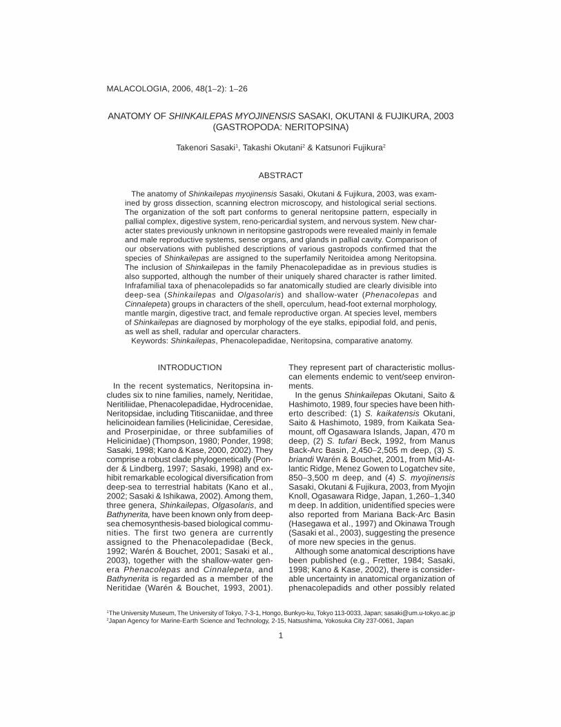
FIG. 1. Dorsal view of animal, male, with mantle removed.

ANATOMY OF SHINKAILEPAS 3
cally identical between both sexes. Only theposterior half of the right eye stalk is coveredwith a patch of tall glandular cells, which arehere termed the “post-tentacular gland” (ptg:Figs. 1, 4F). Black eye spots are visible exter-nally in anterior middle part of the eye stalks(Fig. 1).
The shell muscle is divided into right and leftportions (lsm, rsm: Fig. 1), leaving separatedscars on the internal surface of the shell. Eachmuscle is not subdivided into bundles, norpenetrated by blood vessels. The head retrac-tor muscles are merged with shell muscles anddo not produce independent attachment ar-eas. The mantle is devoid of particular retrac-tor muscle inserted on the shell, but insteadattached to the shell by penetration of pallialprocesses.
Pallial Cavity
The pallial cavity is deep and attains the levelof the posterior end of both shell muscle at-tachments. It contains the ctenidium,osphradium (os: Fig. 4E), anus, the kidneyopening (ko: Fig. 10C), and genital opening(s).
The ctenidium is single, extended from theposterior left to the anterior right (Fig. 1). Thectenidial lamellae are bipectinate in alternating
FIG. 2. Ventral view of animal.
External Anatomy
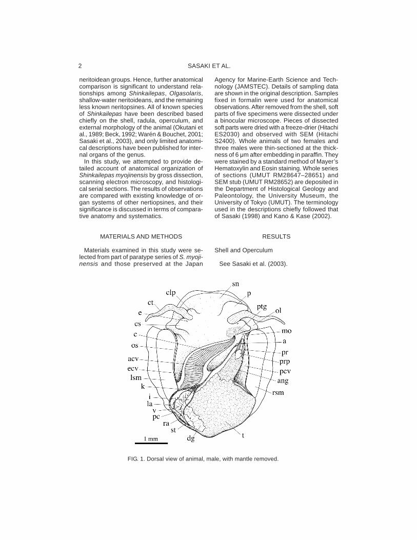
The animal is dorsoventrally flattened andsymmetrical in outline. The visceral mass isnot spirally coiled (Fig. 1). The dorsal surfaceof the animal is covered with a thin mantle.The entire surface of the mantle carries densefilamentous mantle processes (mp: Fig. 3A),which are multicellular and contain fiber-likestructure internally (Fig. 4A). These processesare not branched and some of them penetratethrough whole thickness of the shell at posi-tions corresponding to the shell pores (cf.Sasaki et al., 2003: fig. 12C). The dorsal sideof mantle margin also bears similar processes(Fig. 3B).
The mantle margin (mm: Figs. 2, 4C) isprominently thickened, divided into the outerand inner folds by the periostracal groove (pg:Figs. 3B, 4C). Its inside contains muscle fi-bers and blood sinus. The portion of the mantlecovering the pallial cavity is provided with ex-tensive blood sinus with numerous hemocytesinside (pls: Fig. 4F) and probably functions asa respiratory surface.
Major part of the ventral side of the animal isoccupied by a circular, flattened pedal sole (ps:Fig. 2). Its anterior margin is marked by theanterior pedal groove (apg: Figs. 2, 4B) withthe opening of the anterior pedal gland (apd:Fig. 4B). The ventral side of the head is ex-tended as the oral lappet (ol: Figs. 2, 3C). Thecircumference of the mouth is thickened, papil-late, and wrinkled (Fig. 3C). The lateral foot issmooth without any protrusive structure. Theepipodial fold is extended between posteriordorsal rim of the foot and the mantle marginand provided with triangular tentacles (Figs.2, 3D). The epipodial tentacles lackmicropapillae, sense organ and ciliated struc-ture. Their number ranges from 11 to 19, vary-ing among specimens examined. Theoperculum is firmly attached to the dorsal sur-face of the foot musculature below the visceralmass.
The head consists of the snout, cephalic ten-tacles, cephalic lappets, and eye stalks (Fig.1). The snout is stout, short and not tapered.The cephalic tentacles are paired in equal formand size, symmetrically positioned on eachside of the head, not covered with sensorypapillae, and striated by internal longitudinalmuscle. The cephalic lappets are symmetri-cal, equal in size in female, but the right oneis greatly enlarged in the male as the penis(p: Fig. 1). The eye stalks are posterior to thecephalic tentacles, flattened, and morphologi-

SASAKI ET AL.4
arrangement (Fig. 4D), ridged along midline (Fig.1), and not attached by afferent nor efferentctenidial membranes. The skeletal rods andburiscles (sensory pockets) are absent (Fig. 4D).
A hypobranchial gland is absent. Part of themantle opposing the post-tentacular gland iscovered with a tall glandular epithelium, whichis termed the “anterior pallial gland” (apl: Fig.
4F). This gland is also extended to cover thedistal tip of the pallial gonoduct. Dorsoventrallypaired, two glands, the post-tentacular glandon ventral side and the anterior pallial glandon dorsal side, are developed in the sameposition and size in both sexes. The epithe-lium of equivalent part on the left side is notspecialized as gland.
FIG. 3. Scanning electron micrographs of external and internal organs. A. Mantle processes (mp) onsurface of mantle covering visceral part. B. Dorsal view of mantle margin. C. Ventral view of mouth(m) and oral lappet (ol). D. Ventral view of epipodial fold. E. Tooth of gastric shield (gst) inside ofstomach. F. Left auricle (la) and ventircle (v) penetrated by intestine (i), removed from pericardium.A−F. UMUT RM28652.
ANATOMY OF SHINKAILEPAS 5
Digestive System
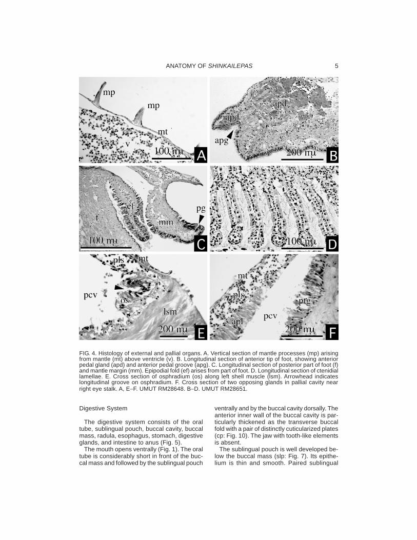
The digestive system consists of the oraltube, sublingual pouch, buccal cavity, buccalmass, radula, esophagus, stomach, digestiveglands, and intestine to anus (Fig. 5).
The mouth opens ventrally (Fig. 1). The oraltube is considerably short in front of the buc-cal mass and followed by the sublingual pouch
ventrally and by the buccal cavity dorsally. Theanterior inner wall of the buccal cavity is par-ticularly thickened as the transverse buccalfold with a pair of distinctly cuticularized plates(cp: Fig. 10). The jaw with tooth-like elementsis absent.
The sublingual pouch is well developed be-low the buccal mass (slp: Fig. 7). Its epithe-lium is thin and smooth. Paired sublingual
FIG. 4. Histology of external and pallial organs. A. Vertical section of mantle processes (mp) arisingfrom mantle (mt) above ventricle (v). B. Longitudinal section of anterior tip of foot, showing anteriorpedal gland (apd) and anterior pedal groove (apg). C. Longitudinal section of posterior part of foot (f)and mantle margin (mm). Epipodial fold (ef) arises from part of foot. D. Longitudinal section of ctenidiallamellae. E. Cross section of osphradium (os) along left shell muscle (lsm). Arrowhead indicateslongitudinal groove on osphradium. F. Cross section of two opposing glands in pallial cavity nearright eye stalk. A, E−F. UMUT RM28648. B−D. UMUT RM28651.
SASAKI ET AL.6
epithelium (Fig. 9B). The dorsal side of buc-cal cavity is thickened by a pair of well-devel-oped dorsal folds (df: Fig. 9D). Salivary glandsare not differentiated around the buccal cav-ity. The radular diverticulum is deep below theesophageal valve.
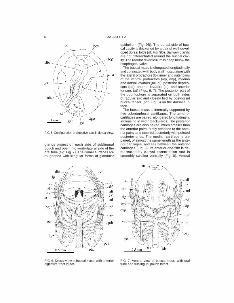
The buccal mass is elongated longitudinallyand connected with body wall musculature withthe lateral protractors (lp), inner and outer pairsof the ventral protractors (ivp, ovp), medianand dorsal levators (ml, dl), posterior depres-sors (pd), anterior levators (al), and anteriortensors (at) (Figs. 6, 7). The posterior part ofthe odontophore is separated on both sidesof radular sac and closely tied by postdorsalbuccal tensor (pdt: Fig. 6) on the dorsal sur-face.
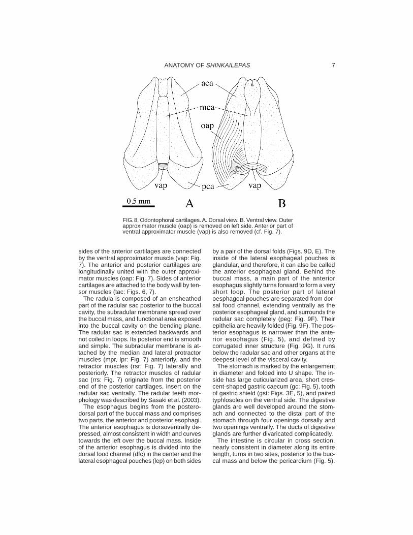
The buccal mass is internally supported byfive odontophoral cartilages. The anteriorcartilages are paired, elongated longitudinally,increasing in width backwards. The posteriorcartilages are also paired, much smaller thanthe anterior pairs, firmly attached to the ante-rior pairs, and tapered posteriorly with pointedposterior ends. The median cartilage is un-paired, of almost the same length as the ante-rior cartilages, and lies between the anteriorcartilages (Fig. 8). Its anterior one-fifth is de-marcated by dorsal constriction and issmoothly swollen ventrally (Fig. 8). Ventral
FIG. 5. Configuration of digestive tract in dorsal view.
FIG. 6. Drosal view of buccal mass, with anteriordigestive tract intact.
glands project on each side of sublingualpouch and open into ventrolateral side of theoral tube (slg: Fig. 7). Their inner surfaces areroughened with irregular forms of glandular
FIG. 7. Ventral view of buccal mass, with oraltube and sublingual pouch intact.
ANATOMY OF SHINKAILEPAS 7
sides of the anterior cartilages are connectedby the ventral approximator muscle (vap: Fig.7). The anterior and posterior cartilages arelongitudinally united with the outer approxi-mator muscles (oap: Fig. 7). Sides of anteriorcartilages are attached to the body wall by ten-sor muscles (tac: Figs. 6, 7).
The radula is composed of an ensheathedpart of the radular sac posterior to the buccalcavity, the subradular membrane spread overthe buccal mass, and functional area exposedinto the buccal cavity on the bending plane.The radular sac is extended backwards andnot coiled in loops. Its posterior end is smoothand simple. The subradular membrane is at-tached by the median and lateral protractormuscles (mpr, lpr: Fig. 7) anteriorly, and theretractor muscles (rsr: Fig. 7) laterally andposteriorly. The retractor muscles of radularsac (rrs: Fig. 7) originate from the posteriorend of the posterior cartilages, insert on theradular sac ventrally. The radular teeth mor-phology was described by Sasaki et al. (2003).
The esophagus begins from the postero-dorsal part of the buccal mass and comprisestwo parts, the anterior and posterior esophagi.The anterior esophagus is dorsoventrally de-pressed, almost consistent in width and curvestowards the left over the buccal mass. Insideof the anterior esophagus is divided into thedorsal food channel (dfc) in the center and thelateral esophageal pouches (lep) on both sides
by a pair of the dorsal folds (Figs. 9D, E). Theinside of the lateral esophageal pouches isglandular, and therefore, it can also be calledthe anterior esophageal gland. Behind thebuccal mass, a main part of the anterioresophagus slightly turns forward to form a veryshort loop. The posterior part of lateraloesphageal pouches are separated from dor-sal food channel, extending ventrally as theposterior esophageal gland, and surrounds theradular sac completely (peg: Fig. 9F). Theirepithelia are heavily folded (Fig. 9F). The pos-terior esophagus is narrower than the ante-rior esophagus (Fig. 5), and defined bycorrugated inner structure (Fig. 9G). It runsbelow the radular sac and other organs at thedeepest level of the visceral cavity.
The stomach is marked by the enlargementin diameter and folded into U shape. The in-side has large cuticularized area, short cres-cent-shaped gastric caecum (gc: Fig. 5), toothof gastric shield (gst: Figs. 3E, 5), and pairedtyphlosoles on the ventral side. The digestiveglands are well developed around the stom-ach and connected to the distal part of thestomach through four openings dorsally andtwo openings ventrally. The ducts of digestiveglands are further divaricated complicatedly.
The intestine is circular in cross section,nearly consistent in diameter along its entirelength, turns in two sites, posterior to the buc-cal mass and below the pericardium (Fig. 5).
FIG. 8. Odontophoral cartilages. A. Dorsal view. B. Ventral view. Outerapproximator muscle (oap) is removed on left side. Anterior part ofventral approximator muscle (vap) is also removed (cf. Fig. 7).
SASAKI ET AL.8
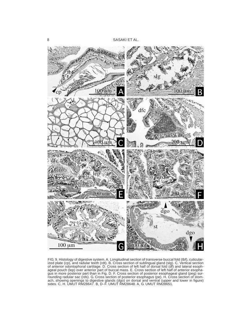
FIG. 9. Histology of digestive system. A. Longitudinal section of transverse buccal fold (tbf), cuticular-ized plate (cp), and radular teeth (rdt). B. Cross section of sublingual gland (slg). C. Vertical sectionof anterior odontophoral cartilage. D. Cross section of left half of dorsal fold (df) and lateral esoph-ageal pouch (lep) over anterior part of buccal mass. E. Cross section of left half of anterior esopha-gus in more posterior part than in Fig. D. F. Cross section of posterior esophageal gland (peg) sur-rounding radular sac (rds). G. Cross section of posterior esophagus (pe). H. Cross section of stom-ach, showing openings to digestive glands (dgo) on dorsal and ventral (upper and lower in figure)sides. C, H. UMUT RM28647. B, D−F. UMUT RM28648. A, G. UMUT RM28651.
ANATOMY OF SHINKAILEPAS 9
Its epithelium is densely ciliated and composedof a layer of prismatic cells (Fig. 10B). Aroundthe second turn, the intestine penetrates theventricle and the pericardium. No anal (rectal)gland was found near the distal part of the in-testine. The anus opens on the right anteriorside of the pallial cavity (Fig. 1).
Circulatory System
The heart is enclosed in the pericardium andconsists of paired auricles of unequal size anda median ventricle (Figs. 1, 3F, 10A). The leftauricle is much larger than the right and con-nected to blood vessels from the ctenidium and
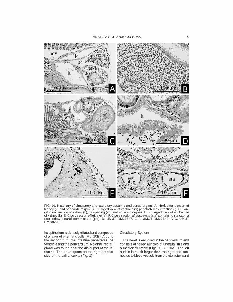
FIG. 10. Histology of circulatory and excretory systems and sense organs. A. Horizontal section ofkidney (k) and pericardium (pc). B. Enlarged view of ventricle (v) penetrated by intestine (i). C. Lon-gitudinal section of kidney (k), its opening (ko) and adjacent organs. D. Enlarged view of epitheliumof kidney (k). E. Cross section of left eye (e). F. Cross section of statosysts (sta) containing statoconia(sc) below pleural commissure (plc). D. UMUT RM28647. E−F. UMUT RM28648. A−C. UMUTRM28651.
SASAKI ET AL.10
the mantle. The margin of the left auricle is rug-ose and constricted to be separated into a fewchambers (Fig. 3F). The right auricle is vesti-gial and situated posterior to the ventricle (Fig.1). The ventricle is voluminous, penetrated bythe intestine, stiffened by cardiac muscles in-side (Fig. 10B). An aorta from the ventricle isshort and opens into haemocoel of the body.An aortic bulb is not formed. Large sinuses aredeveloped among visceral organs and aroundthe buccal mass. Considerable blood sinusesare also found near the lateral wall of the pedalmusculature. Blood space adjacent to the kid-ney is connected anteriorly to the afferentctenidial vessel.
Excretory System
The excretory organ consists of a single kid-ney and the pericardium. The kidney is on theposterior side of the ctenidium; its excretoryopening is located below ctenidial base (ko:Fig. 10C). The inside of the kidney is partlypartitioned into two branches on the anteriorand right sides of the pericardium, respectively(Fig. 10A), but both are indistinguishable inhistology. The epithelium of the kidney is notlamellated throughout the entire area and con-sists of single-layered papillate cells with basalnuclei (Fig. 10D). The renopericardial connec-tion is extended between the two branches ofthe kidney.
Reproductive System
The sexes are separate, and their reproduc-tive organs exhibit striking sexual dimorphism.Large part of the reproductive organ is occu-pied by the gonad and pallial gonoduct. In bothsexes, the gonad develops dorsal to the diges-tive gland, the gonoduct is not connected tothe kidney, and gonopericardial connection isnot present.
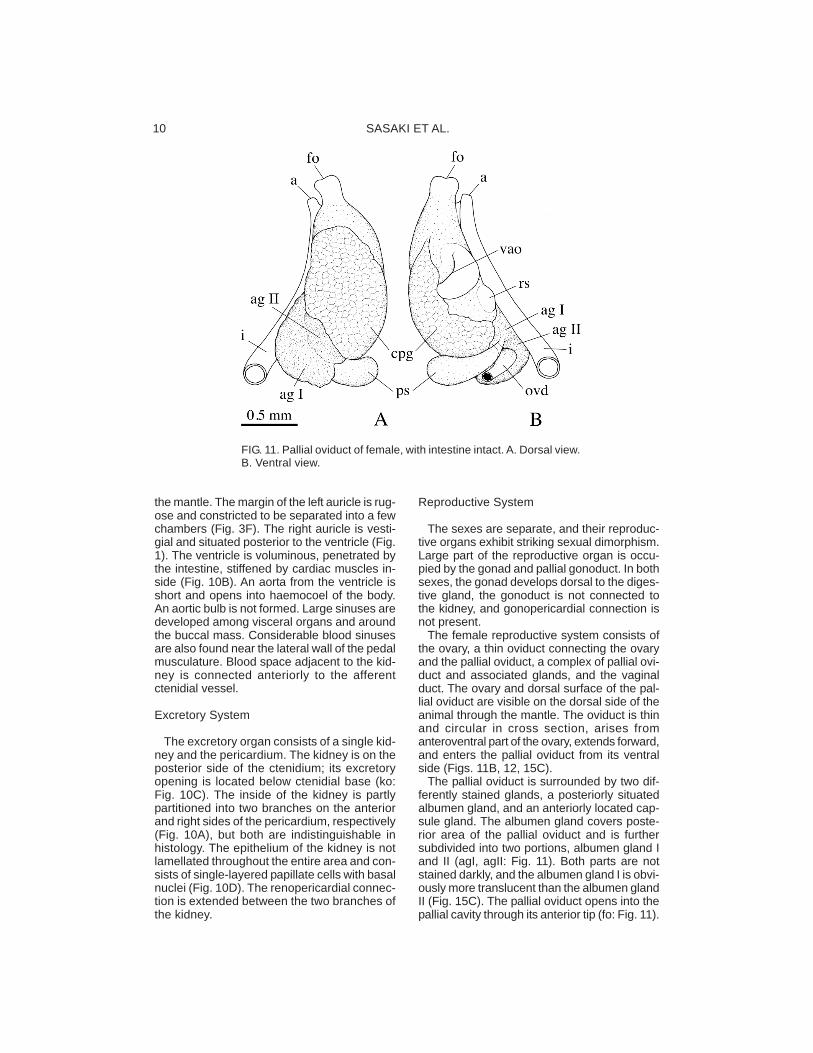
The female reproductive system consists ofthe ovary, a thin oviduct connecting the ovaryand the pallial oviduct, a complex of pallial ovi-duct and associated glands, and the vaginalduct. The ovary and dorsal surface of the pal-lial oviduct are visible on the dorsal side of theanimal through the mantle. The oviduct is thinand circular in cross section, arises fromanteroventral part of the ovary, extends forward,and enters the pallial oviduct from its ventralside (Figs. 11B, 12, 15C).
The pallial oviduct is surrounded by two dif-ferently stained glands, a posteriorly situatedalbumen gland, and an anteriorly located cap-sule gland. The albumen gland covers poste-rior area of the pallial oviduct and is furthersubdivided into two portions, albumen gland Iand II (agI, agII: Fig. 11). Both parts are notstained darkly, and the albumen gland I is obvi-ously more translucent than the albumen glandII (Fig. 15C). The pallial oviduct opens into thepallial cavity through its anterior tip (fo: Fig. 11).
FIG. 11. Pallial oviduct of female, with intestine intact. A. Dorsal view.B. Ventral view.
ANATOMY OF SHINKAILEPAS 11
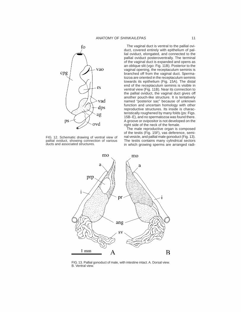
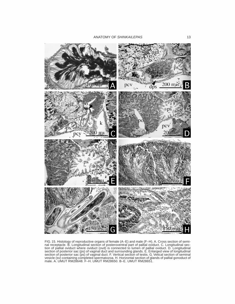
The vaginal duct is ventral to the pallial ovi-duct, covered entirely with epithelium of pal-lial oviduct, elongated, and connected to thepallial oviduct posteroventrally. The terminalof the vaginal duct is expanded and opens asan oblique slit (vgo: Fig. 11B). Posterior to thevaginal opening, the receptaculum seminis isbranched off from the vaginal duct. Sperma-tozoa are oriented in the receptaculum seministowards its epithelium (Fig. 15A). The distalend of the receptaculum seminis is visible inventral view (Fig. 11B). Near its connection tothe pallial oviduct, the vaginal duct gives offanother pouch-like structure. It is tentativelynamed “posterior sac” because of unknownfunction and uncertain homology with otherreproductive structures. Its inside is charac-teristically roughened by many folds (ps: Figs.15B−E), and no spermatozoa was found there.A groove or ovipositor is not developed on theright side of the neck of the female.
The male reproductive organ is composedof the testis (Fig. 15F), vas deference, semi-nal vesicle, and pallial male gonoduct (Fig. 13).The testis contains many cylindrical sectorsin which growing sperms are arranged radi-
FIG. 12. Schematic drawing of ventral view ofpallial oviduct, showing connection of variousducts and associated structures.
FIG. 13. Pallial gonoduct of male, with intestine intact. A. Dorsal view.B. Ventral view.

SASAKI ET AL.12
ally. A thin vas deference originates from theventroanterior side of the testis and extendsanteriorly. The distal part of the vas deferenceis complicatedly folded to form the seminalvesicle, which is filled with filamentous, com-pleted spermatozoa oriented in parallel (Figs.14C−D, 15G). The spermatophore was notobserved in any section of vas deference.
The pallial male gonoduct is greatly enlargedand surrounded by two different glands, theannex gland posteriorly and the prostate glandanteriorly (Figs. 13, 15H). In dorsal view, a partof the prostate pouch is visible on the outersurface (Fig. 13A).
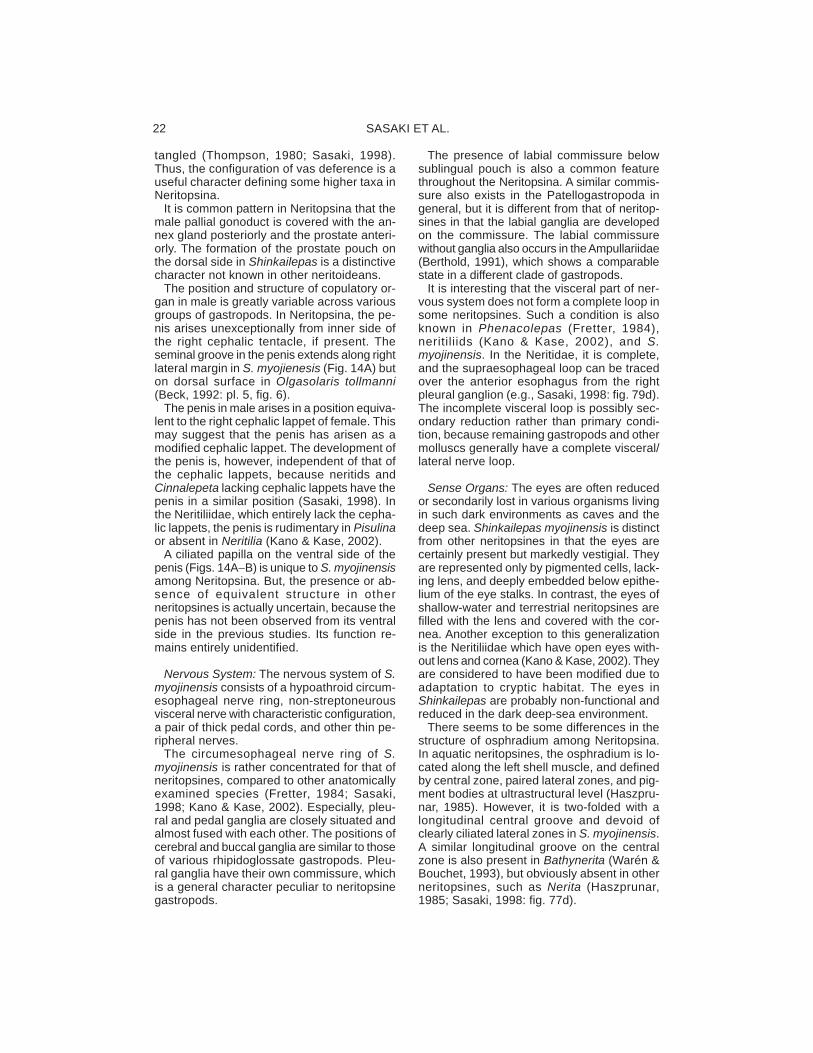
The penis arises from inner side of rightcephalic tentacle, dorsoventrally depressed,lobate with a tapered tip (Fig. 1). Its dorsalsurface is smooth, the right margin is groovedthroughout, and the ventral side has a singleciliated papilla (Figs. 14A, B). A ciliated groovewas not found between the gonoduct openingand the penis.
Nervous System
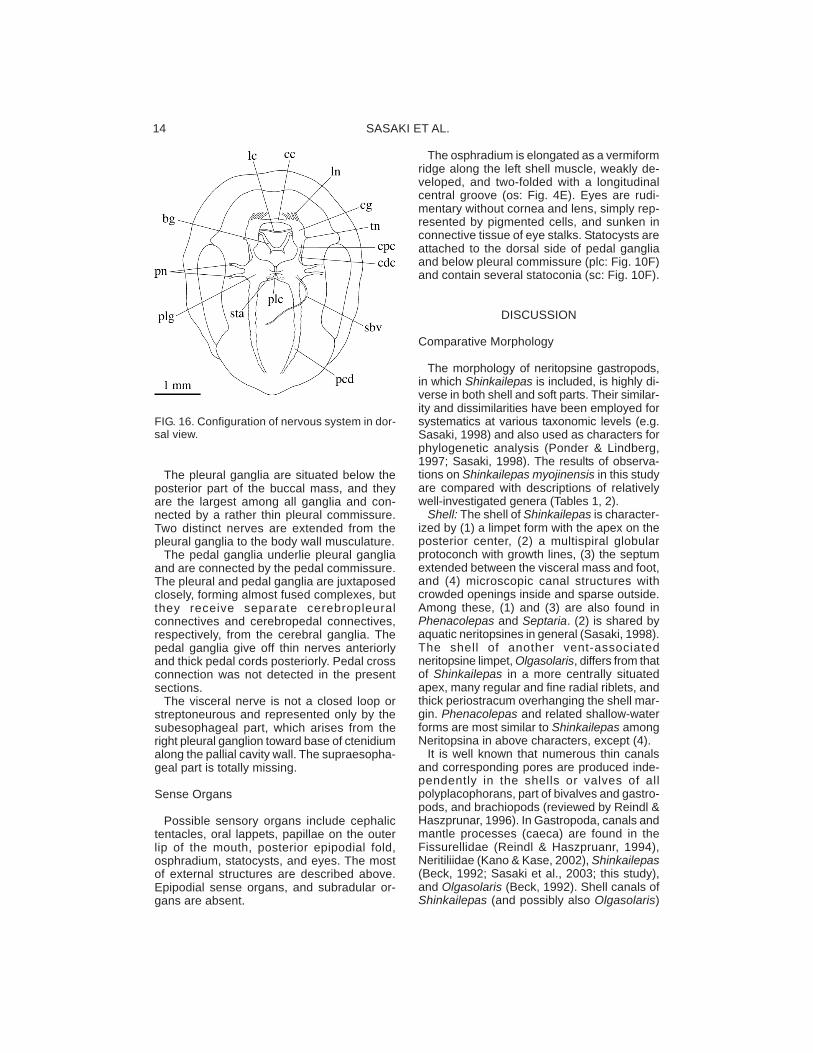
The nervous system consists mainly of thecircumesophageal nerve ring, pedal cords,and visceral nerve. The circumesopahgealnerve ring is formed by pairs of three majorganglia, namely, pleural, pedal, and cerebralganglia. Their configuration is of hypoathroidtype, namely pleural and pedal ganglia aremore adjacent than cerebral ganglia (Fig. 16).
The cerebral ganglia are located on thebases of the cephalic tentacles and sendnerves to the cephalic tentacles and oralarea. The paired ganglia are connected toeach other by the cerebral commissure overthe oral tube and the labial commissure be-low the sublingual pouch. Labial ganglia arenot formed on the labial commissure. Thebuccal ganglia are obliquely extended at thebase of the anterior esophagus and con-nected to the cerebral ganglia through thinconnectives.
FIG. 14. Scannning electron micrographs of male reproductive organs. A. Ventral view of penis re-moved from head. Arrowhead indicates groove along right margin. B. Enlarged view of ciliated pa-pilla on right ventral side. C. Seminal vesicle in convoluted part of vas deference. D. Spermatozoacontained in seminal vesicle. Part of epithelium of seminal vesicle is removed to show inside. A−D.UMUT RM28652.
ANATOMY OF SHINKAILEPAS 13
FIG. 15. Histology of reproductive organs of female (A−E) and male (F−H). A. Cross section of semi-nal receptacle. B. Longitudinal section of posteroventral part of pallial oviduct. C. Longitudinal sec-tion of pallial oviduct where oviduct (ovd) is connected to lumen of pallial oviduct. D. Longitudinalsection of posterior sac (ps) of vaginal duct and surrounding glands. E. Enlarged view of longitudinalsection of posterior sac (ps) of vaginal duct. F. Vertical section of testis. G. Vetical section of seminalvesicle (sv) containing completed spermatozoa. H. Horizontal section of glands of pallial gonoduct ofmale. A. UMUT RM28648. F−H. UMUT RM28650. B−E. UMUT RM28651.
SASAKI ET AL.14
The pleural ganglia are situated below theposterior part of the buccal mass, and theyare the largest among all ganglia and con-nected by a rather thin pleural commissure.Two distinct nerves are extended from thepleural ganglia to the body wall musculature.
The pedal ganglia underlie pleural gangliaand are connected by the pedal commissure.The pleural and pedal ganglia are juxtaposedclosely, forming almost fused complexes, butthey receive separate cerebropleuralconnectives and cerebropedal connectives,respectively, from the cerebral ganglia. Thepedal ganglia give off thin nerves anteriorlyand thick pedal cords posteriorly. Pedal crossconnection was not detected in the presentsections.
The visceral nerve is not a closed loop orstreptoneurous and represented only by thesubesophageal part, which arises from theright pleural ganglion toward base of ctenidiumalong the pallial cavity wall. The supraesopha-geal part is totally missing.
Sense Organs
Possible sensory organs include cephalictentacles, oral lappets, papillae on the outerlip of the mouth, posterior epipodial fold,osphradium, statocysts, and eyes. The mostof external structures are described above.Epipodial sense organs, and subradular or-gans are absent.
The osphradium is elongated as a vermiformridge along the left shell muscle, weakly de-veloped, and two-folded with a longitudinalcentral groove (os: Fig. 4E). Eyes are rudi-mentary without cornea and lens, simply rep-resented by pigmented cells, and sunken inconnective tissue of eye stalks. Statocysts areattached to the dorsal side of pedal gangliaand below pleural commissure (plc: Fig. 10F)and contain several statoconia (sc: Fig. 10F).
DISCUSSION
Comparative Morphology
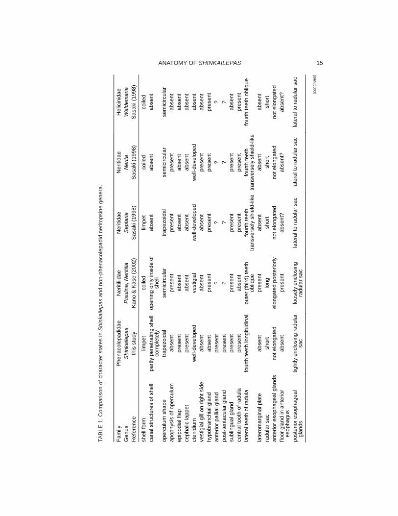
The morphology of neritopsine gastropods,in which Shinkailepas is included, is highly di-verse in both shell and soft parts. Their similar-ity and dissimilarities have been employed forsystematics at various taxonomic levels (e.g.Sasaki, 1998) and also used as characters forphylogenetic analysis (Ponder & Lindberg,1997; Sasaki, 1998). The results of observa-tions on Shinkailepas myojinensis in this studyare compared with descriptions of relativelywell-investigated genera (Tables 1, 2).
Shell: The shell of Shinkailepas is character-ized by (1) a limpet form with the apex on theposterior center, (2) a multispiral globularprotoconch with growth lines, (3) the septumextended between the visceral mass and foot,and (4) microscopic canal structures withcrowded openings inside and sparse outside.Among these, (1) and (3) are also found inPhenacolepas and Septaria. (2) is shared byaquatic neritopsines in general (Sasaki, 1998).The shell of another vent-associatedneritopsine limpet, Olgasolaris, differs from thatof Shinkailepas in a more centrally situatedapex, many regular and fine radial riblets, andthick periostracum overhanging the shell mar-gin. Phenacolepas and related shallow-waterforms are most similar to Shinkailepas amongNeritopsina in above characters, except (4).
It is well known that numerous thin canalsand corresponding pores are produced inde-pendently in the shells or valves of allpolyplacophorans, part of bivalves and gastro-pods, and brachiopods (reviewed by Reindl &Haszprunar, 1996). In Gastropoda, canals andmantle processes (caeca) are found in theFissurellidae (Reindl & Haszpruanr, 1994),Neritiliidae (Kano & Kase, 2002), Shinkailepas(Beck, 1992; Sasaki et al., 2003; this study),and Olgasolaris (Beck, 1992). Shell canals ofShinkailepas (and possibly also Olgasolaris)
FIG. 16. Configuration of nervous system in dor-sal view.
ANATOMY OF SHINKAILEPAS 15
Fam
ily
Phe
naco
lepa
dida
e N
eriti
liidae
N
eriti
dae
Ner
itida
e H
elic
inid
ae
Gen
us
Shi
nkai
lepa
s P
isul
ina,
Ner
itilia
S
epta
ria
Ner
ita
Wal
dem
aria
R
efer
ence
th
is s
tudy
K
ano
& K
ase
(200
2)
Sas
aki (
1998
) S
asak
i (19
98)
Sas
aki (
1998
)
shel
l for
m
limpe
t co
iled
limpe
t co
iled
coile
d ca
nal s
truct
ures
of s
hell
partl
y pe
netra
ting
shel
l co
mpl
etel
y op
enin
g on
ly in
side
of
shel
l ab
sent
ab
sent
ab
sent
oper
culu
m s
hape
tra
pezo
idal
se
mic
ircul
ar
trape
zoid
al
sem
icirc
ular
se
mic
ircul
ar
apop
hysi
s of
ope
rcul
um
abse
nt
pres
ent
pres
ent
pres
ent
abse
nt
epip
odia
l fla
p pr
esen
t ab
sent
ab
sent
ab
sent
ab
sent
ce
phal
ic la
ppet
pr
esen
t ab
sent
ab
sent
ab
sent
ab
sent
ct
enid
ium
w
ell-d
evel
oped
ve
stig
ial
wel
l-dev
elop
ed
wel
l-dev
elop
ed
abse
nt
vest
igia
l gill
on
right
sid
e ab
sent
ab
sent
ab
sent
pr
esen
t ab
sent
hy
pobr
anch
ial g
land
ab
sent
pr
esen
t pr
esen
t pr
esen
t pr
esen
t an
terio
r pal
lial g
land
pr
esen
t ?
? ?
? po
st-te
ntac
ular
gla
nd
pres
ent
? ?
? ?
subl
ingu
al g
land
pr
esen
t pr
esen
t pr
esen
t pr
esen
t ab
sent
ce
ntra
l too
th o
f rad
ula
pres
ent
abse
nt
pres
ent
pres
ent
pres
ent
late
ral t
eeth
of r
adul
a fo
urth
teet
h lo
ngitu
dina
l ou
ter (
third
) tee
th
obliq
ue
four
th te
eth
trans
vers
ely
shie
ld-li
ke
four
th te
eth
trans
vers
ely
shie
ld-li
ke
four
th te
eth
obliq
ue
late
rom
argi
nal p
late
ab
sent
pr
esen
t ab
sent
ab
sent
ab
sent
ra
dula
r sac
sh
ort
long
sh
ort
shor
t sh
ort
ante
rior e
soph
agea
l gla
nds
not e
long
ated
el
onga
ted
post
erio
rly
not e
long
ated
no
t elo
ngat
ed
not e
long
ated
flo
or g
land
in a
nter
ior
esop
hagu
s ab
sent
pr
esen
t ab
sent
? ab
sent
? ab
sent
?
post
erio
r eso
phag
eal
glan
ds
tight
ly e
nclo
sing
radu
lar
sac
loos
ely
encl
osin
g ra
dula
r sac
la
tera
l to
radu
lar s
ac
late
ral t
o ra
dula
r sac
la
tera
l to
radu
lar s
ac
TAB
LE 1
. Com
paris
on o
f cha
ract
er s
tate
s in
Shi
nkai
lepa
s an
d no
n-ph
enac
olep
adid
ner
itops
ine
gene
ra.
(con
tinue
s)

SASAKI ET AL.16
(con
tinue
d)
Fam
ily
Phe
naco
lepa
dida
e N
eriti
liidae
N
eriti
dae
Ner
itida
e H
elic
inid
ae
Gen
us
Shi
nkai
lepa
s P
isul
ina,
Ner
itilia
S
epta
ria
Ner
ita
Wal
dem
aria
R
efer
ence
th
is s
tudy
K
ano
& K
ase
(200
2)
Sas
aki (
1998
) S
asak
i (19
98)
Sas
aki (
1998
)
open
ing(
s) b
etw
een
stom
- ac
h an
d di
gest
ive
glan
ds
paire
d si
ngle
pa
ired
paire
d pa
ired
glan
dula
r and
non
- gl
andu
lar r
egio
ns o
f ki
dney
undi
ffere
ntia
ted
undi
ffere
ntia
ted
diffe
rent
iate
d di
ffere
ntia
ted
diffe
rent
iate
d
right
aur
icle
pr
esen
t pr
esen
t pr
esen
t pr
esen
t ab
sent
er
ythr
ocyt
es
poss
ibly
pre
sent
ab
sent
ab
sent
ab
sent
ab
sent
fe
mal
e re
prod
uctiv
e op
enin
gs
diau
lic
diau
lic
triau
lic
diau
lic
diau
lic
vagi
nal o
peni
ng
slit-
like,
ant
erio
r sl
it-lik
e, p
oste
rior
smal
l por
e, a
nter
ior
smal
l por
e, a
nter
ior
smal
l por
e, p
oste
rior
rece
ptac
ulum
sem
inis
po
ster
ior t
o va
gina
l op
enin
g pa
rt of
spe
rmat
opho
re
sac
conn
ecte
d to
dor
sal
albu
men
gla
nd
conn
ecte
d to
dor
sal
albu
men
gla
nd
abse
nt?
post
erio
r sac
of v
agin
al
duct
pr
esen
t ab
sent
ab
sent
ab
sent
ab
sent
crys
tal s
ac in
fem
ale
abse
nt
abse
nt
pres
ent
pres
ent
abse
nt
neck
furro
w in
fem
ale
abse
nt
pres
ent
abse
nt
abse
nt
abse
nt
fem
ale
flap
abse
nt
pres
ent
abse
nt
abse
nt
abse
nt
sem
inal
ves
icle
en
tang
led
doub
le, u
ncoi
led
enta
ngle
d en
tang
led
sim
ple
ceph
alic
pen
is
pres
ent
rudi
men
tary
or a
bsen
t pr
esen
t pr
esen
t ab
sent
se
min
al g
roov
e on
pen
is
late
ral
? ?
? -
visc
eral
ner
ve lo
op
inco
mpl
ete
inco
mpl
ete
com
plet
e co
mpl
ete
com
plet
e?
eyes
ve
stig
ial,
clos
ed
vest
igia
l, op
en
wel
l-dev
elop
ed, c
lose
d w
ell-d
evel
oped
, clo
sed
wel
l-dev
elop
ed, c
lose
dos
phra
dium
pr
esen
t pr
esen
t pr
esen
t pr
esen
t ab
sent
ce
ntra
l zon
e of
osp
hrad
ium
lo
ngitu
dina
lly g
roov
ed
?
?
sim
ple
- st
atoc
yst p
ositi
on re
lativ
e to
ped
al g
angl
ia
post
erod
orsa
l an
terio
r po
ster
odor
sal
post
erod
orsa
l po
ster
odor
sal
stat
ocys
t con
tent
st
atoc
onia
st
atol
ith
? ?
?
ANATOMY OF SHINKAILEPAS 17
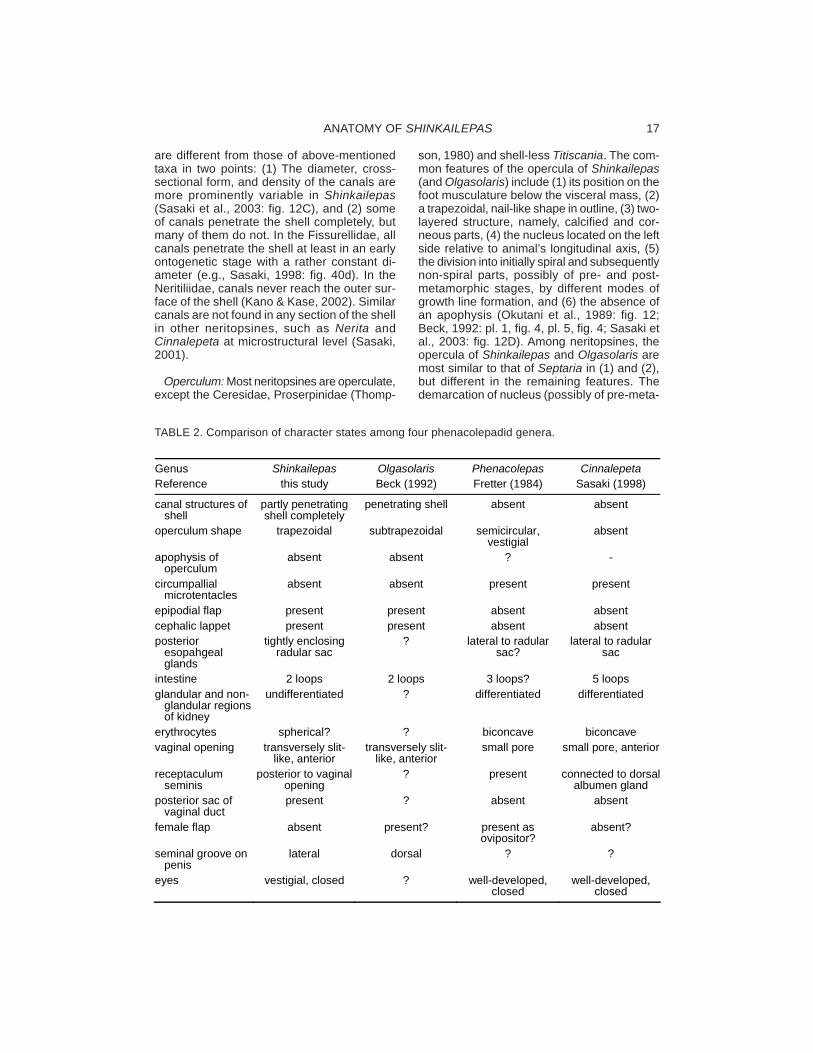
are different from those of above-mentionedtaxa in two points: (1) The diameter, cross-sectional form, and density of the canals aremore prominently variable in Shinkailepas(Sasaki et al., 2003: fig. 12C), and (2) someof canals penetrate the shell completely, butmany of them do not. In the Fissurellidae, allcanals penetrate the shell at least in an earlyontogenetic stage with a rather constant di-ameter (e.g., Sasaki, 1998: fig. 40d). In theNeritiliidae, canals never reach the outer sur-face of the shell (Kano & Kase, 2002). Similarcanals are not found in any section of the shellin other neritopsines, such as Nerita andCinnalepeta at microstructural level (Sasaki,2001).
Operculum: Most neritopsines are operculate,except the Ceresidae, Proserpinidae (Thomp-
son, 1980) and shell-less Titiscania. The com-mon features of the opercula of Shinkailepas(and Olgasolaris) include (1) its position on thefoot musculature below the visceral mass, (2)a trapezoidal, nail-like shape in outline, (3) two-layered structure, namely, calcified and cor-neous parts, (4) the nucleus located on the leftside relative to animal’s longitudinal axis, (5)the division into initially spiral and subsequentlynon-spiral parts, possibly of pre- and post-metamorphic stages, by different modes ofgrowth line formation, and (6) the absence ofan apophysis (Okutani et al., 1989: fig. 12;Beck, 1992: pl. 1, fig. 4, pl. 5, fig. 4; Sasaki etal., 2003: fig. 12D). Among neritopsines, theopercula of Shinkailepas and Olgasolaris aremost similar to that of Septaria in (1) and (2),but different in the remaining features. Thedemarcation of nucleus (possibly of pre-meta-
Genus Shinkailepas Olgasolaris Phenacolepas Cinnalepeta Reference this study Beck (1992) Fretter (1984) Sasaki (1998)
canal structures of shell
partly penetrating shell completely
penetrating shell absent absent
operculum shape trapezoidal subtrapezoidal semicircular, vestigial
absent
apophysis of operculum
absent absent ? -
circumpallial microtentacles
absent absent present present
epipodial flap present present absent absent cephalic lappet present present absent absent posterior
esopahgeal glands
tightly enclosing radular sac
? lateral to radular sac?
lateral to radular sac
intestine 2 loops 2 loops 3 loops? 5 loops glandular and non-
glandular regions of kidney
undifferentiated ? differentiated differentiated
erythrocytes spherical? ? biconcave biconcave vaginal opening transversely slit-
like, anterior transversely slit-
like, anterior small pore small pore, anterior
receptaculum seminis
posterior to vaginal opening
? present connected to dorsal albumen gland
posterior sac of vaginal duct
present ? absent absent
female flap absent present? present as ovipositor?
absent?
seminal groove on penis
lateral dorsal ? ?
eyes vestigial, closed ? well-developed, closed
well-developed, closed
TABLE 2. Comparison of character states among four phenacolepadid genera.

SASAKI ET AL.18
morphic part) is also known in the Neritiliidae(Kano & Kase, 2000, 2003, in press; Kano etal., 2003), Bathynerita (Warén & Bouchet,1993), and Phenacolepas (Kimura & Kimura,1999: fig. 7C−D), and therefore it is not char-acteristic of Shinakailepas and Olgasoralis.The possession of apophysis in the opercu-lum is rather common throughout neritopsinesexcept the Helicinidae, Shinkailepas, andOlgasolaris (cf. Sasaki, 2001). The number ofmicrostructual layers varies from two to fourin neritopsine opercula (Suzuki et al., 1991;Sasaki, 2001), but its phylogenetic or adap-tive significance is uncertain.
External Anatomy: The mantle margin isnormally simple without projective or massiveglandular structure in neritopsines, but it ismodified in shallow-water phenacolepadids.The inner fold of the mantle margin is charac-teristically fringed with retractile microtentaclesin Phenacolepas and Cinnalepeta (Fretter,1984: fig. 5; Sasaki, 1998: fig. 85b). In addi-tion, a thick layer of glandular tissue is devel-oped on ventral surface in Phenacolepas(Fretter, 1984: fig. 5). But, in contrast, such aspecialization does not occur in Shinkailepas.The mantle margin morphology is, therefore,a distinctive character between shallow-wa-ter and deep-water phenacolepadids.
Various forms of folds or tentacles are de-veloped in gastropod epipodium as in vetig-astropods, cocculinifom limpets, “vent-taxa,”deep-sea phenacolepadids, and part ofcerithioideans (Ponder & Lindberg, 1997). InShinkailepas and Olgasolaris, the epipodialfold arises from the latero- to postero-dorsalpart of the foot in Shinkailepas (Okutani et al.,1989; Beck, 1992; Warén & Bouchet, 2001:fig. 33; this study), whereas comparable struc-ture is totally absent in other neritospines in-cluding shallow-water phenacolepadids(Tables 1, 2). Because the degree of epipodialfold development is different from species tospecies, it is a useful taxonomic character atspecies level in Shinkailepas (see below).
The cephalic lappets are small flaps projectedon the inner side of the cephalic tentacles. Theyare found in part of the vetigastropods andneritopsines and may be all regarded as ho-mologue in terms of position and innervation.It is a common feature that the lappets areenlarged and used as a penis in male inneritopsines if present. The cephalic lappetsare present in Shinkailepas (Fig. 1; Okutani etal., 1989: fig. 10), Olgasolaris, and Bathynerita(Warén & Bouchet, 2001), but absent in the
Neritiliidae, shallow-water phenacolepadids,and the Neritidae excluding Bathynerita(Table 1).
Pallial Organs: The pallial cavity of Shin-kailepas contains a set of pallial organs com-mon to the aquatic Neritopsina, namely thectenidium, osphradium, anus, pallial gonod-uct with genital opening(s), and excretory pore,but a hypobranchial gland is absent.
The ctenidium of S. myojinensis has typicalneritopsine elements: (1) a single ctenidiumis situated on the left side, (2) ctenidial lamel-lae are bipectinate in opposing arrangementon either side of ctenidial axis, (3) ctenidiallamellae are centrally ridged, (4) skeletal rodsand (5) sensory pockets are absent. Thesefeatures are not distinguishable from those ofother neritopsines, except for a greatly re-duced ctenidium of the Neritiliidae (Kano &Kase, 2002) and its total absence of in terres-trial groups. A wart-like structure termed “ves-tigial gill” on the right side of the pallial cavity(Fretter, 1965: fig. 1c; Sasaki, 1998: fig. 77c)is lacking in S. myojinensis. The occurrenceof the structure is restricted to members of thegenus Nerita and not universal to neritopsines.
The hypobranchial gland is present on theright pallial roof in some neritopsines, for ex-ample, the Neritidae (Fretter, 1965; Berry etal., 1973; Sasaki, 1998), Neritiliidae (Kano &Kase, 2002), and Helicinidae (Thompson,1980; Sasaki, 1998). However, it is absent incorresponding position in S. myojinensis,Phenacolepas (Fretter, 1984), and Cinnale-peta (Sasaki, 1998).
In S. myojinensis, two glandular zones (thepost-tentacular gland and anterior pallial gland)are observed on the right anterior corner ofthe pallial cavity. These glands may be analo-gous to the hypobranchial gland of other gas-tropods, but probably do not fulfill the functionas a normal hypobranchial gland due to theirrestricted position at the anterior right. The truehypobranchial gland develops in deeper posi-tion in the pallial cavity in other neritoideans.The two glandular zones in S. myojinensispossibly have a function related to reproduc-tion, since the glands are developed near thegonoduct opening in both sexes.
Digestive System: Its has been generallyaccepted that neritopsines lack true jaws(Fretter, 1965). However, paired cuticularizedplates apparently develop on the inner wall ofthe oral tube in S. myojinensis (Fig. 9A). Iden-tical plates are also present generally in other
ANATOMY OF SHINKAILEPAS 19
neritopsines. Sasaki (1998) described theseplates as jaws, because they are located at aposition corresponding to those of other gas-tropods. They may not be regarded as the jawsin that they lack scaly microelements, but suchmicroelements are also lacking in the jaws ofCocculina and Patellogastropods (Sasaki,1998). Although the term “jaw” was not usedin the present description, it is also possiblethat they represent a reduced state of the jaws.Homology can be established between jawsand cuticularized plates under positional cri-terion but is uncertain under structural crite-rion. More extensive comparison should bemade among gastropod jaws for further dis-cussion.
Buccal mass structure is remarkably usefulcharacter to define higher taxa in basal groupsof gastropods (Sasaki, 1998). For example,patellogastropods, vetigastropods, andneritopsines each have their own compositionof musculature and cartilages. The buccalmusculature of Shinkailepas belongs toneritoidean type described for Nerita, Septaria,and Cinnalepeta, and is partially different fromthat of Waldemaria (Sasaki, 1998: table 5).
The configuration of odontophoral cartilagesis invariable throughout the Neritopsina(Sasaki, 1998; Kano & Kase, 2002). Largeanterior and small posterior cartilages arepaired and connected by ventral approximatormuscle, and the median cartilage is situatedbetween the anterior cartilages. The structureof odontophoral cartilages of S. myojienensisconforms entirely to this pattern.
The presence of a pair of sublingual glandsbeside the sublingual pouches is a distinctivecharacter of neritoideans, including theNeritidae, Neritiliidae, and Phenacolepadidae(Sasaki, 1998: Kano & Kase, 2002). They areabsent in the Helicinidae (Sasaki, 1998) orunknown in the rest of neritopsine members.The salivary glands are absent throughout theNeritopsina without exception. Probably theirabsence is compensated by the developmentof sublingual glands and anterior esophagealglands.
The radular formula of Shinkailepas is thesame as that of most members of neritoideans.The radula of Shinkailepas and that ofOlgasolaris (Beck, 1992) are typified by thefollowing six features: (1) the central tooth ispresent, (2) the first lateral teeth are obliquelyelongate, (3) the second and third lateral teethare small, (4) the fourth lateral teeth are longi-tudinally elongate without serration in theircusps, (5) lateromarginal plates are absent,
and (6) the marginal teeth have small triangu-lar projection below their cusps (Sasaki et al.,2003: figs. 12, 14).
Major differences in radular characters existmainly in the central and lateral teeth amongNeritopsina. In contrast to most neritopsines,including Shinkailepas, the central tooth isabsent in the Neritiliidae (Kano & Kase, 2002,2003, in press; Kano et al., 2003), Neritopsis(Warén & Bouchet, 1993: fig. 3D), andTitiscania (Bergh, 1890; Taki, 1955). The lat-eral teeth morphology is considerably variableamong neritopsines and it is difficult to gener-alize. For example, in the Neritidae, the firstlaterals are transversely elongate, and thefourth laterals are thickened in shield-like form.The Neritiliidae (Kano & Kase, 2002, 2003, inpress; Kano et al., 2003) and most helicinoide-ans (e.g., Thompson, 1980; Sasaki, 1998;Richling, 2004) have obliquely elongate out-ermost lateral teeth with sharp serrations, andlateral teeth are more reduced in Neritopsis,Titiscania, and the Hydrocenidae (Ponder,1998: fig. 15.76). The presence of prominencebelow cusps in the marginal teeth (Kano &Kase, 2002: fig. 8) is known in the Neritiliidae,Neritopsis, and Titiscania (Kano & Kase, 2000,2002), but not in others.
The configuration of the radular sac has nothitherto been focused in the studies of theNeritopsina. Recently, Kano & Kase (2002)pointed out that in the Neritopsina the lengthof radular sac can be categorized into twogroups. A long, looped radular sac is typical ofthe Neritiliidae, Neritopsis and Titiscania; bycontrast, a short straight type occurs in theNeritidae, Phenacolepadidae, and Helicinidae(Kano & Kase, 2002). Shinkailepas myojinen-sis (Fig. 5), S. kaikatensis (Okutani et al., 1989:fig. 15), and Olgasolaris tollmanni (Beck, 1992:fig. 3B) have the latter type.
The esophagus of the Neritopsina exhibitssimilar structure throughout the group (Sasaki,1998): (1) It consists of the anterior and pos-terior esophagi only, lacking the differentiationof a mid-esophagus, (2) the anterior esopha-gus is sectioned into a centrally situated, dor-sal food channel and lateral esophagealpouches with the anterior esophageal glandsinside, and (3) the posterior part of the lateralesophageal pouches are separated as theposterior esophageal glands. The esophagusof S. myojinensis matches this generalizationwell.
A peculiar shaped esophagus was recentlydescribed in the Neritiliidae by Kano & Kase(2002). The anterior esophageal glands are
SASAKI ET AL.20
extremely elongated posteriorly as separatedpouches and overlie the posterior esophagealglands. This double structure of esophagealglands is not known in other neritopsines in-cluding Shinkailepas.
The posterior esophageal glands in S.myojinensis are complicatedly infolded andtightly enclose the radular sac with a narrowinterstitial space (Fig. 9F). This morphologyapparently differs from that of otherneritopsines, including the Neritiliidae (Kano& Kase, 2002: fig. 5D). Hence, the structureof this part may be of sufficient systematicvalue, but cross-sectional morphology of theglands have not been described for compari-son in the rest of neritopsines.
Another distinctive character of theNeritiliidae is the presence of the floor glandsarising from anterior esophageal floor (Kano& Kase, 2002). The glands are paired blindsacs that open from the posterior side of thefloor of the anterior esophagus and consist oftwo glandular cells ciliated differently andstained differentially with haematoxylin (Kano& Kase, 2002: fig. 3C). The identical glandswere not found in sections of equivalent posi-tion in S. myojinensis.
The stomach of the Neritopsina consists ofa cuticularized area, gastric shield with a shortreflected tooth, paired (major and minor)typholosoles on the ventral side, a ciliated in-testinal groove between the typhlosoles, anda short gastric caecum (Fretter, 1965; Sasaki,1998; Kano & Kase, 2002). Differences in thestomach structure among the Neritopsina isnot very conspicuous. The Neritiliidae haveonly a single connection between stomach anddigestive glands (Kano & Kase, 2002),whereas two or more openings of digestiveglands occur near sorting area in the stomachof other neritopsines (e.g., Bourne, 1909,1911; Fretter, 1965, 1984; Sasaki 1998). It isuncertain at present whether the number ofthe openings is dependent on body size orphylogentetically fixed.
Excretory System: Neritopsine excretorysystem is composed of the auricle withpodocytes as an ultrafiltration site, renoperi-cardial duct as a conduit of primary urine, anda single left kidney for osmoregulation andexcretion (Estabrooks et al., 1999). Within theNeritopsina, two types of kidneys are knownto date: (1) In the Neritidae, Phenacolepa-didae, and Helicinidae, the kidney is composedof glandular region, non-glandular bladder, andshort ureter (Little, 1972; Sasaki, 1998; Esta-
brooks et al., 1999). (2) In the Neritiliidae, thewall of kidney is simple, not specialized intoglandular and non-glandular areas (Kano &Kase, 2002). The kidney of S. myojinensisbelongs to the latter type. Functional differ-ences of these two types are not clear, thoughdevelopment of infoldings in the glandular areaof the former type is obviously related to func-tional advantage to increase its surface area.
Circulatory System: In the heart of gastro-pods the ventricle is always single, but it isattached by paired or unpaired auricle(s), de-pending on taxa. In S. myojinensis, the peri-cardium contains two (right and left) auriclesand a single median ventricle. The right au-ricle is obviously present in S. myojinensis butgreatly reduced. A vestigial auricle is alsopresent in Cinnalepeta (Sasaki, 1998) andPhenacolepas (Fretter, 1984). In otherneritopsines, the right auricle is present in theTitiscanidae and Cerisidae of the Helicinidaebut absent in the Hydrocenidae, Proserpi-ninae, and Helcinidae (Sasaki, 1998). Theventricle is penetrated by the rectum in theNeritopsina, except the Hydrocenidae andHelicinidae (Sasaki, 1998).
Although details have not been studiedhematologically, the presence of erythrocytesis a possible general feature of phenacole-padids. They are discoidal and biconcave inPhenacolepas (Fretter, 1984) and Cinnalepeta(Sasaki, 1988: fig. 85d). The animals of thesetwo genera are red in fresh live condition, butimmediatedly turned pale after the death byfixation. In Shinkailepas aff. kaikatensis fromthe Mariana Back-arc Basin, the red color isalso very vivid only while living (Hasegawa etal., 1997). Hence, species of Shinkailepas arepresumed to have erythrocytes. The form ofhaemocytes in S. myojinensis, however, do notseem discoidal but spherical (Fig. 10B).
Female Reproductive System: The femaleorgans of neritopsines consist mainly of theovary, oviduct, pallial oviduct with albumen andcapsule glands, and vaginal duct with two sac-like appendages.
It is well known that the number of repro-ductive opening(s) is different in female amongneritopsine taxa. In S. myojinensis, pallial ovi-duct and vaginal duct have their own open-ings (diaulic). In contrast, female reproductivesystem is triaulic with an additional enigmaticduct in Septaria (Sasaki, 1998) and monaulicin Titiscania (Marcus & Marcus, 1967) (neri-tiliids do not have a monaulic system as pre-
ANATOMY OF SHINKAILEPAS 21
viously believed (Kano & Kase, 2002). A diaulicreproductive system is most common amongNeritopsina.
Separation of the vaginal duct from the pal-lial oviduct is a common feature in mostneritopsines, and it is also true of S. myojinen-sis. Characteristically, the vaginal duct in S.myojinensis is associated with three struc-tures: (1) a transverse slit of the vaginal open-ing below the anterior part of pallial oviduct,(2) the receptaculum seminis near the vaginalopening, and (3) the “posterior sac” below theposterior part of the pallial oviduct.
A slit-like vaginal opening in the anterior po-sition is also described in Olgasolaris (Beck,1992). In the Neritiliidae, the vaginal openingis also a slit but located posteriorly (Kano &Kase, 2002). It is a small pore, not a long slit,in Phenacolepas (Fretter, 1984), Cinnalepeta,the Neritidae, and Helicinidae (Sasaki, 1998).
An anteriorly situated receptaculum seminisnear the vaginal opening in S. myojinensis isunique among the Neritopsina. In othergroups, receptaculum seminis has been iden-tified in various positions (e.g., Sasaki, 1998),but it has not always been verified on histo-logical basis. The presence of oriented sper-matozoa in its epithelium (Fig. 15A) is the mostreliable criterion for this identification.
The “posterior sac” of the vaginal duct mayalso be peculiar to S. myojinensis. Its innerwall is heavily folded characteristically. Be-cause no sperm or egg was contained in sec-tioned specimens, its actual function inreproduction could not be detected. An equiva-lent structure is unknown in Olgasolaris (Beck,1992) and Phenacolepas (Fretter, 1984), orlacking in Cinnalepeta (Sasaki, 1998: fig. 84).The spermatophore sac in the Neritidae andCinnalepeta (Sasaki, 1998: figs. 76, 84) can-not be homologized due to the differences intopological relationships with other reproduc-tive organs.
It is uncertain whether S. myojinensis pro-duces spermatophores or not. The spermato-phores are generally known to occur inneritopsine gastropods (Robertson, 1989, re-viewed gastropod spermatophores). In theNeritidae, intact spermatophores are oftencontained in the spermatophore sac in female(Sasaki, 1998: figs. 76c−d), and the formationof the spermatophore sheath is also observ-able in the seminal vesicle of the male. In thiscase, the formation of spermatozoa is un-doubted. But in S. myojinensis, no spermato-phore was found in any section of female andmale reproductive systems.
The pallial oviduct of S. myojinensis is en-closed by two kinds of glands that correspondto the albumen and capsule glands, as is gen-erally found in gastropods that produce eggcapsules. The albumen gland is further divis-ible into two parts in S. myojienesis. The simi-lar division is also known in the Neritiliidae,Neritidae, and shallow-water phenacolepadids(Sasaki, 1998; Kano & Kase, 2002), but not innon-neritopsine gastropods.
Some neritopsines are known to have a min-eral-containing “crystal sac” and cover eggcapsule with minerals from the sac. The ab-sence of the crystal sac was verified in S.moyojinensis in this study and in the Neritiliidaeby Kano & Kase (2002). Meanwhile, the sacis distended with calcified grains in theNeritidae (Sasaki, 1998: fig. 77h). Marcus &Marcus (1967) reported the crystal sac inTitiscania, but it is questionable (Kano & Kase,2002). Probably the possession of the crystalsac is restricted to the family Neritidae.
Internally fertilizing gastropods may developa particular structure conveying eggs from thefemale opening to the foot through the neckregion. The Neritiliidae have the neck furrowand female flap on the right side in the female(Kano & Kase, 2002: figs. 2B, 15B), and pre-sumably eggs are conveyed along a ciliatedfurrow in oviposition. The female flap inNeritiliidae is possibly homologous to the struc-ture identified as the ovipositor in Phena-colepas by Fretter (1984) (Kano & Kase, 2002).In S. myojinensis, a corresponding structurewas not found in the right pedal region. Be-cause actual behavior of egg deposition hasnever been observed in phenacolepadids,functional significance of right neck-foot mor-phology is unclear.
Male Reproductive System: Male reproduc-tive organs of neritopsines generally comprisethe testis, vas deference, seminal vesicle,pallial male gonoduct with prostate, and pe-nis. All of these organs represent a commonelement of the male reproductive system pos-sessed universally by internally fertilizing gas-tropods.
The seminal vesicle is formed in convolutedpart of vas deference in some Neritopsina, forexample, Neritidae including Bathynerita(Warén & Bouchet, 1993), shallow-waterphenacolepadids (Sasaki, 1998), andShinkailepas. By contrast, in the Neritiliidae,the seminal vesicle is double and differentfrom tightly convoluted type (Kano & Kase,2002). In the Helicinidae, it is simple, not en-
SASAKI ET AL.22
tangled (Thompson, 1980; Sasaki, 1998).Thus, the configuration of vas deference is auseful character defining some higher taxa inNeritopsina.
It is common pattern in Neritopsina that themale pallial gonoduct is covered with the an-nex gland posteriorly and the prostate anteri-orly. The formation of the prostate pouch onthe dorsal side in Shinkailepas is a distinctivecharacter not known in other neritoideans.
The position and structure of copulatory or-gan in male is greatly variable across variousgroups of gastropods. In Neritopsina, the pe-nis arises unexceptionally from inner side ofthe right cephalic tentacle, if present. Theseminal groove in the penis extends along rightlateral margin in S. myojienesis (Fig. 14A) buton dorsal surface in Olgasolaris tollmanni(Beck, 1992: pl. 5, fig. 6).
The penis in male arises in a position equiva-lent to the right cephalic lappet of female. Thismay suggest that the penis has arisen as amodified cephalic lappet. The development ofthe penis is, however, independent of that ofthe cephalic lappets, because neritids andCinnalepeta lacking cephalic lappets have thepenis in a similar position (Sasaki, 1998). Inthe Neritiliidae, which entirely lack the cepha-lic lappets, the penis is rudimentary in Pisulinaor absent in Neritilia (Kano & Kase, 2002).
A ciliated papilla on the ventral side of thepenis (Figs. 14A−B) is unique to S. myojinensisamong Neritopsina. But, the presence or ab-sence of equivalent structure in otherneritopsines is actually uncertain, because thepenis has not been observed from its ventralside in the previous studies. Its function re-mains entirely unidentified.
Nervous System: The nervous system of S.myojinensis consists of a hypoathroid circum-esophageal nerve ring, non-streptoneurousvisceral nerve with characteristic configuration,a pair of thick pedal cords, and other thin pe-ripheral nerves.
The circumesophageal nerve ring of S.myojinensis is rather concentrated for that ofneritopsines, compared to other anatomicallyexamined species (Fretter, 1984; Sasaki,1998; Kano & Kase, 2002). Especially, pleu-ral and pedal ganglia are closely situated andalmost fused with each other. The positions ofcerebral and buccal ganglia are similar to thoseof various rhipidoglossate gastropods. Pleu-ral ganglia have their own commissure, whichis a general character peculiar to neritopsinegastropods.
The presence of labial commissure belowsublingual pouch is also a common featurethroughout the Neritopsina. A similar commis-sure also exists in the Patellogastropoda ingeneral, but it is different from that of neritop-sines in that the labial ganglia are developedon the commissure. The labial commissurewithout ganglia also occurs in the Ampullariidae(Berthold, 1991), which shows a comparablestate in a different clade of gastropods.
It is interesting that the visceral part of ner-vous system does not form a complete loop insome neritopsines. Such a condition is alsoknown in Phenacolepas (Fretter, 1984),neritiliids (Kano & Kase, 2002), and S.myojinensis. In the Neritidae, it is complete,and the supraesophageal loop can be tracedover the anterior esophagus from the rightpleural ganglion (e.g., Sasaki, 1998: fig. 79d).The incomplete visceral loop is possibly sec-ondary reduction rather than primary condi-tion, because remaining gastropods and othermolluscs generally have a complete visceral/lateral nerve loop.
Sense Organs: The eyes are often reducedor secondarily lost in various organisms livingin such dark environments as caves and thedeep sea. Shinkailepas myojinensis is distinctfrom other neritopsines in that the eyes arecertainly present but markedly vestigial. Theyare represented only by pigmented cells, lack-ing lens, and deeply embedded below epithe-lium of the eye stalks. In contrast, the eyes ofshallow-water and terrestrial neritopsines arefilled with the lens and covered with the cor-nea. Another exception to this generalizationis the Neritiliidae which have open eyes with-out lens and cornea (Kano & Kase, 2002). Theyare considered to have been modified due toadaptation to cryptic habitat. The eyes inShinkailepas are probably non-functional andreduced in the dark deep-sea environment.
There seems to be some differences in thestructure of osphradium among Neritopsina.In aquatic neritopsines, the osphradium is lo-cated along the left shell muscle, and definedby central zone, paired lateral zones, and pig-ment bodies at ultrastructural level (Haszpru-nar, 1985). However, it is two-folded with alongitudinal central groove and devoid ofclearly ciliated lateral zones in S. myojinensis.A similar longitudinal groove on the centralzone is also present in Bathynerita (Warén &Bouchet, 1993), but obviously absent in otherneritopsines, such as Nerita (Haszprunar,1985; Sasaki, 1998: fig. 77d).
ANATOMY OF SHINKAILEPAS 23
The position of the statocysts is somewhatvariable among neritopsines. They are locatedon the posterodorsal side of the pedal gangliain S. myojinensis, and also the Neritidae(Sasaki, 1998) and Helicinidae (Bourne, 1911).In the Neritiliidae, their position is shifted moreanteriorly (Kano & Kase, 2002).
Each statocyst contains either single statolithor many statoconia, depending on taxa in gas-tropods (Ponder & Lindberg, 1997). Evenwithin Neritopsina, there are both of these twotypes. At least two examples are known: sta-tocysts in the Neritiliidae (Kano & Kase, 2002)and statoconia in S. myojienesis. Their condi-tion in other taxa have not been clearly de-scribed based on histological observations.The systematic and functional significance ofstatocyst contents is still unclear throughoutneritopsines.
Systematic Implications
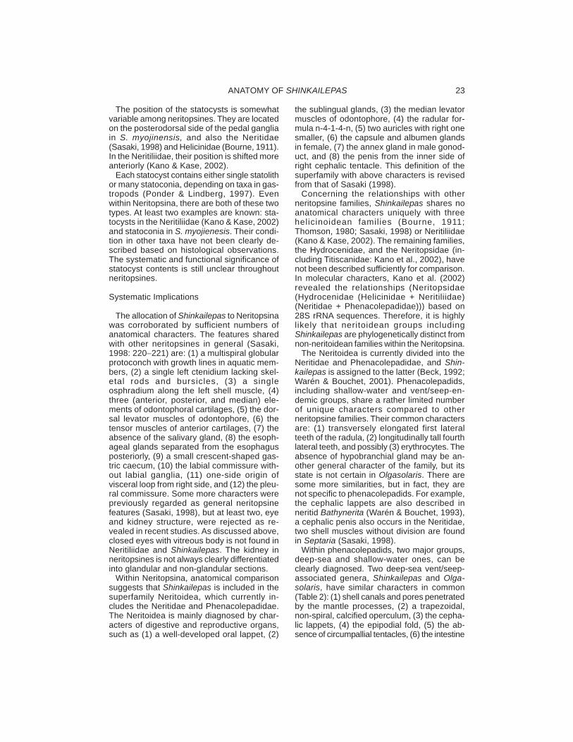
The allocation of Shinkailepas to Neritopsinawas corroborated by sufficient numbers ofanatomical characters. The features sharedwith other neritopsines in general (Sasaki,1998: 220−221) are: (1) a multispiral globularprotoconch with growth lines in aquatic mem-bers, (2) a single left ctenidium lacking skel-etal rods and bursicles, (3) a singleosphradium along the left shell muscle, (4)three (anterior, posterior, and median) ele-ments of odontophoral cartilages, (5) the dor-sal levator muscles of odontophore, (6) thetensor muscles of anterior cartilages, (7) theabsence of the salivary gland, (8) the esoph-ageal glands separated from the esophagusposteriorly, (9) a small crescent-shaped gas-tric caecum, (10) the labial commissure with-out labial ganglia, (11) one-side origin ofvisceral loop from right side, and (12) the pleu-ral commissure. Some more characters werepreviously regarded as general neritopsinefeatures (Sasaki, 1998), but at least two, eyeand kidney structure, were rejected as re-vealed in recent studies. As discussed above,closed eyes with vitreous body is not found inNeritiliidae and Shinkailepas. The kidney inneritopsines is not always clearly differentiatedinto glandular and non-glandular sections.
Within Neritopsina, anatomical comparisonsuggests that Shinkailepas is included in thesuperfamily Neritoidea, which currently in-cludes the Neritidae and Phenacolepadidae.The Neritoidea is mainly diagnosed by char-acters of digestive and reproductive organs,such as (1) a well-developed oral lappet, (2)
the sublingual glands, (3) the median levatormuscles of odontophore, (4) the radular for-mula n-4-1-4-n, (5) two auricles with right onesmaller, (6) the capsule and albumen glandsin female, (7) the annex gland in male gonod-uct, and (8) the penis from the inner side ofright cephalic tentacle. This definition of thesuperfamily with above characters is revisedfrom that of Sasaki (1998).
Concerning the relationships with otherneritopsine families, Shinkailepas shares noanatomical characters uniquely with threehelicinoidean families (Bourne, 1911;Thomson, 1980; Sasaki, 1998) or Neritiliidae(Kano & Kase, 2002). The remaining families,the Hydrocenidae, and the Neritopsidae (in-cluding Titiscanidae: Kano et al., 2002), havenot been described sufficiently for comparison.In molecular characters, Kano et al. (2002)revealed the relationships (Neritopsidae(Hydrocenidae (Helicinidae + Neritiliidae)(Neritidae + Phenacolepadidae))) based on28S rRNA sequences. Therefore, it is highlylikely that neritoidean groups includingShinkailepas are phylogenetically distinct fromnon-neritoidean families within the Neritopsina.
The Neritoidea is currently divided into theNeritidae and Phenacolepadidae, and Shin-kailepas is assigned to the latter (Beck, 1992;Warén & Bouchet, 2001). Phenacolepadids,including shallow-water and vent/seep-en-demic groups, share a rather limited numberof unique characters compared to otherneritopsine families. Their common charactersare: (1) transversely elongated first lateralteeth of the radula, (2) longitudinally tall fourthlateral teeth, and possibly (3) erythrocytes. Theabsence of hypobranchial gland may be an-other general character of the family, but itsstate is not certain in Olgasolaris. There aresome more similarities, but in fact, they arenot specific to phenacolepadids. For example,the cephalic lappets are also described inneritid Bathynerita (Warén & Bouchet, 1993),a cephalic penis also occurs in the Neritidae,two shell muscles without division are foundin Septaria (Sasaki, 1998).
Within phenacolepadids, two major groups,deep-sea and shallow-water ones, can beclearly diagnosed. Two deep-sea vent/seep-associated genera, Shinkailepas and Olga-solaris, have similar characters in common(Table 2): (1) shell canals and pores penetratedby the mantle processes, (2) a trapezoidal,non-spiral, calcified operculum, (3) the cepha-lic lappets, (4) the epipodial fold, (5) the ab-sence of circumpallial tentacles, (6) the intestine
SASAKI ET AL.24
with two loops only, and (7) an anteriorly posi-tioned, slit-like vaginal opening. Most of thesesimilarities are distinctive of these two genera,suggesting their close phylogenetic relation.
In contrast to two deep-sea genera, shallow-water phenacolepadids (Phenacolepas +Cinnalepeta) are united by (1) the absence ofshell pores and mantle processes, (2) a weaklydeveloped, spiral operculum with apophysis, ifpresent, (3) the absence of cephalic lappets,(4) the absence of epipodial folds, (5) the circum-pallial tentacles, (6) complex loops of intestine,and (7) a small pore-like vaginal opening.
At the species level, there are some anatomi-cal differences among four described speciesof Shinkailepas. The comparison with S.myojinensis revealed that in S. briandi Warén& Bouchet, 2001, (1) the eye stalks (“eye-lobes”) are very weakly developed, (2) theepipodial fold in neck region is prominent onthe left and right sides, (3) the penis is moreelongated, and (4) the posterior margin of theepipodial fold is not divided into triangular ten-tacles. Likewise, in S. kaikatensis Okutani, Saito& Hashimoto, 1989, (1) the eye stalks are notdeveloped, (2) the penis is more acutelypointed, and (3) the number of tentacles on theepipodial folds (“pedal papilla”) is smaller (11in S. kaikatensis, 11−19 in S. myojinensis). InS. tufari Beck, 1992, the number of tentacleson epipodial folds is largest (20−22) amongknown species, but other characters have notbeen described in detail. Thus, eye stalks,epipodial folds in neck and posterior pedal re-gions, and penis are useful species-level taxo-nomic characters in the external features of thesoft part. For shell, radular, and opercular char-acters at species level, see Sasaki et al. (2003).
ACKNOWLEDGEMENTS
We are grateful to Prof. George M. Davis(George Washington University Medical Cen-ter), Dr. Eugene Coan (California Academy ofSciences), anonymous reviewers, and Prof.Kazushige Tanabe (University of Tokyo) for in-valuable suggestions for this study and com-ments on the manuscript. The materialexamined in this study was collected in divesdirected by Prof. Toshiyuki Yamaguchi (ChibaUniversity) and Dr. Shinji Tsuchida (JAMSTEC).Sampling operations were kindly supported bycrew of Shinkai 2000 and R/V Natsushima ofJAMSTEC. This study was partly supported bythe Grant-in-Aid from the Japan Society of thePromotion of Science (No. 15740309).
LITERATURE CITED
BECK, L. A., 1992, Two new neritacean limpets(Gastropoda: Prosobranchia: Neritacea:Phenacolepadidae) from active hydrothermalvents at Hydrothermal Field 1 “Wienerwald” inthe Manus Back Arc Basin (Bismarck Sea,Papua New Guinea). Annalen des Naturhisto-rischen Museums, Wien, 93B: 259−275.
BERGH, R., 1890, Die Titiscanien, eine Familieder rhipidoglossen Gastropoden. Morpholo-gisches Jahrbuch, 16: 1−26.
BERRY, A. J., R. LIM & A. S. KUMAR, 1973,Reproductive systems and breeding conditionin Nerita birmanica (Archaeogastropoda:Neritacea) from Malayan mangrove swamps.Journal of Zoology, London, 170: 189−200.
BERTHOLD, T., 1991, Vergleichende Anatomie,Phylogenie und Historische Biogeographie derAmpullariidae. Abhandlungen des Natur-wissenschaftlichen Vereins in Hamburg, (NF)29: 1−256.
BOURNE, G. C., 1909, Contributions to morphol-ogy of the grouop Neritacea of theAspidobranch gastropods. Part I: the Nertidae.Proceedings of the Zoological Society of Lon-don, 1908: 810−887.
BOURNE, G. C., 1911, Contributions to the mor-phology of the group Neritacea of theaspidobranch gastropods. Part II: TheHelicinidae. Proceedings of the ZoologicalSociety of London, 1911: 759−809.
ESTABROOKS, W. A., E. A. KAY & S. A. MCCAR-THY, 1999, Structure of the excretory system ofHawaiian nerites (Gastropoda: Neritoidea).Journal of Molluscan Studies, 65: 61−72.
FRETTER, V., 1965, Functional studies of theanatomy of some neritid prosobranchs. Jour-nal of Zoology, London, 137: 46−74.
FRETTER, V., 1984, The functional anatomy ofthe neritacean limpet Phenacolepas omanen-sis Biggs and some comparison with Septaria.Journal of Molluscan Studies, 50: 8−18.
HASEGAWA, K., K. FUJIKURA & T. OKUTANI,1997, Gastropod fauna associated with hydro-thermal vents in the Mariana Back-Arc Basin:Summary of the results of 1996 “Shinkai 6500”divers. JAMSTEC Journal of Deep Sea Re-search, 13: 69−83.
HASZPRUNAR, G., 1985, The fine morphologyof the osphradial sense organs of the Mollusca.I. Gastropoda, Prosobranchia. PhilosophicalTransactions of the Royal Society of London,B307: 457−496.
KANO, Y., S. CHIBA & T. KASE, 2002, Majoradaptive radiation in neritopsine gastropodsestimated from 28S rRNA sequences and fos-sil records. Proceedings of the Royal Societyof London, B269: 2457−2465.
KANO, Y. & T. KASE, 2000, Taxonomic revisionof Pisulina (Gastropoda: Neritopsina) from sub-marine caves in the tropical Indo-Pacific. Pa-leontological Research, 4: 107−129.
KANO, Y. & T. KASE, 2002, Anatomy and sys-tematics of the submarine-cave gastropodPisulina (Neritopsina: Neritiliidae). Journal ofMolluscan Studies, 68: 365−384.
ANATOMY OF SHINKAILEPAS 25
KANO, Y. & T. KASE, 2003, Systematics of theNeritilia rubida complex (Gastropoda: Neri-tiliidae): three amphidromous species withoverlapping distribution in the Indo-Pacific.Journal of Molluscan Studies, 69: 273−284.
KANO, Y. & T. KASE, in press, Genetic exchangebetween anchialine-cave populations bymeans of larval despersal: the case of a newgastropod species Neritil ia cavernicola .Zoologica Scripta.
KANO, Y., T. KASE & H. KURO, 2003, The uniqueinterstitial habitat of a new neritiliid gastropod,Neritilia littoralis. Journal of Marine BiologicalAssociation of United Kingdom, 83: 835−840.
KIMURA, S. & T. KIMURA, 1999, The gastropodfauna of the marshes of the reed (Phragmitesaustralis (Cav.)) in the estuaries in Mikawa Bayand Ise Bay, Japan. Japanese Journal ofBenthology, 54: 44−56 [in Japanese, with En-glish abstract].
LITTLE, C., 1972, The evolution of kidney func-tion in the Neritacea (Gastropoda, Proso-branchia). Journal of Experimental Biology, 56:249−279.
MARCUS, E. & E. MARCUS, 1967, Americanopisthobranch molluscs. Part 1. Tropical Ameri-can opisthobranchs. Studies in Tropical Ocean-ography, 6: 3−137.
OKUTANI, T., H. SAITO & J. HASHIMOTO, 1989,A new neritacean limpet from a hydrothermalvent site near Ogasawara Islands, Japan. Ve-nus, 48: 223−230.
PONDER, W. F., 1998, Superorder Neritopsina.Pp. 693−702, in: P. L. BEESLEY, G. J. B. ROSS & A.WELLS, eds., Mollusca: the southern systhesis.Fauna of Australia, Volume 5, Part B. CSIRO,Melbourne, Australia. 1234 pp.
PONDER, W. F. & D. R. LINDBERG, 1997, To-wards a phylogeny of gastropod molluscs: ananalysis using morphological characters. Zoo-logical Journal of the Linnean Society, 119: 83−265.
REINDL, S. & G. HASZPRUNAR, 1994, Light andelectron microscopical investigations on shellpores (caeca) of fissurellid limpets (Mollusca:Archaeogastropoda). Journal of Zoology, Lon-don, 233: 385−404.
REINDL, S. & G. HASZPRUNAR, 1996, Shellpores (caea, aesthetes) of Mollusca: a case ofpolyphyly. Pp. 115−118, in: J. D. TAYLOR, ed.,Origin and evolutionary radiation of the Mol-lusca. Oxford University Press, Oxford. 392 pp.
RICHLING, I., 2004, Classification of theHelicinidae: review of morphological charac-teristics based on revision of the Costa Ricanspecies and application to the arrangement ofthe Central American mainland taxa (Mollusca:Gastropoda: Neritopsina). Malacologia, 45(2):195−440.
ROBERTSON, R., 1989, Spermatophores ofaquatic non-stylommatophoran gastropods: areview with new data on Heliacus (Archi-tectonicidae). Malacologia, 30: 341−364.
SASAKI, T., 1998, Comparative anatomy andphylogeny of the recent Archaeogastropoda(Mollusca: Gastropoda). University Museum,University of Tokyo, Bulletin, 38: 1−223 (http://
www.um.u-tokyo.ac.jp/publish_db/Bulletin/no38/no38000.html).
SASAKI, T., 2001, Macro- and microstructure ofshell and operculum in two Recent gastropodspecies, Nerita (Theliostyle) albicilla andCinnalepeta pulchella (Neritopsina: Neritoidea).Paleontological Research, 5: 21−31.
SASAKI, T. & H. ISHIKAWA, 2002, The first oc-currence of a neritopsine gastropod from aphreatic community. Journal of Molluscan Stud-ies, 68: 286−288.
SASAKI, T., T. OKUTANI & K. FUJIKURA, 2003,New taxa and new records of patelliform gas-tropods associated with chemoautosynthesis-based communities in Japanese Waters. TheVeliger, 46(3): 189−210.
SUZUKI, S., Y. TOGO & Y. UOZUMI, 1991, Ex-oskeletal adaptations for life environment inneritacean gastropods. Pp. 397−401, in: S.SUGA & H. NAKAHARA, eds., Mechanisms andphylogeny of mineralization in biological sys-tems. Springer-Verlag, Tokyo. 517 pp.
TAKI, I., 1955, New species of Titiscania, a rarearchaeogastropod mollusc, from Hachijyo-jima.Bulletin of the National Science Museum, To-kyo, 2: 50−53.
THOMPSON, F. G., 1980, Proserpinoid landsnails and their relationships within theArchaeogastropoda. Malacologia, 20: 1−33.
WARÉN, A. & P. BOUCHET, 1993, New records,species, genera, and a new family of gastro-pods from hydrothermal vents and hydrocar-bon seeps. Zoologica Scripta, 22: 1−90.
WARÉN, A. & P. BOUCHET, 2001, Gastropodaand Monoplacophora from hydrothermal ventsand seeps; new taxa and records. The Veliger,44: 116−231.
Revised ms. accepted 28 May 2004
APPENDIX: Abbreviations used in descriptions
a = anusaca = anterior cartilage of odontophoreacv = afferent ctenidial veinag = albumen glandag I = albumen gland Iag II= albumen gland IIal = anterior levator muscle of odontophoreang = annex glandapd = anterior pedal glandapg = anterior pedal grooveapl = anterior pallial glandat = anterior tensor muscle of odontophorebcv = buccal cavitybg = buccal ganglionc = ctenidiumcc = cerebral commissurecdc = cerebropedal connectivecg = cerebral ganglionclp = cephalic lappet
SASAKI ET AL.26
cp = cuticularized platecpc = cerebropleural connectivecpg = capsule glandct = cephalic tentacledbt = dorsal buccal tensor muscledf = dorsal fold of esophagusdfc = dorsal food channel of anterior esophagusdg = digestive glanddgo = opening of digestive gland to stomachdl = dorsal levator muscle of odontophoredps = duct of posterior sace = eyeecv = efferent ctenidial veinef = epipodial foldes = eye stalkev = esophageal valvef = footfo = female opening of gonoductgc = gastric caecumgst = tooth of gastric shieldi = intestineif = inner fold of mantle marginivp = inner ventral protractor muscle of
odontophorek = kidneyko = kidney openingla = left auriclelca = labial cartilagelep = lateral pouch of anterior esophagusln = labial nervelp = lateral protractor muscle of odontophorelpr = lateral protractor muscle of subradular
membranelsm = left shell musclem = mouthmca = median cartilage of odontophoreml = median levator muscle of odontophoremm = mantle marginmo = male opening of gonoductmp = mantle processmpg = male pallial gonoductmpr = median protractor muscle of subradular
membranemt = mantleoap = outer approximator muscle of cartilagesof = outer fold of mantle marginol = oral lappetos = osphradiumot = oral tube
ovd = oviductovp = outer ventral protractor muscle of
odontophorep = penispc = pericardiumpca = posterior cartilage of odontophorepcd = pedal cordpcv = pallial cavitypd = posterior depressor muscle of
odontophorepds = pedal solepdt = postdorsal buccal tensor musclepe = posterior esophaguspg = periostracal grooveplc = pleural commissureplg = pleural ganglionpls = pallial sinuspn = pallial nervepo = pallial oviductpr = prostateprp = prostate pouchps = posterior sac of vaginal ductptg = post-tentacular glandra = right auriclerds = radular sacrdt = radular teethrev = retractor muscle of esophageal valverrs = retractor muscle of radular sacrs = seminal receptaclersm = right shell musclersr = retractor muscle of subradular
membranesbv = subesophageal part of visceral loopsc = statoconiaslg = sublingual glandslp = sublingual pouchsn = snoutsrm = subradular membranest = stomachsta = statocystsv = seminal vesiclet = testistac = tensor muscle of anterior cartilagetbf = transverse buccal foldtn = tentacular nervev = ventriclevad = vaginal ductvao = vaginal openingvap = ventral approximator muscle of cartilages