anatomy and staging breast cancer
TRANSCRIPT

Anatomy, Pathological classification, Investigative workup and Staging of Ca Breast

INTRODUCTION
Is a modified sweat gland. Present in both sexes but rudimentary in males. Is important accessory organ of the female
reproductive system. Provides nutrition to the newborn in the form of
milk.

Anatomy
Lies in the superficial fascia of the pectoral region.
A small extension called the axillary tail of spence, pierces the deep fascia and lies in the axilla.

Extent of breast
2/3rd rests on Pect. Major while 1/3rd on serratus anterior.
Vertically, it extends from the second rib to the sixth rib.
Horizontally, it extends from the lateral border of sternum to the mid-axillary line.

Deep relations of the breast
Lies on deep fascia/pectoral fascia covering the pectoralis major.
Is separated from pectoral fascia by loose areolar tissue- retromammary space.
Clinical importance;- if retromammary space infiltrated. Fixity of breast

Structure of the breast
Can be divided into- Skin The parenchyma The stroma

Skin of the breast
It covers the gland A conical projection called
nipple is present at the level of fouth intercostal space.
Nipple is pierced by 15-20 lactiferous ducts.
Skin surrounding the base of nipple is pigmented and forms the circular area called areola. Rich in modified sebaceous glands

Parenchyma of breast
Is made up of glandular tissues which secrete milk.
Gland consists of 15 to 20 lobes. Each lobe is a cluster of
alveoli/acini, and is drained by lactiferous duct.
Clinical importance;- Infiltration of lactiferous ducts and their consequent fibrosis can cause retratction or puckering of the skin

Stroma of the breast
It forms the supporting framework of the gland.
It is partly fibrous and partly fatty.
Fibrous stroma forms septa k/a suspensory ligaments of cooper- anchor skin and gland to the pectoral fascia
Fatty stroma forms the main bulk, is distirbuted all over breast.
Clinical importance;- if infiltrates suspensory ligaments breast becomes fixed, contraction of ligaments can cause puckering of skin

Primary site
Nipple (areolar) Central portion of breast (subareolar)
area extending 1 cm around areolar complex
Upper inner quadrant (UIQ) of breast Lower inner quadrant (LIQ) of breast Upper outer quadrant (UOQ) of breast Lower outer quadrant (LOQ) of breast Axillary tail of breast Overlapping lesion of breast

Most common site is upper outer quadrant as it has more glandular tissue.
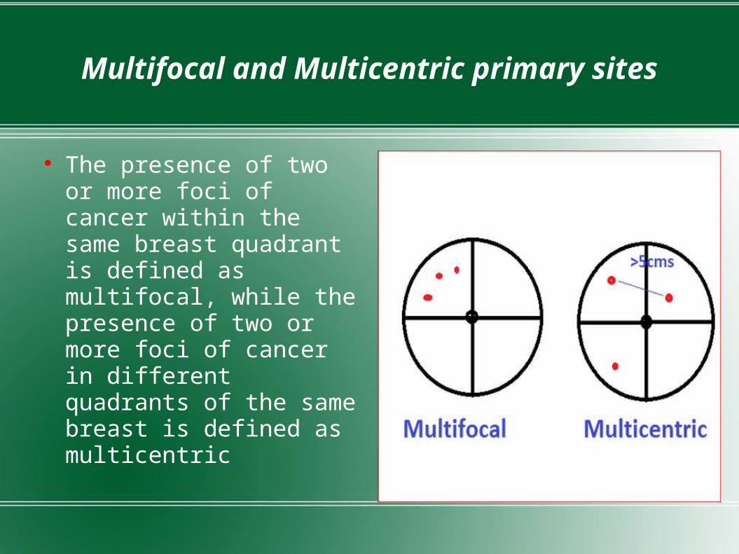
Multifocal and Multicentric primary sites
The presence of two or more foci of cancer within the same breast quadrant is defined as multifocal, while the presence of two or more foci of cancer in different quadrants of the same breast is defined as multicentric

Blood supply
Extremely vascular
Internal thoracic artery – perforating branches
Axillary artery –
Lateral thoracic artery
Superior thoracic artery
Acromiothoracic artery
Posterior intercostal arteries – lateral branches arteries are distributed in the anterior surface.
Posterior surface is relatively avascular.

Axillary artery Superior thoracic
artery
Acromiothoracicartery
Lateral thoracicartery
Posterior intercostalarteries
Internal thoracic artery

Venous drainage
Veins follow the arteries.
First converge around the nipple to form an anastomotic venous circle & then form 02 sets of veins.
Superficial veins: drain into Internal thoracic vein & superficial veins of the lower part of the neck
Deep veins: drain into Internal thoracic , Axillary & Posterior intercostal veins
Clinical importance;-They communicate with vertebral venous plexus- Spread to vertebrae and brain
Axillary vein
Internal thoracic vein
Anastomotic venous
circle

Nerve supply of breast
Is supplied by the anterior and lateral cutaneous branches of the intercostal nerves.
The nerves convey sensory fibres to the skin, and autonomic fibres to smooth muscle and to blood vessels.
The nerves do not control secretion of milk, which is controlled by hormone prolactin.

Lymphatic drainage of breast
Drains by three major routes - Axillary Transpectoral Internal mammary

Lymphatic vessels
The superficial lymphatics drain the skin over the breast except for nipple and areola.
Clinical importance:- obstruction causes peau d'orange
The deep lymphatics drain the parenchyma of the breast, nipple and areola.
Deep lymphatics communicate with with sub diaphragmatic an subperitoneal lymph plexuses
Clinical importance;- cancer may spread to liver and pelvis

Communicates with the superficial lymphatics of breast across the midline
Clinical importance;- spread to contralateral breast About 75% drain axillary nodes, 20% in internal mammary nodes, 5%
in posterior intercostal nodes

Axillary lymph nodes
Clinically divided into 5 groups Anterior, central, posterior,
lateral and apical Lymphatics mostly end in
anterior group and posterior group and through them to apical finally to supraclavicular
clinical importance;- sentinel lymph node is anterior group of axillary lymph nodes mostly and if cancer has not spread that means it has not spread beyond.

Level I- lymph nodes lateral to lateral border of pectoralis minor
Level II- lymph nodes between medial and lateral borders of pectoralis minor and interpectoral lymph nodes.
Lymph III- nodes medial to medial margin of pectoralis minor

Other groups of lymph nodes
Internal mammary lymph nodes- in the intercostal spaces along the edge of the sternum.
Supraclavicular lymph nodes- in the supraclavicular fossa
Intramammary lymph nodes- within the breast are considered as axillary LNs for staging.

CA Breast
Is the most commonly diagnosed malignancy in women in the Western countries, and accounts for more than 25% of cancers diagnosed in women worldwide
Is 2nd most common cancer to be diagnosed overall (11.9%) Is now the most common cancer among women in most cities in
India, and 2nd most common in the rural areas, accounts for 25% to 32% of all female cancers in all cities
It is most common cause cancer related death in females

Risk factors of CA Breast
Established risk factors other reported risk factors possible risk Age alcohol consumption high density breast
Germline mutation birth pills high socioeconomic pos Family history breast-feeding physical activity Benign breast disease BMI dietary factors Radiation exposure Long menstrual history obesity

Female gender- at the age of 50- 1 in 400 women per year, 1% are male breast cancer.
Age – Annual increase in developing breast cancer is approximately 0.07% per yer at 30 yr of age .This increases to 0.44% per year for women in their late 70s.At 40-59 yr age =4% and 60-79 yr of age= 6.9%.
Family history of breast cancer – Ist degree relative risk is 1.7-2.5, 2nd degree relative risk is 1.5. It may be explained in part by inheritance of genetic condition that predisposes an individual to breast cancer development ( e.g., mutaion in BRCA1 and BRCA2); shared lifestyle; and inheritance of genes that affect risk factors, such as body habitus and age at menarche.

Breast Cancer Susceptibility GenesBRCA1 Hereditary breast/ovarian cancer 60%-85% (lifetime); 15-40% risk of ovarian cancer
BRCA2 Hereditary breast/ovarian cancer 60-80%, (lifetime), 15%-40% risk of ovarian cancer
P53 Li-Fraumeni syndrome 50% (by age 50) PTEN1 Cowden syndrome 25% (lifetime) other common genes that can slightly increase a woman's risk of developing
breast cancer CASP8, FGFR2, TNRCP, MAP3K1, rs4973768, LSP1 Rare genes that can also increase breast cancer risk slightly include CHEK2,
ATM (ataxia telangiectasia mutated), BRIP1, PALB2

Child Bearing/Parity/Breastfeeding- linear relation between relative risk of breast cancer and age at first birth, with women age 20 to 25 having nearly a 50 % reduction in relative risk compared to nulliparous.
Ovarian Function- With long menstrual history increases breast cancer risk. In observational studies, removal of ovaries reduces the risk of breast cancer. Women with surgically induced menopause shown to have significantly reduced risk of breast cancer compared to women who has menopause naturally.
Benign Breast Disease- The relative risk of breast cancer developing in women with non proliferative benign breast disease was1.27, for proliferative changes without atypia is 1.88, and for women with proliferative changes with atypia is 4.24

Radiation Exposure- It increases approximately linearly with with increasing dose and was heavily dependent on age at exposure. Multiple flouroscopic examinations for tuberculosis, and multiple examinations for mastitis, radiation therapy at young age for Hodgkin's disease.
Body Mass Index, Physical Activity, and Dietary Factors- the risk of breast cancer to be 30% higher in postmenopausal women with a BMI over 31 kg/m2 compared to women with a BMI of 20 kg/m2 , annual average of at least 1.3 hours of exercise per week from age 10 years onward was associated with a 20% reduction in breast cancer risk .
It is apparent that maintaing sound, varied diet, limiting alcohol intake, avoiding obesity, an getting moderate physical activity are modifiable behaviours that can impact breast cancer risk.

High risk female for breast cancer
>35 yr of age Family history Received therapeutic radiation LCIS Genetic predisposition
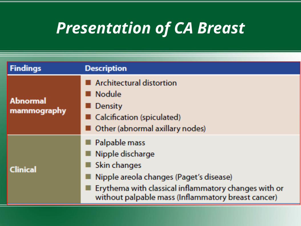
Presentation of CA Breast

Presenting symptoms of breast cancer
The majority of women presents with a lump- hard, painless, immobile, and demonstrates a degree of fixity to surrounding tissues, overlying skin, or the underlying pectoral muscle
Not all cancers are painless may have symptoms of increased discomfort in the lump prior to menstruation. It is, therefore, advisable to undertake histological and cytological examination of any discrete lump in a woman over the age of 25. A diagnosis of a fibroadenoma or 'cystic change' without such confirmation is extremely dangerous.
Up to 15 per cent of women with breast cancer present with a more diffuse process within the breast, in which a lump is not necessarily the presenting feature in lobular carcinoma.
Changes in the skin may be the sole presenting symptom or present with other features of the cancer Puckering maybe present

Peu d'orange is a feature of advanced cancer. The cause of this characteristic clinical sign is oedema of the skin: this is not due to direct infiltration of the skin by tumour but represents lymphatic obstruction of the breast as a result of axillary metastasis
In advanced, untreated cases the skin may be broken, and tumor ulcerates through the skin, with associated haemorrhage and odour
Presentations with changes at the nipple are not uncommon, but these primary tumors may be difficult to diagnose as they are often small and may be easily missed by mammography.
A unifocal or bloodstained nipple discharge is often an intraductal carcinoma developing within the major duct system beneath the nipple. The discharge associated characteristically watery and bloodstained, in contrast to the milk discharge of galactorrhoea and multifocal, tenacious, and coloured discharge of duct ectasia.
It usually represents an underlying intraductal carcinoma that may be quite extensive in the breast and which may also exhibit an invasive component.

Screening of CA Breast
Has resulted in shift in both incidence and stage of patients presenting with breast cancer
Early detection by mammography, followed by appropriate local, regional and systemic treatment is associated with reduced breast cancer mortality rates.
Screening can be Mammogram, Ultrasound of breast, MRI screening, self breast examination.

Mammography Screening
Using low-energy X-rays (usually around 30 kVp) to examine the human breast.
Overall senstivity 75% Two images taken, craniocaudal
and mediolateral oblique

National Cancer Institute recommends baseline mammogram at the age of 35 years , for high risk group it is 30 yrs.
Repeat examination should be carried out every 2 years beginning at 40 yrs of age.
In women older than 50 yrs, mammogram should be performed annually.
Breast imaging reporting and data system has been developed by American College of Radiology to study mammogram

BIRADS Score
Ultrasound screening
It is useful tool to supplement mammography in diagnosis of breast cancer
However use in routine screening of general population has not been established.
It may have role in young patients and high risk patients.

MRI Screening
Role of MRI screening is rapidly evolving however it is unlikely to replace mammography for screening general population.
As it is expensive, not available everywhere and has high false positive rate
It is more sensitive test and there is no risk of radiation exposure
Therefore, can be used for screening high risk patients

Self Breast examination
The estimated overall senstivity is 26% compared with 75% for the combination of clinical breast examination and mammography.
Can be done at home, cost effective.

Algorithm for newly diagnosed breast cancer

Initial work-up
History with emphasis on presenting symptoms, menstrual status, parity, family history of cancer, other risk factors should be done
To clinically rule out metastasis history about bony pain, jaundice, haemoptysis and altered sensorium/ seizures should be asked.
Physical examination with emphasis on breast, axilla, supraclavicular area, abdomen should be done.

Mammography/ultrasonography, Chest radiographsMagnetic resonance imaging of breast
Haemogram/ LFT/KFT/ Serum electrolytes Histopathological examination
- FNAC
- Core Biopsy
- Excisional biopsy
Fine-Needle Aspiration (FNA)
This technique usually is performed when a palpable mass is evident.
The combination of physical examination, mammography, and FNA provides a diagnostic accuracy that approaches 100 percent.
A negative FNA cytology in the presence of a palpable mass, however, does not conclusively exclude carcinoma.
When the mass is clinically and mammographically suspicious, the sensitivity (true-positive) of FNA is 80 to 98 percent. The false- negative rate of FNA is 2 to 10 percent.
The specificity and predictive value of FNA approach 100 percent because false-positive results are rare.

Core Biopsy
False-positive diagnostic rates are lower with tissue procured by cutting needles than with FNA specimens because more tissue is submitted for analysis.
But a core biopsy specimen without malignant tissue cannot conclusively be considered a “negative” biopsy, as it might reflect a sampling error.
Excisional Biopsies Implies removal of the entire lesion and generally a margin of normal breast
parenchyma surrounding the suspicious lesion. False negative cases rare Complete histology before treatment decision May be unnecessary surgery if the condition is benign.

Pathological classification
Histologic sub type In Situ Carcinoma (15-30%)
- Ductal carcinoma in situ (80%)
- Lobular carcinoma in situ(20%) Invasive Carcinoma (70-85%)
- Ductal carcinoma (79%)
- Lobular carcinoma (10%)
- Tubular carcinoma/cribiform carcinoma (6%)
- Mucinous(colloid) carcinoma (2%)
- Medullary carcinoma (2%)
- Papilllary carcinoma (1%)
- Metaplastic carcinoma (<1%)

In Situ Breast Carcinoma
Ductal carcinoma in situ (DCIS)
- Number of cases have increased (5% -15% to 30%) due to screening mammogram.
- It consists of malignant population of cells limited to ducts and lobules by the basement membrane
- Many cases of low grade DCIS and most cases of high grade and extensive DCIS progress to invasive cancer.
Lobular carcinoma in situ (LCIS)
- It is an incidental finding in biopsy performed for another reason as it is not associated with calcification or stromal reaction that would form a density.
- It is bilateral in 20 to 40% of women and more common in young.
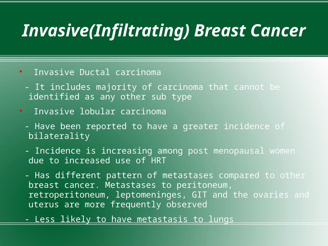
Invasive(Infiltrating) Breast Cancer
Invasive Ductal carcinoma
- It includes majority of carcinoma that cannot be identified as any other sub type
Invasive lobular carcinoma
- Have been reported to have a greater incidence of bilaterality
- Incidence is increasing among post menopausal women due to increased use of HRT
- Has different pattern of metastases compared to other breast cancer. Metastases to peritoneum, retroperitoneum, leptomeninges, GIT and the ovaries and uterus are more frequently observed
- Less likely to have metastasis to lungs

Tumor Grade
Scarff Bloom Richardson grading Combines nuclear grade, tubule formation, and
mitotic rate 85% if grade I, 60% of grade II, 15% of grade III of
early breast cancer survive for 10 years

Biomarkers for breast cancer
In addition to the classical clinical prognostic factors of breast cancer, established molecular biomarkers such as estrogen receptor and progesterone receptor have played a significant role in the selection of patients benefiting from endocrine therapy for many years.
The human epidermal growth factor receptor 2 (HER2) has been validated to be not only a prognostic factor, but also a predictor of response to HER2 targeting therapy.
The marker of proliferation Ki67 has recently emerged as an important marker due to several applications in neoadjuvant therapy in addition to its moderate prognostic value
About 15 biomarkers have been studied like uPA, PAI and TF, h-MAM, osteopontin, snail, twist, zeb-1, FGFR, PTEN and sirtuins and it has been found that combination of all these could be utilized for diagnostic and prognosis.

Immunohistochemistry Status
For assessing hormone responsive receptor status ER/PR/Her2 status Microarray studies have identified subtypes by
morphology (e.g., lobular carcinomas), by protein expression (e.g., ER+ve and Her2 +ve) and by germline mutations (e.g., BRCA1 and BRCA2)
New subtype identified (e.g., Basal like)

ER +ve carcinomas- 70 to 80% breast carcinomas are positive, are usually well to moderately differentiated. Are usually lobular type which infiltrates as single cell.
ER -ve carcinomas- are major 2 types
1. Her 2 +ve carcinomas- tend to be poorly differentiated
2. Basal like- poorly diff, lacking ER/PR/her2, expressing basal like keratins.
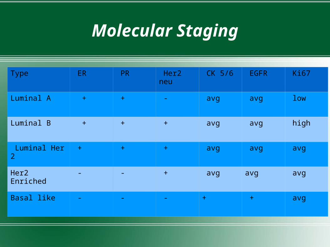
Molecular Staging
Type ER PR Her2 neu CK 5/6 EGFR Ki67
Luminal A + + - avg avg low
Luminal B + + + avg avg high
Luminal Her 2 + + + avg avg avg
Her2 Enriched - - + avg avg avg
Basal like - - - + + avg

Metastatic Work Up
Indicated for N2/N3 patients and T3 orT4 primary lesions
Approximately 6-10% of new breast cancer cases are initially Stage IV or metastatic. This is sometimes called "de novo" metastatic disease
Bone (41.1%)>lung (22.4%)> liver (7.3%)>brain (7.3%).
Bone- scan Computed tomography of chest,
abdomen and pelvis CECT brain

CA breast staging
TNM staging by AJCC latest updated in 2010, given in 7th edition of AJCC cancer staging manual.
Various changes have been made from previous 6th edition reflect advances in both management options and in prognostic information and clarifications in definitions used in the 6th edition.
The changes can be divided into 4 areas:
ƒ Tumor size definition
ƒ Node classification
ƒ Metastasis classification
ƒ Special issues in patients who received neoadjuvant chemotherapy.

Tumor size definition
Invasive tumors should be recorded to the nearest millimeter Small invasive tumors should be defined by microscopic measurement
and larger tumors by gross measurement however, acknowledges that gross measurement may be inaccurate for the following reasons-
1.) the gross appearance of invasive versus non-invasive tumor may not always be distinctive.
2.) invasive tumor may extend microscopically further than is grossly visible .
Multiple cancers as those that are grossly or macroscopically distinct. AJCC defines 0.5 cm as the minimum distance required between two macroscopic cancer foci to call them multiple cancers, anything closer than 0.5 cm likely represents a single cancer .

Primary Tumor (T)
TX Primary tumor cannot be assessed T0 No evidence of primary tumor Tis Carcinoma in situ Tis (DCIS) Ductal carcinoma in situ Tis (LCIS) Lobular carcinoma in situ Tis (Paget’s) Paget’s disease of the nipple NOT associated with invasive carcinoma
and/or carcinoma in situ (DCIS and/or LCIS) in the underlying breast parenchyma. Carcinomas in the breast parenchyma associated with Paget’s disease are categorized based on the size and characteristics of the parenchymal disease, although the presence of Paget’s disease should still be noted

Primary Tumor (T) cont
T1 Tumor ≤ 20 mm in greatest dimension
T1mi Tumor ≤ 1 mm in greatest dimension
T1a Tumor > 1 mm but ≤ 5 mm in greatest dimension
T1b Tumor > 5 mm but ≤ 10 mm in greatest dimension
T1c Tumor > 10 mm but ≤ 20 mm in greatest dimension

Primary Tumor (T) cont
T2 Tumor > 20 mm but ≤ 50 mm in greatest dimension T3 Tumor > 50 mm in greatest dimension

Primary Tumor (T) cont
T4 Tumor of any size with direct extension to the chest wall and/or to the skin (ulceration or skin nodules)
Note: Invasion of the dermis alone does not qualify as T4 T4a Extension to the chest wall, not including only pectoralis muscle
adherence/invasion
(Chest wall includes intercostal muscles and serratus
anterior muscle, but not the pectoral muscles.
Therefore, involvement of the pectoral muscle does
not constitute chest wall invasion)

Primary Tumor (T) cont
T4b Ulceration and/or ipsilateral satellite nodules and/or edema (including peau d’orange) of the skin, which do not meet the criteria for inflammatory carcinoma
Peau d'orange represents lymphatic obstruction of the breast as a result of axillary metastasis
T4c Both T4a and T4b

T4d Inflammatory carcinoma
Inflammatory carcinoma is a
clinical-pathologic entity characterized by
diffuse erythema and edema
involving a third or more of the skin of the
Breast.
- Microscopically, dermal lymphatic invasion
is typically seen in this setting, however dermal
lymphatic invasion alone is NOT sufficient
(nor necessary) to diagnose

Controversy Does not take tumor burden into account as multiple synchronus
tumor has more tumor load as compared to single tumor of same size Does not take immunohistochemistry into account Does not mention how lymphatic invasion should be
handled in terms of tumor size.

Node classification
Classification of isolated tumor cell clusters and single cells is more stringent.
Clusters of cells not greater than 0.2 mm, or non confluent or nearly confluent clusters of cells not exceeding 200 cells in a single histologic lymph node cross section .
When six or more sentinel nodes are identified on gross examination of pathology specimens the (sn) modifier should be omitted.
The separation of pN1mic from pN1 in the overall staging.

Regional Lymph Nodes (N)Clinical
NX Regional lymph nodes cannot be assessed (e.g.,previously removed)
N0 No regional lymph node metastases
N1 Metastases to movable ipsilateral level I, II axillary lymph node(s)
N2 Metastases in ipsilateral level I, II axillary lymph nodes that are clinically fi xed or matted; or in clinically detected * ipsilateral internal mammary nodes in the absence of clinically evident axillary lymph node metastases
N2a Metastases in ipsilateral level I, II axillary lymph nodes fi xed to one another (matted) or to other structures
N2b Metastases only in clinically detected * ipsilateral internal mammary nodes and in the absence of clinically evident level I, II axillary lymph node metastases
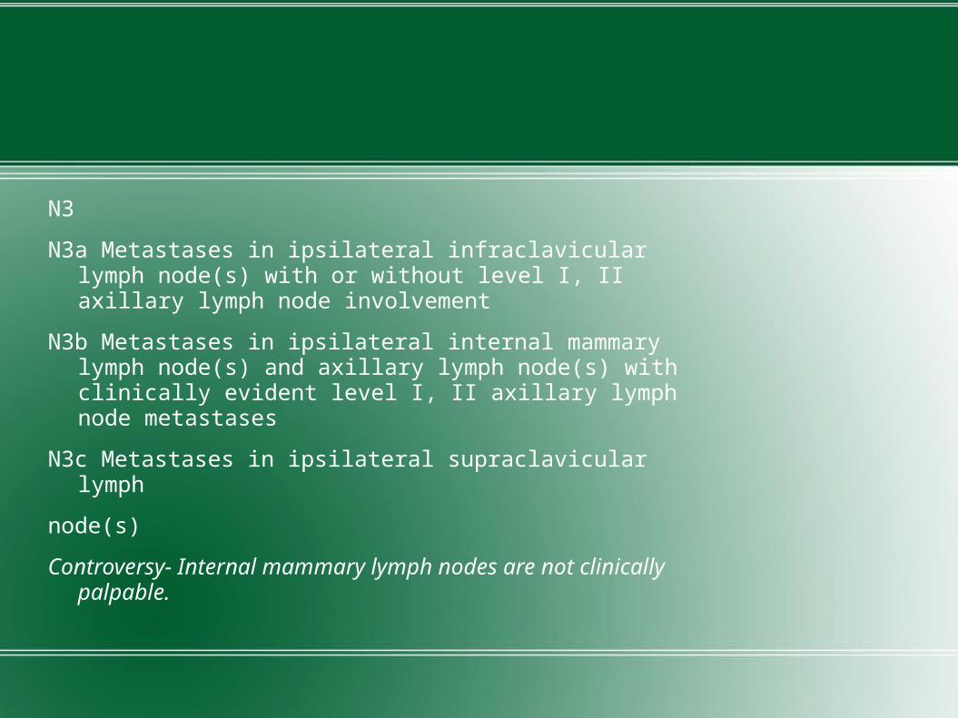
N3
N3a Metastases in ipsilateral infraclavicular lymph node(s) with or without level I, II axillary lymph node involvement
N3b Metastases in ipsilateral internal mammary lymph node(s) and axillary lymph node(s) with clinically evident level I, II axillary lymph node metastases
N3c Metastases in ipsilateral supraclavicular lymph
node(s)
Controversy- Internal mammary lymph nodes are not clinically palpable.


Regional Lymph Nodes (N)Pathologic (pN)
-Level I/II clearance
-Level III if gross nodes in level II
-Atleast 10 LN should be removed
By size of the metastasis
0.2 mm (or >200 cells) up to 2 mm is a
micrometastasis: pN1mic
>2 mm is a macrometastasis: pN1, pN2,
pN3, depending on total number of positive
nodes

By total number of positive nodes
Total of 1-3 positive nodes is pN1a
Total of 4-9 positive nodes is pN2a
Total of >9 positive nodes is pN3a

Regional Lymph Nodes (N)Pathologic (pN)
By specific anatomic nodePositive internal mammary sentinel affects
pN depending on status of other nodes.
pN1b- clinically not detectable but sentinel lymph
node biopsy shows micro or macrometastases
pN2b- clinically detectable in absence of axillary
lymph nodes
pN3b- 1 or more axillary LN + clinically detectable
IMLN or >3 axillary LN + IMLN positive in SLN biopsy
Positive infraclavicular axillary node is pN3a
Positive supraclavicular axillary node is pN3c

Metastasis classification
Now separates out incidentally detected cancer cells (<0.2 millimeter) in distant, non-regional nodal tissue if there is no clinical or radiologic evidence of metastasis.
example raised by AJCC is incidental metastasis <0.2 mm found in a prophylactic oophorectomy; in the absence of any clinical/radiologic evidence of ovarian involvement, this finding should not be staged as pM1 distant metastasis but as pM0i+.
Controversy- Does not differntiate between oligometastasis and multiple metastasis

Staging Patients After Neoadjuvant Therapy
ypT is based on the largest single focus of residual invasive cancer;
Stromal fibrosis in the residual tumor bed should not be used to increase the size beyond that of actual invasive tumor cells
If the residual tumor consists of microscopic nests in fibrotic stroma, ypT should be based on the largest contiguous area of invasive carcinoma
Controversy- may shrink in one of two patterns following
Scenario 1.) concentric shrinkage, resulting in a single residual focus smaller
Scenario 2.) patchy, non-concentric shrinkage, resulting in multifocal residual tumor that may span the same size as the pre-treatment tumor size (but with reduced cellularity) or that may span a smaller size than the pre-treatment tumor size.
Does not provide a definition of what constitutes separate multiple foci or of whether the definition applies to macroscopic tumor or microscopic tumor.
Post-treatment ypN is same as pretreatment.
The M category remains same as pretreatment Identifi cation of distant metastases after the start of therapy is considered progression of disease.

Anatomic Stage
Can be classified as Early breast cancer, Locally invasive breast cancer, Metastatic breast cancer
Early breast cancer- Any T1 0r T2 with N0 or N1 ( IA to IIB)
Locally invasive breast cancer- T3 or T4 , N2 or N3 (IIIA to IIIC)
Metastatic breast cancer- with distant metastasis (IV)
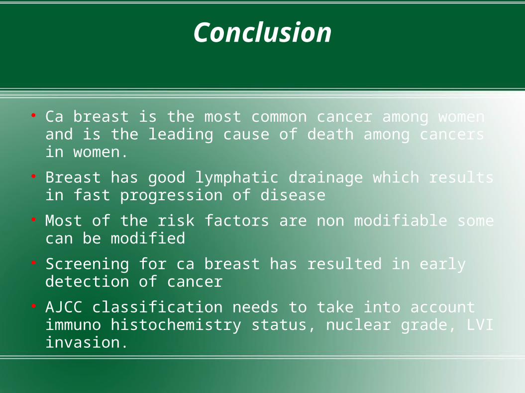
Conclusion
Ca breast is the most common cancer among women and is the leading cause of death among cancers in women.
Breast has good lymphatic drainage which results in fast progression of disease
Most of the risk factors are non modifiable some can be modified
Screening for ca breast has resulted in early detection of cancer
AJCC classification needs to take into account immuno histochemistry status, nuclear grade, LVI invasion.

THANKYOU