anatomy and pathology of the achilles tendon tracy … and pathology of the achilles... · anatomy...
TRANSCRIPT

Anatomy and Pathology of
the Achilles TendonTracy MacNair
Achilles
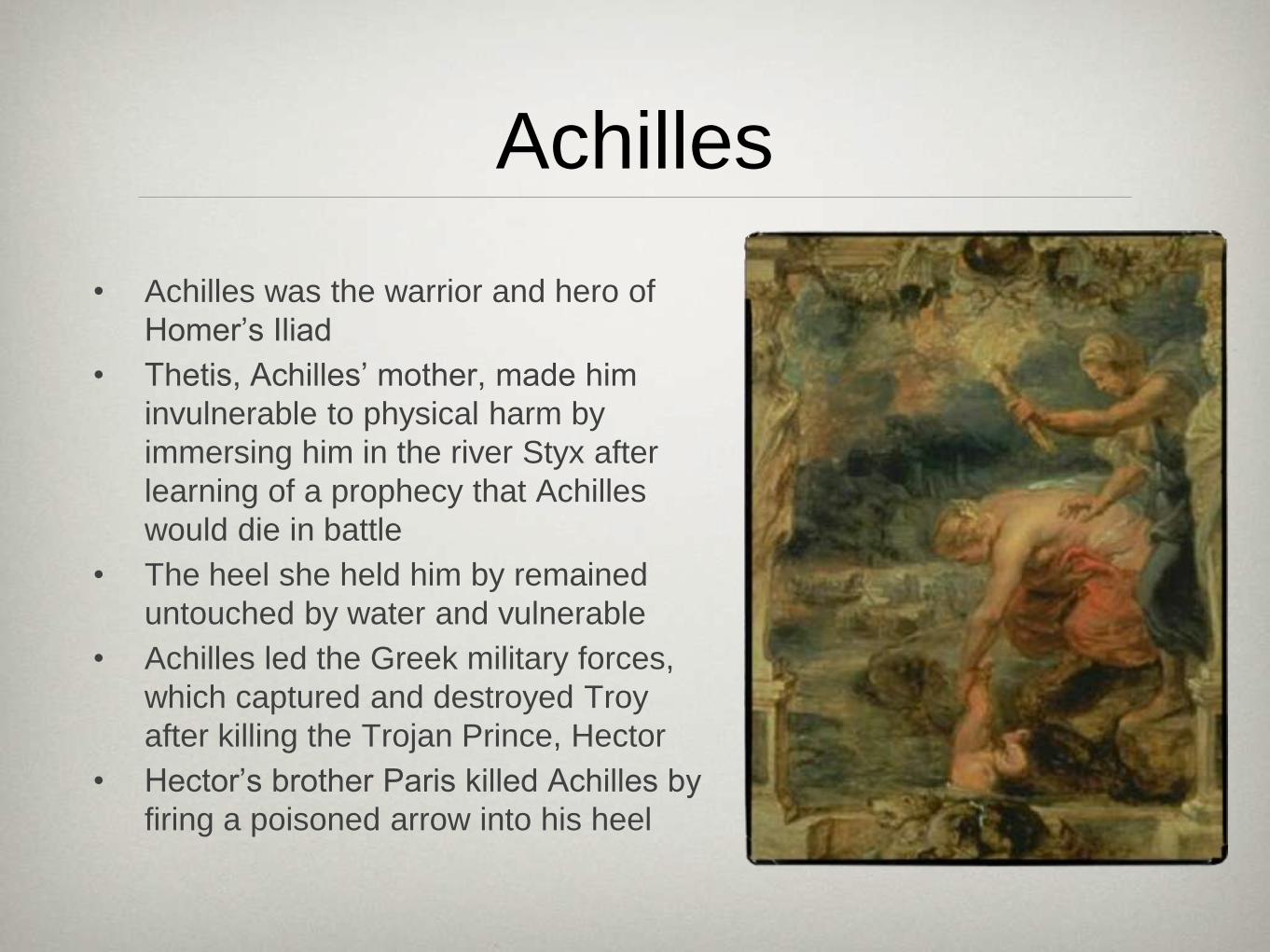
• Achilles was the warrior and hero of
Homer’s Iliad
• Thetis, Achilles’ mother, made him
invulnerable to physical harm by
immersing him in the river Styx after
learning of a prophecy that Achilles
would die in battle
• The heel she held him by remained
untouched by water and vulnerable
• Achilles led the Greek military forces,
which captured and destroyed Troy
after killing the Trojan Prince, Hector
• Hector’s brother Paris killed Achilles by
firing a poisoned arrow into his heel
Outline• Anatomy
o General anatomy
o Gastrocnemius muscle
o Soleus muscle
o Achilles tendon
o Calcaneal tuberosity
o Blood supply
o Retrocalcaneal bursa
o Peritenon
o Plantaris
o Surrounding soft tissues
• Biomechanics
• Epidemiology
• Pathology
o Clinical findings
o Peritendinitis
o Paratendinitis
o Partial & Complete tears
o Muscle atrophy
o Osseous abnormalities
o Insertional pathology
o Myotendinous junction
o Retrocalcaneal bursitis
o Haglands deformity
o Xanthoma
• Post surgical imaging
General Anatomy
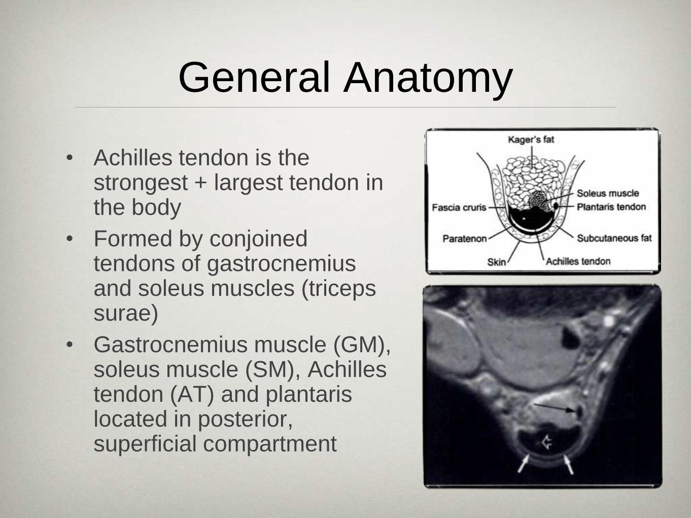
• Achilles tendon is the strongest + largest tendon in the body
• Formed by conjoined tendons of gastrocnemius and soleus muscles (triceps surae)
• Gastrocnemius muscle (GM), soleus muscle (SM), Achilles tendon (AT) and plantaris located in posterior, superficial compartment
Gastrocnemius Anatomy
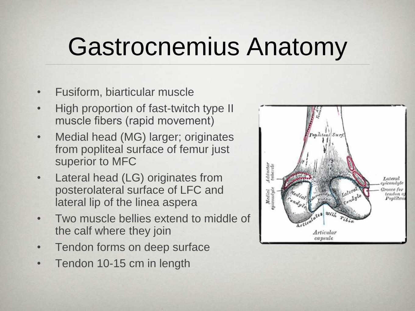
• Fusiform, biarticular muscle
• High proportion of fast-twitch type II muscle fibers (rapid movement)
• Medial head (MG) larger; originates from popliteal surface of femur just superior to MFC
• Lateral head (LG) originates from posterolateral surface of LFC and lateral lip of the linea aspera
• Two muscle bellies extend to middle of the calf where they join
• Tendon forms on deep surface
• Tendon 10-15 cm in length
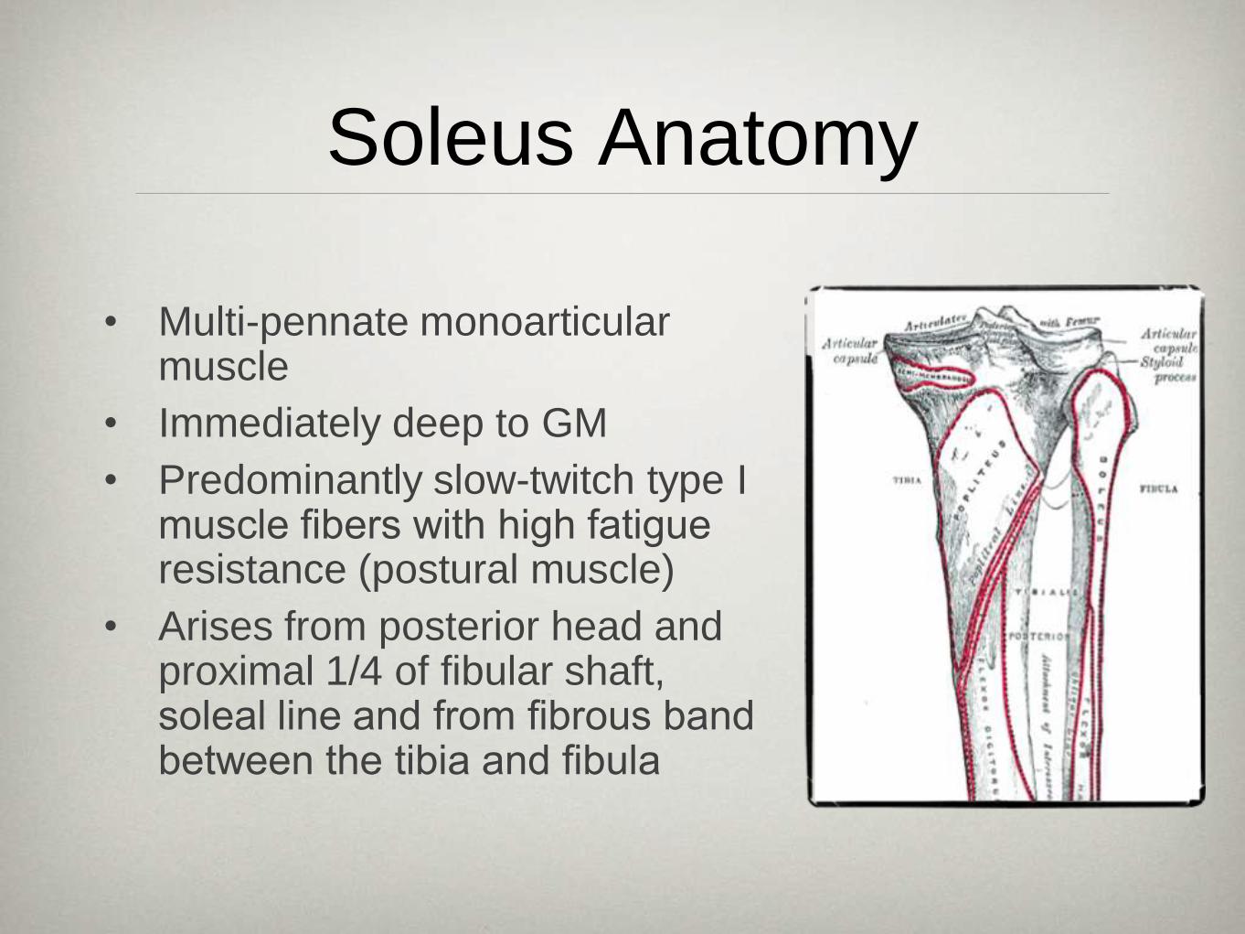
Soleus Anatomy
• Multi-pennate monoarticular muscle
• Immediately deep to GM
• Predominantly slow-twitch type I muscle fibers with high fatigue resistance (postural muscle)
• Arises from posterior head and proximal 1/4 of fibular shaft, soleal line and from fibrous band between the tibia and fibula

Soleus Anatomy
• Muscular fibers terminate in a broad aponeurosis on the posterior surface
• Gastrocnemius and soleus aponeuroses parallel each other for variable distance before uniting
• Large variation in soleus musculotendinous junction
• ? cut off for low lying soleus
o Pichler et al. Anatomic Variations of the Musculotendinous Junction of the Soleus Muscle and Its Clinical Implications. Clinical Anatomy 2007; 20:444–447.
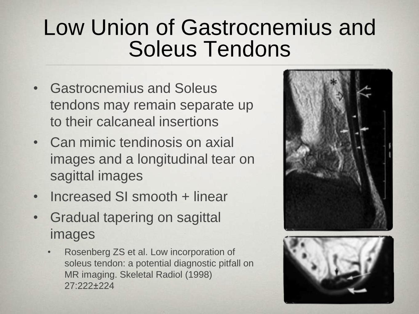
Low Union of Gastrocnemius and Soleus Tendons
• Gastrocnemius and Soleus
tendons may remain separate up
to their calcaneal insertions
• Can mimic tendinosis on axial
images and a longitudinal tear on
sagittal images
• Increased SI smooth + linear
• Gradual tapering on sagittal
images
• Rosenberg ZS et al. Low incorporation of
soleus tendon: a potential diagnostic pitfall on
MR imaging. Skeletal Radiol (1998)
27:222±224

Accessory Soleus
• Rare congenital anatomical variant (0.7%)
• Arises from anterior surface of the soleus,
soleal line of the tibia or proximal fibula
• Inserts as muscle or tendon onto medial
surface of calcaneus or into Achilles' tendon
• Separate blood supply from posterior tibial
artery and separate fascial sleeve
• Manifests in late teens because of muscle
hypertrophy due to increased physical activity
• Majority present with a painful swelling caused
by muscle ischemia or a compressive
neuropathy involving the posterior tibial nerve

Achilles Anatomy
• Begins at junction of
gastrocnemius and soleus
tendons in middle of calf
• Contribution of gastrocnemius
and soleus tendons varies
• Typically 3 to 11 cm in length
• Rotational twist before inserting
on calcaneus
o gastrocnemius fibers insert
laterally
o soleus fibers insert medially
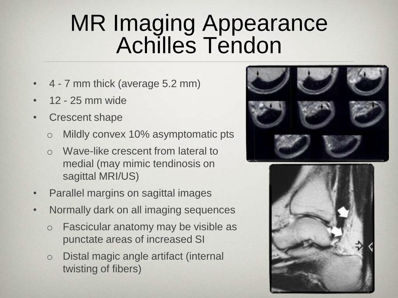
MR Imaging Appearance Achilles Tendon
• 4 - 7 mm thick (average 5.2 mm)
• 12 - 25 mm wide
• Crescent shape
o Mildly convex 10% asymptomatic pts
o Wave-like crescent from lateral to
medial (may mimic tendinosis on
sagittal MRI/US)
• Parallel margins on sagittal images
• Normally dark on all imaging sequences
o Fascicular anatomy may be visible as
punctate areas of increased SI
o Distal magic angle artifact (internal
twisting of fibers)
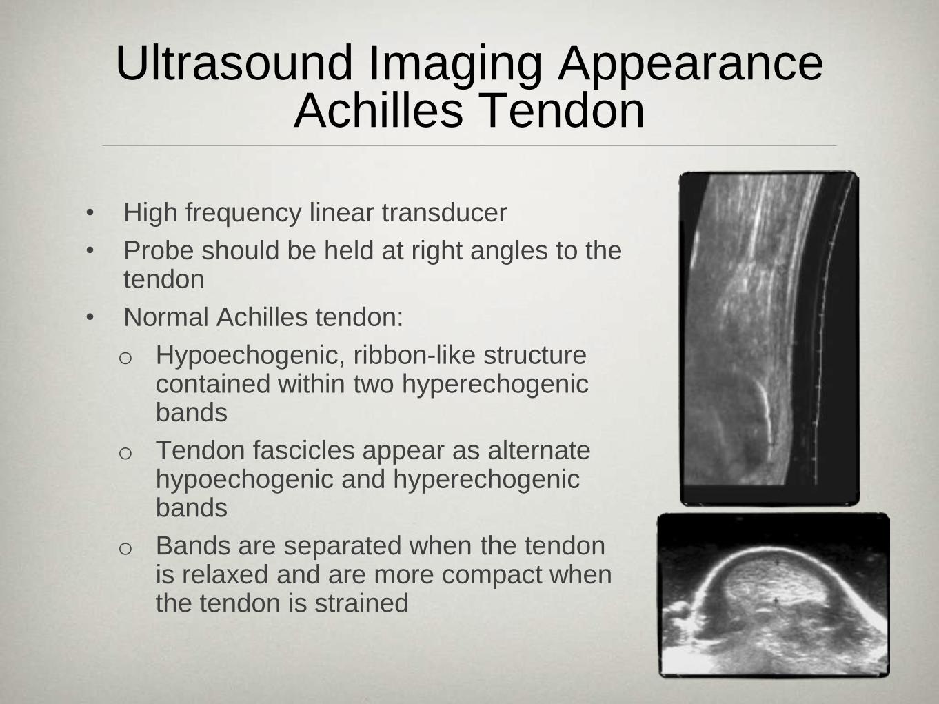
Ultrasound Imaging Appearance Achilles Tendon
• High frequency linear transducer
• Probe should be held at right angles to the tendon
• Normal Achilles tendon:
o Hypoechogenic, ribbon-like structure contained within two hyperechogenic bands
o Tendon fascicles appear as alternate hypoechogenic and hyperechogenic bands
o Bands are separated when the tendon is relaxed and are more compact when the tendon is strained
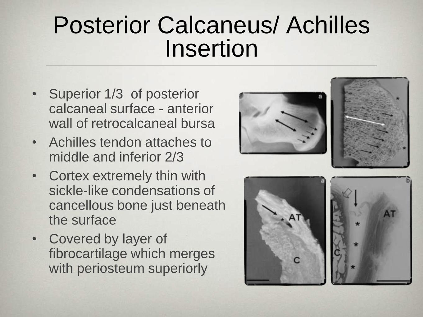
Posterior Calcaneus/ Achilles Insertion
• Superior 1/3 of posterior calcaneal surface - anterior wall of retrocalcaneal bursa
• Achilles tendon attaches to middle and inferior 2/3
• Cortex extremely thin with sickle-like condensations of cancellous bone just beneath the surface
• Covered by layer of fibrocartilage which merges with periosteum superiorly

Blood Supply
• Blood supply from musculotendinous junction, peritenon
and bone-tendon junction
• AT poorly vascularized (like all tendons)
• Dispute regarding the distribution of blood vessels in the
tendon
o Some investigations have shown the density of blood
vessels in the middle of the AT is low compared to
proximal tendon
o Others have shown blood flow is evenly distributed
• Blood flow varies with age and loading conditions

Retrocalcaneal Bursa
• Visible in 96% of patients on MR
• Normally measures < 7 mm SI, 11
mm ML and 1 mm AP
• Margins: calcaneal tuberosity
anterior, AT posterior, Kager’s fat
pad superior
• Protects the distal AT from frictional
wear against calcaneus
• Superior synovial fold with delicate
fascicle of skeletal muscle fibers

Peritenon
• No true synovial sheath surrounding AT
• Enclosed by a peritenon - thin gliding
membrane of loose connective tissue
• Also referred to as paratenon
• Peritenon continuous proximally with the
fascial envelope of GM and SM, and
blends distally with the periosteum of the
calcaneus
• Blood vessels run through the peritenon -
provides nutrition for tendon
• Thin, crescent shaped intermediate SI
posterior, medial + lateral to Achilles
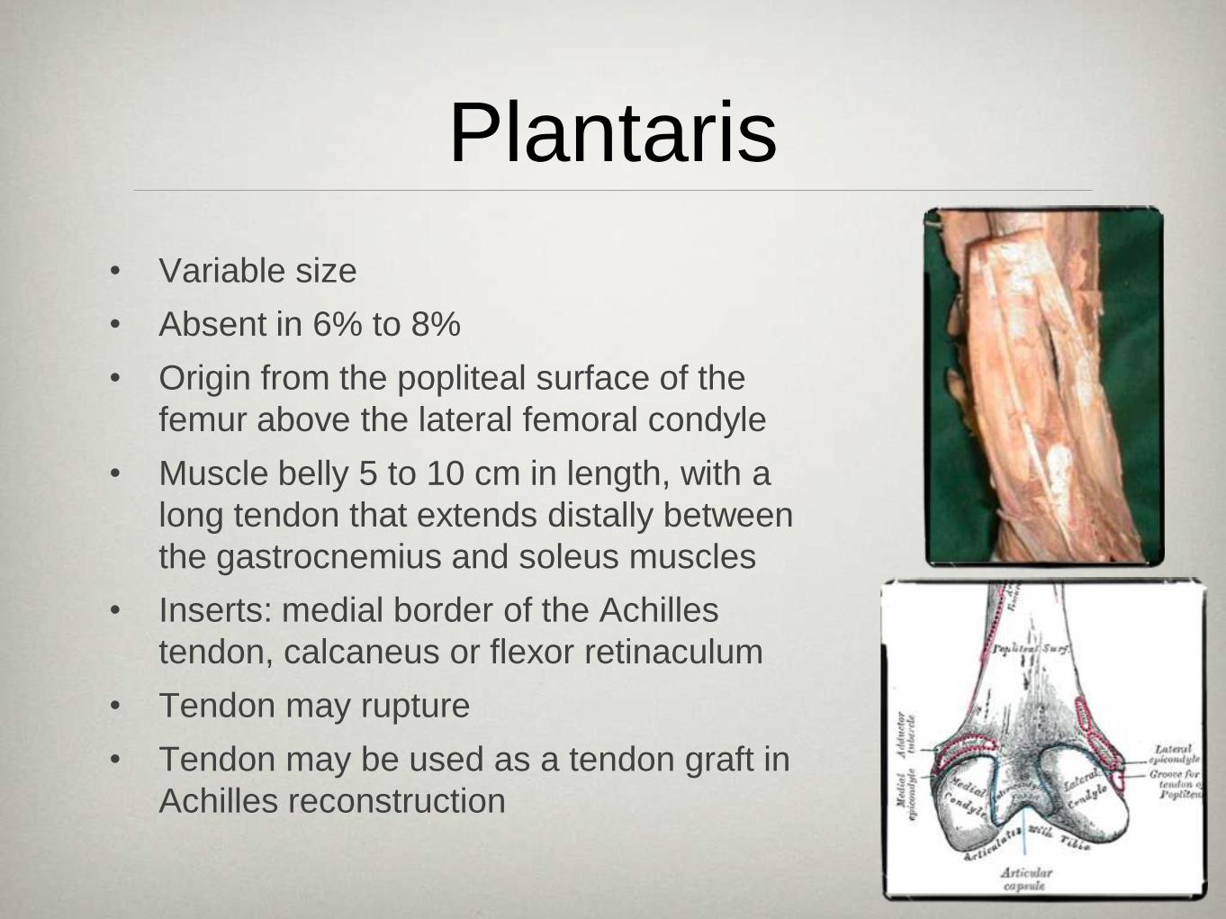
Plantaris
• Variable size
• Absent in 6% to 8%
• Origin from the popliteal surface of the
femur above the lateral femoral condyle
• Muscle belly 5 to 10 cm in length, with a
long tendon that extends distally between
the gastrocnemius and soleus muscles
• Inserts: medial border of the Achilles
tendon, calcaneus or flexor retinaculum
• Tendon may rupture
• Tendon may be used as a tendon graft in
Achilles reconstruction
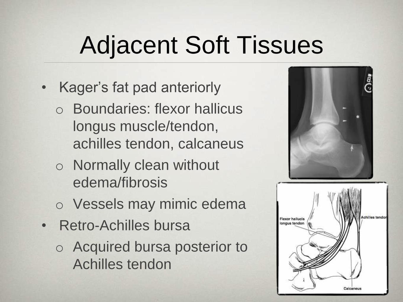
Adjacent Soft Tissues
• Kager’s fat pad anteriorly
o Boundaries: flexor hallicus
longus muscle/tendon,
achilles tendon, calcaneus
o Normally clean without
edema/fibrosis
o Vessels may mimic edema
• Retro-Achilles bursa
o Acquired bursa posterior to
Achilles tendon

“Achilles’ Heel”
• The term “Achilles’ heel”
was first used by a
Dutch anatomist,
Verheyden, in 1693
when he dissected his
own amputated leg
• Expression used for
“area of weakness,
vulnerable spot”

Biomechanics
• AT is subjected to the highest loads in the
body - up to 10x body weight
• Triceps surae primary plantar flexor of foot
o Deep muscles of posterior compartment +
peroneal muscles contribute 15–35%
• Gastrocnemius and Soleus muscles differ in
muscle twitch fibers, muscle length, fascicle
length, and pennation angle
• GM and SM capable of acting individually,
even though they share a common tendon
• Hyperpronation, pes cavus, genu varum
increase tendon stress
Epidemiology
• Achilles tendon pathology rarely reported before 1950s
• Incidence of Achilles tendon tears in industrialized nations is approximately 7/100,000 per year
• Mean age 36; Male predominance (1.7:1 to 12:1)
• Left > Right for unknown reasons
• Etiology of Achilles tendon rupture:
o Repetitive trauma with collagen degeneration
o Also: local steroid injection, oral corticosteroids, fluoroquinolones, inflammatory and autoimmune conditions, collagen abnormalities and neurological conditions
o Violent muscular strain in healthy tendon
Achilles Pathology
• Spectrum of Achilles tendon disorders
and overuse injuries ranges from:
o Inflammation of the peritendinous tissue
(peritendinitis, paratendinitis)
o Degeneration of the tendon (tendinosis)
o Tendon rupture (partial or complete)
o Insertional disorders (retrocalcaneal
bursitis and insertional tendinopathy)
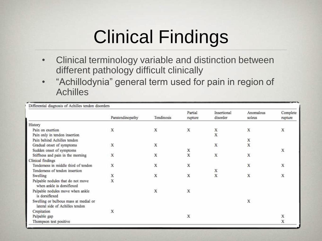
Clinical Findings• Clinical terminology variable and distinction between
different pathology difficult clinically
• “Achillodynia” general term used for pain in region of Achilles
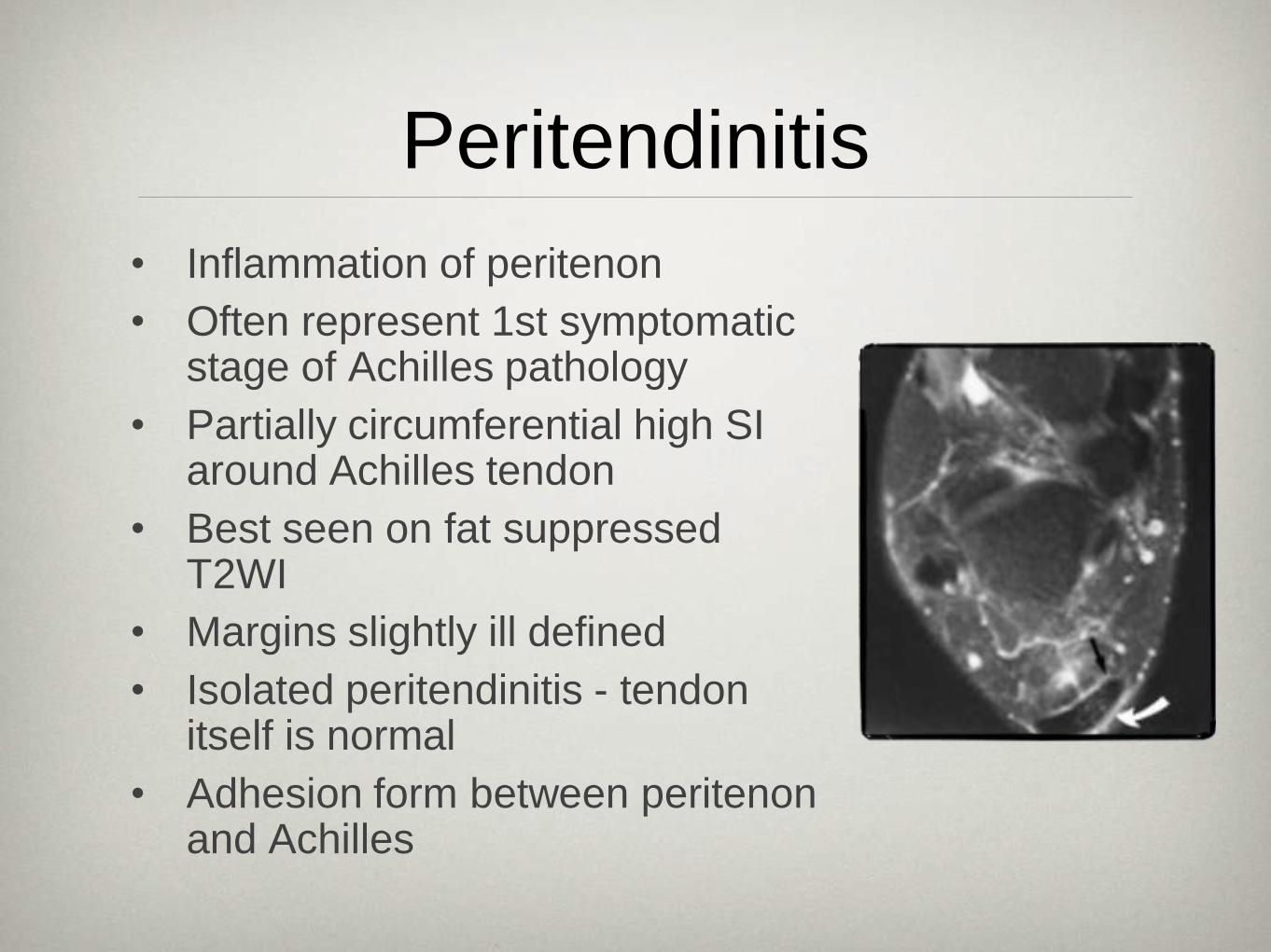
Peritendinitis
• Inflammation of peritenon
• Often represent 1st symptomatic stage of Achilles pathology
• Partially circumferential high SI around Achilles tendon
• Best seen on fat suppressed T2WI
• Margins slightly ill defined
• Isolated peritendinitis - tendon itself is normal
• Adhesion form between peritenon and Achilles
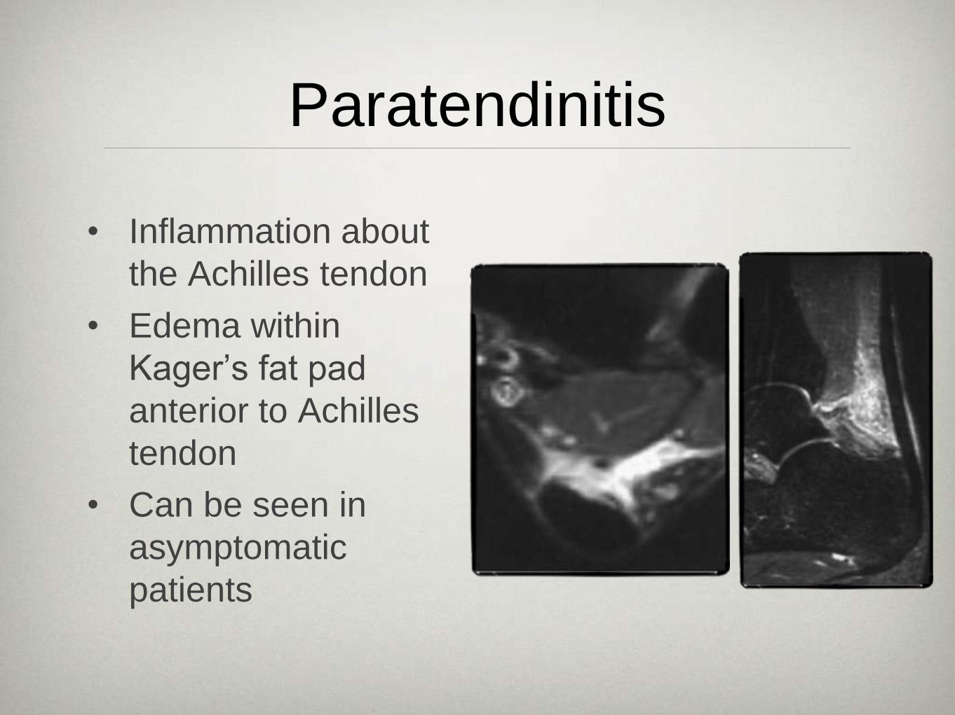
Paratendinitis
• Inflammation about
the Achilles tendon
• Edema within
Kager’s fat pad
anterior to Achilles
tendon
• Can be seen in
asymptomatic
patients
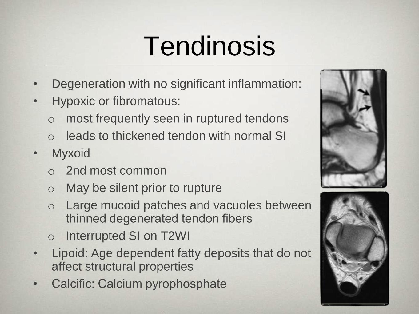
Tendinosis
• Degeneration with no significant inflammation:
• Hypoxic or fibromatous:
o most frequently seen in ruptured tendons
o leads to thickened tendon with normal SI
• Myxoid
o 2nd most common
o May be silent prior to rupture
o Large mucoid patches and vacuoles between thinned degenerated tendon fibers
o Interrupted SI on T2WI
• Lipoid: Age dependent fatty deposits that do not affect structural properties
• Calcific: Calcium pyrophosphate

Tendinosis
• Often accompanied by peritendinitis
• Imaging:
o Diffuse or focal thickening
o Signal intensity generally low
o When intratendinous foci of
increased T2 SI are present an
accompanying partial tear is likely
o Mucoid degeneration junction
entity between tendinosis and
partial tears - focal interrupted
increased T2 SI (coalesce to form
partial tears)

MR Appearance Symptomatic vs Asymptomatic Patients
• Increased thickness in asymptomatic and symptomatic patients relative to previous reports (0.747 cm vs. 0.877 cm)
• Similar incidence of peritendinitis (37% vs. 34%)
• Pre-Achilles edema was more common in asymptomatic patients (40% vs. 28%)
• Symptomatic patient had larger retrocalcaneal fluid volume (0.278 mL vs. 0.104 mL)
• Asymptomatic Achilles tendons frequently demonstrated mild increased intratendon signal (70%)
• Symptomatic patients had more frequent tears (36%) although 7% of asymptomatic patients had interstitial tears
Haims , Schweitzer et al. MR imaging of the Achilles tendon: overlap
of findings in symptomatic and asymptomatic individuals Skeletal
Radiol (2000) 29:640–645

Partial and Complete Tendon Tears
• Spectrum: Microtears -
Interstitial tears - Partial
tears - Complete tears
• Most common site 3-4 cm
proximal to insertion
• Partial tears often lateral
• Discontinuity of fibers
• Intratendinous increased SI
on T2/STIR; heterogeneous
echotexture
• Intratendinous gap

Muscle Atrophy
• Acute atrophy - diffuse edema
throughout muscle belly; best
prognosis after surgery
• Irreversible atrophy - fatty infiltration
• Atrophy occurs first in the soleus -
predominance of slow twitch fibers
• Sagittal images should include at
least 3 cm of distal soleus belly
• Atrophy of gastrocnemius rare even
in remote Achilles tendon tears
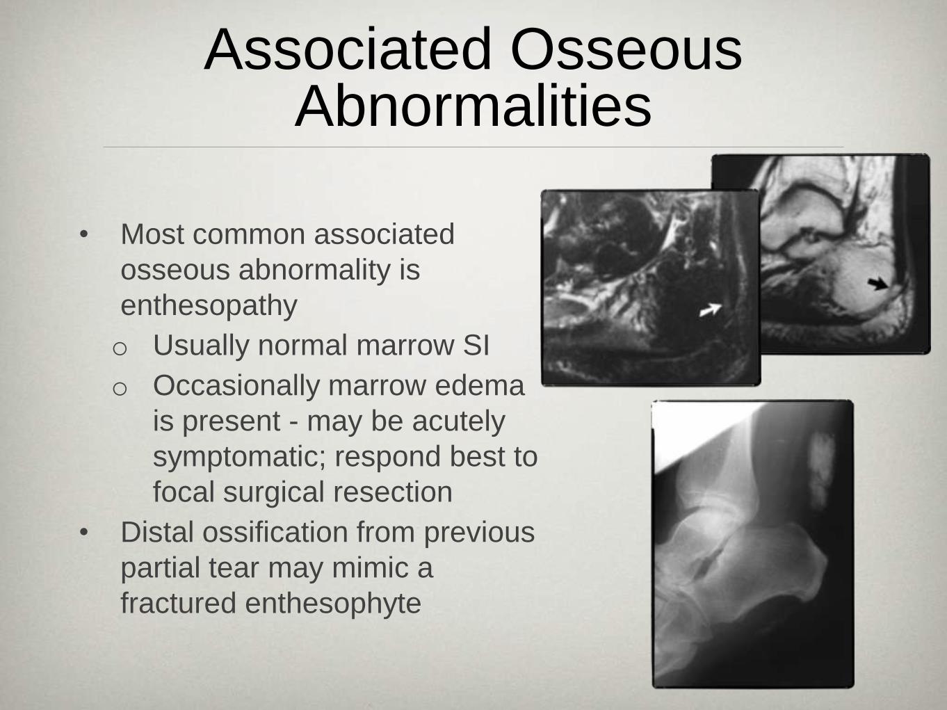
Associated Osseous Abnormalities
• Most common associated
osseous abnormality is
enthesopathy
o Usually normal marrow SI
o Occasionally marrow edema
is present - may be acutely
symptomatic; respond best to
focal surgical resection
• Distal ossification from previous
partial tear may mimic a
fractured enthesophyte
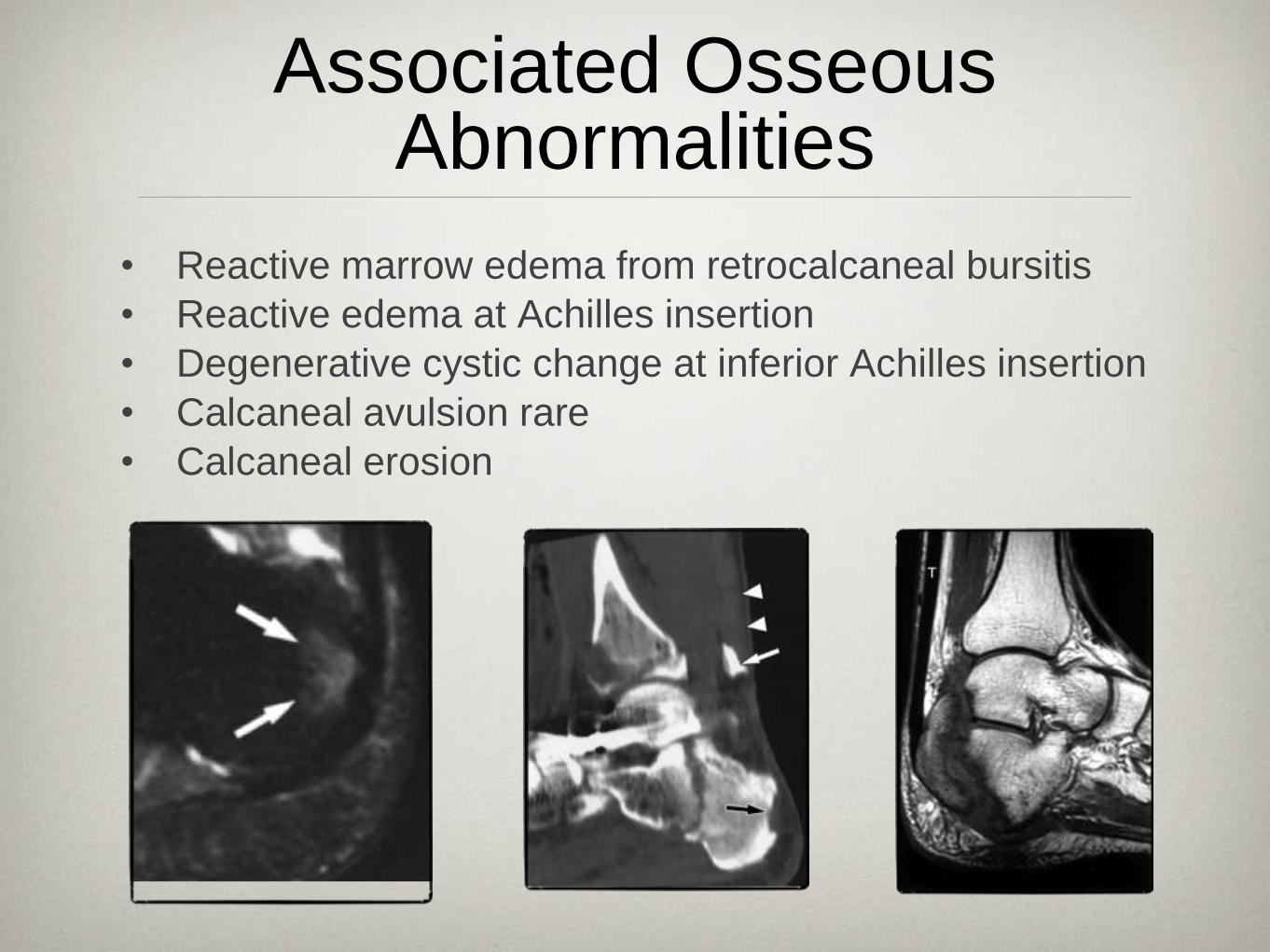
Associated Osseous Abnormalities
• Reactive marrow edema from retrocalcaneal bursitis
• Reactive edema at Achilles insertion
• Degenerative cystic change at inferior Achilles insertion
• Calcaneal avulsion rare
• Calcaneal erosion
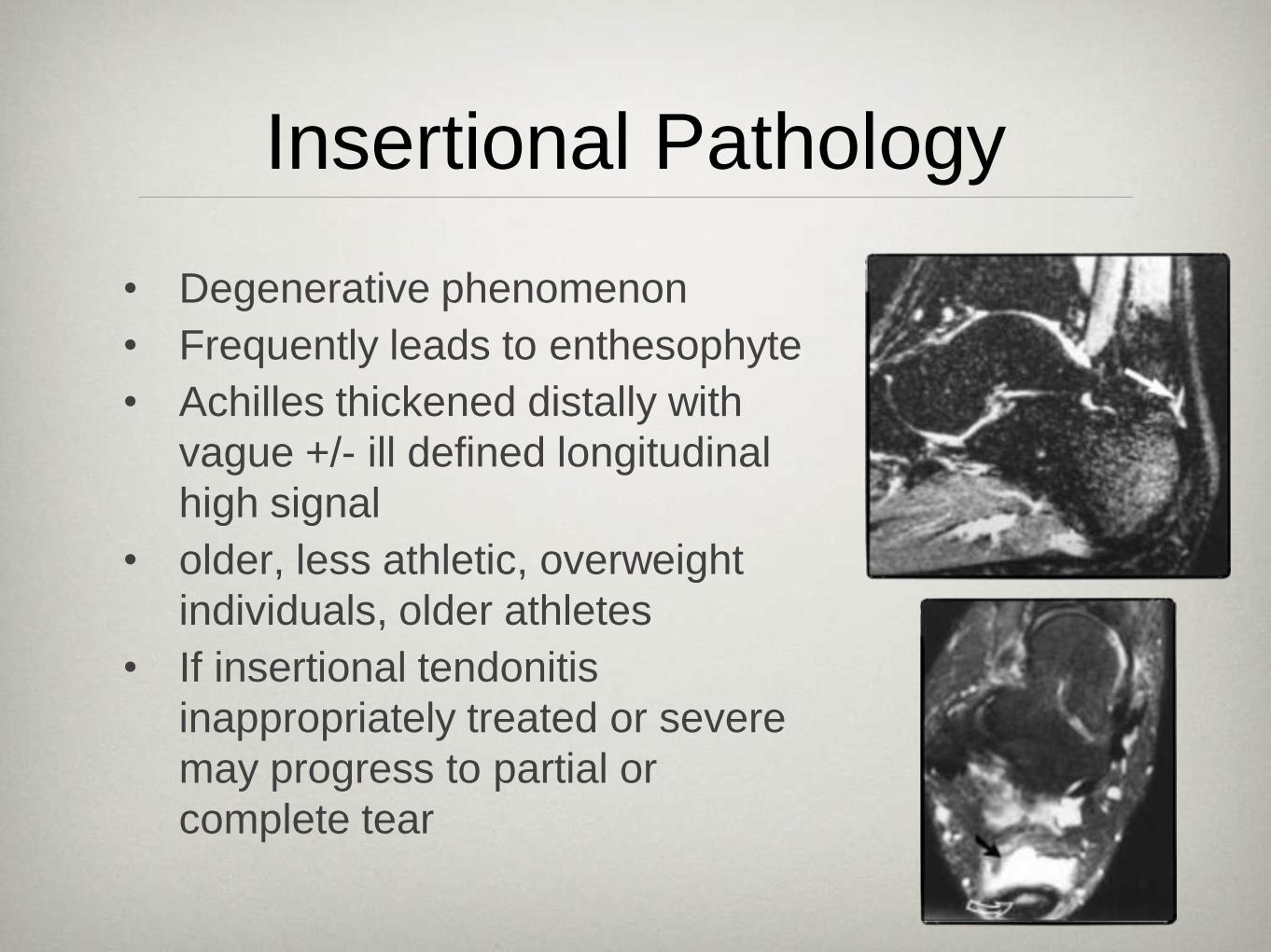
Insertional Pathology
• Degenerative phenomenon
• Frequently leads to enthesophyte
• Achilles thickened distally with
vague +/- ill defined longitudinal
high signal
• older, less athletic, overweight
individuals, older athletes
• If insertional tendonitis
inappropriately treated or severe
may progress to partial or
complete tear
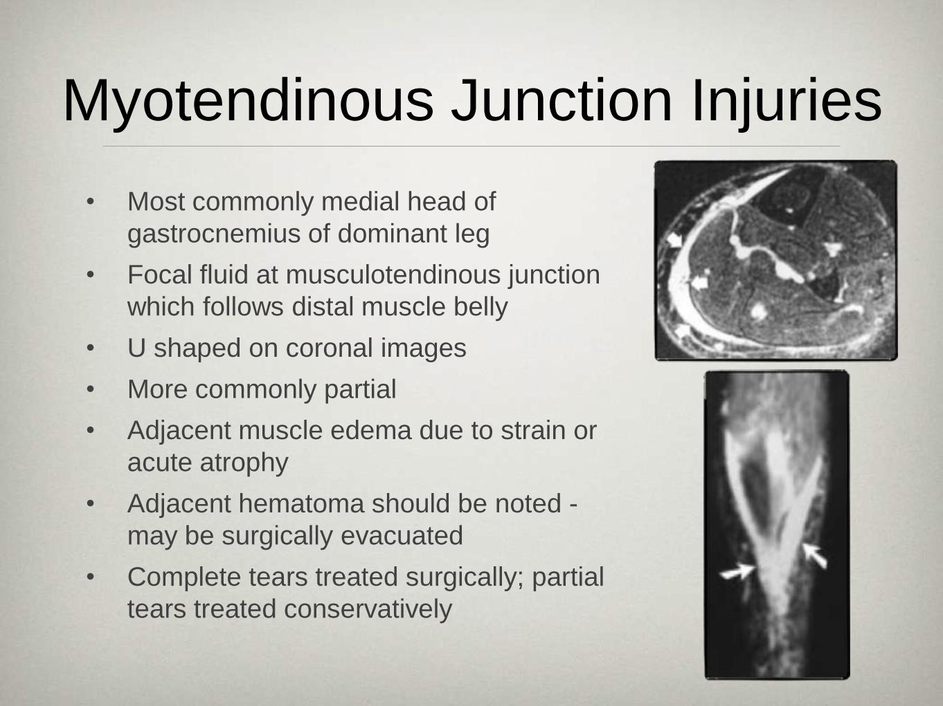
Myotendinous Junction Injuries
• Most commonly medial head of
gastrocnemius of dominant leg
• Focal fluid at musculotendinous junction
which follows distal muscle belly
• U shaped on coronal images
• More commonly partial
• Adjacent muscle edema due to strain or
acute atrophy
• Adjacent hematoma should be noted -
may be surgically evacuated
• Complete tears treated surgically; partial
tears treated conservatively
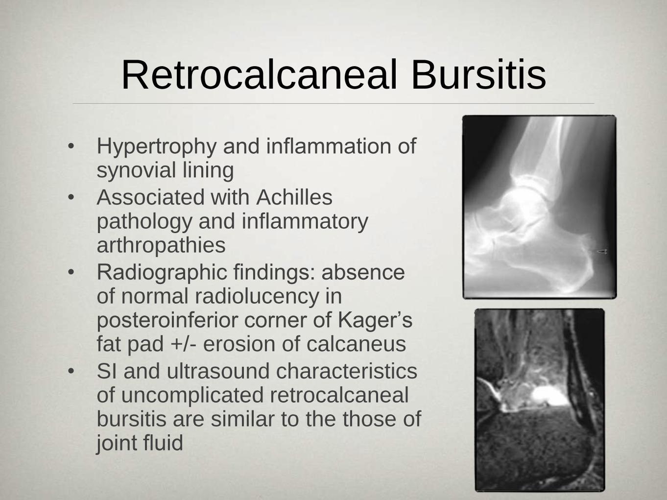
Retrocalcaneal Bursitis
• Hypertrophy and inflammation of synovial lining
• Associated with Achilles pathology and inflammatory arthropathies
• Radiographic findings: absence of normal radiolucency in posteroinferior corner of Kager’s fat pad +/- erosion of calcaneus
• SI and ultrasound characteristics of uncomplicated retrocalcaneal bursitis are similar to the those of joint fluid
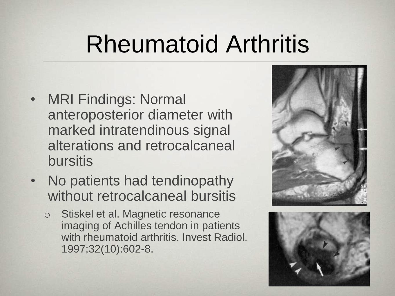
Rheumatoid Arthritis
• MRI Findings: Normal anteroposterior diameter with marked intratendinous signal alterations and retrocalcaneal bursitis
• No patients had tendinopathy without retrocalcaneal bursitis
o Stiskel et al. Magnetic resonance imaging of Achilles tendon in patients with rheumatoid arthritis. Invest Radiol. 1997;32(10):602-8.
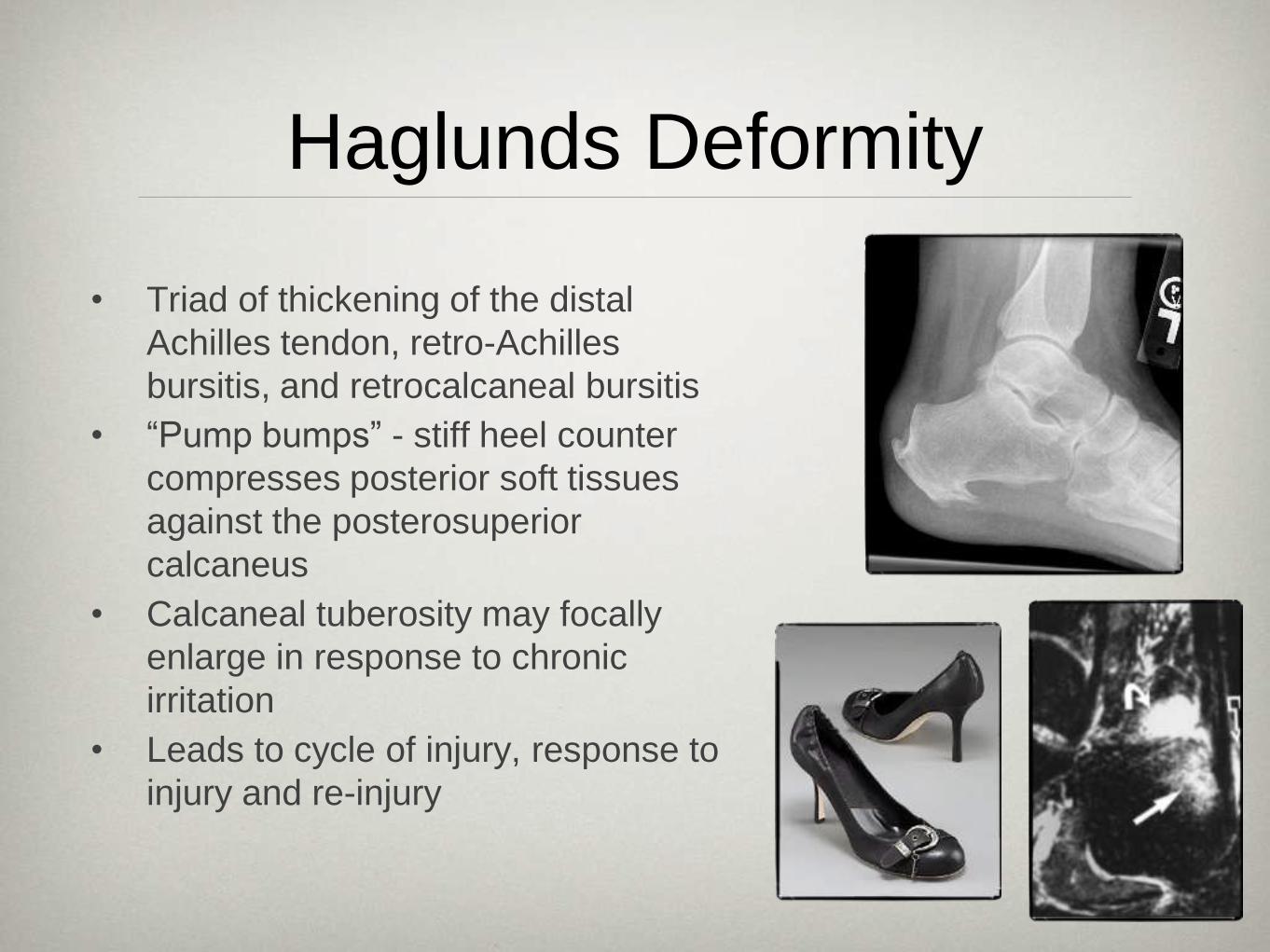
Haglunds Deformity
• Triad of thickening of the distal
Achilles tendon, retro-Achilles
bursitis, and retrocalcaneal bursitis
• “Pump bumps” - stiff heel counter
compresses posterior soft tissues
against the posterosuperior
calcaneus
• Calcaneal tuberosity may focally
enlarge in response to chronic
irritation
• Leads to cycle of injury, response to
injury and re-injury
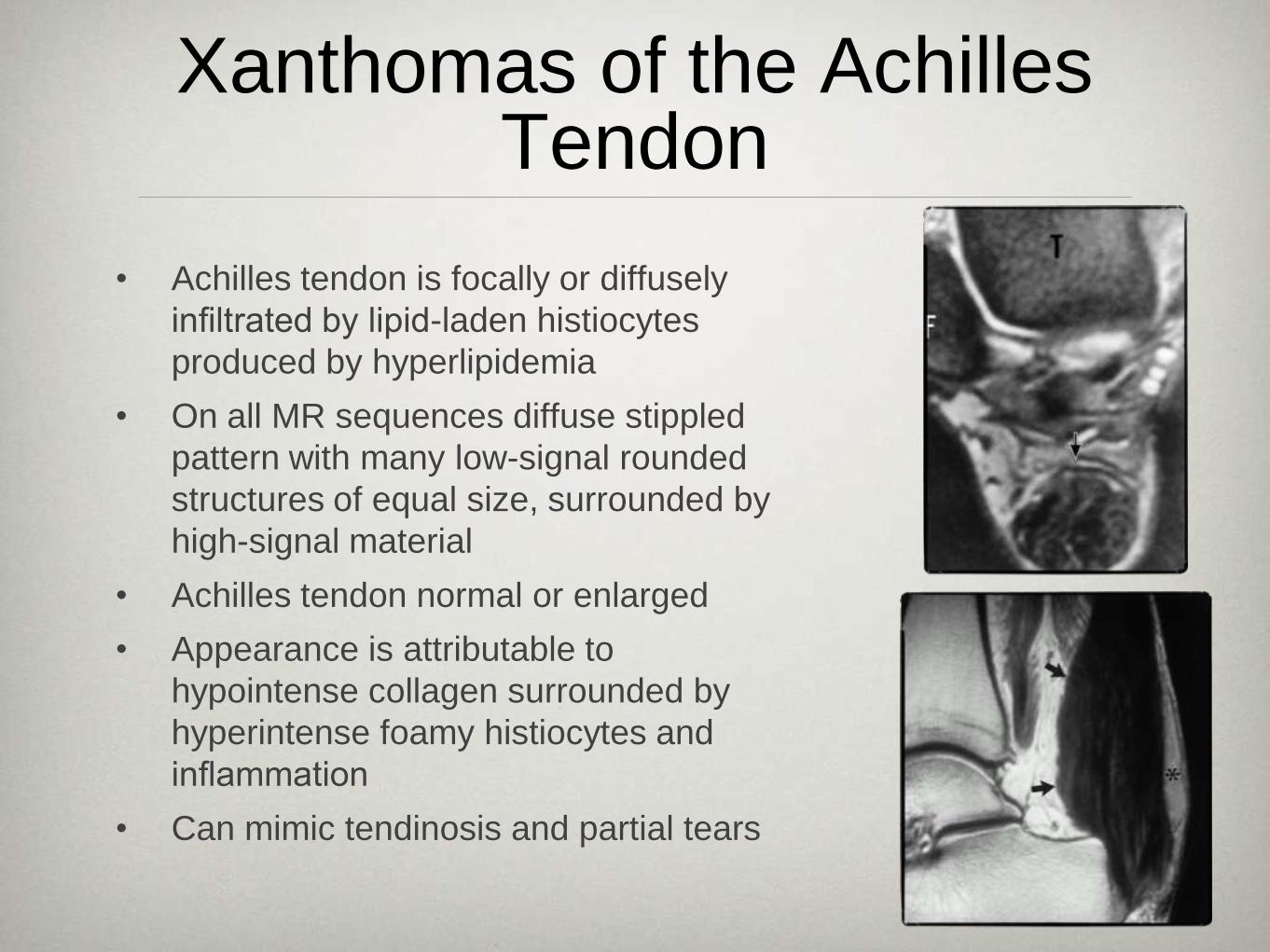
Xanthomas of the Achilles Tendon
• Achilles tendon is focally or diffusely
infiltrated by lipid-laden histiocytes
produced by hyperlipidemia
• On all MR sequences diffuse stippled
pattern with many low-signal rounded
structures of equal size, surrounded by
high-signal material
• Achilles tendon normal or enlarged
• Appearance is attributable to
hypointense collagen surrounded by
hyperintense foamy histiocytes and
inflammation
• Can mimic tendinosis and partial tears

Management

Management Achilles Tendon Ruptures
• Management of complete acute ruptures is controversial
o Operative
• Open: Better functional outcome, lower rate of recurrent rupture, more post-operative complications
• Percutaneous: Higher rate of recurrent rupture, fewer post-operative complications, better cosmetic result
o Nonoperative: High recurrent rupture rate, undesired Achilles lengthening, worse functional outcome
• Treatment for partial ruptures generally conservative
o Surgical debridement when conservative treatment fails
o Confluent areas of intrasubstance signal changes on MRI unlikely to respond to nonoperative treatment

Management Achilles Tendon Ruptures
• Management depends on surgeon and patient preference
• Surgery treatment of choice for athletes, young patients and delayed rupture
• Acute rupture in non-athletes can be treated nonoperatively
• Preoperative MRI/US used to assess:
o Condition of tendon ends
o Orientation of the torn fibers
o Width of diastasis
• With conservative management sagittal imaging may be performed after casting to assess for tendon apposition
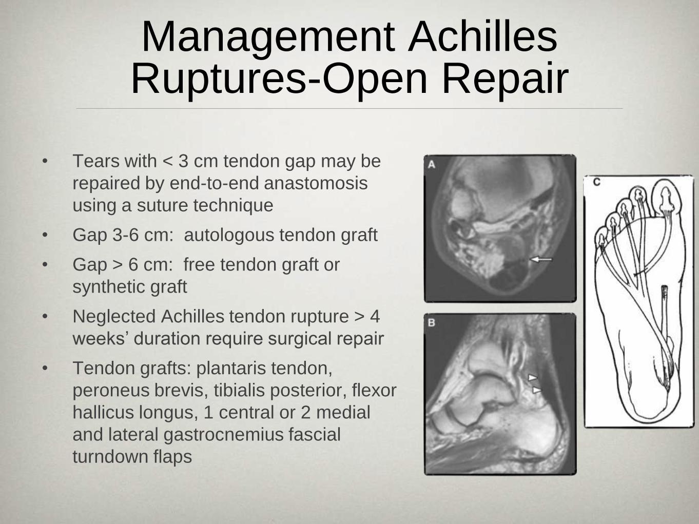
Management Achilles Ruptures-Open Repair
• Tears with < 3 cm tendon gap may be
repaired by end-to-end anastomosis
using a suture technique
• Gap 3-6 cm: autologous tendon graft
• Gap > 6 cm: free tendon graft or
synthetic graft
• Neglected Achilles tendon rupture > 4
weeks’ duration require surgical repair
• Tendon grafts: plantaris tendon,
peroneus brevis, tibialis posterior, flexor
hallicus longus, 1 central or 2 medial
and lateral gastrocnemius fascial
turndown flaps

Management Acute Ruptures-Percutaneous Repair
• Suturing the Achilles tendon and pulling ruptured tendon ends toward each other
• Simpler to perform, better cosmetically outcome and less frequent postoperative infection
• Higher risk of postoperative re-rupture
• Risk of sural nerve injury
• Contact between two ends of the ruptured tendon is incomplete
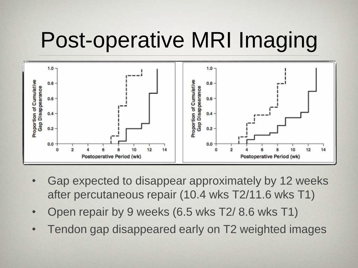
Post-operative MRI Imaging
• Gap expected to disappear approximately by 12 weeks
after percutaneous repair (10.4 wks T2/11.6 wks T1)
• Open repair by 9 weeks (6.5 wks T2/ 8.6 wks T1)
• Tendon gap disappeared early on T2 weighted images

Post-operative MRI Imaging
T2 T1 GAD

The End
Thank you for
providing
original images
Tudor!

References
•Movin et al. Acute Rupture of the Achilles Tendon. Foot Ankle Clin N Am 2005; 10: 331-356
•Young et al. Achilles Tendon Rupture and Tendinopathy: Management of Complications. Foot Ankle Clin N Am. 2005 10: 371-382
•Langber et al. Age related blood flow around the Achilles tendon during exercise in humans. Eur J Appl Physiol 2001; 84: 246-248
•Pichler et al. Anatomic Variations of the Musculotendinous Junction of the Soleus Muscle and Its Clinical Implications. Clinical Anatomy 2007; 20:444–447.
•Ly et al. Anatomy of and Abnormalities Associated with Kager’s Fat Pad. AJR; 182; 147-154
•O’Brien. The Anatomy of the Achilles Tendon. Foot Ankle Clin N Am 2005; 10: 225-238
•Kachlik et al. Clinical anatomy of the calcaneal tuberosity. Annals of Anatomy. 2008
•Kachlik et al. Clinical anatomy of the retrocalcaneal bursa. Surg Radiol Anat 2008.
•Maffulli et al Current Concepts Review: Rupture of the Achilles Tendon. JBJS 1999; 81-A: 1019-1036
•Soila et al. High Resolution MR Imaging of the Asymptomatic Achilles Tendon: New Observations 1999; 173: 1732-323
•Palaniappan et al. Accessory soleus muscle: a case report and review of the literature. Pediatric Radiology 1999; 29: 610-612
•Weishaupt et al. Injuries to Distal Gastrocnemius Muscle: MR Findings. JCAT 2001; 25: 677-682
References
•Kainberger FM. Injury to the Achilles Tendon: DIagnosis with Sonography. AJR 1990; 155:
1031-1036
•Antonios T, et al.. The Medial and Lateral Bellies of Gastrocnemius: A Cadaveric and Ultrasound
Investigation Clinical Anatomy 2008; 21:66–74.
•Karjalainen PT, Aronen HJ, Pihlajamaki HK, Soila K, Paavonen T, Bostman OM. Magnetic
resonance imaging during healing of surgically repaired Achilles tendon ruptures. Am J Sports
Med 1997; 25:164–171
•Maffulli N, Thorpe AP, Smith EW. Magnetic resonance imaging after operative repair of Achilles
tendon rupture. Scand J Med Sci Sports 2001; 11:156–162
•Carr A, Norris S. The blood supply of the calcaneal tendon. J Bone Joint Surg Br 1989;71-B:
100–101
•Frey C, Rosenberg Z, Shereff M, et al. The retrocalcaneal bursa: anatomy and bursography.
Foot Ankle 1982;13:203–207
•Bottger BA, Schweitzer ME, El-Noueam K, Desai M. MR imaging of the normal and abnormal
retrocalcaneal bursae. AJR 1998;170:1239–1241
•Haims A, Schweitzer ME, Patel R, et al. MR imaging of Achilles tendon: overlap of findings in
symptomatic and asymptomatic individuals. Skeletal Radioljuncture of the medial head of the
gastrocnemius muscle. Am J Sports Med 1977;5:191–193
•Bleakne RR et al. Imaging of the Achilles Tendon. Foot Ankle Clin N Am 2005; 10: 239-254