analysis of β-glucans and chitin in a saccharomyces cerevisiae cell wall mutant using...
TRANSCRIPT

YEAST VOL. 10: 1083-1092 (1994)
Analysis of P-Glucans and Chitin in a Saccharomyces cerevisiae Cell Wall Mutant Using High-performance Liquid Chromatography ZHI HONG, PAUL MA", KAREN J. SHAW AND BETH DIDOMENICO*
Chemotherapy and Molecular Genetics, Schering-Plough Research Institute, 2015 Galloping Hill Road, Kenilworth, New Jersey 07033-0539, U. S. A.
Received 24 January 1994; accepted 11 March 1994
We have previously shown that mutations in the yeast KiVR4 gene resulted in pleiotropic cell wall defects, including resistance to killer 9 toxin, elevated osmotic sensitivity to SDS and increased resistance to zymolyase, a (1+3)-P-glucanase. In this report, we further demonstrated that knr4 mutant cells were more permeable to a chromogenic substrate, X-GAL, suggesting that the mutant cell walls were leakier to certain non-permeable molecules. To determine if these defects resulted from structural changes in the cell walls, we analysed the alkali-insoluble cell wall components using HPLC assays developed for this purpose. Comparative analysis using four isogenic strains from a 'knr4 disrupted' tetrad demonstrated that mutant cell walls contained much less (1+3)-P-glucan and (1 +6)-P-glucan; however, the level of chitin, a minor cell wall component, was found to be five times higher in the mutant strains compared to the wild-type strains. The data suggested that the knr4 mutant cell walls were dramatically weakened, which may explain the pleiotropic cell wall defects.
KEY WORDS - Cell wall; glucan; chitin; killer toxin; HPLC; S. cerevisiae.
INTRODUCTION The yeast cell wall is composed of an ever-changing molecular scaffold of polysaccharides, proteins and lipids that serve as an outer envelope, protecting the enclosed protoplast. The major cell wall com- ponents, usually comprising greater than 80% of the cell wall materials, are polysaccharides, fol- lowed by proteins and lipids. The polysaccharides consist primarily of three classes of polymers: poly- mers of mannose covalently linked to peptides (mannoproteins), polymers of glucose (p-glucans), and polymers of N-acetylglucosamine (chitins) (Cabib et al., 1982; Fleet, 1991). p-glucans may be divided into two subtypes that can be structurally distinguished by their predominant linkages be- tween monosaccharide units: the more abundant glucan is (1 --+ 3)-p-glucan, a p-( 1 -+ 3)-linked poly- mer containing a small amount of branches (3%) through p-(1-+6)-~ linkages (Manners et al., 1973a); the minor glucan, (1 +6)-p-glucan7 is a p-( 1 -+6)-linked polymer, containing a somewhat
*Corresponding author.
0 1994 by John Wiley & Sons Ltd CCC 0749-503)094/08 1083-1 0
higher degree of branching (14Yo) through F-(l-+))-D Iinkages (Manners et al., 1973b). It is generally believed that the p-glucans and chitin play very important roles in determining cell morphology, cell division, and osmotic stability of the organism, all of which are critical for survival of the yeast in its natural environment (Bacon et al., 1969, 1970; Bulawa and Osmond, 1990; Cabib et al., 1988; Fleet, 1991; Shaw et al., 1991).
Genetic and molecular analysis of different classes of cell wall components has begun to address some of the questions concerning the complexity and the coordination involved in as- sembling a scaffold of mixed cell wall polymers. Genes involved in cell wall biosynthesis, as well as mutants harboring cell wall defects have been isolated and characterized (Al-Aidroos and Bussey, 1978; Ballou, 1981; Boone et al., 1990, 1991; Brown et al., 1993; Bulawa et al., 1986; Meaden et al., 1990; Ribas et al., 1991; Roemer and Bussey, 1991; Sburlati and Cabib, 1986; Shaw et al., 1991; Shiota et al., 1985; Valdivieso et al., 1991). Recently, we reported on the isolation and

1084 Z. HONG ET AL.
Table 1. Yeast strains used.
Strains Genotype Reference
JY102 MATa, leu2-3,112, lys2-801, ura3-52 J. Greene PM413- 1A MATa, his3, l e d , KRE5, canl’ This study PM413- 1C MATa, his3, l e d , kreSAI::HIS3, c a d r This study ZH40 1 A ZH40 1 B ZH40 1 C ZH40 1 D
MATa, led-3.112, lys2-801, ura3-52 MATa, his4-619, led-3,112, ura3-52 MATa, his4-619, leU2-3,112, ura3-52, knr4::LEUZ MATa, led-3.112, lys2-801, ura3-52, knr4::LEU2
Hong et al. (1994) Hong et al. (1 994) Hong et al. (1994) Hong et al. (1994)
characterization of KNR4 by complementation of a mutant defective in (1 +3)-P-glucan synthase activity with a concomitant 50% reduction in the amount of cell wall (1 +3)-P-glucan (Hong et al., 1994). To verify and further investigate the role of the KNR4p in yeast cell wall biosynthesis, we analysed the carbohydrates of mutant cell walls and compared them to the wild-type cells. Current studies on yeast cell wall structure are generally based on fractionation techniques using specific enzymes and chemicals to separate and measure different cell wall polymers (Fleet, 1991; Manners et al., 1973a,b). The complexity of the cell wall makes it difficult to obtain a homogeneous prep- aration of a specific polymer. Furthermore cross- contamination by other carbohydrates in the enzyme preparations, as well as interference by other cellular components often complicate inter- pretation of the data. Therefore, we developed a series of HPLC assays that permits us to distin- guish between newly synthesized (1 -+3)-P-glucan, (1 +6)-P-glucan and chitin, and to evaluate the relative contribution of each of these polymers in mutant and wild type strains of Saccharomyces cerevisiae.
MATERIALS AND METHODS
Strains, plasmid and media The yeast strains used in this study are listed
in Table 1. ZH401A, ZH401B, ZH401C and ZH40 1 D were spores derived from a single tetrad. PM413- 1 A and PM4 13- 1 C were spore clones from a tetrad derived from a heterozygous diploid which was kindly provided by Dr H. Bussey (McGill University, Montreal, Canada). The plasmid pLGA312 was obtained from Dr L. Guarente (MIT, Cambridge).
Yeast cultures were grown and maintained in YPD medium (1% yeast extract, 2% bacto- peptone, 2% glucose). Yeast transformants were grown in synthetic medium (0.67% yeast nitrogen base, 2% glucose) supplemented with appropriate nutrients. Yeast cells were labeled in a ‘low glucose’ medium (1% yeast extract, 2% bacto- peptone, 0.2% glucose). The X-GAL plates used for the colony color assays have been described previously (Rose and Botstein, 1983).
Chemicals and enzymes All of the isotopes were purchased from NEN
Research Products Co. and Amersham Corp. Zymolyase- 1 OOT was from ICN Pharmaceuticals. Chitinase (Serratia marcescens), laminarinase, a-amylase, X-GAL and sodium-m-periodate were obtained from Sigma Co.
Yeast cell permeability assay Yeast cells were transformed with the plasmid
pLGA312, a CYC1-lacZ construct expressing basal level of P-D-galactosidase activity in the presence of glucose (Guarente et al., 1984), using the lithium acetate method (It0 et al., 1983). Colony-color assays were carried out by growing the transformants on X-GAL plates; the relative cell permeability was estimated visually by the intensity of blue staining of colonies on the plates. The level of P-galactosidase activity in the trans- formants was quantitated as described (Sherman et al., 1986) using chlorophenol red P-D-galacto- pyranoside as the substrate.
Yeast cell wall labeling Yeast cells (-lo4) from a fresh overnight
culture were inoculated into 50ml fresh YPD medium and grown in a 30°C shaker for 16 h to
SACCHAROMYCES CEREVISIAE CELL WALL
Yeast Cells Labeled with 3-[3H]-D-glucose
1085
' (1,6)-B-glucan Total Total (1,3)-O-glucan (1,6)-8-g~ucan Chitin & Glycogen (1,6)-B-glucan Chitin
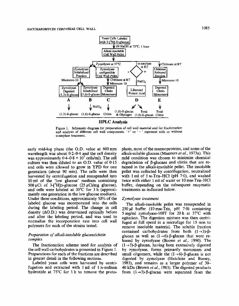
HF'LC Analysis Figure 1. Schematic diagram for preparation of cell wall material and for fractionation and analysis of different cell wall components. '+' or ' - ' represent with or without a-amylase treatment.
early mid-log phase (the O.D. value at 6OOmm wavelength was about 0 . 2 4 4 and the cell density was approximately 04-04 x lo' cells/ml). The cell culture was then diluted to an O.D. value of 0.15 and cells were allowed to grow in YPD for one generation (about 90 min). The cells were then harvested by centrifugation and resuspended into 10ml of the 'low glucose' medium containing 500 pCi of 3-[3H]~-glucose (25 pCi/mg glucose), and cells were labeled at 30°C for 3 h (approxi- mately one generation in the low glucose medium). Under these conditions, approximately 50% of the labeled glucose was incorporated into the cells during the labeling period. The change in cell density (A0.D.) was determined optically before and after the labeling period, and was used to normalize the incorporation rate into cell wall polymers for each of the strains tested.
Preparation of alkali-insoluble glucanslchitin complex
The fractionation scheme used for analysis of the cell wall carbohydrates is presented in Figure 1. Preparations for each of the fractions are described in greater detail in the following sections.
Labeled yeast cells were harvested by centri- fugation and extracted with 1 ml of 1 N-sodium hydroxide at 75°C for 1 h to remove the proto-
plasts, most of the mannoproteins, and some of the alkali-soluble glucans (Manners et al., 1973a). This mild condition was chosen to minimize chemical degradation of p-glucans and chitin that are re- tained in the alkali-insoluble pellet. The insoluble pellet was collected by centrifugation, neutralized with 1 ml of 1 ~-Tris-HCl (PH 7-0), and washed twice with either 1 ml of water or 10 m-Tris-HC1 buffer, depending on the subsequent enzymatic treatments as indicated below.
Zymolyase treatment The alkali-insoluble pellet was resuspended in
250 pl buffer (10 mM-Tris, pH 7.0) containing 5mg/ml zymolyase-100T for 20 h at 37°C with agitation. The digestion mixture was then centri- fuged at full speed in a microfuge for 15 min to remove insoluble material. The soluble fraction contained carbohydrates from both (1 -3)-P- glucan as well as (1+6)-P-glucan that were re- leased by zymolyase (Boone et al., 1990). The (1 -3)-p-glucan, having been extensively digested by zymolyase, forms primarily monomers and small oligomers, while the (1 +6)-P-glucan is not digested by zymolyase (Hutchins and Bussey, 1983), and remains as a larger polymer of 30- 40 kDa (Brown et al., 1983). The digested products from (1 -+3)-P-glucan were separated from the

1086 2. HONG ET A L
(1 +6)-P-glucan by passing the zymolyase-soluble fraction through a Microcon-10 membrane (MW cut off-10000 daltons, Amicon Co.). The run- through (Figure 1, fraction A) containing the degradation products from (1 -+3)-P-glucan was subjected to HPLC analysis as described below. The carbohydrate retained by the column repre- sents the (1 -+6)-P-glucan released by zymolyase (Figure 1, fraction B), which most closely re- sembles the fraction used by others for (1 -+6)-P- glucan analysis (Boone et al., 1990). This material was further degraded by periodate prior to HPLC (see next section).
Periodate oxidation Carbohydrate to be used for (1 -+6)-P-glucan
analysis was treated with 5 0 0 ~ 1 5% sodium- m-periodate at room temperature for 20 h with agitation. Insoluble material was removed by centrifugation; the soluble fraction containing the labeled formic acid liberated from the (1 +6)-P- glucan was analysed and quantitated by HPLC as described below.
Chitinase treatment Most of the chitin (-80%) was retained in the
zymolyase-resistant cell wall pellet. It was washed once with 1 ml of 10 m-Tris-HC1 (pH 6.0), resus- pended in 2 5 0 ~ 1 of the same buffer containing 50 mg/ml chitinase (lyophilized enzyme mixture prepared from Serratia marcescens, Sigma Co.) and digested at room temperature for 20 h with agitation. Insoluble material was removed by cen- trifugation, the supernatant was passed through a Microcon- 10 membrane and precipitated with 70% acetonitrile. The resulting supernatant containing the digested chitin products (Figure 1, fraction C) was suitable for HPLC analysis as described below.
a-Amylase digestion 2.5 mg/ml a-amylase, a (1 +4)-a-glucanohy-
drolase, was added to the alkali-insoluble cell wall pellet. The reaction was carried out in 2 5 0 ~ 1 10 mM-Tris-HC1 (pH 7-0)) at room temperature for 16 h and insoluble material was collected by centrifugation for further analysis.
Carbohydrate analysis by HPLC HPLC was performed using a Waters 680 auto-
mated gradient controller coupled with a Waters
712 WISP autosampler and a Waters 590 pro- grammable pump (Waters Chromatography, Millipore Corp.). The HPLC effluent was monitored by a radioactive flow detector (Flo- One/Beta A200 from Radiomatic Co.) using the Flo-Scint A scintillation liquid (Radiomatic Co.). Cell wall degradation products from zymolyase or chitinase digestion were separated and analysed using a Waters Carbohydrate Analysis column (part no. 84038). The chromatography was carried out at room temperature using 70% acetonitrile as the mobile phase and the flow rate was set at 1 mumin. The formic acids liberated from (1 +6)- P-glucan by periodate oxidation were analysed using a Waters Ionpak KC-811 column (part no. 342980 coupled with a KC-810P precolumn (part no. 35501). The column temperature was set at 60°C and the flow rate was 1 ml/min. The mobile phase was 0.1% phosphoric acid.
RESULTS AND DISCUSSION We have previously shown that the yeast KNR4 gene is likely to be involved in cell wall bio- synthesis. Mutations in this gene resulted in pleio- tropic cell wall defects, including resistance to k9 toxin which inhibits (1 +3)-P-glucan synthesis (Yamamoto et al., 1986, 1988), sensitivity to an osmotic destabilizing agent, SDS, and resistance to enzymatic digestion by a (1 +3)-P-glucanase (Hong et al., 1994). To further investigate changes associated with the knr4 mutants, we analysed the relative permeability and carbohydrate compo- sition of four isogenic sibling strains derived from a knr4::LEU2 disrupted tetrad: ZH401A and ZH401 B containing the wild-type alleles; ZH401C and ZH401D containing the mutant alleles. The KNR4 gene product was determined to be com- pletely absent in the mutant strains by Western analysis (Hong et al., 1994).
Relative cell permeability of knr4 mutants To demonstrate that the knr4 mutant strains
had acquired a functional cell wall defect, we measured cell permeability using a colony-color assay (Materials and Methods). For this exper- iment, each strain from the tetrad was transformed with a plasmid, pLGA312, which expresses a basal level of P-galactosidase in the presence of glucose (Guarente et al., 1984). When grown on plates containing the chromogenic substrate X-GAL, knr4-2 mutants (ZH401C and ZH401D) turned

SACCHAROMYCES CEREVISIAE CELL WALL 1087
ZH401A ZH401B
8-galactosidase 218 237 (units)
ZH40 1 C ZH40 1 D
243 225 Figure 2. Yeast cell permeability assay. pLGA312 transformants of both the wild-type strains (ZH401A and ZH401B) and the knr4 mutant strains (ZH401C and ZH401D) were plated on a medium containing 2% dextrose and 50 pg/ml X-GAL. The plate was incubated at 30°C or 37°C for 2-3 days. The number below each colony represents the corresponding intracellular P-galactosidase activity measured in units.
blue, while wild-type cells (ZH401A and ZH401B) remained white (Figure 2). In addition, the blue staining of the knr4 mutants could be reversed by the presence of a plasmid containing the KNR4 gene (data not shown). Since color development is determined by either the level of intracellular P-galactosidase activity or permeability to the substrate, we quantitated the enzyme activity in cell lysates from each of the strains. The data showed that there was little difference in enzyme activity between the four strains (Figure 2), sug- gesting that the enhanced blue color observed in the mutant cells was not due to changes in Lac2 expression, rather, it was likely due to an increase in cell permeability. The increase in cell permeability may result from a dysfunctional cell wall which could lead to partial or transient cell membrane lysis.
Measurement of ( I -3)-P-glucan content To analyse the cell wall structure, labeled degra-
dation products from (1 +3)-p-glucan were pre- pared from each of the four sibling strains, as described in Materials and Methods and outlined in Figure 1, fraction A. The results of the HPLC
analysis are shown in Figure 3. For each of the mutant and wild-type strains tested, at least three major peaks (I, I1 and 111) were eluted off the HPLC column. The first peak was the most abun- dant and had the same retention time (7.3 min) as the standard, [3H]~-glucose, suggesting that it rep- resents the glucose monomer derived from (1 +3)- P-glucan. Under the column conditions used, the monomer eluted first, followed by larger oligo- mers. The second and third peaks (having reten- tion times of 9.4 and 14.9 min, respectively) were most likely glucose oligomers since we were able to convert them to glucose by a com- bined digestion with zymolyase and laminari- nase, or by total acid hydrolysis (data not shown). It is known that zymolyase is not very active on soluble substrates and produces a mix- ture of degradation products of different sizes (Kitamura, 1982; Kitamura et al., 1972). Further- more, previous studies demonstrated that the (1 +3)-P-glucan polymer from S. cerevisiae contains very few (1 +3)4 1-6) branches (only 3%) (Manners et al., 1973a; Shiota et al., 1985). Thus, it is likely that most of the oligomers in peaks I1 and I11 were incomplete zymolyase
Glc ~~~~1 <rIl: ylI!l , c 4 B , : h Z y l : ~~~~1 : r : l D ,
14.9 m X
OO 10 20 0 10 20 OO 10 20 0 10 20 Minutes Minutes Minutes Minutes
A B C D Figure 3. HPLC analysis of zymolyase-released (1 +3)-P-glucan products. (A-D) Zymolyase- digested (1 +3)-P-glucan products from spore clones ZH401A, ZH401B, ZH401C and ZH401D respectively. Samples loaded onto the HPLC column were normalized to cell growth rate (AOD) during the labeling period.

1088 2. HONG ET AL.
Table 2. Measurement of newly sythesized P-glucans and chitin.
Strains (1 +3)+glucan* (1 +6)-P-glucan Chitin?
ZH40 1 A ZH40 1 B ZH40 1 C ZH40 1 D
174.2 f 30.6 168.9 f 23.9 106.4 f 17.3 103.7 f 10-6
21.9 f 4.0 21.3 f 2.8
5.7 f 1.5 5.3 f 1.3
9.0 f 2.4 8.2 f 2.4
42.4 f 2.4 40.8 f 4.9
*Total radioactivity incorporated into p-glucans was converted to milligrams of newly synthesized p-glucans, based on the assumption that the specific radioactivity in newly synthesized glucans should be very similar to that in the labeling medium (lo6 cpm-45 pg glucose). The amount of glucans was then normalized to grams of dry weight of the cells in units of mglg cell dry weight as described in the text. The data here represent averages of at least three independent labeling of experiments and HPLC analysis. ?Total radioactivity incorporated into chitin was also converted to milligrams of de n o w sythesized chitin. In this case, lo6 cpm are equivalent to 56 pg N-acetylglucosamine. The amount of chitin was also normalized and expressed in mglg cell dry weight.
digestion products rather than (1 +3)/(1+6) branch points.
A comparison between the HPLC tracings showed that there was a significant reduction in the peak areas from mutant cells compared to wild- type cells (Figure 3), suggesting that the mutant strains contained a reduced amount of (1 +3)-P- glucan linkages relative to wild-type strains. The total amount of radioactivity loaded onto the column for each strain was normalized to the growth rate of the cells during the labeling period to account for any difference in glucose uptake. In fact, there was little difference in the rate of growth or glucose uptake between the strains. Since ac- tively growing yeast cells have a very small intra- cellular pool of glucose (Becker and Betz, 1972; Jaspers and van Steveninck, 1975), the specific radioactivity incorporated in the newly synthesized P-glucans should be similar to that in the labeling medium (about lo6 cpm-45 pg glucose). The total radioactivity can therefore be converted into milligrams of P-glucan per gram of dry weight of cells that was synthesized during the labeling period. We observed a near two-fold reduction in the amount of (1 +3)-P-glucan synthesized in knr4 mutants compared with the wild-type strains (Table 2). This correlated well with our previous studies measuring (1 +3)-P-glucan synthase ac- tivity from mutant cells compared with wild-type cells (Hong et al., 1994).
Quantitation of ( 1 +6)-fi-glucan Approximately 90% of the total (1 +6)-P-glucan
in the alkali-insoluble cell wall pellet is released
into the zymolyase-soluble fraction (H. Bussey, personal communication). However, we were un- able to quantify this polymer directly by HPLC due to its large size, which prohibits the carbo- hydrate from entering the column. Instead, we took advantage of the specificity of periodate oxidation on (1 +6)+-glucan. Since each glucose residue in a linear (1+6)-P-glucan has three hy- droxyl groups on adjacent carbon atoms (C2, C, and C4), periodate can oxidize and cleave C-C bonds between C, and C , as well as between C, and C,, resulting in the liberation of the C, atom in the form of formic acid. The amount of formic acid can then be quantitated using HPLC and represents the relative molar concentration of glu- cose with linear P-(1+6) linkages. It should be noted that periodate oxidation will also release formic acid from the non-reducing ends of (1 +3)- P-glucan and from glycogen. Both sources of contamination in fraction B (see Figure 1) are negligible due to zymolyase degradation and prior size separation. However, the contamination is significant when analysing (1 +6)-(3-glucan in fraction D as explained below.
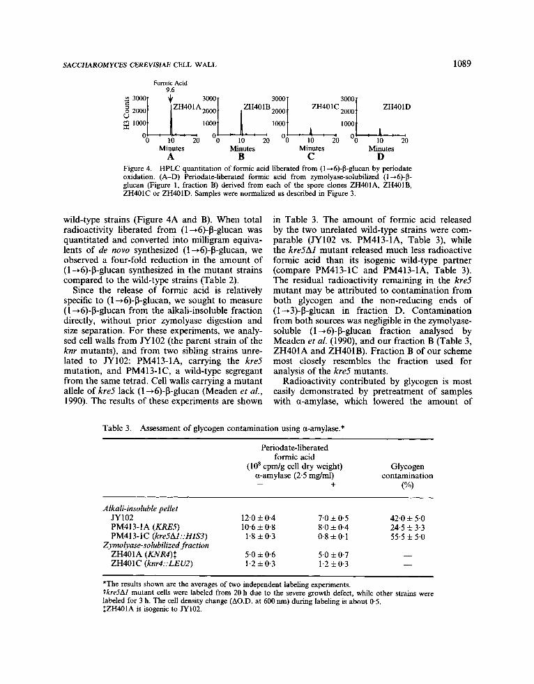
Carbohydrate from each of the four strains was prepared and treated as described. The results from HPLC analysis of (1 -6)-P-glucan in fraction B are shown in Figure 4. The standard, formic acid, has a column retention time of 9.6 min. The HPLC analysis revealed that the major product released from each strain by periodate co-eluted with formic acid. Comparative analysis of the four isogenic strains showed a significant reduction in the quantity of formic acid released from the knr4 mutant strains (Figure 4C and D) compared to the
SACCHAROMYCES CEREVISlAE CELL WALL 1089
Formic Acid 9.6
Minutes Minutes Minutes Minutes
Figure 4. HPLC quantitation of formic acid liberated from (1 -6)-P-glucan by periodate oxidation. (A-D) Periodate-liberated formic acid from zymolyase-solubilized (1 +6)-p- glucan (Figure 1, fraction B) derived from each of the spore clones ZH401A, ZH4OlB, ZH401C or ZH401D. Samples were normalized as described in Figure 3.
A B C D
wild-type strains (Figure 4A and B). When total radioactivity liberated from (1 +6)-P-glucan was quantitated and converted into milligram equiva- lents of de now synthesized (1 +6)-P-glucan, we observed a four-fold reduction in the amount of (1 +6)-P-glucan synthesized in the mutant strains compared to the wild-type strains (Table 2).
Since the release of formic acid is relatively specific to (1 +6)-P-glucan, we sought to measure (1 +6)-P-glucan from the alkali-insoluble fraction directly, without prior zymolyase digestion and size separation. For these experiments, we analy- sed cell walls from JY102 (the parent strain of the knr mutants), and from two sibling strains unre- lated to JY102: PM413-1A, carrying the kre5 mutation, and PM413-lC, a wild-type segregant from the same tetrad. Cell walls carrying a mutant allele of kre5 lack (1 +6)-P-glucan (Meaden et al., 1990). The results of these experiments are shown
in Table 3. The amount of formic acid released by the two unrelated wild-type strains were com- parable (JY102 vs. PM413-1A, Table 3), while the kre5AZ mutant released much less radioactive formic acid than its isogenic wild-type partner (compare PM413-1C and PM413-1A, Table 3). The residual radioactivity remaining in the kre5 mutant may be attributed to contamination from both glycogen and the non-reducing ends of (1 -+3)-P-glucan in fraction D. Contamination from both sources was negligible in the zymolyase- soluble (1 +6)-P-glucan fraction analysed by Meaden et al. (1990), and our fraction B (Table 3, ZH401A and ZH401B). Fraction B of our scheme most closely resembles the fraction used for analysis of the kre5 mutants.
Radioactivity contributed by glycogen is most easily demonstrated by pretreatment of samples with a-amylase, which lowered the amount of
Table 3. Assessment of glycogen contamination using a-amylase.* ~ ~~~
Periodate-liberated formic acid
(10' c p d g cell dry weight) Glycogen a-amylase (2.5 mg/ml) contamination
+ ("w -
Alkali-insoluble pellet JY102 12.0 f 0.4 7.0 f 0.5 42.0 f 5.0 PM413-1A (KRE5) 10.6 f 0.8 8.0 f 0.4 24.5 * 3.3 PM413-IC (kreSAI::HZS3) 1.8 f 0.3 0.8 f 0.1 55.5 f 5.0
Zymolyase-solubilized fraction ZH401A (KNR4)S 5.0 f 0.6 5.0 f 0.7 -
1.2 f 0.3 - ZH401C (knr4::LEU2) 1.2 f 0.3
*The results shown are the averages of two independent labeling experiments. tkre5Al mutant cells were labeled from 20 h due to the severe growth defect, while other strains were labeled for 3 h. The cell density change (A0.D. at 600 nm) during labeling is about 0.5. IZH401A is isogenic to JY102.
1090 Z. HONG ET AL..
0 10 20 OO 10 20 0 10 20 0 10 20 Minutes Minutes Minutes Minutes
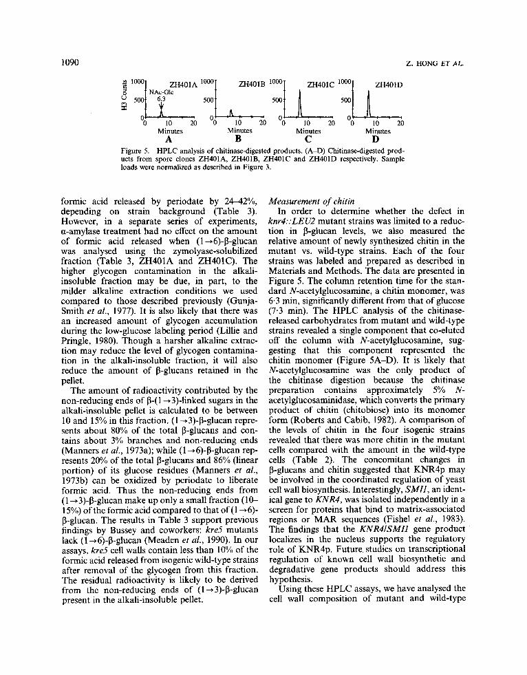
Figure 5. HPLC analysis of chitinase-digested products. (A-D) Chitinase-digested prod- ucts from spore clones ZH4OIA, ZH401B, ZH401C and ZH401D respectively. Sample loads were normalized as described in Figure 3.
A B C D
formic acid released by periodate by 2442Y0, depending on strain background (Table 3). However, in a separate series of experiments, a-amylase treatment had no effect on the amount of formic acid released when (1 +6)-P-glucan was analysed using the zymolyase-solubilized fraction (Table 3, ZH401A and ZH401C). The higher glycogen contamination in the alkali- insoluble fraction may be due, in part, to the milder alkaline extraction conditions we used compared to those described previously (Gunja- Smith et al., 1977). It is also likely that there was an increased amount of glycogen accumulation during the low-glucose labeling period (Lillie and Pringle, 1980). Though a harsher alkaline extrac- tion may reduce the level of glycogen contamina- tion in the alkali-insoluble fraction, it will also reduce the amount of P-glucans retained in the pellet.
The amount of radioactivity contributed by the non-reducing ends of 0-( 1 +3)-linked sugars in the alkali-insoluble pellet is calculated to be between 10 and 15% in this fraction. (1 -+3)-P-glucan repre- sents about 80% of the total P-glucans and con- tains about 3% branches and non-reducing ends (Manners et al., 1973a); while (1 +6)-P-glucan rep- resents 20% of the total P-glucans and 86% (linear portion) of its glucose residues (Manners et al., 1973b) can be oxidized by periodate to liberate formic acid. Thus the non-reducing ends from (1 43)-P-glucan make up only a small fraction (10- 15%) of the formic acid compared to that of (1 +6)- P-glucan. The results in Table 3 support previous findings by Bussey and coworkers: kre5 mutants lack (1 +6)-P-glucan (Meaden et al., 1990). In our assays, kre5 cell walls contain less than 10% of the formic acid released from isogenic wild-type strains after removal of the glycogen from this fraction. The residual radioactivity is likely to be derived from the non-reducing ends of (1 +3)-P-glucan present in the alkali-insoluble pellet.
Measurement of chitin In order to determine whether the defect in
knr4::LEU2 mutant strains was limited to a reduc- tion in P-glucan levels, we also measured the relative amount of newly synthesized chitin in the mutant vs. wild-type strains. Each of the four strains was labeled and prepared as described in Materials and Methods. The data are presented in Figure 5 . The column retention time for the stan- dard N-acetylglucosamine, a chitin monomer, was 6-3 min, significantly different from that of glucose (7.3 min). The HPLC analysis of the chitinase- released carbohydrates from mutant and wild-type strains revealed a single component that co-eluted off the column with N-acetylglucosamine, sug- gesting that this component represented the chitin monomer (Figure 5A-D). It is likely that N-acetylglucosamine was the only product of the chitinase digestion because the chitinase preparation contains approximately 5% N- acetylglucosaminidase, which converts the primary product of chitin (chitobiose) into its monomer form (Roberts and Cabib, 1982). A comparison of the levels of chitin in the four isogenic strains revealed that there was more chitin in the mutant cells compared with the amount in the wild-type cells (Table 2). The concomitant changes in P-glucans and chitin suggested that KNR4p may be involved in the coordinated regulation of yeast cell wall biosynthesis. Interestingly, SMZl, an ident- ical gene to KNR4, was isolated independently in a screen for proteins that bind to matrix-associated regions or MAR sequences (Fishel et al., 1983). The findings that the KNRIISMIl gene product localizes in the nucleus supports the regulatory role of KNR4p. Future. studies on transcriptional regulation of known cell wall biosynthetic and degradative gene products should address this hypothesis.
Using these HPLC assays, we have analysed the cell wall composition of mutant and wild-type

SACCHAROMYCES CEREVISIAE CELL WALL 1091
strains. These assays are simple to run and yield easily quantifiable results. We demonstrated that strains defective in knr4 contained less 0-glucans and more chitin than their wild-type counterparts. However, one of the difficulties in cell wall carbo- hydrate analysis has been the inability to distin- guish between absolute changes in composition versus changes in structure, so that an apparent polymer decrease in one fraction may really reflect its change in solubility. This is particularly crucial in the analysis of cell wall P-glucans where there is a distinction between alkali-soluble and alkali- insoluble glucan fractions. Evidence from several laboratories has suggested that the insoluble fraction is cross-linked to chitin: treatments with chitinase or nitrous acid increase the solubility of P-glucans (Mol and Wessels, 1987; Sietsma and Wessels, 1981, 1990). Furthermore, strains de- ficient in chitin synthase I11 activity (call mutants) contain a much higher percentage of alkali-soluble glucan than their wild-type counterparts (Roncero et al., 1988). We would argue that in the case of the knr4 mutants, the apparent decrease in the alkali- insoluble P-glucans is probably not a consequence of changes in solubility, since total sugar analysis on the alkali-soluble fraction revealed a 33% reduc- tion in the amount of hexose present compared with isogenic wild-type strains. Indirect methods of determining mannose composition reflected no dif- ferences between the strains (data not shown). In addition, the reduction of glucan synthase activity in these strains supports the finding that there is less (1 -3)-P-glucan in the mutants. Even with the substantial decrease in the major wall polymer, knr4 mutants display nearly normal growth rates at all temperatures tested, causing us to postulate that the increase in chitin strengthens the cell wall scaf- fold. Further studies of the composition of this and other cell wall-defective mutants should lead to a more complete understanding of the structure of the yeast cell wall.
ACKNOWLEDGEMENT We thank Roberta S. Hare, Jonathan R. Greene and Alan Cooper for helpful discussions and inter- est in this work. We also thank Jonathan Pachter for help with the HPLC analysis.
REFERENCES Al-Aidroos, K. and Bussey, H. (1978). Chromosomal
mutants of Saccharomyces cerevisiae affecting the cell
wall binding site for killer factor. Can. J. Microbiol.
Bacon, J. S. D., Farmer, V. C., Jones, D. and Taylor, I. F. (1969). The glucan components of the cell wall of baker’s yeast considered in relation to its ultrastruc- ture. Biochem. 114, 557-567.
Bacon, J. S. D., Gordon, A. H., Jones, D., Taylor, I. F. and Webley, D. M. (1970). The separations of P-glucanases produced by Cytophaga johnsonii and their role in the lysis of yeast cell walls. Biochem. 120, 67-78.
Ballou, C. E. (1981). Yeast cell wall and cell surface. In Strathem, J. N., Jones, E. W. and Broach, J. R. (Eds), Molecular Biology of the Yeast Saccharomyces: Me- tabolism and Gene Expression. Cold Spring Harbor Laboratories, Cold Spring Harbor, New York, pp. 335-360.
Becker, J. U. and Betz, A. (1972). Membrane transport as controlling pacemaker of glycolysis in Saccharomy- ces carlsbergensis. Biockim. Biophys. Acta 274, 5 8 4 597.
Boone, C., Sdicu, A.-M., Laroche, M. and Bussey, H. (1991). Isolation from Candida albicans of a func- tional homolog of the Saccharomyces cerevisiae KREl gene, which is involved in cell wall P-glucan synthesis. J. Bacteriol. 173, 685S6864.
Boone, C., Sommer, S. S., Hensel, A. and Bussey, H. (1990). Yeast KRE genes provide evidence for a pathway of cell wall P-glucan assembly. J. Cell Biol.
Brown, J . L., Kossaczka, Z. , Jiang, B. and Bussey, H. (1993). A mutational analysis of killer toxin resistance in S. cerevisiae identifies new genes involved in cell wall (1,6)-P-glucan synthesis. Genetics 113, 837-849.
Bulawa, C. E. and Osmond, B. C. (1990). Chitin syn- thase I and chitin synthase I1 are not required for chitin synthesis in vivo in Saccharomyces cerevisiae. Proc. Natl. Acad. Sci. USA 87, 7424-7428.
Bulawa, C. E., Slater, M. L., Cabib, E., Au-Young, J., Sburlati, A., Adair, W. L. and Robbins, P. W. (1986). The S. cerevisiae structural gene for chitin synthase is not required for chitin sythesis in vivo. Cell 46, 213- 225.
Cabib, E., Bowers, B., Sburlati, A. and Silverman, S. J. (1988). Fungal cell wall synthesis: the construction of a biological structure. Microbiol. Sci. 5, 370-375.
Cabib, E., Roberts, R. and Bowers, B. (1982). Synthesis of yeast cell wall synthesis and its regulation. Annu. Rev. Biochem. 51, 763-793.
Fishel, B. R., Sperry, A. 0. and Garrard, W. T. (1993). Yeast calmodulilr and a conserved nuclear protein participate in the in vivo binding of a matrix associ- ation region. Proc. Natl. Sci. Acad. USA 90, 5623- 5627.
Fleet, G. H. (1991). Cell walls. In Rose, A. H. and Harrison, J. S. (Eds), The Yeasts. Academic Press, New York, pp. 41199-277.
24, 228-237.
110, 1833-1843.

1092 Z. HONG ET AL.
Guarente, L., Lalonde, B., Gifford, P. and Alani, E. (1984). Distinctly regulated tandem upstream acti- vation sites mediate catabolite repression of the CYCI gene of Saccharomyces cerevisiae. Cell 36, 503-5 1 1.
Gunja-Smith, Z., Patil, N. B. and E., S. E. (1977). Two pools of glycogen in Saccharomyces. J. Bacteriol. 130,
Hong, Z., Mann, P., Brown, N. H., Tran, L. E., Shaw, K. J., Hare, R. S. and DiDomenico, B. (1994). Clon- ing and characterization of m R 4 : a yeast gene in- volved in (1,3)-P-glucan synthesis. Mol. Cell. Biol. 14,
Hutchins, K. and Bussey, H. (1983). Cell wall receptor for yeast killer toxin: Involvement of (1 -6)-P-~-glucan. J. Bacteriol. 154, 161-169.
Ito, H., Fukuda, Y., Murata, K. and Kimura, A. (1983). Transformation of intact yeast cells treated with alkali cations. J. Bacteriol. 153, 163-168.
Jaspers, H. T. A. and van Steveninck, J. (1975). Transport-associated phosphorylation of 2-dideoxy- D-glUCOSe in Saccharomyces fragilis. Biochim. Biophys. Acta 406, 370-385.
Kitamura, K. (1982). Re-examination of zymolyase puri- fication. Agric. Biol. Chem. 46,963-969.
Kitamura, K., Kaneko, T. and Yamamoto, Y. (1972). Lysis of viable yeast cells by enzymes of Anthrobacter luteus. I. Isolation of lytic strain and studies on its lytic activity. J. Gen. Appl. Microbiol. 18, 57-71.
Lillie, S. H. and Pringle, J. R. (1980). Reserve carbo- hydrate metabolism in Saccharomyces cerevisiae: response to nutrient limitation. J. Bacteriol. 143, 1384-1394.
Manners, D. J., Masson, A. J. and Patterson, J. C. (1973a). The structure of a P-(l-3)-~-glucan from yeast cell walls. Biochem. J. 135, 19-30.
Manners, D. J., Masson, A. J., Patterson, J. C., Bjorndal, J. and Lindberg, B. (1973b). The structure of a P-(l-6)-~-glucan from yeast cell walls. Biochem. J.
Meaden, P., Hill, K., Wagner, J., Slipeta, D., Sommer, S. S. and Bussey, H. (1990). The yeast KRE.5 gene encodes a probable endoplasmic reticulum protein required for (1-6)-P-~-glucan synthesis and normal cell growth. Mol. Cell. Biol. 10, 3013-3019.
Mol, P. C. and Wessels, J. G. H. (1987). Linkages between glucosaminoglycan and glucan determine alkali-insolubility of the glucan in walls of Saccharo- myces cerevisiae. FEMS Microbiol. Lett. 41, 95-99.
Ribas, J. C., Kiaz, M., Duran, A. and Perez, P. (1991). Isolation and characterization of Schizosaccharomy- ces pombe mutants defective in cell wall (1-3)-P-~- glucan. J. Bacteriol. 173, 34563462.
8 18-825.
1017-1025.
135, 31-36.
Roberts, R. L. and Cabib, E. (1982). Serratia marcescens chitinase: one-step purification and use for the deter- mination of chitin. Anal. Biochem. 127,402412.
Roemer, T. and Bussey, H. (1991). Yeast P-glucan synthesis: KRE6 encodes a predicted type I1 mem- brane protein required for glucan synthesis in vivo and for glucan synthase activity in vitro. Proc. Natl. Acad. Sci. USA 88, 11295-1 1299.
Roncero, C., Valdivieso, M. H., Ribas, J. C. and Duran, A. (1988). Isolation and chracterization of Saccharo- myces cerevisiae mutants resistant to calcofluor white. J. Bacteriol. 170, 1950-1954.
Rose, M. and Botstein, D. (1983). Construction and use of gene fusions to Lac2 (P-galactosidase) that are expressed in yeast. Methods Enzymol. 101, 167-180.
Sburlati, A. and Cabib, E. (1986). Chitin synthetase 2, a presumptive participant in septum formation in Saccharomyces cerevisiae. J. Biol. Chem. 261, 15147- 15152.
Shaw, J. A., Mol, P. C., Bowers, B., Silverman, S. J., Valdivieso, M. H., Duran, A. and Cabib, E. (1991). The function of chitin synthase 2 and 3 in the Sac- charomyces cerevisiae cell cycle. J. Cell Biol. 114,
Sherman, F., Fink, G. R. and Hicks, J. B. (1986). Methods in Yeast Genetics. Cold Spring Harbor Lab- oratory, Cold Spring Harbor, New York.
Shiota, M., Nakajima, T., Satoh, A., Shida, M. and Matsuda, K. (1985). Comparison of p-glucan struc- tures in a cell wall mutant of Saccharomyces cerevisiae and the wild type. J. Biochem. 98, 1301-1307.
Sietsma, J. H. and Wessels, J. G. H. (1981). Solubility of (1,3)-p-D/( 1,6)-P-~-glucan in fungal walls: importance of presumed linkage between glucan and chitin. J. Gen. Microbiol. 125, 209-212.
Sietsma, J. H. and Wessels, J. G. H. (1990). The occurrence of glucosaminoglycan in the wall of Schizosaccharomyces pombe. J. Gen. Microbiol. 136,
Valdivieso, M. H., Mol, P. C., Shaw, J. A., Cabib, E. and Duran, A. (1991). CAL1, a gene required for activity of chitin synthase 3 in Saccharomyces cere- visiae. J. Cell Biol. 114, 101-109.
Yamamoto, T., Hiratani, T., Hirata, H., Imai, M. and Yamaguchi, H. (1986). Killer toxin from Hansenula mrakii selectively inhibits cell wall synthesis in a sensitive yeast. FEBS 197, 50-54.
Yamamoto, T., Uchida, K. and Hiratani, T. (1988). In vitro activity of the killer toxin from yeast Hansenula mrakii against yeasts and molds. J. Antibiot. (Tokyo) 41, 398403.
11 1-123.
2261-2265.