an assemblage of lizards from the early cretaceous of japan · pared manually. this delicate and...
TRANSCRIPT
Palaeontologia Electronica palaeo-electronica.org
An assemblage of lizards from the Early Cretaceous of Japan
Susan E. Evans and Ryoko Matsumoto
ABSTRACT
The Early Cretaceous deposits of the Tetori Group of western Japan have yieldeda diverse wetland vertebrate fauna including both aquatic and terrestrial components.The latter include several lizards, three of which have been named and described indetail: Kaganaias hakusanensis, a long-bodied aquatic lizard; Kuwajimalla kagaensis,a herbivorous borioteiioid; and Sakurasaurus shokawensis, a relative of the ChineseJehol genus Yabeinosaurus. Here we describe lizard material from the Shiraminelocality representing five or six additional taxa, three of which are named herein: asmall lizard represented by two associations, but of unresolved phylogenetic position;a slightly larger lizard with tricuspid teeth that is related to borioteiioids; and a bizarrelizard with bicuspid teeth represented by a single, but morphologically unique, jaw. Thethree additional lizard morphotypes are unnamed. One has bicuspid teeth butunspecialised jaws. The second has small unicuspid teeth in a dentary bearing a deepcoronoid process and resembling the dentary of the enigmatic Late Cretaceous Mon-golian Myrmecodaptria microphagosa. The third morphotype is represented by a singlefragmentary specimen and has small teeth in a deep jaw. Together, the Kuwajima liz-ards form a phylogenetically and morphologically diverse assemblage.
Susan E. Evans. Department of Cell and Developmental Biology, University College London, Gower Street, London WC1E 6BT, UK. [email protected] (corresponding author)Ryoko Matsumoto. Kanagawa Prefecture Museum of Natural History, 499 Iryuuda, Odawara, Kanagawa Prefecture, 250-0035, Japan. [email protected]
Keywords: Squamata; new genus; new species; Japan; Asia; Cretaceous
Submission: 18 October 2014. Acceptance: 8 July 2015
INTRODUCTION
In the Early Cretaceous, Japan lay on theedge of the main Asian landmass, adjacent to whatis now the Korean Peninsula, and therefore rela-tively close to the deposits of the Chinese Yixianand Jiufotang formations that have yielded the
exceptional Jehol Biota (Chang et al., 2003).Although fossil remains from Japan are generallyless complete than specimens from China, depos-its of the Tetori Group have yielded a rich anddiverse vertebrate assemblage that combinesaquatic and terrestrial components including fish
http://zoobank.org/FB40EDBC-4B8B-4E0F-857E-C5AE6CC78C5A
Evans, Susan E. and Matsumoto, Ryoko. 2015. An assemblage of lizards from the Early Cretaceous of Japan. Palaeontologia Electronica 18.2.36A: 1-36palaeo-electronica.org/content/2015/1271-japanese-fossil-lizards
Copyright: Palaeontological Association July 2015

EVANS AND MATSUMOTO: JAPANESE FOSSIL LIZARDS
(Yabumoto, 2000), rare amphibians (Evans andManabe, 1998; Matsuoka, 2000a), synapsids(mammals and tritylodonts) (Setoguchi et al.,1999a, 1999b; Manabe et al., 2000a; Matsuoka,2000b; Takada et al., 2001; Rougier et al., 2007),turtles (Hirayama, 1996, 1999, 2000), choristode-res (Evans and Manabe, 1999a; Matsumoto et al.,2002, 2007, 2014), pterosaurs (Unwin et al., 1996,1997; Unwin and Matsuoka, 2000), non-avian dino-saurs (Azuma and Tomida, 1995; Hasegawa etal.,1995; Manabe, 1999; Manabe and Barrett,2000; Manabe et al., 2000b), birds (Unwin andMatsuoka, 2000), and squamates (Evans andManabe, 1999b, 2000, 2008, 2009; Evans et al.,1998, 2006).
Most of the small tetrapods described fromthe Tetori Group were recovered from one of twolocalities: Shokawa, Takayama City, Gifu Prefec-ture, and Shiramine, Hakusan City, Ishikawa Pre-fecture. However, other localities have beenreported. Shikama (1969) briefly described anarticulated skeleton from Fukui Prefecture, whichhe interpreted as a lizard and named Tedorosaurusasuwaensis. This specimen is in a private collec-tion and is inaccessible, leaving its identificationuncertain. The lizard species Sakurasaurusshokawensis was first described from Shokawa(Evans and Manabe, 1999b), and the same localityhas yielded more fragmentary remains of one ormore additional lizard taxa. More recently, squa-mate remains have also been recovered from theslightly younger (early Albian) Sasayama Group ofHyogo Prefecture (Ikeda and Saegusa, 2013;Ikeda et al., 2015). Shiramine has been more pro-ductive, with a diverse squamate assemblage(Evans and Manabe, 2000) that includes Saku-rasaurus sp. (Evans and Manabe, 2009); a long-
bodied aquatic lizard (Kaganaias hakusanensis,Evans et al., 2006); an herbivorous borioteiioid(Kuwajimalla kagaensis, Evans and Manabe,2008), and several smaller taxa. The latter aredescribed herein.
Geological Setting
Rocks of the Tetori Group outcrop in CentralHonshu, Japan (Maeda, 1961; Matsukawa andObata, 1994; Kusuhashi et al., 2002; Fujita, 2003;Isaji et al., 2005), and are represented by theKuzuryu, Itoshiro, and Akaiwa Subgroups inascending order (Maeda, 1961). All the fossil mate-rial described in this paper came from the upperpart of the Kuwajima Formation, Itoshiro Subgroup,at a single locality, the Kaseki-kabe or "Fossil-cliff",in the village of Shiramine, Hakusan City, IshikawaPrefecture (Figure 1). In earlier accounts, thesedeposits were dated variously as Kimmeridgian toHauterivian (Matsukawa and Obata, 1994), earlyNeocomian (Kusuhashi et al., 2002), Valanginian(Isaji et al., 2005), and late Hauterivian (Fujita,2003). The latter age estimate (~132 Ma, Grad-stein and Ogg, 2004) was based on the finding of aHauterivian ammonite in a marine horizon (Kami-hambara Formation) in the central part of theItoshiro Subgroup at another locality. This date wasin agreement with Zircon fission-track dates of135±7 Ma for the laterally equivalent (Maeda,1961) Okuradani Formation at Shokawa, Gifu Pre-fecture (Gifu-ken Dinosaur Research Group,1992). A Berriasian to Hauterivian age for theKuwajima Formation was also supported by a Mid-dle to Late Jurassic (Callovian-Oxfordian) age forthe underlying Kuzuryu Subgroup (based onammonites), and a Barremian (or late Hauterivian)to Aptian age for the overlying Akaiwa Subgroup
FIGURE 1. Map showing the location of the Kaseki-Kabe fossil site, Ishikawa Prefecture.
2

PALAEO-ELECTRONICA.ORG
(based on non-marine molluscs, pollen and sporeassemblages, and Zircon fission-track dates, Mat-sukawa and Obata, 1994; Fujita, 2003; Isaji et al.,2005). If correct, the fossil vertebrates from theKuwajima Formation would have been slightlyolder (~10 million years) than specimens from theYixian Formation of China. However, Kusuhashi etal. (2006) reported zircon U-Pb ages of 132.9 ± 0.9Ma and 117.5 ± 0.7 Ma for the Okuradani Forma-tion, and concluded the deposits correlated to theBarremian-Aptian. More recently, Sha and Hirano(2012) placed the Okuradani Formation (and there-fore its lateral equivalent, the Kuwajima Formation)in the Aptian (117.5 ± 0.7 Ma). Overall, thereremains a degree of uncertainty as to the age ofthe Kuwajima Formation, but most recent analysesplace it within the Barremian-Aptian interval, and itis therefore probably close in age to the ChineseYixian and Jiufotang Formations of China (Changet al., 2009).
In the Kaseki-Kabe, the Kuwajima Formationis represented by alternating beds of sandstonesand mudstones with three dominant facies (Isaji etal., 2005), each representing a different palaeoen-vironment with its own taphonomic characteristicsand assemblage. Facies 1 (peat marsh) andFacies 2 (shallow lake) yield a vertebrate assem-blage dominated by freshwater aquatic taxa (fish,turtles, and choristoderes). Facies 3 has beeninterpreted as representing a vegetated subaerialswamp environment, subject to occasional flooding(Isaji et al., 2005). This facies has produced mostof the terrestrial vertebrate remains (tritylodonts,mammals, lizards, dinosaurs).
MATERIALS AND METHODS
The fossiliferous deposits were extracted inbulk (~16, 700 m³) during the construction of a roadtunnel through the fossil cliff in 1997. Of this mate-rial, a sample of 210 m³ of facies 2 and 3 wasretained and examined. As the matrix is acid resis-tant, each block has to be broken into manageablepieces, examined for traces of bone, and then pre-pared manually. This delicate and time-consumingprocess is still ongoing but has yielded over 2,000catalogued vertebrate specimens. Of these speci-mens, more than 270 have been attributed to liz-ards, including many isolated elements (jaws,vertebrae, osteoderms) but also one articulatedpartial skeleton (the holotype of Kaganaias, Evanset al., 2006) and several associations. In some ofthe latter, the bones are jumbled together and havethe appearance of either oral or faecal pellets, pos-sibly of one of the contemporaneous theropod
dinosaurs (Azuma and Tomida, 1995; Manabe,1999; Manabe and Barrett, 2000).
Specimens were photographed using a NikonD800 digital camera and drawn under a Wild binoc-ular microscope with drawing tube. Selected speci-mens were also subjected to X-raymicrotomography using a TESCO, Microforcus CTTXS 320-ACTIS machine, at 312 kv, 202µA. How-ever, the images from CT scanning yielded littleuseful information.
In order to determine the phylogenetic posi-tion of the more complete specimens, we ran phy-logenetic analyses using the morphological matrixof Gauthier et al. (2012), as revised and extendedby Longrich et al. (2012: 622 characters, 221 spe-cies [219 originals + two from Japan]). The speci-mens were coded based on detailed examinationof the original specimens (Appendix 1 and Appen-dix 2). In many cases, individual elements are pre-served at an angle to the rock surface andalthough we have tried as far as possible to illus-trate them in a planar view, it was not always possi-ble. This should be borne in mind when trying tocode characters based only on the two dimen-sional figures. We designated the basal rhyncho-cephalian Gephyrosaurus bridensis as outgrouptaxon rather than the more derived Sphenodonpunctatus (as used by Gauthier et al., 2012). Anexhaustive search was performed (unordered,equally weighted, unconstrained) using a protocolin which the command 'xmult' is employed to findthe shortest tree (MPT) 50 times independently(Daza et al., 2013). In this protocol, each run ofxmult, TBR (20 independent Wagner trees) is fol-lowed by sectorial searching, ratchet and tree drift-ing [100 iterations], and then, for each set of fivetrees obtained, tree fusion (Goloboff, 1999). The 50resulting MPTs are then subjected to further TBRbranch swapping to find additional collapsiblebranches, until 1000 MPTs are recovered. Treesupport is estimated using Bremer support indices(Bremer, 1994), jackknifing, and symmetric resam-pling (expressed as GC values reflecting differencein frequencies for groups supported/contradicted;Goloboff et al., 2003), and values plotted on thestrict consensus tree.
However, the above analysis did not includethe character ordering used by Gauthier et al.(2012). Moreover, all recent molecular phylogeniesdiffer significantly from those based on morphologi-cal characters, notably in the placement of igua-nians and gekkotans. For that reason, we ran aseries of additional analyses with varying proto-cols. All analyses were run using TNT New Tech-
3

EVANS AND MATSUMOTO: JAPANESE FOSSIL LIZARDS
nology search with Sectorial search, Ratchet (20iterations), and Tree Fusion, set at 1000 RandomAddition Sequences. Separate runs were madewith characters unordered and then with charac-ters ordered according to Gauthier et al. (2012)and Longrich et al. (2012). In each case (orderedor unordered), the analyses were run both equallyweighted and with Goloboff’s implied weighting(k=3, 7,15, and 30), which down weights homo-plastic characters (Goloboff et al., 2008). All analy-ses were then repeated using a molecularbackbone constraints tree (Wiens et al., 2010;Pyron et al., 2013): (Gekkota, Dibamidae (Scin-coidea [Xantusiidae, Cordylidae, Scincidae](Lacertoidea [Teiidae+Gymnophthalmidae, Lacerti-dae+ Amphisbaenia] (Iguania, Anguimorpha, Ser-pentes)). In total, 27 taxa were included in theconstraint tree as follows: Gekkota (Gekko gecko,Eublepharis macularius, Lialis burtonensis),Dibamidae (Dibamus novaeguineae, Anelytropsispapillosus), Cordylidae (Cordylus mossambicus,Platysaurus imperator), Xantusiidae (Xantusia vigi-lis, Cricosaura typica), Scincidae (Acontias per-civali, Scincus sp.), Teiidae (Tupinambis teguixin),Gymnophthalmidae (Pholidobolus montium),Lacertidae (Lacerta viridis, Takydromus ocellatus),Amphisbaenia (Bipes biporus, Rhineura floridana),Anguimorpha (Xenosaurus grandis, Anniella pul-chra, Varanus salvator), Iguania (Anolis carolinen-sis, Oplurus cyclurus, Sauromalus ater, Calotesemma, Uromastyx aegypticus), Serpentes (Cylin-drophis ruffus, Python molurus). All remaining taxawere allowed to float.
Institutional and Anatomical Abbreviations
SBEI, collection of the Shiramine Board of Educa-tion, Shiramine Institute of Paleontology, HakusanCity Board of Education, Hakusan, Japan (formerlyShiramine Village Board of Education, Shiramine,Japan). Shiramine specimens (SBEI) have a dualcataloguing system with a general collection num-ber and also a taxonomic group collection number(e.g., Li=lizard). Thus, for example, SBEI 190 (Li1)was the first lizard specimen catalogued from thelocality.Art-Pa, articular-prearticular; a.tth, anteriormosttooth position; Cor.ft, coronoid facet; Fr, frontals;Hy, hyoid ceratobranchial; Il, ilium; ims, intraman-dibular septum; J, jugal; L.Cor, left coronoid; L.D,left dentary; L.Fe, left femur;L.Fi, left fibula; L.Fr,left frontal; L.J, left jugal; L.Mx, left maxilla; L.Pe,left pelvis; L.Pofr, left postorbitofrontal; L.Prf, leftprefrontal; L.Ti, left tibia; Mx, maxillary facet; P,parietal; P.D, postdentary element; Ph, phalanx;
Po.ft, position of postorbital facet; Pofr.ft, positionof postorbitofrontal facet; Px.Fe, proximal head offemur; Q, quadrate; R.An, right angular; R.Cor,right coronoid; R.D, right dentary; R.Fe, rightfemur; R.Fr, right frontal; R.J, right jugal; R.Mx,right maxilla; R.Pa, right prearticular; R.Pal, rightpalatine; R.Pofr, right postorbitofrontal; R.Pt, rightpterygoid; R.Px, right premaxilla; R.Spl, right splen-ial; R.Su, right surangular; Sa.rb.ft, sacral rib facet;Sq.ft, squamosal facet; sym, symphysis; Vert, ver-tebra; zyg, zygosphene.
SYSTEMATIC PALAEONTOLOGY
There is currently a discordance between theinterfamilial relationships of squamates as yieldedby morphology based analysis (e.g., Estes et al.,1988; Conrad, 2008; Gauthier et al., 2012) andthose using molecular data sets or combined evi-dence (e.g., Townsend et al., 2004; Vidal andHedges, 2009; Wiens et al., 2006, 2010; Müller etal., 2011; Jones et al., 2013; Pyron et al., 2013).Most notably, molecular analyses have not sup-ported the monophyly of several higher groups inwidespread use since the work of Estes et al.(1988), notably Scleroglossa (all squamatesexcept Iguania), Scincomorpha (lacertoids andscincoids), and Autarchoglossa (non-gekkotan,non-iguanian squamates). However, the followingsquamate clades are generally recovered by bothmolecular and morphological data sets: Iguania,Gekkota, Scincoidea, Anguimorpha, Serpentes,and Lacertoidea (sensu Pyron et al., 2013). Thelatter includes teiids, gymnophthalmids, lacertids,and, in molecular and combined evidence trees,amphisbaenians (e.g., Müller et al., 2011). The fos-sil group Borioteioiidea, encompassing the Asiangilmoreteiids (=macrocephalosaurs) and Eurameri-can polyglyphanodonts (sensu Nydam et al.,2007), is also included within Lacertoidea by someanalyses (e.g., Nydam et al., 2007; Conrad, 2008),but not others (e.g., Gauthier et al., 2012; Longrichet al., 2012).
SQUAMATA Oppel, 1811Family Indet.
Genus KUROYURIELLA gen. nov.
zoobank.org/6609C8A9-B39E-4CF6-B676-9ABBF371BC0F
Type species. Kuroyuriella mikikoiEtymology. From Kuro-yuri, the Black or Choco-late Lily that is the prefectural flower of IshikawaDiagnosis. As for type and only species
Kuroyuriella mikikoi sp. nov.Figures 2.1–7, 3.1–9, 4.1–10
zoobank.org/90BE9C80-6176-496E-982A-293725876EEE
4
PALAEO-ELECTRONICA.ORG
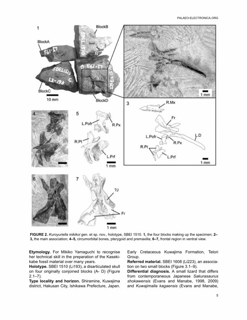
Etymology. For Mikiko Yamaguchi to recogniseher technical skill in the preparation of the Kaseki-kabe fossil material over many years.Holotype. SBEI 1510 (Li193), a disarticulated skullon four originally conjoined blocks (A- D) (Figure2.1–7).Type locality and horizon. Shiramine, Kuwajimadistrict, Hakusan City, Ishikawa Prefecture, Japan.
Early Cretaceous Kuwajima Formation, TetoriGroup.Referred material. SBEI 1608 (Li223), an associa-tion on two small blocks (Figure 3.1–9).Differential diagnosis. A small lizard that differsfrom contemporaneous Japanese Sakurasaurusshokawensis (Evans and Manabe, 1998, 2009)and Kuwajimalla kagaensis (Evans and Manabe,
FIGURE 2. Kuroyuriella mikikoi gen. et sp. nov., holotype, SBEI 1510. 1, the four blocks making up the specimen; 2–3, the main association; 4–5, circumorbital bones, pterygoid and premaxilla; 6–7, frontal region in ventral view.
5

EVANS AND MATSUMOTO: JAPANESE FOSSIL LIZARDS
2008); Chinese Jehol Dalinghosaurus longidigitus(Ji and Ji, 2004; Evans and Wang, 2005),Liushusaurus acanthocaudata (Evans and Wang,2010), and Yabeinosaurus tenuis (Evans et al.,2005; Evans and Wang, 2012; Wang and Evans,2011); and from other known Jurassic and EarlyCretaceous lizards (e.g., Euramerican Paramacel-
lodus spp., Evans and Chure, 1998; Meyasaurusspp., Evans and Barbadillo, 1997) in the distinctiveposterior margin of the dentary and strongly con-cave narial margin of maxilla; differs from Yabeino-saurus tenuis in parietal shape and in lackingparietal foramen; differs from Late CretaceousMongolian Slavoia darevskii (Alifanov, 2000a; Gao
FIGURE 3. Kuroyuriella mikikoi gen. et sp. nov., referred specimen, SBEI 1608. 1–2, main association; 3–4, offsetassociation; 5–6, detail of parietal and frontal; 7–8, detail of quadrate; 9, detail of postdentary bones. For abbrevia-tions, see Material and Methods.
6

PALAEO-ELECTRONICA.ORG
and Norell, 2000) in having proportionally moregracile jaw, proportionally longer and narrowerfrontals and parietals, and absence of parietal fora-men; resembles the Late Cretaceous Carusia inter-media (Borsuk-Białynicka, 1985) and Exostinusserratus (Bhullar, 2010) in having frontals that areanteriorly narrow and posteriorly broad, with strongsub-olfactory processes, in the strong concavity ofthe maxillary narial margin, and, for Carusia but notExostinus, in the shape of the dentary coronoidprocess, but Kuroyuriella differs from both in thatthe frontals are paired rather than fused and lack
coarse tubercular sculpture, there is no sculptureon the maxilla, the parietal is of a different morphol-ogy (square and largely excluded from the uppertemporal fenestra by postorbitofrontal facets, noparietal foramen) and sculpture pattern (low reliefrather than coarse and tubercular); resembles LateCretaceous genera Parmeosaurus scutatus andHymenosaurus clarki (Gao and Norell, 2000) ingracile dentary with straight long axis, but differsfrom former in having uni- rather than tricuspidteeth, free posterior edge on dentary coronoid pro-cess, greater orbital emargination of frontals,
FIGURE 4. Kuroyuriella mikikoi gen. et sp. nov., jaw elements. 1–2, SBEI 1510, right maxilla in labial view; 3–4, SBEI1608, left dentary in labial view; 5–6, SBEI 1608, right dentary in lingual view; 7–10, SBEI 1510, right mandible in 7–8,labial view, and 9–10, ventrolateral view. For abbreviations, see Material and Methods.
7

EVANS AND MATSUMOTO: JAPANESE FOSSIL LIZARDS
deeply concave narial margin of maxilla, and inlacking cranial osteoderms. Hymenosaurus clarkiis very poorly preserved, but as described, it differsfrom Kuroyuriella in having a prefrontal/postfrontalcontact that excludes the short wide frontals fromthe orbital margin and a putative parietal foramenclose to the posterior parietal margin.Material. The holotype skull, SBEI 1510, was con-tained within a single block that was fragmentedduring preparation so that the bones are nowspread over four smaller blocks (Figure 2.1) bear-ing: A, the orbital process of a left maxilla; B, anincomplete left dentary in labial view; C, a rightsplenial; and D, an association of right maxilla andpremaxilla, right and left frontals, left prefrontal andpostorbitofrontal, right pterygoid, and right mandi-ble. The bones are mostly disarticulated but asso-ciated, suggesting some postmortem decay butlittle transport.
A second association (SBEI 1608) isattributed to Kuroyuriella mikikoi on the basis ofdentary characters (tooth number and shape,curved free posterior edge), and complements theholotype in preserving the parietal, the dentary inlingual view, and more details of the accessory jawbones (notably coronoids and articular). The speci-men is on two blocks, also divided during prepara-tion. Block A bears the left dentary in labial view(Figure 3.3–4), as well as a poorly preserved elon-gate right postdentary mass and a right quadrate(Figure 3.7–8). Block B preserves the parietal anda partial frontal, as well as the right dentary, andpostdentary bones including both coronoids, andthe left surangular-angular-prearticular-articularassociation (Figure 3.1–2, 3.6, 3.9). No osteo-derms are preserved in association with thesespecimens, nor are there isolated osteoderms ofappropriate size on any of the many matrix sam-ples from this locality.
The individual bones on both blocks are simi-lar in size to comparable bones of extant lizardswith an adult skull length of ~10 mm, a snout-ventlength (SVL) of 45–50 mm, and a total length of110–125 mm. For their size, the skull bones arerobust and fully ossified with weak sculpturing onthe parietal and complex sutures, but given the dis-articulation, the skeleton may have been that of asub-adult.Description. The holotype (SBEI 1510) preservesa right maxilla (5.8 mm long) in lateral view with 19functional teeth and spaces for at least two more(Figures 2.3, 4.1–2). The bone is distinctive in hav-ing a strongly concave narial margin, caused partlyby the dorsal curvature of the tip of the premaxillary
process but also the anterior margin of the verticalfacial process. Further posteriorly, the facial pro-cess is broken along its base, but the line of break-age clearly demarcates the original antero-posterior length. Posteriorly, the bone tapers into arather short, suborbital process. The partial leftmaxilla (not figured) bears a medial shelf that sup-ported the jugal, but the relative contributions ofthe two bones to the ventral orbital margin cannotbe determined. A right premaxilla underlies the leftpostorbitofrontal (Figure 2.5). Three small teeth arevisible with a gap representing a fourth tooth posi-tion. Further teeth may be hidden by the overlyingelement.
Paired frontals with deep subolfactory pro-cesses (=cristae cranii) that curve slightly inwardand strong midline interdigitations are preserved inventral aspect (Figure 2.6–7). They have sepa-rated slightly in the midline, and the right is twistedslightly on its long axis, making the anterior part ofthe bone appear somewhat narrower than it reallyis. Anteriorly each bone narrows into a straightedge that underlay the nasal. Posteriorly, both fron-tals bear ventrolateral and ventromedial parietalfacets, suggesting the frontoparietal joint wasinflexible rather than mesokinetic. The large pre-frontal facet on the right bone is incised posteriorlybut becomes shallower anteriorly. It extendsroughly halfway along the bone. The prefrontalbone of that side is not preserved, but the left pre-frontal has been displaced laterally and is pre-served in medial view (Figure 2.4–5). Its frontalprocess is posteriorly tapered and relatively flatmedially. The body is expanded and has a deeplyconcave medial surface. The posteroventral mar-gin appears to bear a small facet, possibly for alacrimal bone. Adjacent to the orbital edge of theleft frontal in SBEI 1510 is a curved bar of bonewith a mediolaterally compressed blade at one endand a thicker bar (rounded cross-section) at theother (Figure 2.6–7). This element may be part of asickle-shaped jugal.
A near-complete left postfrontal or, more prob-ably, postorbitofrontal (based on size and articula-tions) is preserved between the frontals and thepterygoid (Figure 2.4–5). It is roughly rectangularwith a straight lateral margin and a more convexmedial margin that would have clasped the fronto-parietal suture (and matches the size and shape ofthe corresponding facet on the parietal of SBEI1608 (see below) and the smaller shallow facet onthe frontal of SBEI 1510. Judging from the arrange-ment of these bones, the jaw adductor muscles
8

PALAEO-ELECTRONICA.ORG
originated from the ventral surface of the parietaland postorbitofrontals.
SBEI 1510 preserves the only palatal ele-ment, a right pterygoid (Figure 2.4–5) with an ante-rior facet for the palatine and a sharply pointedlateral process bearing a narrow slot facet for theectopterygoid. The posterior quadrate process isincomplete but it is narrow and shallow with a dis-tinct dorsal pit (fossa columellae) for the epiptery-goid flanked by a low crest that continuesposteriorly along the dorsolateral edge of the bone.
An almost complete parietal is preserved onSBEI 1608 (Figure 3.5–6). The exposed dorsal sur-face bears weakly pustulate sculpture anteriorly,but is smooth posteriorly; it lacks any trace of headscale markings. The anterior margin (uppermost inFigure 3.5–6) is irregular rather than straight and aparietal foramen is not evident. The posterior mar-gin bears shallow smooth nuchal shelves for neckmuscle attachment, separated by a small mediannotch, probably for the processus ascendens of thesupraoccipital. The postparietal (=supratemporal)processes are broken, revealing bases that aredorso-ventrally shallow in cross-section. On theright side, the parietal margin is partially overlain bya postorbitofrontal (Figure 3.5–6). On the left, theexposed margin is subdivided into a long anteriorconcavity that accommodated the postorbitofrontaland a shorter posterior edge that bordered areduced upper temporal fenestra. Lateral to theright parietal margin, and displaced slightly posteri-orly, is a partial right frontal, also lightly sculptured.On superficial examination, this element might bemistaken for an expanded jugal, but it lacks any ofthe requisite facets or thickenings. SBEI 1608 alsopreserves a right quadrate in association with theposterior end of the right mandible (Figure 3.7–8).It is small (2.3 mm tall) with a short lateral conchsupported by a robust curved posterior pillar. Thedorsal head is wider than the ventral one, the latterbeing anteroposteriorly short and weakly dividedinto medial and lateral condyles.
The lower jaw of Kuroyuriella mikikoi is repre-sented on the holotype (SBEI 1510) by the left den-tary and an almost complete right mandible, and onSBEI 1608 by right and left dentaries and parts ofthe postdentary series (Figure 4.3–10). The leftdentary of SBEI 1510 (5.9 mm long, not figured) ispreserved in labial view and is broken posterior tothe tooth row. It bears 18 teeth with positions fortwo or three more in replacement and an edentu-lous region at the symphysial end that originallyprobably accommodated four or five small teeth.The right dentary (7.4 mm long) is also preserved
in labial view. It is slender with a rounded sym-physial end and a subtle muscle attachment scaranteroventrally (Figure 4.7–10). The Meckelianfossa, exposed by preparation runs medially andthen ventrally. At its posterior end, the dentarybears a small rounded coronoid process with aslightly recurved tip. From this tip, the edge of thedentary curves posteroventrally before extendinginto a process that meets the surangular. The tip ofthis surangular process and the posteroventral endof the dentary are damaged, but the bone clearlyextended beyond the coronoid process to bracethe postdentary bones. The posterodorsal dentarycurvature appears to be a distinctive feature ofKuroyuriella mikikoi (resembling the condition insome acontine skinks, Evans, personal observa-tion). A narrow, posteriorly angled coronoid boneprojects above the dentary coronoid process. Thesurangular is relatively shallow throughout. A nar-row rod of bone runs above it from the tip of thecoronoid lappet. This rod cannot be part of the jawand may be a displaced hyoid ceratobranchial, orpossibly an epipterygoid. The relatively deep angu-lar bears a shallow anterolateral facet for the pos-teroventral ramus of the dentary. The ventromedialsurface of the mandible has been prepared (Figure4.10). A small mylohyoid foramen perforates theangular just posterior to the level of the coronoidprocess. The angular met the splenial medially butits posterior limit is uncertain. The right splenial fillsthe Meckelian fossa posteriorly but terminatessome distance from the symphysis, leaving thefossa open for about one-third of its length. The leftsplenial is preserved in isolation on SBEI 1510C(not figured). It is a relatively short bone with ablunt anterior margin. It fully encloses a small infe-rior alveolar foramen.
The jaw material of SBEI 1608 complementsthat of SBEI 1510. An almost complete left dentary(Figure 4.3–4) is preserved in labial view on Block1608A. There is a posteroventral lappet (missing inSBEI 1510 although there is an impression for it onthe angular). The dentary is 5.7 mm long parallel toits alveolar margin and 6.1 mm along the ventraledge. It is shallow with a relatively horizontal infe-rior margin, a tapering anterior end, and a conspic-uous muscle scar along the anteroventral border.There are 21 teeth, with one space for a non-implanted replacement. The labial surface is perfo-rated by seven large, closely spaced neurovascu-lar foramina. The right dentary (Figure 4.5–6) is inassociation with postdentary elements of bothmandibles including the right and left coronoid, andthe left surangular, articular and angular. It is pre-
9
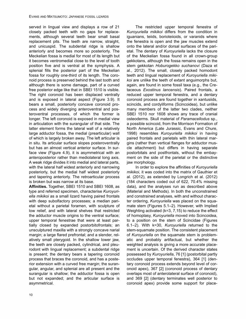
EVANS AND MATSUMOTO: JAPANESE FOSSIL LIZARDS
served in lingual view and displays a row of 21closely packed teeth with no gaps for replace-ments, although several teeth bear small basalreplacement pits. The teeth are narrow, straight,and unicuspid. The subdental ridge is shallowanteriorly and becomes more so posteriorly. TheMeckelian fossa is medial for most of its length butit becomes ventromedial close to the level of toothposition five and is ventral at the symphysis. Asplenial fills the posterior part of the Meckelianfossa for roughly one-third of its length. The coro-noid process is preserved behind the last tooth andalthough there is some damage, part of a curvedfree posterior edge like that in SBEI 1510 is visible.The right coronoid has been displaced ventrallyand is exposed in lateral aspect (Figure 3.9). Itbears a small, posteriorly concave coronoid pro-cess and widely diverging anteroventral and pos-teroventral processes, of which the former islonger. The left coronoid is exposed in medial viewin articulation with the surangular of that side. Thelatter element forms the lateral wall of a relativelylarge adductor fossa, the medial (prearticular) wallof which is largely broken away. The left articular isin situ. Its articular surface slopes posteroventrallybut has an almost vertical anterior surface. In sur-face view (Figure 4.9), it is asymmetrical with ananteroposterior rather than mediolateral long axis.A weak ridge divides it into medial and lateral parts,with the lateral half widest anteriorly and narrowingposteriorly, but the medial half widest posteriorlyand tapering anteriorly. The retroarticular processis broken but was narrow at its base.Affinities. Together, SBEI 1510 and SBEI 1608, astype and referred specimen, characterise Kuroyuri-ella mikikoi as a small lizard having paired frontalswith deep subolfactory processes; a median pari-etal without a parietal foramen, with sculpture oflow relief, and with lateral shelves that restrictedthe adductor muscle origins to the ventral surface;upper temporal fenestrae that were at least par-tially closed by expanded postorbitofrontals; anunsculptured maxilla with a strongly concave narialmargin; a large flared prefrontal; and a slender, rel-atively small pterygoid. In the shallow lower jaw,the teeth are closely packed, cylindrical, and pleu-rodont with lingual replacement; a subdental ridgeis present; the dentary bears a tapering coronoidprocess that braces the coronoid, and has a poste-rior extension with a curved free margin; the suran-gular, angular, and splenial are all present and thesurangular is shallow; the adductor fossa is openbut not expanded; and the articular surface isasymmetrical.
The restricted upper temporal fenestra ofKuroyuriella mikikoi differs from the condition iniguanians, teiids, borioteiioids, or varanids wherethe fenestra is open and the jaw muscles extendonto the lateral and/or dorsal surfaces of the pari-etal. The dentary of Kuroyuriella lacks the closureof the Meckelian fossa found in all crown-groupgekkotans, although the fossa remains open in thestem gekkotan Hoburogekko suchanovi (Daza etal., 2012). The small, closely packed homodontteeth and lingual replacement of Kuroyuriella miki-koi are unlike the teeth of extant anguimorphs but,again, are found in some fossil taxa (e.g., the Cre-taceous Exostinus lancensis). Paired frontals, areduced upper temporal fenestra, and a dentarycoronoid process are found together in xantusiids,scincids, and cordyliforms (Scincoidea), but unlikemany members of the latter two clades, neitherSBEI 1510 nor 1608 shows any trace of cranialosteoderms. Skull material of Paramacellodus sp.,a possible scincoid, from the Morrison Formation ofNorth America (Late Jurassic, Evans and Chure,1998) resembles Kuroyuriella mikikoi in havingpaired frontals and parietals with thin lateral mar-gins (rather than vertical flanges for adductor mus-cle attachment) but differs in having separatepostorbitals and postfrontals, without the embay-ment on the side of the parietal or the distinctivejaw morphology.
In order to explore the affinities of Kuroyuriellamikikoi, it was coded into the matrix of Gauthier etal. (2012), as extended by Longrich et al. (2012)(184 characters coded out of 622, 70.4% missingdata), and the analyses run as described above(Material and Methods). In both the unconstrainedand constrained analyses, with and without charac-ter ordering, Kuroyuriella was placed on the squa-mate stem (Figures 5.1–2). However, with ImpliedWeighting activated (k=3, 7,15) to reduce the effectof homoplasy, Kuroyuriella moved into Scincoidea,to a position on the stem of Scincidae (Figures6.1–2). With k=30, Kuroyuriella returned to thestem-squamate position. The consistent placementof Kuroyuriella on the squamate stem is problem-atic and probably artifactual, but whether theweighted analysis is giving a more accurate place-ment is uncertain. Of the derived character statespossessed by Kuroyuriella, 76 [1] (postorbital partlyoccludes upper temporal fenestra), 364 [1] (den-tary coronoid process extends beyond level of cor-onoid apex), 367 [2] (coronoid process of dentaryoverlaps most of anterolateral surface of coronoid),and 369 [2] (dentary terminates well posterior tocoronoid apex) provide some support for place-
10

PALAEO-ELECTRONICA.ORG
ment of Kuroyuriella on the stem of scincids, and129 [1] (prefrontal extends to mid-orbit), 104 [1](absence of parietal foramen) and 385 [1] (poste-rior mylohyoid foramen posterior to coronoid apex)would be consistent with that placement. However,given the considerable difference between theresults using equal weighting and Implied Weight-ing, Kuroyuriella remains incertae sedis pendingrecovery of more complete material.
SQUAMATA Oppel, 1811Family indet.
Genus ASAGAOLACERTA gen.nov.
zoobank.org/4CF086BA-7B06-46A6-B8F2-D99F923B38B7
Type species. Asagaolacerta tricuspidensEtymology. From Asagao, the Japanese MorningGlory flower, the symbol of Hakusan City, IshikawaPrefecture
Diagnosis. As for type and only species
Asagaolacerta tricuspidens sp. nov.Figures 7–10
zoobank.org/B80126CA-645A-44A8-A5D7-556512A4EF4B
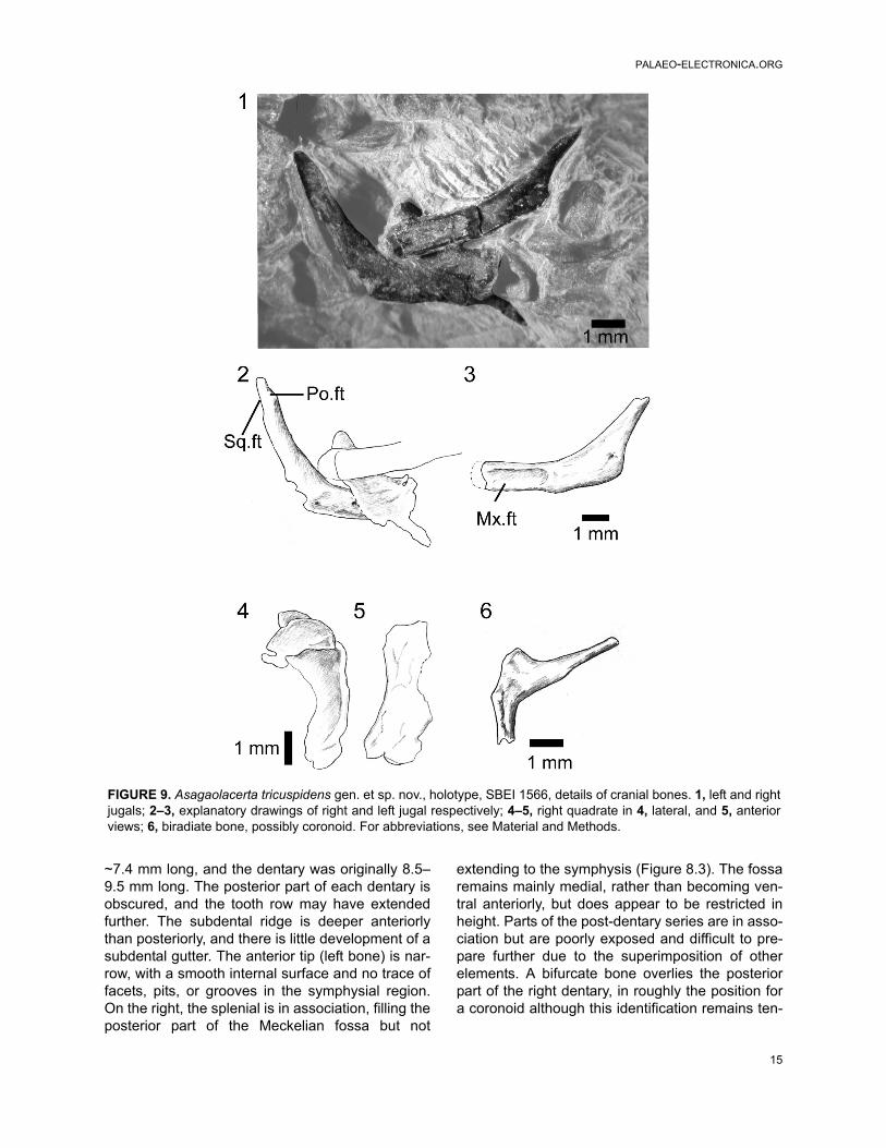
Etymology. For the tricuspid teethHolotype. SBEI 1566 An association of skull, jawsand postcranial bones of a small lizard with facet-ted tricuspid teeth (Figure 7).Type locality and horizon. The Kaseki-Kabe, Shi-ramine, Hakusan city, Ishikawa Prefecture, Japan.Early Cretaceous Kuwajima Formation, TetoriGroup.Referred material. Possibly SBEI 1621, a rightmaxilla (not figured).Differential diagnosis. A small lizard character-ised by the presence of sharply pointed tricuspidteeth with a central cusp and flanking anterior and
FIGURE 5. Phylogenetic position of Kuroyuriella mikikoi gen. et sp. nov. and Asagaolacerta tricuspidens gen. et sp.nov. in squamate trees, using the morphological data matrix of Gauthier et al. (2012), extended by Longrich et al.(2012). 1, Strict consensus of 1000 trees using unordered equally weighted characters, and run using TNT with 'mini-search.run'. Major clades are condensed. 1Note that Iguania was not monophyletic and comprised four smaller cladeswhose position was unresolved in relation to Borioteiioidea. Support values at nodes are Bremer/Jacknife/Symmetricresampling; 2, one of three trees (identical at this level) with clades condensed, run using the same matrix as in Fig-ure 5.1 and analysed with TNT (with sectorial search, ratchet [20 iterations], and tree fusion all activated), but with themolecular tree of Wiens et al. (2010) and Pyron et al. (2013) providing the backbone constraint.
11

EVANS AND MATSUMOTO: JAPANESE FOSSIL LIZARDS
posterior cuspules; tooth shaft cylindrical ratherthan bulbous, and tooth crown and shaft of similarwidth (not apically flared nor strongly labiolinguallyflattened). These dental features distinguishAsagaolacerta from other Kuwajima Formation liz-ards and from described Late Jurassic and EarlyCretaceous taxa from Europe (e.g., Hoffstetter,1967; Evans and Searle, 2002), North America(Nydam, 2002; Nydam and Cifelli, 2002a, 2002b),and China (Evans and Wang, 2005, 2010, 2012;Evans et al., 2005). Asagaolacerta tricuspidensresembles North American Late Cretaceous borio-teiioids like Obamadon gracilis, Tripennaculus sp.,Socognathus brachyodon and Chamops segnis(Longrich et al., 2012) in having tricuspid teeth, butdiffers in lacking any swelling of the tooth bases,and in having tooth crowns that are proportionallysmaller in relation to the tooth bases.
Asagaolacerta tricuspidens also resembles Mon-golian Late Cretaceous borioteiioid lizards (sensuNydam et al., 2007) Altanteius facilis, Mongolo-chamops reshetovi, Pyramicephalosaurus cher-minicus, and Tchingisaurus multivagus (Alifanov,2000b; Gao and Norell, 2000) in having tricuspidteeth, and in having a slender jugal with long dorsalramus forming most of posterior orbital border, butdiffers in having lower tooth count and step-likejugo-maxillary suture; further differs from Pyram-icephalosaurus and Tchingisaurus in lacking anyflaring or labiolingual flattening of tooth crowns (Ali-fanov, 2000b; Gao and Norell, 2000); resemblesLate Cretaceous "mongolochamopine" Cyclurasiamultidentata (Alifanov, 2000b) in jugo-maxillarysuture shape but differs in having fewer, largerteeth. Asagaolacerta tricuspidens also differs frommany Asian borioteiioids in lacking a hypertrophied
FIGURE 6. The phylogenetic position of Kuroyuriella mikikoi gen. et sp. nov. as recovered by analyses run withImplied Weighting (k=7), and character ordering. 1, no backbone constraint; 2, backbone constraint tree based on themolecular trees of Wiens et al. (2010) and Pyron et al. (2013). Only the scincoid section of each tree is shown as in allanalyses with equally weighted taxa, Kuroyuriella lies in the stem-squamate position shown in Figure 5.
12

PALAEO-ELECTRONICA.ORG
splenial and build-up of cementum around toothbases.Material. SBEI 1566 (Figure 7) is a small block inwhich cranial and postcranial elements are super-imposed, possibly as an oral pellet, but the speci-men appears to represent a single individual (onthe basis of non-repetition of parts and consistentsize). Of the skull, the elements exposed includethe left maxilla, the right and left dentaries andsome postdentary bones, the left and right jugal, apartial left frontal, a palatine, and a quadrate, aswell as some undetermined elements. The postcra-nial bones include several vertebrae, parts of the
left pelvis, both femora, and scattered phalanges.The pectoral girdle and forelimbs seem to be miss-ing. X-ray microtomography of the block revealedno other significant elements and no associatedosteoderms. In comparison with modern skeletalmaterial, the size of the jaws, pelvis, and limb ele-ments are matched by a specimen of the extantteiid lizard Aspidoscelis tigris of 90 mm SVL (totallength 290 mm). The body proportions ofAsagaolacerta tricuspidens, at least for those ele-ments preserved, seem to have been broadly simi-lar to the proportions of the living species.
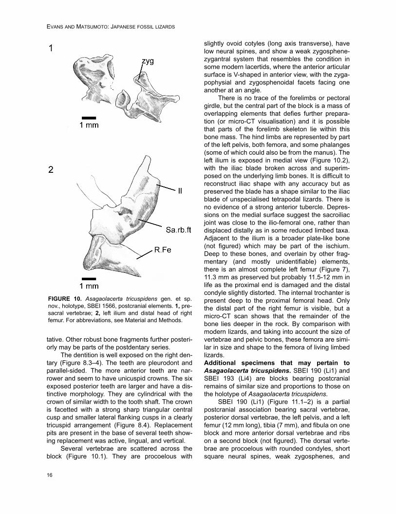
FIGURE 7. Asagaolacerta tricuspidens gen. et sp. nov., holotype, SBEI 1566. 1, the main association with the offsetsecond bone group in the smaller image; 2, explanatory drawing of the same. For abbreviations, see Material andMethods.
13

EVANS AND MATSUMOTO: JAPANESE FOSSIL LIZARDS
Description. The anterior end of the left maxilla isexposed in labial view (Figure 8.1). The narial mar-gin is oblique (unlike the deeper concavity ofKuroyuriella mikikoi). As preserved, the first eightteeth are slender and gradually increase in length,but the crowns are damaged, and it is not possibleto determine whether there was a change from uni-cuspid to tricuspid within this series.
Of the other clearly identifiable skull elements,the right and left jugals lie together near the lowerjaws (Figure 9.1–3). They are roughly L-shaped,but with an oblique angle between the ventral andpostorbital rami. The left bone preserves a nearlycomplete ventral ramus and part of the postorbitalramus, whereas the right preserves most of thepostorbital ramus and the ventral ramus, but thelatter is overlain by a left palatine. The left ventralramus bears an elongate lateral facet for the max-illa and was clearly partially overlapped by thatbone in its anterior half, leaving a thin strip of thejugal along the ventral margin of the orbit. Thisarrangement would have given the jugal-maxillarysuture a step-like structure in lateral view. It alsosuggests that the suborbital ramus of the maxillaextended only to mid-orbit. The jugal lacks a poste-rior spur or tubercle, but the thin bone is drawn intoa slight angle. The postorbital ramus is slender andlonger than the ventral ramus. The facet for thepostorbital (or postorbitofrontal) is confined to thedorsal tip, suggesting that the jugal formed most ofthe posterior orbital margin and that the postorbitalitself was not large. This interpretation is supportedby a slight recess on the posterodorsal edge of thejugal that may have received the anterior tip of thesquamosal, excluding the postorbital from the mar-gin of the lower temporal fenestra.
Part of a left frontal is preserved in dorsal viewat the edge of the association (Figure 7.2), butshows only that the frontals were paired andweakly sculptured with fine lines. A partial left pala-tine overlaps the ventral ramus of the right jugal. Itis preserved in dorsal view and shows only that aforamen perforated the base of the maxillary pro-cess, as it does in most lizards. A robust curvedbone close to the anterior end of the maxilla isidentified as the right quadrate (Figure 9.4–5). Ithas a narrow conch and a posteriorly extendedhead, but no obvious notch or pit for the squamo-sal.
Both dentaries are preserved in association,the right in lingual view, the left in dorsolingual view(Figures 7, 8.2–3). The right shows 12 tooth posi-tions with space for at least three further smallteeth in the symphysial region. The tooth row is
FIGURE 8. Asagaolacerta tricuspidens gen. et sp. nov.,holotype, SBEI 1566. 1, Left partial maxilla in labialview; 2, left dentary in lingual view; 3, right dentary in lin-gual view; 4, details of teeth on the right dentary(enlarged from 3). For abbreviations, see Material andMethods.
14
PALAEO-ELECTRONICA.ORG
~7.4 mm long, and the dentary was originally 8.5–9.5 mm long. The posterior part of each dentary isobscured, and the tooth row may have extendedfurther. The subdental ridge is deeper anteriorlythan posteriorly, and there is little development of asubdental gutter. The anterior tip (left bone) is nar-row, with a smooth internal surface and no trace offacets, pits, or grooves in the symphysial region.On the right, the splenial is in association, filling theposterior part of the Meckelian fossa but not
extending to the symphysis (Figure 8.3). The fossaremains mainly medial, rather than becoming ven-tral anteriorly, but does appear to be restricted inheight. Parts of the post-dentary series are in asso-ciation but are poorly exposed and difficult to pre-pare further due to the superimposition of otherelements. A bifurcate bone overlies the posteriorpart of the right dentary, in roughly the position fora coronoid although this identification remains ten-
FIGURE 9. Asagaolacerta tricuspidens gen. et sp. nov., holotype, SBEI 1566, details of cranial bones. 1, left and rightjugals; 2–3, explanatory drawings of right and left jugal respectively; 4–5, right quadrate in 4, lateral, and 5, anteriorviews; 6, biradiate bone, possibly coronoid. For abbreviations, see Material and Methods.
15
EVANS AND MATSUMOTO: JAPANESE FOSSIL LIZARDS
tative. Other robust bone fragments further posteri-orly may be parts of the postdentary series.
The dentition is well exposed on the right den-tary (Figure 8.3–4). The teeth are pleurodont andparallel-sided. The more anterior teeth are nar-rower and seem to have unicuspid crowns. The sixexposed posterior teeth are larger and have a dis-tinctive morphology. They are cylindrical with thecrown of similar width to the tooth shaft. The crownis facetted with a strong sharp triangular centralcusp and smaller lateral flanking cusps in a clearlytricuspid arrangement (Figure 8.4). Replacementpits are present in the base of several teeth show-ing replacement was active, lingual, and vertical.
Several vertebrae are scattered across theblock (Figure 10.1). They are procoelous with
slightly ovoid cotyles (long axis transverse), havelow neural spines, and show a weak zygosphene-zygantral system that resembles the condition insome modern lacertids, where the anterior articularsurface is V-shaped in anterior view, with the zyga-pophysial and zygosphenoidal facets facing oneanother at an angle.
There is no trace of the forelimbs or pectoralgirdle, but the central part of the block is a mass ofoverlapping elements that defies further prepara-tion (or micro-CT visualisation) and it is possiblethat parts of the forelimb skeleton lie within thisbone mass. The hind limbs are represented by partof the left pelvis, both femora, and some phalanges(some of which could also be from the manus). Theleft ilium is exposed in medial view (Figure 10.2),with the iliac blade broken across and superim-posed on the underlying limb bones. It is difficult toreconstruct iliac shape with any accuracy but aspreserved the blade has a shape similar to the iliacblade of unspecialised tetrapodal lizards. There isno evidence of a strong anterior tubercle. Depres-sions on the medial surface suggest the sacroiliacjoint was close to the ilio-femoral one, rather thandisplaced distally as in some reduced limbed taxa.Adjacent to the ilium is a broader plate-like bone(not figured) which may be part of the ischium.Deep to these bones, and overlain by other frag-mentary (and mostly unidentifiable) elements,there is an almost complete left femur (Figure 7),11.3 mm as preserved but probably 11.5-12 mm inlife as the proximal end is damaged and the distalcondyle slightly distorted. The internal trochanter ispresent deep to the proximal femoral head. Onlythe distal part of the right femur is visible, but amicro-CT scan shows that the remainder of thebone lies deeper in the rock. By comparison withmodern lizards, and taking into account the size ofvertebrae and pelvic bones, these femora are simi-lar in size and shape to the femora of living limbedlizards.Additional specimens that may pertain toAsagaolacerta tricuspidens. SBEI 190 (Li1) andSBEI 193 (Li4) are blocks bearing postcranialremains of similar size and proportions to those onthe holotype of Asagaolacerta tricuspidens.
SBEI 190 (Li1) (Figure 11.1–2) is a partialpostcranial association bearing sacral vertebrae,posterior dorsal vertebrae, the left pelvis, and a leftfemur (12 mm long), tibia (7 mm), and fibula on oneblock and more anterior dorsal vertebrae and ribson a second block (not figured). The dorsal verte-brae are procoelous with rounded condyles, shortsquare neural spines, weak zygosphenes, and
FIGURE 10. Asagaolacerta tricuspidens gen. et sp.nov., holotype, SBEI 1566, postcranial elements. 1, pre-sacral vertebrae; 2, left ilium and distal head of rightfemur. For abbreviations, see Material and Methods.
16

PALAEO-ELECTRONICA.ORG
large oblique rib synapophyses. The sacrum isdamaged. The pelvis is preserved in medial viewand its components are co-ossified. The ilium is ofsimilar shape to the ilium on the holotype ofAsagaolacerta tricuspidens; the sacral rib facet ispositioned just above the level of the acetabulum.The pubis is only partially preserved, but wasclearly tapering rather than broad, and the ischiumwas flask-shaped. The femur is robust and has athick circular shaft that has only a slight sigmoidcurvature. The proximal and distal ends are fullyossified, with a rounded proximal femoral head, awell-developed tuber-like greater trochanter, and adistinct intertrochanteric fossa. The bone is rela-tively stouter than the femur of the holotype ofAsagaolacerta tricuspidens (overall length/distalwidth ~ 4.7 compared to 6.2 in Asagaolacerta tri-cuspidens), although only slightly longer. The tibiais shorter than the femur (~ 58%) but again robustwith a wider proximal end and a flange-like cnemialcrest that is separated from the proximal end. The
fibula was probably slight longer than the tibia butits distal end is broken. There are no associatedosteoderms.
SBEI 193 (Li4) (Figure 11.3–4) is a secondpartial postcranial association, preserved in ventralview. The sacral vertebrae are fused and the con-joined distal ends of the sacral ribs enclose a fora-men sacrale on each side. Rather unusually, thefirst sacral, though incomplete, appears to be lessrobust than the second. Behind them, the first cau-dal has a slight keel and robust transverse pro-cesses, each of which bears a deep linear grooveproximally. The left pelvis is complete with the com-ponents conjoined. The right ilium is preserved inmedial view. Its elongate blade lies at roughly 45°degrees to the long axis of the acetabular region,with the sacral rib facet lying just above the level ofthe acetabulum. There is a slight anterodorsalexpansion rather than a tubercle. Both femora (~8.7 mm) are preserved to one side of the block,although they are partially covered by disarticu-
FIGURE 11. Associated postcranial specimens that may be referable to Asagaolacerta gen. nov. 1–2, SBEI 190; 3–4,SBEI 193. For abbreviations, see Material and Methods.
17

EVANS AND MATSUMOTO: JAPANESE FOSSIL LIZARDS
lated dorsal vertebrae. They are smaller than fem-ora on the holotype of Asagaolacerta tricuspidensbut they are otherwise similar in proportions (over-all length/distal width ~6.0 in both SBEI 193 andSBEI 1566). Again, there are no associated osteo-derms.
On the basis of the femoral and, to a lesserdegree, iliac shape, one or both specimens may beattributable to Asagaolacerta tricuspidens but with-out more complete material they cannot bereferred with confidence and characters from thesespecimens were not included in the diagnosis orphylogenetic analyses.Affinities. Among extant lizards, tricuspid teeth arefound mainly in iguanians, lacertids, and teiioids,although they may also occur in other groups. Inthe Mesozoic, tricuspidy is rarer (Nydam, 2002),but again occurs most commonly in taxa referred toIguania or Borioteiioidea (sensu Nydam et al.,2007, =Polyglyphanodontia of Gauthier et al., 2012and Longrich et al., 2012). One exception is theLate Cretaceous Mongolian Parmeosaurus scuta-tus (Gao and Norell, 2000), which our analysisgrouped with scincoids. Of described Jurassic andEarly Cretaceous lizard taxa, only Ptilotodon wil-soni from the Aptian-Albian Antlers Formation ofTexas (Nydam and Cifelli, 2002b) approaches tri-cuspidy in having small anterior and posteriorexpansions that form shoulders on either side of amuch larger central cusp. The teeth ofAsagaolacerta tricuspidens differ in having distinctcuspules that are almost the same height as thecentral cusp. In the Late Cretaceous, tricuspidybecame more common. It has been recorded inseveral Campanian-Maastrichtian genera fromNorth America (Nydam, 2002; Longrich et al.,2012) that were once considered teiioid but havemore recently (Nydam et al., 2007; Longrich et al.,2012) been classified as borioteiioid - notablyChamops segnis and Leptochamops denticulatus(Estes, 1964), Meniscognathus altmani (Nydamand Voci, 2007), Socognathus brachyodon (Gaoand Fox, 1996; Longrich et al., 2012), Tripennacu-lus eatoni (Nydam and Voci, 2007), and Obama-don gracilis (Longrich et al., 2012), The first four ofthese have been grouped (with others) into thefamily Chamopsiidae (e.g., Nydam et al., 2010;Longrich et al., 2012), but they did not alwaysemerge as a monophyletic unit in our analyses(Figure 12). Tricuspidy is also found in a range ofborioteiioid taxa from Late Cretaceous deposits inMongolia (Alifanov, 2000b), including Altanteiusfacilis, Cyclurasia multidentata, Mongolochamopsreshetovi, Piramicephalosaurus cherminicus, and
Tchingisaurus multivagus. A borioteiioid attributionfor Asagaolacerta would be consistent with the het-erodonty and tricuspidy (e.g., Nydam and Cifelli,2002a, 2002b), the paired unsculptured frontals,and the long postorbital ramus of the jugal. How-ever, Asagaolacerta tricuspidens differs from manymore derived borioteiioids in lacking a hypertro-phied splenial and a heavy deposition of cemen-tum around the tooth bases, characters that havebeen cited as diagnostic of both teiioids and borio-teiioids (e.g., Denton and O'Neill, 1995; Gao andFox, 1996; Nydam et al., 2007).
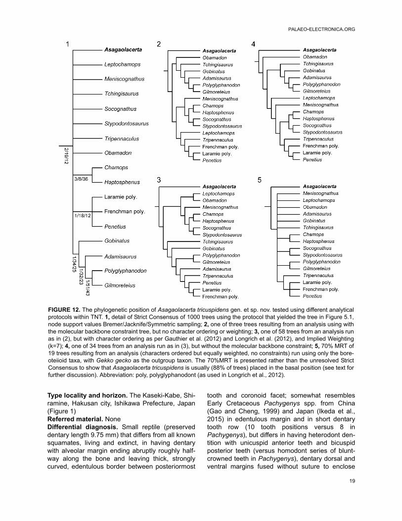
As for Kuroyuriella mikikoi, we codedAsagaolacerta tricuspidens into the matrix ofGauthier et al. (2012) and Longrich et al. (2012)(94/622 characters, 85% missing data). In all anal-yses, weighted and unweighted, constrained orunconstrained, ordered or unordered, Asagoal-acerta was consistently placed on the stem of Bori-oteiioidea (Figure 12), although the position of theclade within the squamate tree varied with differentprotocols (e.g., Figure 5), as did the resolution andpositions of in-group taxa (Figure 12). Although thestrict consensus of the exhaustive analysis (uncon-strained, unordered, equally weighted) placedAsagaolacerta in an unresolved position in relationto the fragmentary North American taxa (Figure12.1), it is important to note that Asagaolacerta didnot group with any one of these taxa in the moreresolved individual trees from analyses using dif-ferent protocols (e.g., Figures 12.2–12.4), norwhen the borioteiioid taxa were run on their own(Figure 12.5).
SQUAMATA Oppel, 1811Family indet.
Genus HAKUSEPS gen. nov.
zoobank.org/6AD065FC-1D2E-488D-ABBB-5C27E306057D
Type species. Hakuseps imberisEtymology. From Hakusan, white mountain, thename of the city of which Shiramine village forms apart and of the regional volcanic peak, and seps(L), variably used for lizard.Diagnosis. As for type and only species
Hakuseps imberis sp. nov.Figure 13
zoobank.org/11AC5FFE-4BA8-4D27-B149-A326E5AF2921
Etymology. From imber, imberis (L) meaning ashower, in reference to the shape of the dentarywhich resembles that of an inverted shower-head.Holotype. SBEI 2086. An almost complete leftdentary (Figure 13).
18
PALAEO-ELECTRONICA.ORG
Type locality and horizon. The Kaseki-Kabe, Shi-ramine, Hakusan city, Ishikawa Prefecture, Japan(Figure 1)Referred material. NoneDifferential diagnosis. Small reptile (preserveddentary length 9.75 mm) that differs from all knownsquamates, living and extinct, in having dentarywith alveolar margin ending abruptly roughly half-way along the bone and leaving thick, stronglycurved, edentulous border between posteriormost
tooth and coronoid facet; somewhat resemblesEarly Cretaceous Pachygenys spp. from China(Gao and Cheng, 1999) and Japan (Ikeda et al.,2015) in edentulous margin and in short dentarytooth row (10 tooth positions versus 8 inPachygenys), but differs in having heterodont den-tition with unicuspid anterior teeth and bicuspidposterior teeth (versus homodont series of blunt-crowned teeth in Pachygenys), dentary dorsal andventral margins fused without suture to enclose
FIGURE 12. The phylogenetic position of Asagaolacerta tricuspidens gen. et sp. nov. tested using different analyticalprotocols within TNT. 1, detail of Strict Consensus of 1000 trees using the protocol that yielded the tree in Figure 5.1,node support values Bremer/Jacknife/Symmetric sampling; 2, one of three trees resulting from an analysis using withthe molecular backbone constraint tree, but no character ordering or weighting; 3, one of 58 trees from an analysis runas in (2), but with character ordering as per Gauthier et al. (2012) and Longrich et al. (2012), and Implied Weighting(k=7); 4, one of 34 trees from an analysis run as in (3), but without the molecular backbone constraint; 5, 70% MRT of19 trees resulting from an analysis (characters ordered but equally weighted, no constraints) run using only the bore-oteiioid taxa, with Gekko gecko as the outgroup taxon. The 70%MRT is presented rather than the unresolved StrictConsensus to show that Asagaolacerta tricuspidens is usually (88% of trees) placed in the basal position (see text forfurther discussion). Abbreviation: poly, polyglyphanodont (as used in Longrich et al., 2012).
19

EVANS AND MATSUMOTO: JAPANESE FOSSIL LIZARDS
Meckelian fossa (versus deep fossa filled by sepa-rate splenial in Pachygenys), and posterior edentu-lous region completely separated from tooth row(contiguous with it in Pachygenys).Material. SBEI 2086 (Li275) is a left dentary, origi-nally preserved in labial view but prepared from thematrix to reveal its lingual aspect.Description. The bone is divided into two parts ofroughly equal length (Figure 13). The alveolar(dental) margin is limited to the anterior half of thebone. It has a total of ten tooth positions, five ofwhich bear complete teeth. The implantation ispleurodont, but the teeth protrude well above thelabial wall of the jaw. The anteriormost tooth posi-tion lies immediately adjacent to the symphysisand is visible only in occlusal view (Figure 13.7).The teeth are heterodont, unicuspid anteriorly, witha concave lingual surface flanked by weak mesial
and distal crests, and distinctly bicuspid posteriorly,with a small divergent mesial cusp and a broaderdistal cusp. The tooth in the fifth position (thirdcomplete) is intermediate in crown morphology,essentially resembling the anterior teeth but bear-ing a slight protrusion on the mesial edge in theposition occupied by the cuspule in more posteriorteeth. The teeth also increase in basal diameteralong the row, with the missing penultimate toothrepresented by a gap that is more than twice thediameter of anterior teeth. The last tooth position ismuch smaller. Tooth replacement was lingual,replacement pits being present at the bases of theteeth in positions five and seven. The anterior tip ofthe bone narrows dorsoventrally, but maintains itswidth labiolingually to form a distinct symphysialsurface. This surface is supported by a deep sub-dental ridge below which the Meckelian fossa
FIGURE 13. Hakuseps imberis gen. et sp. nov., holotype left dentary, SBEI 2086. 1–2, labial view; 3–4, lingual view;5–6, occlusal view; and 7, ventral view. For abbreviations, see Material and Methods.
20

PALAEO-ELECTRONICA.ORG
opens for the short distance anteroventrally (Figure13.3–4). However, at the level of the sixth toothposition, the dorsal and ventral margins of the den-tary fuse to fully enclose the Meckelian canal.
Half way along the bone, as preserved, thelabial wall supporting the alveolar margin endsabruptly, creating a distinct step in the dorsal edgeof the bone. This morphology is not the result ofpost-mortem damage, the bone surfaces are intactand smooth (unfacetted). The posterior half of thedentary is composed only of its cylindrical ventralportion, enclosing the Meckelian canal, the inferioralveolar nerve, and blood vessels. As a result, thedentary as a whole rather resembles a shower-head or a small hand brush. The "handle" curvesposterodorsally but the posterior end of the bone ismissing. The posterolingual wall is deeply incised.There is no trace of a splenial facet and this ele-ment was either absent or fully fused into the den-tary. Immediately above the posteromedial incision,the rising dorsal edge of the bone bears a veryshallow depression, flanked dorsolabially by aslight ridge. This depression may represent a weakcoronoid facet, but this interpretation is tentative.Posteroventral to it, a vertical sheet of boneextends between the incised lingual wall of thedentary and the rounded labial one. This sheetappears to have a free ventral margin (although itcannot be fully prepared out from the matrix fillingthe Meckelian fossa as the walls are thin) and mayrepresent an intramandibular septum, separatingthe Meckelian canal from the inferior alveolar canallaterally. The dorsomedial edge of the septumbears a facet, again possibly for part of the coro-noid. The remaining postdentary bones (surangu-lar, angular, prearticular), separately or conjoined,would have slotted into the back of the dentary, butthey must have done so at a distinct angle to thehorizontal given the strong curvature of the ventraldentary margin.
Four small nutrient foramina pierce the labialsurface.Affinities. This unusual dentary is attributed to theSquamata on the basis of tooth implantation andthe general morphology of the anterior part of thejaw. However, it is unlike the dentary of any knownlizard, with the partial exception of the roughly con-temporaneous species Pachygenys thlastesa (Gaoand Cheng, 1999) (Figure14.1–2). Pachygenyswas based on two mandibles from Early Creta-ceous deposits in Shandong Province, China.Recently, however, closely similar jaws were recov-ered from the Sasayama Group of Hyogo Prefec-ture, Japan (Ikeda et al., 2015), dated as early
Albian (112 Ma, Kusuhashi et al., 2013) and namedPachygenys adachii. As in Hakuseps imberis, thedentary of Pachygenys thlastesa is curved and hassmall number of teeth (eight) concentrated at theanterior end of the dentary. However, although theposterior region of the dentary of Pachygenys isalso edentulous, the labial wall remains intactrather than being stepped (Figure 14.1). Further-more, below the tooth row, the lingually openMeckelian fossa is filled by a large free posteriorlydeep splenial (Figure 14.2). This splenial is piercedjust posterior to the level of the last tooth by theanterior inferior alveolar foramen, behind which isthe anterior mylohyoid foramen. Pachygenys alsohas blunt-crowned homodont teeth, unlike the het-erodont dentition of Hakuseps with its stronglybicuspid posterior teeth. The two genera could berelated, but given that both are represented only bypartial mandibles, their phylogenetic position withinSquamata is difficult to ascertain. Pachygenys hasbeen referred to "scincomorphs" (Gao and Cheng,1999) or lacertoids (Ikeda et al., 2015). Hakusepsimberis was not included in the phylogenetic analy-ses as it preserves too few codable characters.
Among extant lizards, some xantusiids havea short edentulous region behind the tooth row.However, this morphology more closely resemblesthe condition in Pachygenys thlastesa thanHakuseps imberis, and the tooth row is longer andthe edentulous region shorter. Xantusiids alsoshow the complete dentary enclosure of the Meck-elian fossa seen in Hakuseps, but this featureoccurs convergently in gekkotans and in somescincids, gymnophthalmids, and occasional mem-bers of other clades, and seems to be a way ofstrengthening the jaw against bending. An intra-mandibular septum that extends to the posteriorend of the dentary (and has a free margin) hasbeen considered an anguimorph synapomorphyassociated with a reduced overlap of dentary andpostdentary bones (e.g., Estes et al., 1988),although it can occur in other taxa (e.g., some cha-meleons, Estes et al., 1988). However, mesio-dis-tally bicuspid teeth with lingual tooth replacementare more usually associated with lacertoids (nota-bly teiids, gymnophthalmids, and lacertids),although bicuspidy can occur in other groups. Infossil lizards, it is recorded in the Early CretaceousMeyasaurus spp. (=Ilerdaesaurus, Hoffstetter,1966) from Spain (e.g., Richter, 1994; Evans andBarbadillo, 1997) and Britain (Sweetman, 2009;Sweetman and Evans, 2011). Like Hakuseps, theanterior teeth in all Meyasaurus species are uni-cuspid and the posterior teeth bicuspid, but there
21

EVANS AND MATSUMOTO: JAPANESE FOSSIL LIZARDS
the resemblance ends. Meyasaurus has variouslybeen placed with teiids (Evans and Barbadillo,1997), anguimorphs (Richter, 1994; Conrad, 2008;Bolet and Evans, 2011), cordyliforms (Müller et al.,2011), or in an unresolved position amongst crownsquamates (Bolet and Evans, 2010). Bicuspidy hasrecently been reported in a second Spanish taxon,Pedrerasaurus latifrontalis (Bolet and Evans,2010), in jaws (unnamed) that occur with Meyasau-rus sp. in the Barremian Wessex Formation (UK,Sweetman and Evans, 2011), and in another Tetoritaxon (see below) with "normal" jaws. In all of thesetaxa, a small anterior cusp precedes a large poste-rior cusp, but the anterior cusp is divergent only inHakuseps imberis, and none of the other taxashares the atypical jaw morphology.
A jaw morphology somewhat similar to that ofHakuseps imberis, with the posterior half dentarybeing much shallower than the anterior half, andhaving a strong upward curvature, is found in somescolecophidian snakes (e.g., Leptotyphlops dulcis,Kley, 2014). However, without further material, thisstrange squamate remains an enigma. The sharppointed teeth suggest a diet of small invertebrates,but their unusual arrangement (and the overallshape of the jaw) implies a specialised feedingstrategy. In the absence of the maxilla and premax-illa, however, it is difficult to understand how thejaws might have been used.
SQUAMATA Oppel, 1811Family indet.
Morphotype AFigure 15
Material. Within the Shiramine lizard collectionthere are several maxillae and dentaries withbicuspid teeth, notably SBEI 1501 (Li191) and1525 (Li193), a right and left maxilla respectively(Figure 15.1–4); SBEI 197 (Li8) and 808 (Li92)(Figure 15.5–10), both left dentaries; and SBEI1487 (Li190), a right dentary. In all, the accessorycusp is smaller and less divergent than the corre-sponding cusp of Hakuseps imberis.Description. SBEI 1501 (Li191) and SBEI 1525 (Li199) both represent the anterior maxilla. SBEI1525 is a left maxilla, 4.7 mm long, with 10 toothpositions (Figure 15.1–2). The last four preservedteeth are bicuspid (from tooth position five or six).Although the dorsal margin of the premaxillary pro-cess is oblique, the anterior tip is recurved. SBEI1501 (Figure15.3–4) is a right maxilla, 2.6 mmlong, that preserves 12 tooth positions. The ante-rior teeth are damaged but the posterior ones (fromtooth position 8) are bicuspid. The anterior narialmargin of the bone is smoothly oblique, with nostep or angulation between ventral and posteriorcomponents. In this respect it resembles the max-illa of Asagaolacerta tricuspidens, but is less thanhalf of its size. It is possible that this maxilla origi-nally bore a recurved tip like that of SBEI 1525.
SBEI 808 (Li92) is a shallow left dentary, pre-served in two pieces, both in labial view (Fig-ure15.5–10). The reconstructed length is ~7.9 mm,with a posterior depth of ~1.6 mm. The bone tapersat the symphysis, and there are six large labialneurovascular foramina. Fourteen teeth are pre-served with at least 10 empty tooth positions, morethan twice the number in Hakuseps imberis. The
FIGURE 14. Comparison of Pachygenys thlastesa (Gao and Cheng, 1999) and Hakuseps imberis gen. et sp. nov.Labial and lingual views respectively of 1-2, Pachygenys thlastesa (redrawn from Gao and Cheng, 1999), 3-4,Hakuseps imberis gen. et sp. nov.
22

PALAEO-ELECTRONICA.ORG
dorsal margin of the dentary is scalloped, with theedge expanding slightly at the base of each tooth.There is also some cementum around the toothbases. Most of the tooth crowns are preserved andthey are visibly bicuspid with a small anterior cuspand a large posterior one (Figure 15.9–10). The
second preserved tooth, which is probably thefourth or fifth in position (allowing for at least twosmall symphysial tooth positions), is already bicus-pid. The teeth are quite long and narrow in labialview (exposed height/width 1.9). SBEI 808 is sup-plemented by two smaller specimens, SBEI 197
FIGURE 15. Shiramine Morphotype A, bicuspid dentition. 1–2, left maxilla, SBEI 1525 in labial view; 3–4, right max-illa, SBEI 1501, in labial view; 5–10, left dentary, SBEI 808, in two parts, in labial view, with 5–6, symphysial region, 7–8, posterior dentary, and 9–10, detail of bicuspid teeth.
23

EVANS AND MATSUMOTO: JAPANESE FOSSIL LIZARDS
(Li8) and SBEI 1487 (Li190) (not illustrated), withsimilar teeth.Affinities. Given that these bicuspid jaw remainsare generally smaller than the jaws ofAsagaolacerta tricuspidens, one possibility is thatthey represent juveniles of that taxon. Ontogeneticvariation in cusp number is known to occur in somemodern lizards (e.g., lacertids, Barahona andGreen, 1997), and variation can also occur alongthe tooth row. However, although none of thebicuspid jaws is complete, SBEI 808 has at least24 tooth positions, almost double the numberfound in Asagaolacerta tricuspidens, and none istricuspid. This high tooth count also rules out rela-tionships with Hakuseps imberis and suggests thesecond kind of bicuspid jaws may represent a dis-tinct lizard taxon, intermediate in size betweenKuroyuriella mikikoi and Asagaolacerta tricuspi-dens.
As noted above, mesio-distal bicuspidy is rel-atively rare in Mesozoic lizards. The dentition ofthis second bicuspid Japanese lizard differs fromthe condition in Meyasaurus in which the anteriordentary teeth are unicuspid and become bicuspidhalfway along the tooth row. SBEI 808 showsbicuspidy in anterior dentary teeth (from at leasttooth position four). The dentary of Pedrerasauruslatifrontalis is poorly known but, as in the Japaneselizard, it is relatively shallow and bicuspid teethwere present more anteriorly in the tooth row thanin Meyasaurus spp. (Bolet and Evans, 2010). In themaxilla, both have unicuspid anterior teeth, withbicuspidy occurring at about tooth position 8-9 inPedrerasaurus latifrontalis and at 5-6 in the Japa-nese lizard. However, without more complete spec-imens, it would be premature to attach a name toany of these bicuspid jaws as they cannot be diag-nosed with assurance.
SQUAMATA Oppel, 1811Family indet.
Morphotype BFigure 16
Material. SBEI 827 (Li100) is a small right mandi-ble separated into its dentary and postdentarycomponents (Figure 16). The dentary is 4.5 mmlong as preserved but is missing the symphysialregion and posterior margin (estimated originaltotal length ~5.7 mm). The postdentary com-plex is 4.8 mm long, giving an overall original jawlength of ~10.5 mm. The specimen is delicate andis held together by a preservative that obscuressome of the detail but which cannot be removedwithout risk of damage.
Description. The dentary bears at least 15 small,closely-packed unicuspid pleurodont teeth (Figure16.3–4). Allowing for the missing symphysialregion, there may originally have been about 20teeth. These teeth are slightly spatulate linguallybut taper abruptly into small pointed tips (SEM notpossible due to the fragility of the specimen andpreservative covering). Where visible anteriorly,the subdental ridge is deep and there is a narrowgutter between the tooth bases and the edge of theridge. The Meckelian fossa seems to be closedanteriorly by the dentary alone but behind this clo-sure, a long splenial obscures the rest of the fossa.The splenial is slightly disarticulated and has beendisplaced anteromedially, probably by about fourtooth positions. It is narrow anteriorly and deepensposteriorly, but a dorsal embayment at the poste-rior end appears to be real. By comparison withmodern lizards, this embayment probably originallyunderlay the coronoid region. The dentary extendsbehind the tooth row, expanding dorsally into alarge coronoid process that would have covered atleast part of the lateral surface of the coronoidbone. Seen in lateral view (Figure 16.1–2), andallowing for preservational artefact, the dentaryshows a marked anterior to posterior increase inheight.
The postdentary bones appear, at first, to beco-ossified, with a deep anterior notch betweendorsal and ventral processes (Figure 16.5–10).However, although the surangular and articular/prearticular are fused, the narrow angular is sepa-rate and has disarticulated labially so that it formsthe lower margin of the apparent notch. A thin frag-ment of bone anterior to this notch may be part ofthe posterior process of the dentary. The surangu-lar is shallow and bears lateral facets for the den-tary, and angular and medial facets for thecoronoid. Along its lateral face is a low surangularcrest marking the ventral limit of the superficialadductor muscle mass. Even allowing for somedistortion, the postdentary bones at their anteriormargin are much shallower than the posterior partof the dentary, and a large coronoid presumablyfilled part of the gap between them. The surangularhas a tongue-like anterior extension but this flangeis short and is unlikely to have penetrated theMeckelian fossa of the dentary to any significantdegree. Medially, the surangular and articular/prearticular enclose a small shallow posterioradductor fossa. The articular surface for the quad-rate is small, broad, and almost vertical, suggest-ing that the quadrate was oriented at an obliqueangle to the rest of the skull. There is no evidence
24

PALAEO-ELECTRONICA.ORG
of a retroarticular process but, if thin, this processmay have broken off without leaving an obviousedge.Affinities. In tooth number and the presence of acoronoid process, SBEI 827 resembles the jaws ofKuroyuriella mikikoi, but the jaw shape is com-
pletely different (anterior/posterior height differ-ence), the coronoid process is larger and broader,the splenial is deeper, and the angular is shallower.The tightly packed pleurodont teeth, partial dentaryclosure of the Meckelian fossa, posterior extensionof the dentary, co-ossification of the surangular and
FIGURE 16. Shiramine Morphotype B, right mandible SBEI 827. 1–2, dentary in labial view; 3–4, dentary and associ-ated splenial in lingual view; 5–11, postdentary bones in 5–6 dorsomedial view; 7, ventral view; 8, dorsal view; 9,medial view; and 10, posterior view of articular surface. For abbreviations, see Material and Methods.
25

EVANS AND MATSUMOTO: JAPANESE FOSSIL LIZARDS
articular/prearticular, and small size of the adductorfossa are features shared with extant gekkotans,and this jaw was tentatively attributed to that groupin a previous review (Evans and Manabe, 2000).However, gekkotans generally have larger num-bers of teeth; do not have a large dentary coronoidprocess; lose or reduce the splenial; and lose afree angular in all except some eublepharids. Gek-kotans are rare in the Mesozoic fossil record. Theearliest recorded taxon is Hoburogekko suchanovifrom Höovor (Aptian-Albian) in Mongolia (Alifanov1989, 1990; Daza et al., 2012, 2014), but the asso-ciated dentary lacks postdentary bones and has afully open Meckelian fossa. Another possible stem-gekkotan (as yet unnamed) was described (Conradand Norell, 2006) from Öösh, Mongolia (Berriasianto Albian, Andres and Norell, 2005; Berriasian toBarremian, Turner et al., 2007). Like the Tetorispecimen, it has a large splenial covering a largelyopen Meckelian fossa and a small angular, but itlacks a dentary coronoid process and the surangu-lar and articular/prearticular are not fused. TheLate Cretaceous Gobekko (Borsuk-Białynicka,1990; Daza et al., 2013) has a very poorly pre-served jaw making comparison difficult.
Many characters of SBEI 827 (fusion or partialfusion of postdentary bones, partial closure of theMeckelian fossa by the dentary, posterior exten-sion of the dentary, reduced angular) occur in indi-vidual members of other squamate cladesincluding pleurodont iguanians, scincids, cordyli-forms, xantusiids, gymnophthalmids, and xeno-saurs (Evans, 2008). A subset of these cladesshare other features of SBEI 827, notably the well-developed dentary coronoid process (scincids,cordyliforms, and xantusiids), the small adductorfossa (scincids, xenosaurs, gerrhosaurid cordyli-forms), and a sublingual gutter (xantusiids, cordyli-forms), but in xantusiids the adductor fossa is largeand the Meckelian fossa is usually closed by thedentary and a co-ossified splenial.
Myrmecodaptria microphagosa Gao andNorell, 2000 is an enigmatic Late Cretaceous Gobilizard currently represented by a single skull. Gaoand Norell (2000) referred it to Gekkota, but it lacksgekkotan characters and subsequent phylogeneticanalyses have not supported this attribution (Con-rad and Norell, 2006; Conrad, 2008; Gauthier etal., 2012; Daza et al., 2014). Conrad (2008) placedit on the stem of Autarchoglossa (scincomorphs +anguimorphs, sensu Estes et al., 1988), whereasGauthier et al. (2012) sited it on the scincid stem,and our analyses recover it in the same position -usually in a clade with Carusia (Figure 6). SBEI
827 resembles Myrmecodaptria (Figure 17.1–2), inhaving a jaw that deepens strongly from anterior toposterior, as well as a large dentary coronoid pro-cess, a large splenial, fusion of the postdentarybones, the near vertical orientation of the articularsurface for the quadrate, and a posterior extensionof the dentary. Some of these features are alsopresent in Carusia intermedia (Borsuk-Białynicka,1985), although the jaw shape is markedly differ-ent. The jaw of Myrmecodaptria microphagosadoes have a retroarticular process, apparentlyabsent in the Japanese jaw. Both fossils showsome resemblance in jaw morphology (posteriordeepening, large dentary coronoid process, near-vertical articular surface) to the rare living bur-rower, Dibamus spp.
Two other disarticulated elements in the Shi-ramine collection also resemble bones in Myrme-codaptria microphagosa and are of comparablesize to SBEI 827. SBEI 2407 (LI304: Figure 17.4)is an almost complete left maxilla preserved inlabial view (3.35 mm along the alveolar margin). Itpreserves only six sharp unicuspid teeth but hasspaces for up to 18 others. The premaxillary pro-cess is oblique but, as preserved, the facial pro-cess extends anterodorsally creating an overhangthat partially closes the narial opening. Thisarrangement could be a preservational artefact, butclosely resembles the same region in Myrme-codaptria microphagosa (Gao and Norrell, 2000,Figure 17.1). In SBEI 2407, the posterior margin ofthe facial process inclines gradually to form a deeporbital process. Only at the end of the bone is therea sharp step. The maxilla probably formed most ofthe ventral orbital margin in this lizard with much ofthe jugal hidden in lateral view by the deep maxilla.
SBEI 1803 (Li253, Figure 17.5) is an almostcomplete median frontal (~2 mm across the pre-served anterior margin, and 1.3 mm between theorbits). There is no trace of a midline suture andthe facets for adjacent bones are well formed, sug-gesting maturity despite the very small size. Thedorsal surface bears weak tuberculate sculpture.Small shelves on the anterior margins are probablythe edges of the nasal facets. Prefrontal facetsextend along the anterolateral margins of the bonefor almost two-thirds of its preserved length,although it is not clear how much of the bone hasbeen lost posteriorly. The bone is embedded inmatrix and the small size limits preparation. How-ever, exposure of the ventrolateral edges showsthat the subolfactory crests (cristae cranii)extended medially as well as ventrally, although itis not clear how far. Asagaolacerta tricuspidens,
26

PALAEO-ELECTRONICA.ORG
Kuroyuriella mikikoi, and Sakurasaurus shokawen-sis all have paired frontals, and their larger sizeprecludes their paired condition being a juvenilefeature. SBEI 1803 is also too small for either thelong-bodied Kaganaias hakusanensis or the borio-teiioid Kuwajimalla kagaensis, but again it resem-bles the frontal of Myrmecodaptria microphagosaand could pertain to Shiramine morphotype B.
Whether these elements belong to a singletaxon, and whether that taxon is related to Myme-codaptria, remains speculative pending recovery ofmore complete material.
SQUAMATA Oppel, 1811Family indet.
Morphotype CFigure 18
Material. Many of the other isolated lizard maxillaeand dentaries in the Shiramine collection can beattributed to Sakurasaurus, which is the most com-mon lizard taxon at this locality. However, SBEI1277 (Li177) is the posterior end of a deep (2.25mm) left dentary preserved in labial view and bear-ing seven teeth with spaces for 4–5 others (Figure18).Description. The jaw is characterised by a deepposterior incisure that extends anteriorly below theend of the tooth row, and in having teeth that arevery small in relation to the height of the jaw (toothheight/jaw height 0.08-0.11 v. 0.21-0.34 in othersmall Tetori lizards). It does not match any of thenamed Shiramine taxa, and seems to represent yetanother squamate morphotype, but is too fragmen-tary for further identification. In the deep posteriorincisure, it resembles some of the "scincomorph"dentaries described by Ikeda and Saegusa (2013)from the Sasayama Group, but the teeth of theTetori lizard are relatively much smaller.
SQUAMATA Oppel, 1811Family indet.
In addition to the two partial postcranial skele-tons that may pertain to Asagaolacerta tricuspi-dens, the Shiramine material includes furtherisolated postcranial elements. A large proportion ofthe vertebrae can be attributed to the long-bodiedKaganaias hakusanensis (Evans et al., 2006), butsmaller ones include: SBEI 292 (Li157), the ante-rior part of an autotomous caudal vertebra; SBEI831 (Li104), a procoelous vertebra with a roundedcondyle and no zygosphenes; and SBEI 833(Li106), a fragmentary vertebra with small zyga-pophyses and a more dorsoventrally depressedcondyle. These vertebrae are difficult to attribute
FIGURE 17. Comparison of Shiramine Morphotype Band Myrmecodaptria microphagosa (Gao and Norell,2000) from the Late Cretaceous of Mongolia. 1-2, leftlateral and dorsal views respectively of the skull of Myr-mecodaptria microphagosa, redrawn from Gao andNorell (2000). Scale bar equals 5mm; 3, dentary of mor-photype B, SBEI 827, labial view; 4, isolated left max-illa, SBEI 2407, in labial view; 5, isolated median frontal,SBEI 1803, in dorsal view.
27

EVANS AND MATSUMOTO: JAPANESE FOSSIL LIZARDS
but presumably belong to one or more of the smalllizards described above.
In addition to the vertebrae, the most commonpostcranial elements are osteoderms (e.g., SBEI200-226, 247-255, 276, 556, 559-61, 563, 565,568-69, 571-79, 807, 811-813, 819, 839-841, 857,865-883, 893-899, 1218-1220, 1230, 1280-81,1296-97, 1299-1300, 1509, 1523, 1516-17, 1539,1544, 1547, 1560, 1564, 1580, 1584, 1586,1594,1599, 1634, 1640, 1648, 1671-73, 1694,1715, 1723, 1728, 1731, 1741, 1804-1806), pre-served either individually or in groups. Theseosteoderms are all of the same type. They are rect-angular and broadly resemble the osteoderms ofparamacellodid lizards (e.g., Paramacellodus spp.,Becklesius spp., Sharovisaurus karatauensis,Mimobecklesisaurus gansuensis) from the LateJurassic and Early Cretaceous of Europe (e.g.,Guimarota, Portugal; Purbeck Limestone Group ofEngland: Hoffstetter, 1966; Estes, 1983; Bro-
schinski, 2000; Evans, personal observation); Asia(e.g., Transbaikalian Russia: Averianov and Fayn-gertz, 2001; Averianov and Skutschas, 1999;Kazakhstan: Hecht and Hecht, 1984; China: Li,1985); and North America (e.g., Morrison Forma-tion: Evans and Chure,1998). However, the Tetoriosteoderms are too large to belong to any of thelizard taxa described herein and none of the lizardspecimens from the Tetori deposits preservesosteoderms in association with other skeletal mate-rial. They therefore remain something of a mystery.
DISCUSSION
The small lizard specimens described hereinadd a minimum of five taxa to the Tetori squamateassemblage: Asagaolacerta tricuspidens, Kuroyuri-ella mikikoi, Hakuseps imberis, the second bicus-pid form (Morphotype A), and the Myrmecodaptria-like Morphotype B (SBEI 827), with the potential ofat least one other, the lizard represented by SBEI1277 (Morphotype C). The latter three cannot benamed without more suitable type material, butwith the previously described. Sakurasaurus sp.(Evans and Manabe, 2009), Kaganaias hakusan-ensis (Evans et al., 2006), and Kuwajimallakagaensis (Evans and Manabe, 2008), the newmaterial places eight or nine different lizard taxa inthe Tetori deposits at Shiramine. Sakurasaurusshokawensis and bones of one or more unnamedtaxa are known from roughly contemporaneousdeposits at Shokawa (Evans and Manabe, 1999a).Moreover, new squamate material has recentlybeen recovered from slightly younger (early Albian)deposits in Hyogo Prefecture. In addition to thePachygenys adachii jaw mentioned above (Ikedaet al., 2015), the "Lower Formation" of the Sas-ayama Group in Hyogo Prefecture has yielded sev-eral isolated "scincomorph" dentaries (Ikeda andSaegusa, 2013). One of these (Hyogo Type A)shows some resemblance to Sakurasaurusshokawensis, but the others are distinct and showno obvious affinity to the taxa described herein intheir combination of dental and mandibular charac-ters.
At the time of Tetori Group deposition, Japanlay on the eastern edge of the Asian continent,adjacent to what is now the Korean Peninsula andnorth eastern China. Earlier age estimates for theKuwajima and Okurodani formations suggestedthey were significantly older than the Jehol depos-its, but the new age estimates for these TetoriGroup horizons have largely removed the age gap.The relative age and geographical proximity of theJapanese and Chinese assemblages is reflected
FIGURE 18. Shiramine Morphotype C, SBEI 1277, pos-terior region of a left dentary in 1–2, labial view.
28

PALAEO-ELECTRONICA.ORG
by the presence of some shared, or closely related,taxa, notably of fish (Yabumoto, 2000, 2014), cho-ristoderan reptiles (Evans and Manabe, 1999a;Matsumoto et al., 2007, 2014), and multitubercu-late mammals (Kusuhashi, 2008). As yet, only twoof the Japanese lizard genera are known to beclosely related to Chinese taxa. One isPachygenys, originally described from ShandongProvince, China (Gao and Cheng, 1999). The otheris Sakurasaurus, which seems to be the sistertaxon of the Jehol (Yixian and Jiufotang Fm) genusYabeinosaurus (Evans et al., 2005; Evans andManabe, 2009; Evans and Wang, 2012). However,others of the Tetori assemblage may represent lin-eages that came to dominate Asia (Mongolia,China) in the Late Cretaceous (e.g., Alifanov,2000a, 2000b; Gao and Norrell, 2000), most nota-bly the borioteiioids. The multicuspidate Kuwa-jimalla kagaensis was interpreted as an earlyborioteiioid (Evans and Manabe, 2008), and thisgroup is now also represented in Japan byAsagaolacerta tricuspidens.
Borioteiioids were also highly successful inthe Late Cretaceous of North America, mainly inthe form of the large, broad-toothed Polyglyphano-don sternbergi and the smaller chamopsiids andchamopsiid-like taxa (as discussed above). Thefirst records of borioteiioids in North America datefrom the Coniacian (Longrich et al., 2012), and itseems likely that this group entered North Americafrom Asia when the Bering Strait land bridgeopened in the Aptian-Albian or Albian-Cenomanian(both dates have been proposed, Zanno andMakovicky, 2011). Kuwajimalla kagaensis isalready specialised, but Asagaolacerta tricuspi-dens may be closer to the ancestral borioteiioidmorphotype. Borioteiioids have also been reportedfrom the Aptian-Albian deposits of Höovor, Mongo-lia (Alifanov, 1993), although none has teeth likeAsagaolacerta.
The remaining Kuwajima Formation lizardsinclude the bizarre Hakuseps imberis, the tantalis-ing Myrmecodaptria-like Morphotype B, and theproblematic Kuroyuriella mikikoi. The stem squa-mate placement of the latter taxon seems likely tobe due to 'stemward slippage' (sensu Sansom etal., 2010), with too few definitive characters avail-able for coding. A stem-scincid position was recov-ered with Implied Weighting, and is moreconsistent with the preserved characters, but mustbe regarded as provisional at best. Most recentmolecular analyses have recognised the mono-phyly of Scincidae and many place its origin in theJurassic, ~175 Ma (e.g., Kumazawa, 2007; Albert
et al., 2008; Vidal and Hedges, 2009). Wiens et al.(2006) estimated that stem scincids separatedfrom cordyliforms and xantusiids in the Late Juras-sic (~155 Ma), with crown-group scincids diversify-ing in the Late Cretaceous from around 95 Ma.Jones et al. (2013) obtained a similar date for thediversification of crown-scincids but placed the ori-gin of the scincid stem in the Early Cretaceous(~135 Ma). The Mesozoic record of scincids is sur-prisingly poor and reported occurrences are limitedto isolated jaws from the Late Jurassic (Kimmeridg-ian) of Guimarota, Portugal (Kosma, 2004) and theLate Cretaceous (Campanian) of North America(Gao and Fox, 1996), with possible scincoids fromthe Early Cretaceous of Mexico (Tepexisaurustepexii, Reynoso and Callison, 2000) and Utah(Nydam, 2002). The least derived genera of extantscincids (the "Eumeces group") are found in North-ern continents (Eastern Asia, North America), lead-ing Greer (1970) to suggest an Old World origin ofscincids, with later entry into North America via theBering Strait land bridge. Nydam (2002) made asimilar inference. The reported presence of Sla-voia-like stem-scincids in the Aptian-Albian of Mon-golia (Höövör: Alifanov, 2000a), as well as the LateCretaceous of the same region (Gao and Norell,2000), would be consistent with this hypothesis, aswould a stem-scincid from the Barremian-Aptian ofJapan.
The long-bodied, putative mosasauroidKaganaias hakusanensis (Evans et al., 2006) wasprobably at least semi-aquatic. Unlike the JeholBiota, where the shinisaur or stem-shinisaur Dal-inghosaurus longidigitus is relatively common, ter-restrial anguimorphs have yet to be recovered fromJapan unless the parietal recovered from Shokawa(Evans and Manabe, 1998) belongs to a represen-tative of this group. Moreover, neither lizard faunacontains any certain representation of iguanians,despite their reported presence in the Early Creta-ceous (Aptian-Albian) of Mongolia (Alifanov,2000a) and Central Asia (Kirghizia, Uzbekistan,Kazakhstan: Nessov, 1988; Gao and Nessov,1998), and the Late Cretaceous (e.g., Gao andHou, 1995, 1996; Gao and Norell, 2000) of bothChina and Mongolia. The Jehol gliding lizard Xian-glong zhaoi was interpreted as an acrodont igua-nian (Li et al., 2007), but its skull and dentition aretoo poorly preserved for confident attribution(Evans, personal observation), and adult materialneeds to be recovered.
It is clear that western Japan in the Barre-mian-Aptian hosted a rich and diverse lizardassemblage of herbivores (Kuwajimalla), insecti-
29

EVANS AND MATSUMOTO: JAPANESE FOSSIL LIZARDS
vores (Hakuseps, Kuroyuriella, Asagoalacerta,Morphotype A, Sakurasaurus, and the SasayamaGroup lizards), and small carnivores (Kaganaias).Although many remain difficult to place in a phylog-eny, they seem to include freshwater mosasau-roids (Kaganaias) and terrestrial borioteiioids orstem-borioteiioids (Asagaolacerta, Kuwajimalla). Atleast some have relatives or congenerics in China(Pachygenys, Sakurasaurus). Together with theanatomically more complete, but currently lessdiverse, Jehol lizard assemblage, these taxa pro-vide an indication of squamate diversity in Asia atan important time in its history – presaging the LateCretaceous Asian lizard diversity that is far betterknown (e.g., Alifanov, 2000a, 2000b; Gao andNorell, 2000). Moreover, if the Bering Strait landbridge opened up in Aptian-Albian or Albian-Ceno-manian times, then these Early Cretaceous Asianlizard faunas may include the ancestors of somelater North American clades.
CONCLUSIONS
Lizard assemblages from the Late Cretaceousof eastern Asia are exceptionally rich and diverse,with representatives of pleurodont and acrodontiguanians; monstersaur, shinisaur and varanoidanguimorphs; scincoids; and a wide range of borio-teiioids. Our knowledge of the antecedent faunasof the same region remains incomplete, but overthe last two decades, Chinese, Mongolian, andJapanese localities have gradually begun to fillsome of the gaps. The Japanese lizard specimensfrom the Kuwajima Formation are more fragmen-tary than specimens from the Yixian and JiufotangFormations of China, but they make an importantcontribution in providing the earliest records ofborioteiioids, a group that subsequently radiated inboth Asia and North America.
ACKNOWLEDGEMENTS
This work began during Royal Society-JapanSociety for the Promotion of Science funded jointprojects coordinated by S.E. Evans and M.Manabe. To M. Manabe we extend our thanks forhis collegiality, and continued support and encour-agement over almost two decades. Research onthe faunal assemblage at Shiramine was initiatedby I. Yamaguchi and later continued by T. Saku-moto, T. Hibino, T. Sonoda, and K. Otsuka, withmuch of the exquisite and delicate preparation byM. Yamaguchi, S. Isaji, H. Matsuoka, and, morerecently, A. Wakimoto (dentary of Hakuseps). Workon the Kaseki-Kabe assemblage has been sup-
ported by the Shiramine Board of Education and,more recently, the Board of Education for HakusanCity, notably H. Imai and Y. Kobarashi. To them weexpress our gratitude. The work was also funded inpart by Japan Society for the Promotion of Science(JSPS) grants 12800018 (2000-2003) and15340179 (2003-2006) to M. Manabe. TheHakuseps specimen was found by a young schoolstudent, Teruko Ueno, during an Excavation Expe-rience course at the Natural History Museum andInstitute, Chiba, in 2003. Other materials were col-lected during fieldwork and subsequent prepara-tion. The authors thank S. Isaji, Natural HistoryMuseum and Institute, Chiba, Japan, for discussionof geology, and Y. Wang, Institute of VertebratePaleontology and Paleoanthropology, Beijing,P.R.China, for access to Jehol lizard specimens.We also thank J. Daza, Villanova University, USA,for comments on an earlier version of the manu-script, for discussion of gekkotan fossils, for helpwith aspects of the phylogenetic analysis, and forsupplying the 'minisearch' macro developed by S.Arias (INSUE, Universidad Nacional de Tucumán,Argentina). The Willi Hennig Society is acknowl-edged for making TNT freely available to research-ers. The digital images used in this paper weremostly taken by M. Okura (Aichi Prefecture,Japan).
REFERENCES
Albert, E.M., San Mauro, D., García-París, M., Rüber, L.,and Zardoya, R. 2008. Effect of taxon sampling onrecovering the phylogeny of squamate reptiles basedon complete mitochondrial genome and nuclear genesequence data. Gene, 441:12-21.
Alifanov, V.R. 1989 The oldest gecko (Lacertilia, Gek-konidae) from the Lower Cretaceous of Mongolia.Paleontologicheskii Zhurnal, 1989:124-126. (In Rus-sian)
Alifanov, V. R. 1990. The oldest gecko (Lacertilia, Gek-konidae) from the Lower Cretaceous of Mongolia.Paleontological Journal, 23:128-131. (English trans-lation of Alifanov, 1989)
Alifanov, V.R. 1993. Some peculiarities of the Creta-ceous and Palaeogene lizard faunas of the Mongo-lian People’s Republic. Kaupia, 3:9-13.
Alifanov, V.R. 2000a. The fossil record of Cretaceous liz-ards from Mongolia, p. 368-389. In Benton, M.J.,Shishkin, M.A., Unwin, D.M., and Kurochkin, E.N.(eds.), The Age of Dinosaurs in Russia and Mongo-lia. Cambridge University Press, Cambridge.
Alifanov, V.R. 2000b. Macrocephalosaurs and the earlyevolution of lizards of Central Asia. Trudi Paleonto-logicheskoyo Instituta, 272:1-126. (In Russian)
Andres, B. and Norell, M.A. 2005. The first record of apterosaur from the Lower Cretaceous sediments of
30
PALAEO-ELECTRONICA.ORG
Öösh (Omnogov: Mongolia). American MuseumNovitates, 3472:1-6.
Averianov, A.O. and Fayngertz, A.V. 2001. Lizards of theEarly Cretaceous of Western Siberia, p. 6-8. In Anan-jeva, N.B. (ed.), Questions of Herpetology. Materialsof the First Congress of the A.M. Nikol’skii Herpeto-logical Society. Pushchino-Moskva MGU. (In Russianwith English summary)
Averianov, A.O. and Skutschas, P.P. 1999. Paramacello-did lizard (Squamata, Scincomorpha) from the EarlyCretaceous of Transbaikalia. Russian Journal of Her-petology, 6:115-117.
Azuma, Y. and Tomida, Y. 1995. Early Cretaceous dino-saur fauna of the Tetori Group in Japan, p.125-131.In Sun, A. and Wang, Y. (eds.), Sixth Symposium onMesozoic Terrestrial Ecosystems and Biota, Shortpapers. China Ocean Press, Beijing.
Barahona, F. and Green, B. 1997. Postnatal ontogeny ofthe dentition in lacertid lizards, p.13. In Roček, Z. andHart, S. (eds.), Herpetology 97, Abstracts of the ThirdWorld Congress of Herpetology, Prague.
Bhullar, B-A. S. 2010. Cranial osteology of Exostinusserratus (Squamata: Anguimorpha), fossil sistertaxon to the enigmatic clade Xenosaurus. ZoologicalJournal of the Linnean Society, 159:921-953.
Bolet, A. and Evans, S.E. 2010. A new lizard from theEarly Cretaceous of Catalonia (Spain), and theMesozoic lizards of the Iberian Peninsula. Creta-ceous Research, 31:447-457.
Bolet, A. and Evans, S.E. 2011. New material of theenigmatic Scandensia, an Early Cretaceous lizardfrom the Iberian Peninsula. Special Papers in Palae-ontology, 86:99-108.
Borsuk-Białynicka, M. 1985. Carolinidae, a new family ofxenosaurid-like lizards from the Upper Cretaceous ofMongolia. Acta Palaeontologica Polonica, 30:151-176.
Borsuk-Białynicka, M. 1990. Gobekko cretacicus gen. etsp. n., a new gekkonid lizard from the Cretaceous ofthe Gobi Desert. Acta Palaeontologica Polonica,35:67-76.
Bremer K. 1994. Branch support and tree stability. Cla-distics, 10:295–304.
Broschinski, A. 2000. The lizards from the Guimarotamine, p. 59-68. In Martin, T. and Krebs, B. (eds.),Guimarota, a Jurassic Ecosystem. Verlag Dr Fried-rich Pfeil, Mϋnchen.
Chang, M., Chen, P., Wang, Y., and Wang, Y. 2003. TheJehol Biota: Emergence of Feathered Dinosaurs andBeaked Birds. Shanghai Scientific and TechnicalPublishers, Shanghai, China.
Chang, S., Zhang, H., Renne, P.R., and Fang, Y.2009. High precision 40Ar/39Ar age for the JeholBiota. Palaeogeography, Palaeoclimatology,Palaeoecology, 280:94-104.
Conrad, J. 2008. Phylogeny and systematics of Squa-mata (Reptilia) based on morphology. Bulletin of theAmerican Museum of Natural History, 310:1-182.
Conrad, J. and Norell, M.A. 2006. High-resolution X-raycomputed tomography of an Early Cretaceous gek-konomorph (Squamata) from Öösh (Övörkhangai,Mongolia). Historical Biology, 18:405-431.
Daza, J.D., Alifanov, V.R., and Bauer, A.M. 2012. A rede-scription and phylogenetic reinterpretation of the fos-sil lizard Hoburogekko suchanovi Alifanov, 1989(Squamata, Gekkota), from the Early Cretaceous ofMongolia. Journal of Vertebrate Paleontology,32:1303-1312.
Daza, J.D., Bauer, A.M., and Snively, E.D. 2013.Gobekko cretacicus (Reptilia: squamata) and itsbearing on the interpretation of gekkotan affinities.Zoological Journal of the Linnean Society, 167:430-448.
Daza, J.D., Bauer, A.M., and Snively, E.D. 2014. On thefossil record of the Gekkota. Anatomical Record,297:433-462.
Denton, R.K. and O'Neill, R.C. 1995. Prototeius stageri,gen. et sp. nov., a new teiid lizard from the UpperCretaceous Marshaltown Formation of New Jersey,with a preliminary phylogenetic revision of the Teii-dae. Journal of Vertebrate Paleontology, 15:235-253.
Estes, R. 1964. Fossil vertebrates from the Late Creta-ceous Lance Formation eastern Wyoming. Universityof California Publications in Geological Science,49:1-180.
Estes, R., 1983. Sauria Terrestria, Amphisbaenia, p.1-245. In Wellnhofer, P. (ed.), Handbuch der Paläoher-petologie, 10A. Gustav Fischer Verlag, Stuttgart.
Estes, R., De Queiroz, K., and Gauthier, J. 1988. Phylo-genetic relationships within Squamata, p.119-281. InEstes, R. and Pregill, G. (eds.). Phylogenetic Rela-tionships of the Lizard Families. Stanford UniversityPress, Stanford.
Evans, S.E. 2008. The skull of lizards and Tuatara, p.1-347. In Gans, C., Gaunt, A.S., and Adler, K. (eds.),Biology of the Reptilia, Vol. 20, The Skull of Lepido-sauria. Society for the Study of Amphibians and Rep-tiles, Ithaca, New York.
Evans, S.E. and Barbadillo, L.J. 1997. Early Cretaceouslizards from las Hoyas, Spain. Zoological Journal ofthe Linnean Society, 119:23-49.
Evans, S.E. and Chure, D. 1998. Paramacellodid lizardskulls from the Jurassic Morrison Formation at Dino-saur National Monument, Utah. Journal of VertebratePaleontology, 18:99-114.
Evans, S.E. and Manabe, M. 1998. Early Creta-ceous frog remains from the Okurodani Forma-tion, Tetori Group, Japan. PaleontologicalResearch, 2:275-278.
Evans, S.E. and Manabe, M. 1999a. Early Cretaceouslizards from the Okurodani Formation of Japan. Geo-bios, 32:889-899.
Evans, S.E. and Manabe, M. 1999b. A choristoderanreptile from the Lower Cretaceous of Japan. SpecialPapers in Palaeontology, 60:101-119.
Evans, S.E. and Manabe, M. 2000. Fossil lizards, p.105-106. In Matsuoka, H. (ed.), Fossils of the Kuwajima
31

EVANS AND MATSUMOTO: JAPANESE FOSSIL LIZARDS
‘Kaseki-kabe’ (Fossil-bluff). Scientific Report on aNeocomian (Early Cretaceous) Fossil Assemblagefrom the Kuwajima Formation, Tetori Group, Shi-ramine, Ishikawa, Japan. Shiramine Village Board ofEducation, Ishikawa Prefecture, Japan. (In Japanesewith English abstract)
Evans, S.E. and Manabe, M. 2008. A herbivorous lizardfrom the Early Cretaceous of Japan. Palaeontology,51:487-498.
Evans, S.E. and Manabe, M. 2009. The Early Creta-ceous lizards of eastern Asia: new material of Saku-rasaurus from Japan. Special Papers inPalaeontology, 81:43-59.
Evans, S.E., Manabe, M., Cook, E., Hirayama, R., Isaji,S., Nicholas, C.J., Unwin, D., and Yabumoto, Y.1998. An Early Cretaceous assemblage from GifuPrefecture, Japan. Bulletin of the New MexicoMuseum of Natural History and Science, 14:183-186.
Evans, S.E., Manabe, M., Noro, M., Isaji, S., and Yama-guchi, M. 2006. A long-bodied lizard from the Creta-ceous of Japan. Palaeontology, 49:1143-1165.
Evans, S.E. and Searle, B. 2002. The lepidosaurianassemblage of the Purbeck Limestone Group. Spe-cial Papers in Palaeontology, 68:145-159.
Evans, S.E. and Wang, Y. 2005. The Early CretaceousDalinghosaurus from China. Acta PalaeontologicaPolonica, 50:725-742.
Evans, S.E. and Wang, Y. 2010. A new lizard (Reptilia:Squamata) with exquisite preservation of soft tissuefrom the Lower Cretaceous of Inner Mongolia, China.Journal of Systematic Palaeontology, 8:81-95.
Evans, S.E. and Wang, Y. 2012. New material of theEarly Cretaceous lizard Yabeinosaurus from China.Cretaceous Research, 34:48-60.
Evans, S.E., Wang, Y., and Li, C. 2005. The Early Creta-ceous lizard Yabeinosaurus from China: resolving anenigma. Journal of Systematic Palaeontology, 3:319-335.
Fujita, M. 2003. Geological age and correlation of thevertebrate-bearing horizons in the Tetori Group.Memoir of the Fukui Prefectural Dinosaur Museum,2:3-14.
Gao, K. and Cheng, Z. 1999. A new lizard from theLower Cretaceous of Shandong, China. Journal ofVertebrate Paleontology, 19:456-465.
Gao, K. and Fox, R.C. 1996. Taxonomy and evolution ofLate Cretaceous lizards (Reptilia: Squamata) fromwestern Canada. Bulletin of the Carnegie Museum ofNatural History, 33:1-107.
Gao, K. and Hou, L. 1995. Iguanians from the UpperCretaceous Djadochta Formation, Gobi Desert,China. Journal of Vertebrate Paleontology,15:57-78.
Gao, K. and Hou, L. 1996. Systematics and diversity ofsquamates from the Upper Cretaceous DjadochtaFormation, Bayan Mandahu, Gobi Desert, People’sRepublic of China. Canadian Journal of Earth Sci-ences, 33:578-598.
Gao, K. and Nessov, L.A. 1998. Early Cretaceous squa-mates from the Kyzylkum Desert, Uzbekistan. Neues
Jahrbüch für Geologische und PaläontologischeAbhandlungen, 207: 289-309.
Gao, K. and Norell, M.A. 2000. Taxonomic compositionand systematics of Late Cretaceous lizard assem-blages from Ukhaa Tolgod and adjacent localities,Mongolian Gobi Desert. American Museum of Natu-ral History Bulletin, 249:1-118.
Gauthier, J., Kearney, M., Maisano, J.A., Rieppel,O., and Behlke, A.D.B. 2012. Assembling thesquamate tree of life: perspectives from thephenotype and the fossil record. Bulletin of thePeabody Museum of Natural History, 53:3-308.
Gifu-Ken Dinosaur Research Group 1992. The TetoriGroup in Ogamigo area, Shokawa-mura, Gifu Pre-fecture, Japan. Bulletin of the Gifu PrefecturalMuseum, 13:9-16. (In Japanese)
Goloboff, P.A. 1999. Analyzing large data sets in reason-able times: Solutions for composite optima. Cladis-tics, 15:415-428.
Goloboff, P.A., Farris, J.S., and Nixon, K.C. 2003. T.N.T.:tree analysis using new technology, V. 1.0., down-loaded February 2015. www.zmuc.dk/public/phylog-eny
Goloboff, P.A., Farris, J.S., and Nixon, K.C. 2008. TNT, afree program for phylogenetic analysis. Cladistics,24:774-786.
Gradstein, F.M. and Ogg, J.G. 2004. Geologic time scale2004, why, how, and where next? Lethaia, 37:175-181.
Greer, A.M. 1970. A subfamilial classification of scincidlizards. Bulletin of the Museum of Comparative Zool-ogy, Harvard, 139:151-183.
Hasegawa, Y., Manabe, M., Isaji, S., Ohkura, M., Shi-bata, I., and Yamaguchi, I. 1995. Terminally resorbediguanodontid teeth from the Neocomian Tetori Group,Ishikawa and Gifu Prefecture, Japan. Bulletin of theNational Science Museum, Tokyo, Series C, 21:35-49.
Hecht, M.K. and Hecht, B.M. 1984. A new lizardfrom Jurassic deposits of Middle Asia. Paleon-tologicheskii Zhurnal, 3:135-138.
Hirayama, R. 1996. Fossil land turtles from the EarlyCretaceous of Central Japan. Journal of VertebratePaleontology, 16(Supplement to 3):41A.
Hirayama, R. 1999. Testudinoid turtles from the EarlyCretaceous (Neocomian) of Central Japan. Journalof Vertebrate Paleontology, 19(Supplement to 3):51-52A.
Hirayama, R. 2000. Fossil turtles, p.75-92. In Matsuoka,H. (ed.), Fossils of the Kuwajima ‘Kaseki-kabe’ (Fos-sil-bluff). Scientific report on a Neocomian (Early Cre-taceous) Fossil Assemblage from the KuwajimaFormation, Tetori Group, Shiramine, Ishikawa,Japan. Shiramine Village Board of Education, Ishi-kawa Prefecture, Japan. (In Japanese with Englishabstract)
Hoffstetter, R. 1966. Les Sauria (=Lacertilia) du Juras-sique supérieur du Montsech (Espagne). Bulletin dela Societe Géologique de France, 7:549-557.
32

PALAEO-ELECTRONICA.ORG
Hoffstetter, R. 1967. Coup d’oeil sur les Sauriens (Lacer-tiliens) des couches de Purbeck (Jurassiquesupérieur d’Angleterre). Problèmes Actuels de Palé-ontologie (Evolution des Vertébrés) Centre Nationalde la Recherche Scientifique, 163:349-371.
Ikeda, T., Ota, H., and Saegusa, H. 2015. A new fossillizard from the Lower Cretaceous Sasayama Groupof Hyogo prefecture, western Honshu, Japan. Jour-nal of Vertebrate Paleontology, 35:e885032.
Ikeda, T. and Saegusa, H. 2013. Scincomorphan lizardsfrom the Lower Cretaceous Sasayama Group,Hyogo, Japan. Journal of Fossil Research, 46:2-14.
Isaji, S., Okasaki, H., Hirayama, R., Matsuoka, H., Bar-rett, P., Tsubamoto, T., Yamaguchi, M., Yamaguchi, I.,and Sakumoto, T. 2005. Depositional environmentsand taphonomy of the bone-bearing beds of theLower Cretaceous Kuwajima Formation, TetoriGroup, Japan. Bulletin of the Kitakyushu Museum ofNatural History and Human History, Series A NaturalHistory, 3:123-133.
Ji, S. and Ji, Q. 2004. Postcranial anatomy of the Meso-zoic Dalinghosaurus (Squamata): evidence from anew specimen of western Liaoning. Acta GeologicaSinica, 78:897-906.
Jones, M.E.H., Anderson, C.L, Hipsley, C.A., Müller, J.,Evans, S.E., and Schoch, R.S. 2013. Integration ofmolecules and new fossils supports a Triassic originfor Lepidosauria (lizards, snakes, and tuatara). BMCEvolutionary Biology (Section: Phylogenetics andPhylogeography), 13:208. doi:10.1186/1471-2148-13-208
Kley, N. 2004. The Deep Scaly Project, "Leptotyphlopsdulcis" (Online), Digital Morphology. Accessed July4th, 2014, at http://digimorph.org/specimens/Leptoty-phlops dulcis/
Kosma, R. 2004. The Dentitions of Recent and FossilScincomorphan Lizards (Lacertilia, Squamata): Sys-tematics, Functional Morphology, Palecology. Ph.D.Thesis, University of Hannover. Digital publication,University of Hannover website: www.tib.uni-han-nover.de/Digitale Bibliothek/Elektronische Hoch-schulschriften.html
Kumazawa, Y. 2007 Mitochondrial genomes from majorlizard families suggest their phylogenetic relation-ships and ancient radiations. Gene, 388:19-26.
Kusuhashi, N. 2008. Early Cretaceous multituberculatemammals from the Kuwajima Formation (TetoriGroup), Central Japan. Acta Palaeontologica Polon-ica, 53:379-390.
Kusuhashi, N., Matsuoka, H., Kamiya, H., and Setogu-chi, T. 2002. Stratigraphy of the late Mesozoic TetoriGroup in the Hakusan Region, central Japan: anoverview. Memoirs of the Faculty of Science, KyotoUniversity, Geology and Mineralogy Series, 59:9-31.
Kusuhashi, N., Matsumoto, A., Murakami, M., Tagami, T.,Hirata, T., Iizuka, T., Handa, T., and Matsuoka, H.2006. Zircon U−Pb ages from tuff beds of the upperMesozoic Tetori Group in the Shokawa district, Gifu
Prefecture, central Japan. The Island Arc, 15:378-390.
Kusuhashi, N., Tsutsumi, Y., Seagusa, H., Horie, K.,Ikeda, T., Yokoyama, K., and Shiraishi, K. 2013. Anew Early Cretaceous eutherian mammal from theSasayama Group, Hyogo, Japan. Proceedings of theRoyal Society B, 280(1759). doi: 10.1098/rspb.2013.0142
Li, J. 1985. A new lizard from the Late Jurassic of Subei,Gansu. Vertebrata PalAsiatica, 23: 13-18. (In Chi-nese with English summary)
Li, P., Gao, K., Hou, L., and Xu, X. 2007. A gliding lizardfrom the Early Cretaceous of China. Proceedings ofthe National Academy of Sciences of the UnitedStates of America, 104:5507-5509.
Longrich, N.A., Bhullar, B.A., and Gauthier, J.A. 2012.Mass extinction of lizards and snakes at the Creta-ceous-Paleogene boundary. Proceedings of theNational Academy of Sciences of the United Statesof America, 109:21396-21401.
Maeda, S. 1961. On the geological history of the Meso-zoic Tetori Group in Japan. Journal of the College ofArts and Science, Chiba University, 3:369-426. (InJapanese with English abstract)
Manabe, M. 1999. The early history of the Tyrannosauri-dae in Asia. Journal of Paleontology, 13:477-482.
Manabe, M. and Barrett, P.M. 2000. Dinosaurs, p.93-98.In Matsuoka, H. (ed.), Fossils of the Kuwajima‘Kaseki-kabe’ (Fossil-bluff). Scientific Report on aNeocomian (Early Cretaceous) Fossil Assemblagefrom the Kuwajima Formation, Tetori Group, Shi-ramine, Ishikawa, Japan. Shiramine Village Board ofEducation, Ishikawa Prefecture, Japan. (In Japanesewith English abstract)
Manabe, M., Rougier, G.W., Isaji, S., and Matsuoka, H.2000a. Fossil mammals, p.107-108. In Matsuoka, H.(ed.), Fossils of the Kuwajima ‘Kaseki-kabe’ (Fossil-bluff). Scientific Report on a Neocomian (Early Creta-ceous) Fossil Assemblage from the Kuwajima For-mation, Tetori Group, Shiramine, Ishikawa, Japan.Shiramine Village Board of Education, Ishikawa Pre-fecture, Japan. (In Japanese with English abstract)
Manabe, M., Barrett, P.M., and Isaji, S. 2000b. A refu-gium for relicts? Nature, 404:953.
Matsukawa, M. and Obata, I. 1994. Dinosaurs and sedi-mentary environments in the Japanese Cretaceous:a contribution to dinosaur facies in Asia based onmolluscan palaeontology and stratigraphy. Creta-ceous Research, 15:101-125.
Matsumoto, R., Evans, S.E., and Manabe, M. 2007.Monjurosuchus (Reptilia: Choristodera) from theLower Cretaceous of Japan. Acta PalaeontologicaPolonica, 52:329-350.
Matsumoto, R., Evans, S.E., and Shimojima, S. 2002.The dentary of a choristodere (Reptilia: Archosauro-morpha) from the Okurodani Formation, Tetori Group(Early Cretaceous), Japan. Bulletin of the NationalScience Museum, Tokyo, 28:43-48.
33
EVANS AND MATSUMOTO: JAPANESE FOSSIL LIZARDS
Matsumoto, R., Manabe, M., and Evans, S.E. 2014. Thefirst record of a long-snouted choristodere (Reptilia,Diapsida) from the Early Cretaceous of Ishikawa Pre-fecture, Japan. Historical Biology, 27:109-114.
Matsuoka, H. 2000a. A frog fossil, p.50-52. In Matsuoka,H. (ed.), Fossils of the Kuwajima ‘Kaseki-kabe’ (Fos-sil-bluff). Scientific report on a Neocomian (Early Cre-taceous) fossil assemblage from the KuwajimaFormation, Tetori Group, Shiramine, Ishikawa,Japan. Shiramine Village Board of Education, Ishi-kawa Prefecture, Japan. (In Japanese)
Matsuoka, H. 2000b. Tritylodonts (Synapsida, Therap-sida), p.53-74. In Matsuoka, H. (ed.), Fossils of theKuwajima ‘Kaseki-kabe’ (Fossil-bluff). ScientificReport on a Neocomian (Early Cretaceous) FossilAssemblage from the Kuwajima Formation, TetoriGroup, Shiramine, Ishikawa, Japan. Shiramine Vil-lage Board of Education, Ishikawa Prefecture, Japan.(In Japanese)
Müller, J., Hipsley, C.A., Head, J.J., Kardjilov, N., Hilger,A., Wuttke, M., and Reisz, R.R. 2011. Eocene lizardfrom Germany reveals amphisbaenian origins.Nature, 473:364-367.
Nessov, L.A. 1988. Late Mesozoic amphibians and liz-ards of Soviet Middle Asia. Acta Zoologica Cracovi-ensia, 31:475-486.
Nydam, R.L. 2002. Lizards of the Mussentuchit localfauna (Albian-Cenomanian boundary) and commentson the evolution of the Cretaceous lizard fauna ofNorth America. Journal of Vertebrate Paleontology,22:645-600.
Nydam, R.L., Caldwell, M.W., and Fanti, F. 2010. Borio-teiioidean lizard skulls from Kleskun Hill (Wapiti For-mation; upper Campanian), west-central Alberta,Canada. Journal of Vertebrate Paleontology,30:1090-1099.
Nydam, R.L. and Cifelli, R.L. 2002a A new teiid lizardfrom the Cedar Mountain Formation (Albian-Ceno-manian boundary) of Utah. Journal of VertebratePaleontology, 22:276-285.
Nydam, R.L. and Cifelli, R.L. 2002b. Lizards from theLower Cretaceous (Aptian–Albian) Antlers and Clo-verly Formations. Journal of Vertebrate Paleontology,22:286-298.
Nydam, R.L., Eaton, J.G., and Sankey, J. 2007. Newtaxa of transversely-toothed lizards (Squamata: Scin-comorpha) and new information on the evolutionaryhistory of ‘teiids’. Journal of Vertebrate Paleontology,81:538-549.
Nydam, R.L. and Voci, G.E. 2007. Teiid-like scincomor-phan lizards from the Late Cretaceous (Campanian)of southern Utah. Journal of Herpetology, 41:211-219.
Oppel, M. 1811. Die Ordnungen, Familien und Gattun-gen der Reptilien, als Prodrom einer Naturgeschichtederselben. Joseph Lindauer, Munich.
Pyron, R.A., Burbrink, F.T., Wiens, J.J. 2013. A phylog-eny and revised classification of Squamata, including
4161 species of lizards and snakes. BMC Evolution-ary Biology, 13:93: 1-53.
Reynoso, V.H. and Callison, G. 2000. A new scinco-morph lizard from the early Cretaceous of Puebla,México. Zoological Journal of the Linnean Society,130:183-212.
Richter, A. 1994. Die problematische Lacertilier Ilaerdae-saurus (Reptilia; Squamata) aus der Unter-Kreidevon Uña and Galve (Spanien). Berliner geowissen-schaftliche Abhandlungen, 13:135-161.
Rougier, G.W., Isaji, S., and Manabe, M. 2007. An EarlyCretaceous mammal from the Kuwajima Formation(Tetori Group), Japan, and a reassessment oftriconodont phylogeny. Annals of the CarnegieMuseum, 76:73-115.
Sansom, R.S., Gabbott, S.E., and Purnell, M.A. 2010.Non-random decay of chordate characters causesbias in fossil interpretation. Nature, 463:797-800.
Setoguchi, T., Matsuoka, H., and Matsuda, M. 1999.New discovery of an early Cretaceous tritylodontid(Reptilia, Therapsida) from Japan and the phyloge-netic reconstruction of Tritylodontidae based on den-tal characters, p.117-124. In, Wang, Y. and Deng, T.(eds.), Proceedings of the Seventh Annual Meetingof the Chinese Society of Vertebrate Paleontology.China Ocean Press, Beijing.
Setoguchi, T., Tsubamoto, T., Hanamura, H., andHachiya, K. 1999. An early Late Cretaceous mammalfrom Japan, with reconsideration of the evolution oftribosphenic molars. Paleontological Research, 3:18-28.
Sha, J. and Hirano, H. 2012. A revised Barremian-Aptianage for the Mitarai Formation (lower Tetori Group,Makito area of central Japan), previously consideredMiddle Jurassic-earliest Cretaceous. Episodes,35:431-437.
Shikama, T. 1969. On a Jurassic reptile from Miyama-cho, Fukui Prefecture, Japan. Science Reports ofYokohama National University, Section 2, 15:25-34.
Sweetman, S.C. 2009. A new species of the plagiaula-coid multituberculate mammal Eobaatar from theearly Cretaceous of southern Britain. Acta Palaeonto-logica Polonica, 54:373-384.
Sweetman, S.C. and Evans, S.E. 2011. Lissamphibians(frogs, salamanders and albanerpetontids), p.240-263. In Batten, D.J. (ed.), Palaeontological Associa-tion Field Guide to Fossils, 14. English Wealden fos-sils. The Palaeontological Association, London.
Takada, T., Matsuoka, H., and Setoguchi, T. 2001. Thefirst multituberculate from Japan, p. 55-58. In Deng,T. and Wang, Y. (eds.), Proceedings of the EighthAnnual Meeting of the Chinese Society of VertebratePaleontology. China Ocean Press, Beijing.
Townsend, T.M., Larson, A., Louis, E., and Macey, J.R.2004. Molecular phylogenetics of Squamata: theposition of snakes, amphisbaenians, and dibamids,and the root of the squamate tree. Systematic Biol-ogy, 53:735-757.
34

PALAEO-ELECTRONICA.ORG
Turner, A.H., Hwang, S.H., and Norell, M.A. 2007. Asmall derived theropod from Öösh, Early Cretaceous,Baykhangor Mongolia. American Museum Novitates,3551:1-27.
Unwin, D.M., Bakhurina, N.N., Lockley, M.G., Manabe,M., and Lü, J. 1997. Pterosaurs from Asia. Journal ofthe Palaeontological Society of Korea, 2:43-65.
Unwin, D.M., Manabe, M., Shimizu, K., and Hasegawa,Y. 1996. First record of pterosaurs from the EarlyCretaceous Tetori Group: a wing-phalange from theAmagodani Formation in Shokawa, Gifu Prefecture,Japan. Bulletin of the National Science Museum,Tokyo, Series C, Geology and Paleontology, 22:37-46.
Unwin, D.M. and Matsuoka, H. 2000. Pterosaurs andbirds, p. 51-54. In Matsuoka, H. (ed.), Fossils of theKuwajima ‘Kaseki-kabe’ (Fossil-bluff). ScientificReport on a Neocomian (Early Cretaceous) FossilAssemblage from the Kuwajima Formation, TetoriGroup, Shiramine, Ishikawa, Japan. Shiramine Vil-lage Board of Education, Ishikawa Prefecture, Japan.(In Japanese)
Vidal, N. and Hedges, S.B. 2009. The molecular evolu-tionary tree of lizards, snakes, and amphisbaenians.Comptes Rendus Biologie, 332:129-139.
Wang, Y. and Evans, S.E. 2011. A gravid lizard from theEarly Cretaceous of China: insights into the history of
squamate viviparity. Naturwissenschaften, 98:739-743.
Wiens, J.J., Brandley, M.C., and Reeder, T.W. 2006.Why does a trait evolve multiple times within a clade?repeated evolution of snake like body form in squa-mate reptiles. Evolution, 60:123-141.
Wiens, J.J., Kuczynski, C.A., Townsend, T., Reeder,T.W., Mulcahy, D.G., and Sites, J.W. Jnr. 2010. Com-bining phylogenomics and fossils in higher-levelsquamate reptile phylogeny: molecular data changethe placement of fossil taxa. Systematic Biology,59:674-688.
Yabumoto, Y. 2000. Fossil fishes, p.46-49. In Matsuoka,H. (ed.), Fossils of the Kuwajima ‘Kaseki-kabe’ (Fos-sil-bluff). Scientific Report on a Neocomian (EarlyCretaceous) Fossil Assemblage from the KuwajimaFormation, Tetori Group, Shiramine, Ishikawa,Japan. Shiramine Village Board of Education: Ishi-kawa Prefecture, Japan. (In Japanese)
Yabumoto, Y. 2014. Sinamia kukurihime, a new EarlyCretaceous amiiform fish from Ishikawa, Japan.Paleontological Research, 18: 211-223.
Zanno, L.E. and Makovicky, P.J. 2011. On the earliestrecord of Cretaceous tyrannosauroids in westernNorth America: implications for an Early CretaceousLaurasian interchange event. Historical Biology,23:317-325.
35

EVANS AND MATSUMOTO: JAPANESE FOSSIL LIZARDS
APPENDIX
APPENDIX 1. Codings for Kuroyuriella and Asagaolacerta in Gauthier et al. (2012) and Longrich et al.(2012).Codings in Gauthier et al. (2012) and Longrich et al. (2012) - A=0&1, B=1&2; C=2&3; D=3&4:Kuruyuriella (70.4% missing data)0????????? ??????0?0? ?????????? ?????0?1?0 -00-000420 ??0000100? ?20?0?C00? ????01???1 ??????01?2 -?00-?A??00111-00??? 00?A00?0?0 0?0000001? 0000?????? ??????????????????0? ?????????? ????????00 1200?0???? ???????????????????? ?????????? ?????????? ?????????? 2???????????????1?1- 00?0?0???0 0????????? ??????0?00 ????1??????????????? ?????????? ?????????? ?????????? ??????????????100001 4001002020 00000??0?0 ?0B?101000 0??0-??0?00??A0????? ?00-0000D3 3000000000 00?0000??? ???????????????????? ?????????? ?????????? ?????????? ???????????????????? ?????????- -1??0-0000 0?0??????? ???????????????????? 00?0000100 00Asagaolacerta (85% missing data)?????????? ?????????? ?????????? ?????0???? ???????????????????? ?????????? ?????????? ?B10?????? ???????????????????? 00?????1?? ?011?????? ?????????? ?00??10011000020?00? ?????????? ??????1?00 1B00?02??? ???????????????????? ?????????? ?????????? ??0?????0? ???0?1- -???????????? ?????????? ?????????? ?????????? ???????????????????? ?????????? ?????????? ?????????? ??????????????1000?1 ????0????? 000000?-?? ?????????? ???????????????????? ????0??1?C 2000000000 00?20?0??? ???????????????????? ??????11?? ?????????? ?????????? ???????????????????? ?????????0 000??????? ?????????? ???????001 ?????????? ?????????? ?0???????? ?????????? ???????????????????? ?01?0??110 00
APPENDIX 2. Full nexus file of data matrix, see online at palaeo-electronica.org/content/2015/1271-japa-nese-fossil-lizards
36