an analysis of upper gastro intestinal bleeding
TRANSCRIPT

AN ANALYSIS OF
UPPER GASTRO INTESTINAL BLEEDING
Dissertation Submitted to
THE TAMIL NADU DR. M.G.R. MEDICAL UNIVERSITY
in partial fulfillment of the regulations
for the award of the degree of
M.S. BRANCH – I GENERAL SURGERY
GOVT. STANLEY MEDICAL COLLEGE & HOSPITAL THE TAMIL NADU DR. M.G.R. MEDICAL UNIVERSITY
CHENNAI, INDIA.
SEPTEMBER 2006

CERTIFICATE
This is to certify that the dissertation titled “AN ANALYSIS
OF UPPER GASTRO INTESTINAL BLEEDING” of
Dr. S.K. SUBHA KANESH in partial fulfilment of the
requirements for M.S. Branch – I (General Surgery) Examination
of the Tamilnadu Dr. M.G.R. Medical University to be held in
September 2006. The period of study was from January 2004 to
January 2006.
UNIT CHIEF
HEAD OF THE DEPARTMENT
DEAN Govt. Stanley Medical College & Hospital,
Chennai-600 001.

DECLARATION
I, Dr. S.K. SUBHA KANESH solemnly declare that dissertation
titled, “AN ANALYSIS OF UPPER GASTRO INTESTINAL
BLEEDING” is a bonafide work done by me at Govt. Stanley Medical
College & Hospital during 2004-2006 under the guidance and
supervision of my Unit Chief
Prof. S. DEIVANAYAGAM, M.S.,
Additional Professor of Surgery..
The dissertation is submitted to Tamilnadu, Dr. M.G.R. Medical
University, towards partial fulfillment of requirement for the award of
M.S. Degree (Branch – I) in General Surgery.
Place : Chennai.
Date :
(Dr. S.K. SUBHA KANESH)

ACKNOWLEDGEMENT I owe my thanks to the Dean, Govt. Stanley Medical College and
Hospital, Dr. M. VASANTHA, M.D. for allowing me to avail the
facilities needed for my dissertation work.
I am grateful to Prof. Dr. D.R. GUNASEKARAN, M.S, FICS,
Professor and Head of the Department of Surgery, Govt. Stanley
Medical College for permitting me to do the study and for his constant
encouragement.
I am extremely thankful to my unit Chief
Prof. S. DEIVANAYAGAM, M.S., for his guidance and
encouragement.
I am so thankful to our former unit chief
Prof. Dr. M. AMARANATHAN, M.S. for his valuable guidance and
suggestions.
I sincerely thank Dr. GUNASEELAN, M.S, Surgical Registrar,
Stanley Medical College for his valuable guidance.
I owe my sincere thanks to our Assistant Professors
Dr. Zahir Hussain, M.S., Dr. Manivannan, M.S. and
Dr. Manoharan, M.S., for their valuable guidance and appropriate
suggestions.
I thank my seniors colleagues, my fellow postgraduates and my
junior colleagues, without whose help this study would not have been
possible.
Last but not the least, my sincere thanks to all the patients who
cooperated for this study, without whom this study could not have been
possible.

BIBLIOGRAPHY
1. Russell, R.C.G. and N.S. Williams – Bailey and Love’s short
practice of surgery 23rd & 24th Edition.
2. Textbook of surgery – 17th edition, Sabiston.
3. Hamilton Bailey’s Emergency Surgery – 13th Edition. Brain. W.
Ellis., Simon Paterson – Brown.
4. Upper GI bleeding in an urban hospital,Annals of surgery 1990,
Oct,212(4) 521 – 6. Sugawa C et al.
5. Palmr. ED UGI Haemorrhage JAMA 1975: 231:853.
6. Sugawa. C. Steffes CP Nakamura. R. et al. UGI bleeding in urban
hospital. Ann Sur. 1990: 215: 52%.
7. Fleischer D. Endoscopic therapy of UGI bleed in humans
gastroententign 1986 : 217.
8. Upper GI bleed an etiological survey ,Journal of Pakistan
institute of medical sciences,July 2004, 15 (1) : 845 –8.
9. Practical GI endoscopy by peter B.Cotton Second edition 1983.
10. Endoscopy interpretation, normal and Pathological appearance of
GIT. Michael, 6. Black stone M.D. 1984.
11. Acute Upper GI Bleeding in Kuwait, 1995, Kuwait Medical
Journal, 2001, 33(2) (44-14) Abdul Karee Y. Khajah et al.

12. UGI Bleeding in Medical Intensive Care Unit, Doha, Qatar-
Kamaha A et al, Dept. of Medicine, Medical Intensive Care Unit.
Hamad Medical Corporation, Doha, Qatar. QMJ, June 2003.
13. M.Kekki. P. Sipponen et al. Peptic ulcer and Chronic Gastritis
Gastroenterology Clinical Biology, 1990, 14: 217 – 223.
14. Stefes C. Fromm D, The current diagnosis and management of
UGI bleeding Adv. Surg. 1992; 25:31.
15. Macleod. IA, Mills PR. Factors identifying the probability of
further Hrrge after acute G/T Hrrg. Br. J. Surg. 1982: 69: 256.
16. Sugawa C. Joseph Ac, Endoscopic management of bleeding
Duodenal and Gastric ulcer. Surgical Clinic of North America.
1992; 72:317.
17. Peterson WL, Laine L. GI bleeding in Sleisenger MH, Ford trans.
JS(eds) GI disease Philadelphia. PA WB Saunders 1993.
18. Moreno. Otero, R. et al. Acute UGI bleed as Symptom of Gastric
Cancer J. Surg. Oncol. 1987, 36: 130-133.
19. Allum W.H. et al Acute Hrrg from gastric malignancy. Br. J.
Surg. 1990, 77; 19-20.
20. Panes, J. et al. Controlled trial of endoscopic sclerosis in bleeding
peptic ulcer. Lancet 1987, 1: 1292-1294.
21. Lin H.J.et al – Octreotide for arrest of peptic ulcer. Haemorrhage
a prospective randomized controlled trial. Hepatogastro
entgerology 1995, 42(6): 856-60.

22. Imperiale, T.F. and S. Birgisson somatostatin or octreotide
compared with H2 antagonist and placebo in management of
acute non variceal upper GI Haemorrhage a meta analysis. Ann.
Int. Med. 1997, 127: 1062-71.
23. WIGJD. D Sis and Mgmt of UGI Haemorrhage SGE GDH J.
Edition 2: 291-311. 1986.
24. Practice of therapeutic Endoscopy G.N.J. Tytgat and M. Classen.
25. Text Book of Gastroenterology. Volume Two. Tadataka Yamada,
David H. Alpers Fred. E. Siverstein.
26. GI Diseases – Sleinsenger and Fordtran. Vol.1, Fifth Edition.
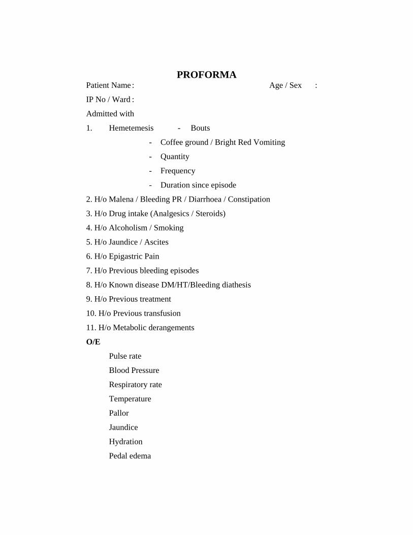
PROFORMA Patient Name : Age / Sex :
IP No / Ward :
Admitted with
1. Hemetemesis - Bouts
- Coffee ground / Bright Red Vomiting
- Quantity
- Frequency
- Duration since episode
2. H/o Malena / Bleeding PR / Diarrhoea / Constipation
3. H/o Drug intake (Analgesics / Steroids)
4. H/o Alcoholism / Smoking
5. H/o Jaundice / Ascites
6. H/o Epigastric Pain
7. H/o Previous bleeding episodes
8. H/o Known disease DM/HT/Bleeding diathesis
9. H/o Previous treatment
10. H/o Previous transfusion
11. H/o Metabolic derangements
O/E
Pulse rate
Blood Pressure
Respiratory rate
Temperature
Pallor
Jaundice
Hydration
Pedal edema

P/A Inspection
Dilated veins
Distension
Shape
Scar
Palpation
Epigastric tenderness
Hepatomegaly
Splenomegaly
Ascites
Any mass
Auscultation
Bowel sounds
Per rectal examination
Investigation
1. Urine
Albumin
Sugar
Deposits
2. Blood
Hemoglobin
Grouping and typing
Bleeding time
Clotting time
Prothrombin time
TC, DC, ESR.

3. Liver function tests S. Bilirubin - Total
Con jugated
Unconjugated
S. Proteins - Total
Albumin
Globulin
S. Enzymes - SGOT, SGPT,
SAP
4. OGD Scopy Oesophagus varices – Grade – Columns
Oesophagitis
Mallory weiss tear
Stomach Diffuse erosion
Ulcer
Growth
Treatment
Conservative
Blood transfusion
Vasopressin
Sengs taken
Somatostatin
Saline lavage
EST
Surgery
Risk factor for rebleeding
Risk factors for morbidity and mortality

CONTENTS
Sl.No. Page No.
1) INTRODUCTION 1
2) AIM OF THE STUDY 3
3) THEORETICAL CONSIDERATION 4
4) ANATOMIC CONSIDERATION 5
5) DEFINITION & INCIDENCE 15
6) ETIOLOGY 16
7) PATHOGENESIS 17
8) CLINICAL FEATURES AND DIAGNOSIS
25
9) INITIAL EVALUATION AND TREATMENT OF PATIENTS WITH UPPER GI BLEED
28
10) UPPER GI ENDOSCOPY 32
11) ALGORITHM FOR MANAGEMENT OF UPPER GI BLEED
42
12) MANAGEMENT 43
13) CAUSES OF MORBIDITY AND MORTALITY
51
14) REVIEW OF LITERATURE 53
15) MATERIALS AND METHODS 60
16) OBSERVATION AND RESULTS 61
17) DISCUSSION 66
18) CONCLUSION 69
PROFORMA
MASTER CHART
BIBLIOGRAPHY

INTRODUCTION
This dissertation is a randomized study conducted among patients with upper GI bleeding predominantly belonging to low socio economic status in Stanley Medical College during period January 2004 to January 2006.
UGI bleeding is one of the common and serious clinical problem
related to severity of bleeding and its etiology.
The diagnosis and management of this emergency, role of general
surgeon, remarkable value of endoscopy and other investigations are
highlighted in this study.
The sex ratio, etiologic factors, key factors in management also
high lighted.
This is one of the common surgical emergency with extremely
frightening symptoms to the patients and relatives.
Alertness of surgeon with thorough understanding of causes of
pathological and physiological factors, that determine the course, which
will decide the outcome.
The ageing population, chronicity of disease like asthma, low
backache, osteo arthritis, migrain, intake of aspirin, cortisone, NSAIDS,
purchase of drugs across counter and using for longer duration are the
accountable factors .

Its our aim to study incidence of etiology, clinical manifestation,
course of illness, management of cases admitted to our hospital.

AIM OF THE STUDY
This study of upper gastro intestinal bleeding has been undertaken
with view of analyzing the
Etiology
Various clinical presentations
Methods of investigations
Study of upper GI scopy
Treatment in our hospital from January 2004 to January 2006.
To determine age, dietary habits in distribution of disease
categories.
To analyse efficiency of investigations in localizing bleeding
sites.
To summarise treatment undertaken and to report their outcome.
To study association of mortality with age, sex distribution of the
disease.

THEORETICAL CONSIDERATION
Incidence varies from country to country. It is 1-2% of acute
hospital admissions with an incidence of 170 per 100,000 adults per
year.
The diversity of etiology with important causes being ulcer
disease, erosive disease, esophageal varices due to portal hypertension.
CAUSES OF UPPER GI BLEEDING
Condition % ULCERS 60%
Esophageal 6% Gastric 21% Duodenal 33%
EROSION 26% Oesophageal 13% Gastric 9% Duodenal 4%
OESOPHAGEAL VARICES 4% MALLORY WEISS TEAR 4% TUMORS 0.5% VASCULAR LESIONS 0.5% OTHERS 5%
Despite advanced sophisticated diagnostic and therapeutic techniques the overall mortality of UGI bleeding remain unchanged at 8-10%. The constancy of mortality rate probably reflects overall improved care of an aged, less fit patients population.
Virtually all studies indicate advanced age as predictor of
mortality. Death among patients younger than 50 year becomes a rarity.

ANATOMIC CONSIDERATION
ANATOMY OF UPPER GI TRACT
Upper GI Tract consist of esophagus, stomach and duodenum.
OESOPHAGUS
Commences at lower edge of cricoid cartilage (C6 vertebra) and
ends at oesophago gastric junction (T 11 vertebra).
This is a muscular tube, 25cm long, occupying the posterior
mediastinum, 2cm lies below diaphragm.
It is closed at upper end by cricopharyngeus muscle 18 cm from the incisor teeth and at lower end by lower esophageal sphincter approximately 40 cm from incisor teeth.
The musculature of
Upper 5% - including upper esophageal sphincter is striated.
Middle 40% - mixed striated and smooth muscle.
Distal 55% - is entirely smooth muscular.

LAND MARKS DURING ENDOSCOPY
Cricopharyngeal sphincter which opens and closes intermittently.
Left atrial distension may compress esophagus and displace it
posteriorly.
Left main bronchus – a tumor may cause narrowing of
esophagus.
NERVE SUPPLY OF OESOPHAGUS
Vagus is the parasympathetic nerve supply which has synaptic
connections to myentric (Auerbach’s plexus).
In the body – there are only cholinergic receptors and vagal
stimulation results in contractions.
In inferior oesophageal sphincter there are both cholinergic and
adrenergic receptors, vagal stimulation results in relaxation of sphincter.
Afferent visceral pain impulses pass along sympathetic nerves
which are closely related to somatic sensory fibres of phrenic and
intercostal nerves in posterior horn of spinal cord.

ARTERIAL SUPPLY
Upper part – Inferior thyroid artery.
In its main extent – Oesophageal branches of aorta.
Lower part – Gastric and inferior phrenic arteries.
VENOUS DRAINAGE
Cervical esophagus drain into inferior thyroid veins and then the
brachiocephalic veins.
Thoracic esophagus drains on left side into brachiocephalic vein
via left hemiazygos and on right side through azygos system to
superior vena cava.
Abdominal esophagus drain into coronary, splenic,
retroperitoneal and inferior phrenic veins which connect with
portal and caval system.

LYMPHATIC DRAINAGE
Is longitudinal rather than segmental.
Lymphatic vessels in submucosa may run for some distances up and
down before penetrating the muscle layers to join lymphatics in adventitia
which drain to adjacent lymph nodes.
I) Para Oesophageal Nodes
Situated on the wall of esophagus include the cervical, upper,
middle and lower thoracic para-esophageal and para cardial nodes.
II) Peri-Oesophageal Nodes
Located on structures immediately adjacent to esophagus, they
include the deep cervical, scalene, paratracheal, subcarinal, posterior
mediastinal, diaphragmatic, left gastric, lesser curvature and coeliac
nodes.
III) Lateral Esophageal Nodes
Found lateral to esophagus and receive lymph from para and peri esophageal nodes. Includes posterior triangle, hilar, suprapyloric, common hepatic and greater curvature lymph nodes.

STOMACH
Main function is to act as reservoir for ingested food. It also serves to
break down food stuffs mechanically and commence the process of digestion.
It originates as a dilatation in tubular embryonic foregut during
5th week of gestation. By 7th week, it descends rotates and further
dilates with a disproportionate elongation of greater curvature into its
normal anatomic shape and position.
Stomach is divided into
The Lesser Curvature
Continuous with the right free border of oesophagus, it is the
concave border.
The Greater Curvature
Starts at cardiac notch,forms the convex border.
The Fundus
Area above the horizontal line from the cardiac notch. So it lies
superior to oesophago gastric junction.

The Incisura Angularis
This is the junction of vertical and horizontal parts of lesser curvature clearly seen at gastroscopy.
The Body
Portion of stomach lying between fundus and pyloric portion.
The Pyloric Portion
Approximately distal one fifth of stomach consist of pyloric
antrum, canal and sphincter. The pyloro duodenal junction is identified
by vein of mayo.
HISTOLOGY
Histologically three types of mucosa can be recognized.
The Cardia
A small area of stomach around oesophago gastric junction,
contains mucus-secreting glands only.
Fundic Gland Area
Lies between the pyloric gland area and cardia.
Gastric pits are shallow and mucosa contains parietal (acid
secreting) and chief (pepsin secreting) cells.

Pyloric Gland Area
Forms mucosa of the pylorus. This area has deep gastric pits and
contains mucus-secreting cells.
BLOOD SUPPLY
Stomach is richly endowed with an arterial supply on both lesser
and greater curves. Most of the supply is from the Coeliac Artery.
On the lesser curve
The left gastric artery, a branch of celiac axis anastamosis with,
The right gastric artery arises from the common hepatic artery.
On the greater curver
The right gastroepiploic artery
Arises from gastroduodenal artery which arises from hepatic
artery and pass behind duodenum. It is often this artery that is eroded in
a bleeding duodenal ulceration.
The left gastroepiploic artery
Arises from splenic artery and contributes to arterial archade along the greater curvature.

Fundus of stomach is supplied by vasa brevia or short gastric
arteries which arise from near the termination of the splenic artery.
VENOUS DRAINAGE
Veins are equivalent to arteries, those along lesser curve ending
in the portal vein and those along greater curve joining splenic vein.
Of particular importance is the left gastric or coronary vein, which receives branches from esophagus. This becomes markedly dilated in portal HT, and must be divided specifically in operations for bleeding esophageal varices.
LYMPHATIC DRAINAGE
Lymph nodes concerned in drainage of stomach are.
Hepatic Group
Lie in lesser omentum along bile ducts receive lymph from liver and
gall bladder. An outlying member along cystic artery is cystic node.
The Subpyloric Nodes
Lie in angle between the 1st and 2nd parts of duodenum on head of
pancreas in relation to bifurcation of gastroduodenal artery. They
receive lymph from right 2/3rd of greater curve through inferior
gastric nodes.

Gastric Group
Superior node – along left gastric artery and paracardial nodes
around cardiac end.
Inferior node – lie along greater curve between layers of greater
omentum.
Pancreaticolienal Group
Lie along splenic artery in relation to upper border of pancreas.
Some occur in gastrosplenic ligament in relation to short gastric
branches.
NERVE SUPPLY
Has intrinsic and extrinsic supply
Intrinsic Nerves
Principally the myenteric plexus of Auerbach and submucosal plexus
of Meissner.
Extrinsic Nerve
Derived mainly from vagus
Vagal plexus around esophagus condenses to form anterior and posterior
vagus. These are both afferent (sensory) and efferent fibres. Efferent
fibres are involved in receptive relaxation of stomach and stimulation of

gastric motility as well as well known secretory function. The
sympathetic supply is derived mainly from the celiac ganglion.
DUODENUM
It is 25 cm. Forms a ‘C’ loop with head of pancreas.
The first 2-5 cm of first part is entirely covered by peritoneum
duodenal ulcers usually occur in this part of duodenum.
Ulcers in posterior wall penetrates, massive bleeding may occur
if it erodes gastroduodenal artery.
Ulcers in anterior wall may perforate and cause generalized
peritonitis.
Duodenum is lined by mucus secreting columnar epithelium. In
addition Brunner’s gland lie beneath the mucosa and are similar to
pyloric glands in pyloric part of stomach. Endocrine cells in duodenum
produce cholecystokinin and secretin.
LIGAMENT OF TREITZ
This is a supportive ligament containing unstriped muscle which
pass to it from the region of left crus of diaphragm and the tissue about
the celiac plexus. This is suspensory ligament of duodenum.

DEFINITION AND INCIDENCE
Upper gastro intestinal bleeding is defined as bleeding from a
source proximal to ligament of Treitz.
It is a common and potentially deadly condition accounting for
85% of hospital admission for gastro intestinal bleeding.
Despite the availability of effective antiulcer medications and
improved understanding of pathogenesis of ulcer disease,
gastroduodenal ulcer disease remains the most common cause,
responsible for half of bleeding episodes.
CLINICAL PRESENTATIONS
Hematemesis and melena are the most frequent clinical finding. However, massive bleeding from an upper source may also cause hematochezia.

ETIOLOGY
Important causes of upper gastro intestinal bleeding
1) Peptic ulcer disease which includes
Gastric ulcer
Duodenal ulcer
Ulcer in stoma/anastamotic site
Reflux esophagitis
Ulcer in Meckel’s
2) Oesophago gastric varices due to portal HT
3) Mucosal Erosions
Gastric
Duodenal
Esophageal
4) NSAID – Associated disorder
Rare causes
1) Vascular Malformations
2) Dieulafoy’s lesion
3) Mallory weiss tear
4) Aorto enteric fistula
5) Tumors of GIT
6) Hemobilia / Hemosuccus pancreaticus / pancreaticpseudocyst /
aneurysm
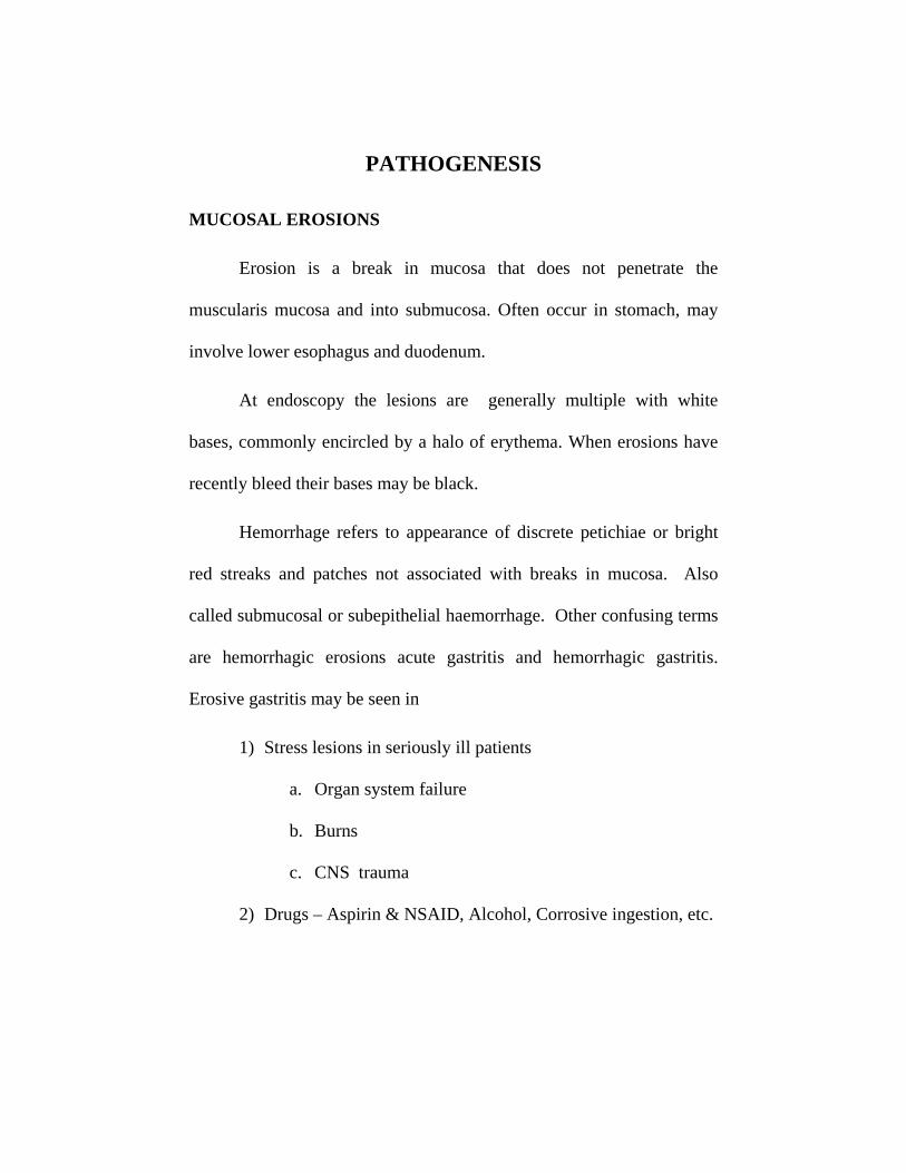
PATHOGENESIS
MUCOSAL EROSIONS
Erosion is a break in mucosa that does not penetrate the
muscularis mucosa and into submucosa. Often occur in stomach, may
involve lower esophagus and duodenum.
At endoscopy the lesions are generally multiple with white
bases, commonly encircled by a halo of erythema. When erosions have
recently bleed their bases may be black.
Hemorrhage refers to appearance of discrete petichiae or bright
red streaks and patches not associated with breaks in mucosa. Also
called submucosal or subepithelial haemorrhage. Other confusing terms
are hemorrhagic erosions acute gastritis and hemorrhagic gastritis.
Erosive gastritis may be seen in
1) Stress lesions in seriously ill patients
a. Organ system failure
b. Burns
c. CNS trauma
2) Drugs – Aspirin & NSAID, Alcohol, Corrosive ingestion, etc.
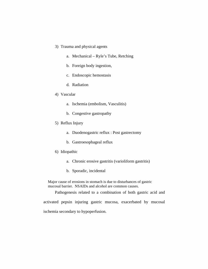
3) Trauma and physical agents
a. Mechanical – Ryle’s Tube, Retching
b. Foreign body ingestion,
c. Endoscopic hemostasis
d. Radiation
4) Vascular
a. Ischemia (embolism, Vasculitis)
b. Congestive gastropathy
5) Reflux Injury
a. Duodenogastric reflux : Post gastrectomy
b. Gastroesophageal reflux
6) Idiopathic
a. Chronic erosive gastritis (varioliform gastritis)
b. Sporadic, incidental
Major cause of erosions in stomach is due to disturbances of gastric mucosal barrier. NSAIDs and alcohol are common causes.
Pathogenesis related to a combination of both gastric acid and
activated pepsin injuring gastric mucosa, exacerbated by mucosal
ischemia secondary to hypoperfusion.

PEPTIC ULCER DISEASE
This term includes gastric, duodenal, oesophageal, stomal ulcers,
anastamotic ulcer, ulcer in Meckel’s due to ectopic gastric mucosa.
Gastric and duodenal ulcer are common causes of UGI bleed. Duodenal
bleed is more common than gastric ulcer.
H.Pylori infection remains most common etiological agent,
followed by ingestion of ulcerogenic drugs and then stress. Other
uncommon specific forms of peptic ulcer.
1) Acid hyper secretion
a. Gastrinoma - inherited MEN I , sporadic
b. Increased mast cells / basophils
c. Antral G cell hyperfunction / hyperplasia
2) Other infections
a. Viral infection – HSV Type I, CMV.
3) Duodenal obstruction / Disruption
a. Congenital bands / Annular pancreas
4) Vascular insufficiency
a. Crack cocain associated perforation
5) Radiation induced
6) Chemotherapy induced (hepatic artery infusions)

Major complications of peptic ulcer disease are intractability,
hemorrhage, perforation, penetration and obstruction.
Bleeding ulcer is caused by acid-peptic erosions into submucosal
or extraluminal vessels.
In stomach, bleeding vessel is typically a small submucosal
artery.
In duodenum, posterior ulcer bleeds because of erosion of ulcer
into retro peritoneal, extraluminal vasculature supplying duodenum and
pancreas.
PORTAL HYPERTENSION
Normal portal venous pressure is 7-8 mm HG. When exceeds 10mm it is portal hypertension. In clinical practice portal pressure is measured indirectly as hepatic venous pressure gradient which is wedged hepatic minus free hepatic vein pressure.
Presence of varices at endoscopy or contrast imaging studies
indicates portal hypertension
Portal venous system is entirely devoid of valves. Numerous
small tributaries connect portal and systemic venous systems and evolve
into major collateral channel when portal HT supervenes. Formation of
collaterals is triggered when portal pressure rises above 12 mm Hg.

Important porto systemic channels that gives rise to UGI bleed
Left Gastric /coronary vein which connects oesophago cardiac
venous plexus with splenic or portal vein. This is responsible for
esophageal varices.
Short gastric and left gastro epiploic veins which connect the
esophageal and gastric plexus with splenic vein. Responsible for
formation of fundal varices.
HYPER DYNAMIC CIRCULATION OF PORTAL HYPERTENSION
Cirrhosis and other causes of portal HT ↓
Increased portal systemic resistance ↓
Portal Hypertension ↓
Increased production of vasoactive substances ↓
Portosystemic collateral formation ↓
Peripheral vasodilatation and increased plasma volume ↓
Hyperdynamic circulation

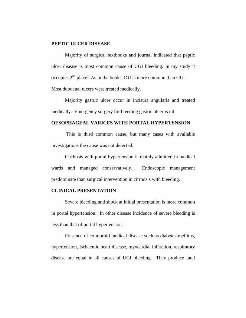
OESOPHAGEAL VARICES
Rupture of esophageal varices is more common than gastric
fundal varices. Elegant studies revealed the peculiar portosystemic
connection which occurs in distal 2-5 cm of esophagus.
Four distinct layers of veins noted.
Intra epithelial veins connected to deep intrinsic vein via
superior venous plexus.
Deep intrinsic vein which is located in submucosa
connected to para esophageal vein via perforating veins.
Major bleeding mainly due to rupture of deep intrinsic vein.
RUPTURE OF ESOPHAGEAL VARICES
Erosive Theory
Due to reflux esophagitis.
This theory was not accepted as antiulcer drug does not reduce
incidence of ruptures.
Transmural Tension
The difference in pressure between the esophageal lumen and that of
the varix.

Tension was inversely related to variceal wall thickness, derived by
Laplace law T = TPX r/w
TP - Transluminal pressure
r - Radius of lumen
w - Wall thickness
MALLORY WEISS SYNDROME
Forceful vomiting with a contracted cricopharyngeus and marked
diaphragmatic fixation may lead to rise in intra abdominal and
intragastric pressure that causes esophageal rupture.
This is prone in alcoholics. Lesion is characterized by a tear in
proximal gastric mucosa near esophago gastric junction.
DIEULAFOY’S LESIONS
Rare cause of GI bleed.
lesions are unusually large submucosal or mucosal vessels found in
gastric mucosa, most commonly along lesser curvature in mid-
stomach.
AORTOENTERIC FISTULA
Uncommon condition, in which an inflammatory tract develops
between aorta and gastro intestinal tract.

Develop as primary process resulting from infections aortitis, or
inflammatory aortic aneurysm or a secondary process following aortic
replacement with synthetic graft.
HEMOBILIA
Rare cause that results from blunt or penetrating hepatic injury
with fistula formation with in the liver between vascular structure and
biliary ductal system . Bleeding is usually mild and accompanied by
jaundice and right hypochondrial pain.
HEMOSUCCUS PANCREATICUS / PANCREATIC
PSEUDOCYST AND PSEUDO ANEURYSMS
UGI bleed can occur from variety of mechanism that are consequent to chronic pancreatitis. The autodigestive process imparted by pancreatitis can lead to pseudoaneurysm formation of any arteries such as splenic artery, gastroduodenal artery, pancreaticoduodenal artery, left gastric artery. Rupture of an artery results in bleeding.
UNCOMMON CAUSES
1. Chronic renal failure
2. Typhoid fever
3. Hemophilia A & B
4. Thrombocytopenia
5. Collagen Vascular Disease
6. Anti coagulant therapy.

CLINICAL FEATURES AND DIAGNOSIS
Thin, dark, granular coffee ground vomiting is important
diagnostic feature of UGI bleeding.
Melena, hematochezia, blood streaked stools and occult blood in
stools are clinical features but they may be confused with lower GI
bleeding.
CLINICAL SIGNS OF BLOOD LOSS
American college of Surgery Committee on Trauma 1997 estimated
fluid and blood loss in shock.
< 1000ml 1000 – 1500 1500 – 2000 > 2000 BP Normal > 90. Sys. 70 – 90 Sys < 90
HR Normal 100 – 120 > 120 bpm > 140
RR Normal > 24 ↑ ↑ / Hyperventilation
CRT Normal > 3 SLC Cool, Pale skin
Cold, Skin
Urine > 30ml/hr 25 – 30 ml/hr 5 – 15 cc ml/hr
Minimal
Mental changes
No Weakness, Anxiety
+ Lethargic
Sources
1. Conrad, S.A. (2002) Acute upper GI bleeding in critically ill patients
cause and modalities criteria care Med. 30 (6), S. 365.
2. Gosch, S. Loatt S.D. and Kinnear M. (2002) Management of GI
Hemorrhage. Postgraduate Med. J. 78 (915).

Nasogastric Aspirate Colour
Stool Colour Mortality Rate (%)
Clear Brown or Red 6 Red 19.1 Coffee ground Brown / Black 8.2
Red Blood Black Brown Red
Comparison Of UGI & LGI Symptoms
UGI LGI
Hemetemesis Assured Ruled out
Melena Probable Possible
Hematochezia Unlikely Highly probable
Blood streaked stools Ruled out Assured
Occult blood in motion Possible Possible
Once diagnosis of UGI bleed was made first priority is rapid
assessment of hemodynamic status.
Early symptoms of shock appears with blood losses of 15 – 20%
includes weakness, cold skin, diaphoresis and pallor. These symptoms
are due to sympathetic stimulation resulting from peripheral
vasoconstriction.

When volume loss of 20-40% occur symptoms such as
tachycardia, hypotension, mental confusion and oliguria (<400 ml/day)
occurs.
When volume loss more than 40% profound shock manifested by
prostration, depressed level of consciousness, thready pulse and blood
pressure not recordable. Myocardial infarction followed by arrhythmias
will be sequelae of profound shock in patient with old age with coronary
artery disease.
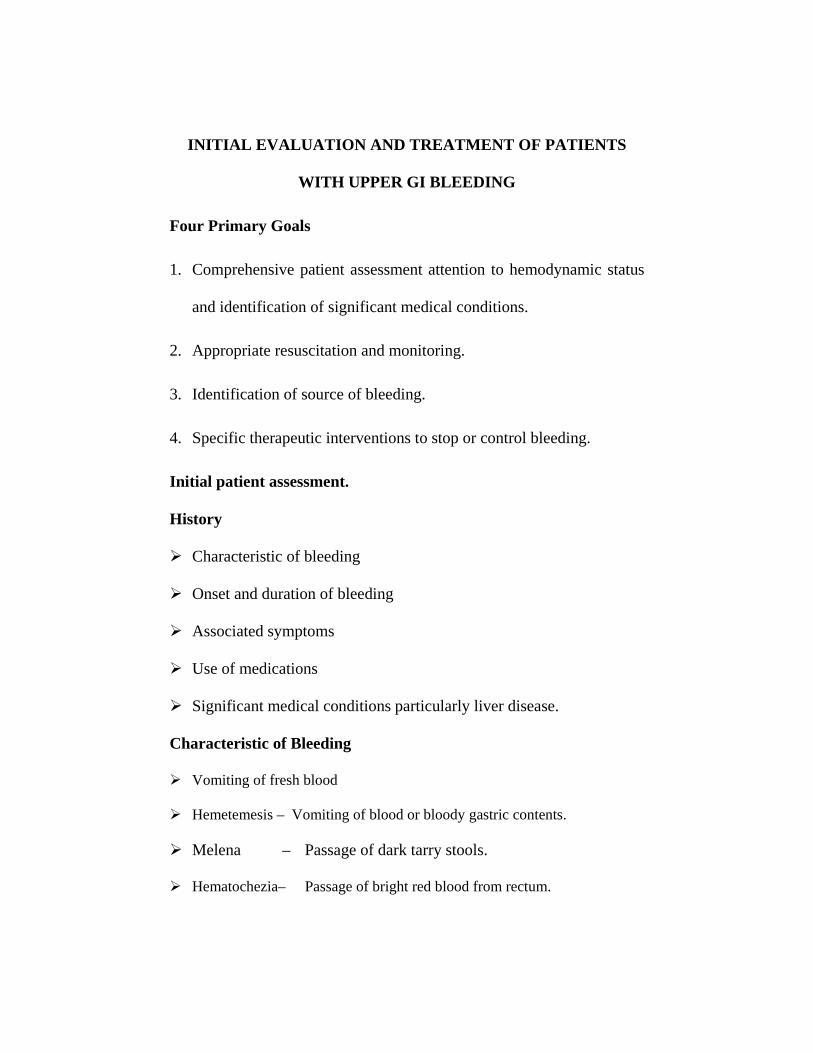
INITIAL EVALUATION AND TREATMENT OF PATIENTS
WITH UPPER GI BLEEDING
Four Primary Goals
1. Comprehensive patient assessment attention to hemodynamic status
and identification of significant medical conditions.
2. Appropriate resuscitation and monitoring.
3. Identification of source of bleeding.
4. Specific therapeutic interventions to stop or control bleeding.
Initial patient assessment.
History
Characteristic of bleeding
Onset and duration of bleeding
Associated symptoms
Use of medications
Significant medical conditions particularly liver disease.
Characteristic of Bleeding
Vomiting of fresh blood
Hemetemesis – Vomiting of blood or bloody gastric contents.
Melena – Passage of dark tarry stools.
Hematochezia– Passage of bright red blood from rectum.

Associated Symptoms
H/O orthostatic dizziness or syncope indicate rapid and profound
blood loss.
Antecedent dyspepsia – suggestive of peptic ulcer disease.
Crampy abdominal pain – more consistent with upper GI
bleeding.
Vomiting – may suggest Mallory – weiss tear.
History of Medications
Salicylates or NSAIDS - may impair platelet function and
contribute to poor coagulation.
Warfarin
Low molecular weight Heparin.
Corticosteroids
Past Medical History
Previous episodes of GI bleeding.
H/O dysphagia or reflux esophagitis.
Recent GI distress with vomiting.
Peptic ulceration / H.Pylori infection.
Liver disease, alcohol abuse.

IBD, intestinal polyps, malignancy.
Co-Morbid Medical Conditions
Renal insufficiency
Atherosclerotic disease
Congestive cardiac failure
Chronic respiratory conditions
Preexisting liver disease
CNS disability
Physical Examination
Major initial objective is to determine the degree of blood loss
and volume depletion.
LABORATORY ASSESSMENT.
Blood Investigation
Hemoglobin
Hematocrit
Coagulation Profile
Blood grouping and typing
Bleeding Time
Clotting Time
Prothrombin Time
Platelet count

These help to plan for transfusion to rule out bleeding and
collagen vascular disease.
Liver Function Test
S. Bilirubin
SGOT
SGPT
SAP
GGT
Help for management
Gives clue to diagnosis and etiology
Abnormal LFT indicates bad prognosis
Child Pugh’s Criteria
5-6 (A) 7-9 (B) 10 – 15 (C) S. Bilirubin (mg/dl) <2 2-3 >3
S. Albumin (gm/dl) > 3.5 2.8-3.5 >3
Prothrombin Time 1-4 4-6 >6
Ascites Absent Slight Moderate
Encephalopalthy None Minimal to moderate Coma A - Good liver function
B - Moderate impairment
C - Poor liver function
A & B - 70 – 80% survival rate
C - 30% survival rate
- S. Electrolytes
- Renal function tests
- Upper GI scopy
- Angiography
UPPER GI ENDOSCOPY
Gold Standard in diagnosis of upper GI bleeding.
INDICATION FOR EMERGENCY ENDOSCOPY
Patients with active hematemesis and massive melena with hypotension
and hemodynamic instability should undergo endoscopy as soon as
possible after proper resuscitation.
Patient Preparation
First establish a patent airway as massive bleeding may predispose to
aspiration.
Hemodynamic status assessed
Gastric Lavage
Not necessary in a majority of patients because blood tends to
pool in the fundus when patient is lying in left lateral position.
Since most causes of bleeding are located in lesser curve and
duodenum, adequate visualization possible without disturbing the pool
of blood.
If gastric lavage necessary a large bore sump tube with large side
holes should be used rather than the small nasogastric tubes which can
be blocked easily by clots.

Sedation for Endoscopy
Sedation necessary to calm and relax the patient especially since
therapeutic endoscopy is longer compared to diagnostic endoscopy.
Commonly IV benzodiazepines are used.
Procedure
Endoscopy is performed under IV sedation and with topical pharyngeal
anaesthesia.
Patient in the left lateral position.
Adequate suctioning is important to remove oropharyngeal
secretion and in case patient vomits.
Passing the endoscope
There are two methods 1) Blind method, 2) Direct vision method. Blind Method
Tip of endoscopy passed into patients mouth through a bite guard
and advancing the tip approximately 18-20 cm from incisor teeth.
Patient is asked to swallow, cricopharyngeal sphincter opens tip is
advanced into esophagus by bending the tip of endoscope.
Direct Vision Method :
Passing endoscope through a bite guard and watching
endoscopically to observe anatomy of hypopharynx.
After passing through cricipharyngeus, important land marks are

Oesophago gastric junction
Cardia
Incisura
Pylorus
Superior duodenal flexure
OESOPHAGUS (20-40cms from incisor)
Important landmark are
Indentation of left main bronchus
Pulsation of left atrium
Pulsation of aorta
Mucosa
Mucosal colour
The folds which run parallel and converge to become cardiac
rosette.
Oesophago Gastric Junction
Seen where pale pink mucosa meets the dark red gastric mucosa,
often seen as irregular ‘Z’ line- ora serrata.
The diaphragm normally clasps oesophago gastric junction and position of diaphragm and hiatus should be noted. When patient is taking deep breath, varices are seen as red / blue raised columns in lower oesophagus.

STOMACH
With slight angulation to left and anteriorly endoscope is passed in to
stomach. Fluid removed to improve visualization and to reduce aspiration. It
is important to note presence of bile, food or blood and the following
Ulcer
Growth
Fundal varices are diagnosed
Fundus
On retroflexion, fundus appear as hollow rounded area with flat
mucosa. Mucosal capillaries are prominent while venules are smaller
and straight. Gastric varices are wide and appear serpigenous.
Gastric Ulcer
With patient lying on his left side fluid will collect along most
dependent portion of greater curve. This is called as gastric lake.
The fluid is usually grayish in the absence of bile but greenish
yellow in presence of bile.
Body
Rugal folds principally located in greater curve. In lesser curve
they are vertically placed.

Gastric Angle
Major landmark for lesser curve and separation of body and
antrum.
Lesser curve ends distally as a cresent shaped mucosal fold
called incisura.
Pylorus
Starts from incisura
Pyloric antrum is identified by horizontally placed rugal folds.
Pyloric channel is within 2 cm of pylorus with enormous
variable.
DUODENUM
Duodenal bulb
Triangular base is demonstrated by angular fornices which
are best seen during retroflexion. .
Mucosa of Duodenum
Mucosa is smooth. Arranged in concentric fold or triangular fold
called “valves of Kerking”.
In emergency after adequate suctioning
Oesophageal pathologies such as varices, ulcers, tumours Mallory-
weiss tears should be obvious.
Fresh blood refluxing into esophagus indicate active bleeding lesion
in stomach.
With endoscope in stomach air is insufflated to distend stomach
lumen and provide an adequate endoscopic view.
In most cases it is useless to attempt removal of blood or clots in
fundus, rather examine by sliding the endoscope along lesser curve
above the pool of blood until it reaches II part of duodenum.
Most bleeding pathologies readily seen since they lie along lesser
curve in distal antrum and duodenum.
If no pathology is found, return the endoscope into stomach aspirate
the fluid portion of blood pooled in corpus and fundus. Turn the
patient to right lateral position. This shifts the blood and clot in to
distal stomach to allow better examination of fundus.
Adherent clots removed to expose the underlying bleeding
pathology.
PROGNOSTIC FINDINGS AT ENDOSCOPY FOR PEPTIC ULCER
Actual appearance of ulcer at endoscopy is the most important
predictor of rebleeding.

APPEARANCES OF ULCERS
A clean ulcer base
A flat ulcer
Pigmented spot (Purple / Browns / Black)
Adherent clot
A visible vessel which may be smooth surfaced or tubular
protuberance on ulcer surface or active bleeding with sprouting
blood, continuous oozing or oozing around an adherent clot.
Later four appearances are considered stigmata of hemorrhage
PROBALILITY OF REBLEEDING.
Clear ulcer base – rarely bleeds.
Flat / pigmented spot – 10%
Adherent non bleeding clot – 20%
Visible vessel – 40 – 80%
Ulcer size >1 cm

STIGMATA OF RECENT BLEEDING
Forrest Classification System
Descriptive identification of ulcer characteristics
F Ia - Active spurting bleeding ulcer
F Ib - Non-spurting active bleeding ulcer
F II a - Ulcer with a visible vessel or pigmented protuberance.
F II b - Ulcer with adherent clot
F II c - Ulcer with pigmented spot
F III - Clean ulcer base without stigmata of bleeding
An adherent clot and a non-bleeding visible vessel are
considered major stigmata and carry high incidence of rebleeding
(30-40%).
Ulcer with clean base the risk of rebleeding is less than 5%.

FINDINGS AT ENDOSCOPY FOR ESOPHAGEAL VARICES
GRADING OF OESOPHAGEAL VARICES
Grade I - Varices can be depressed by endoscope
Grade II - Varices cannot be depressed by endoscope
Grade III - Varices are confluent around the circumference of
the esophagus.
GRADING OF VARICES AT 2 CM ABOVE THE OESOPHAGO GASTRIC JUNCTION
Grade Bite width of 1.7mm, biopsy forceps 5mm
I 1/4th bite width
II ½ bite width
III 3/4th bite width
IV 1 or more bite width
PREDICTORS OF BLEEDING VARICES
Size of varix
Directly proportional to rupture of varix
Colour
Red colour
Important predictor

Cherry red spot
Dilated subepithelial veins may appear as raised cherry red spot.
The hemocystic spot is 4 mm in diameter. It typically lies in summit of
dilated veins. It represents blood coming from deeper extrinsic veins of
esophagus straight out toward the lumen through communicating vein
into superficial submucosal vein.
These 3 signs are highly positive predictors.
SIGNS OF RECENT BLEEDING
Fresh clot in the base
Pulsation of vessel
VARIOUS TECHNIQUES CURRENTLY AVAILABLE FOR
ACHIEVING HAEMOSTASIS
Injection of vasoactive agents.
Injection of sclerosing agents
Bipolar electrocoagulation
Band ligation
Thermal probe coagulation
Constant probe press tamponade
Argon plasma coagulator
Laser photo coagulation
Rubber band ligation
Application of haemostatic material including biologic glue.

ALGORITHM FOR MANAGEMENT OF UPPER GI BLEED
ACUTE UPPER GASTRO INTESTINAL HAEMORRHAGE
Consider Orotracheal intubation Fluid Resuscitation Nasogastric Tube Labs – Typing & Cross Matching Hematocrit BT, CT, PT History – Liver Failure Alcohol Peptic Ulcer disease NSAID intake
Monitored setting / IV
H2 blocker / PPI foley Massive Haemorrhage preventing endoscopy
Endoscopy Surgery Positive Negative Varices Duodenal gastric
ulcer AGML Mallory
Weiss tear Evaluate for
LGIB Endoscopic control
by injection or electrocoagulation
Medical Management
Endoscopic therapy
Succeed Fails
Suceeds Fails Rebleeds
Surgery
Observe Surgery AGML: Active Gastric Mucosal Lesion LGIB : Lower gastrointestinal Bleeding

MANAGEMENT OF VARIOUS CAUSES
Mucosal Erosive Disease
Refers to endoscopically visualized lesions (subepithelial haemorrhage
and erosions) that may cause bleeding. As these lesions are strictly mucosal
lesions whereas all blood vessels are significant size or in submucosa and
below they never have life threatening bleeding or large transfusion
requirement
Main mode of management is conservative line of medical
management. Endoscopic therapy is unlikely to be helpful or needed because
of the diffuse, superficial nature.
Peptic Ulcer
Peptic ulcer and duodenal ulcers are most common cause of acute
hemorrhage in upper gastrointestinal tract accounting for 25% of cases.
About 5% of patients with peptic ulcer disease have hemorrhage
as the initial manifestation of the condition.
Hemorrhage remains most lethal form of complicated ulcer
disease.

JOHNSON’S CLASSIFICATION OF PEPTIC ULCER
Type I
Gastric Ulcer
60 – 70%
Location - lesser curvature at or proximal to incisura, near
junction of oxyntic and antral mucosa.
Associated with diffuse antral gastritis or multifocal atrophic
gastritis.
Type II
Gastric ulcer associated with active or chronic duodenal ulcer
Location – same site
Type III
Pyloric Channel Ulcer
Location with in 2 cm of pylorus
Type IV
Gastric ulcer
Located in proximal stomach or in the gastric cardia.
Type 2 and 3 gastric ulcers appear to behave more like duodenal
ulcers and associated with excess acid.
Type I and IV are not associated with excess acid.

NSAID induced ulcer. They are typically multiple and located
anywhere in stomach where protective mucosal barrier was damaged.
PORTAL HT GASTROPATHY
Seen largely in fundus, but can extend through out stomach.
Shown as mosaic – like pattern with small polygonal areas surrounded
by whitish yellow depressed border. Red point lesion and cherry red
spots predict high risk of bleeding. Black brown spots are due to intra
mucosal hemorrhage.
Sclerotherapy increases the gastropathy in portal hypertension.
Management
Resuscitation
IV cannulation – fluids
Blood transfusion
Oxygen mask / Nasal cannula
Ryle’s Tube
Continuous Bladder Drainage
Endoscopy
o Clean based ulcer-discharged home after resuscitation,
stabilization and institution of ulcer therapy.
o Flat spots or adherent clots – watched in hospital , as recurrent
bleeding is low

o Visible vessel and active bleeding- to undergo endoscopic
hemostatic therapy.
Surgery for Gastric Ulcer
Patients with severe ongoing ulcer bleeding despite attempt at
endoscopic therapy.
Procedures used
Ulcer excision with or without antrectomy
Partial gastrectomy, with Billroth I anastomosis
Partial gastrectomy with Billroth II anastomosis
Cseudes procedure
Surgery for bleeding duodenal ulcer
Suture ligation of bleeding artery with vagotomy with
drainage procedure.
LONG TERM PREVENTION OF ULCER BLEEDING
Conservative medical therapy
Full course of antiulcer therapy

OESOPHAGO GASTRIC VARICES
Bleeding from osophagogastric varices responsible for one third
of all deaths in patients with cirrhosis and portal hypertension.
90% of cirrhotic patients develop esophageal varices and 25-30% develop hemorrhage. Repeated hemorrhage develop in 70% of patients.
MANAGEMENT
Upper GI scopy remains main stay in investigation and treatment
MEDICAL MANAGEMENT
Vitamin k IM route
Platelets, fresh frozen plasma infusion
Octreotide infusion
Sengstaken – Blackmore tube
Minnesota tube
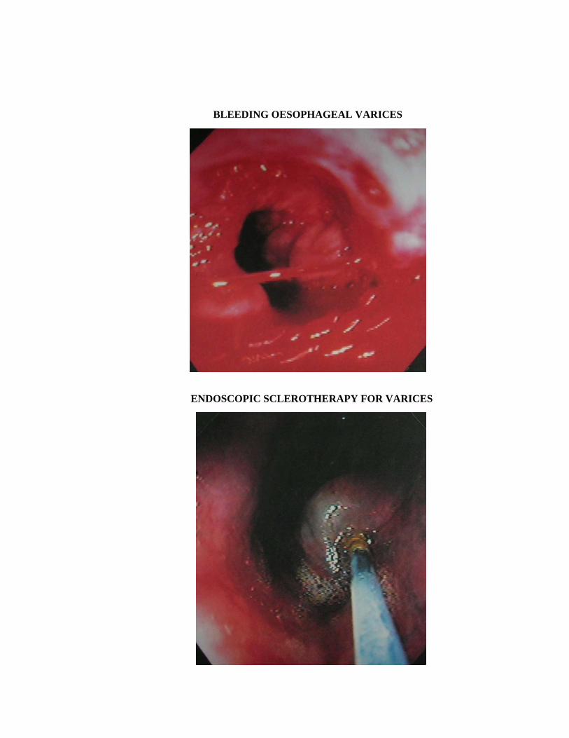
Endoscopic – Sclerotherapy
- Band ligation
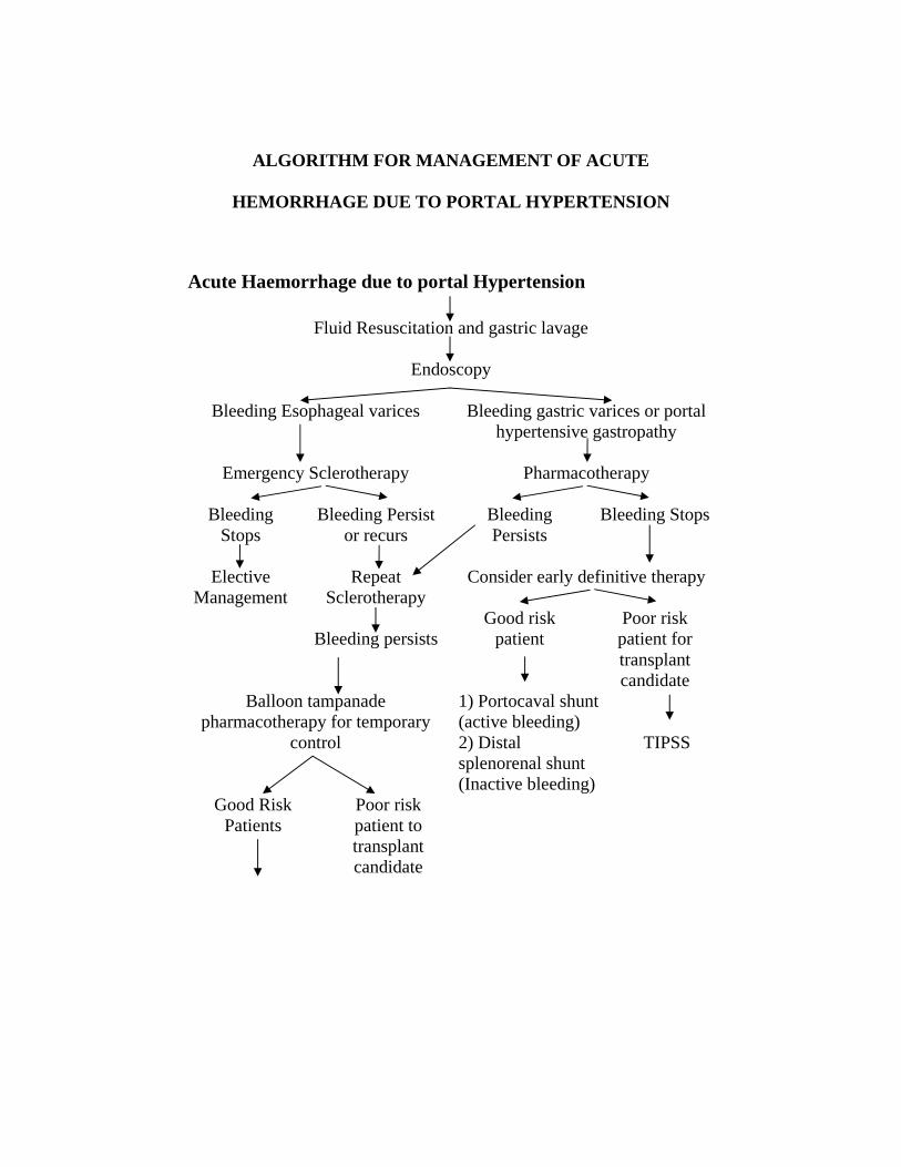
ALGORITHM FOR MANAGEMENT OF ACUTE
HEMORRHAGE DUE TO PORTAL HYPERTENSION
Acute Haemorrhage due to portal Hypertension
Fluid Resuscitation and gastric lavage
Endoscopy
Bleeding Esophageal varices Bleeding gastric varices or portal hypertensive gastropathy
Emergency Sclerotherapy Pharmacotherapy
Bleeding
Stops Bleeding Persist
or recurs Bleeding Persists
Bleeding Stops
Elective
Management Repeat
Sclerotherapy Consider early definitive therapy
Bleeding persists
Good risk patient
Poor risk patient for transplant candidate
Balloon tampanade pharmacotherapy for temporary
control
1) Portocaval shunt (active bleeding) 2) Distal splenorenal shunt (Inactive bleeding)
TIPSS
Good Risk Patients
Poor risk patient to transplant candidate

1. Portocaval shunt (active bleeding,) 2. Distal splenorenal shunt (Inactive bleeding) 3. Esophageal transection.
TIPSS
TIPSS : Trans jugular intrahepatic porto systemic shunt surgery
SURGICAL MANAGEMENT
1. Oesophago gastric devascularisation with or without splenectomy
(Nonshunt)
2. Surgical decompression of portal venous system by shunt operation.
Indication : Two sessions of failed sclerotherapy with reasonable liver
failure,
Porto systemic shunt surgeries
End to side portocaval
Proximal splenorenal shunt
Distal splenorenal shunt (Warrant’s shunt)
Transjugular intrahepatic portacaval shunt (TIPS)

NON SHUNT SURGERIES
Splenic vein thrombosis with isolated fundal varices are best
treated by splenectomy with or without devascularisation procedure.
OTHER OPERATIONS
Oesophageal transection
Porto Azygos disconnection by Hassab procedure –
Devascularisation of upper half of stomach and esophagus.
Sugiura’s procedures
I Stage :
Devascularisation of upper half of stomach and esophagus as in Hassab
procedure.
II Stage :
Vagotomy with pyloroplasty was performed.

CASUES OF MORBIDITY AND MORTALITY
Most important cause of death in upper GI bleed is shock and its
sequelae. Comorbid medical diseases particularly myocardial ischaemia
and infarction are aggrevated due to shock leading to fatal arrhythmias.
Mortality associated with underlying medical illness and gastro
intestinal bleeding.
Condition Mortality Rate (%) Renal disease 29
Acute renal failure 63
Liver disease 25
Jaundice 42
Pulmonary disease 23
Respiratory failure 57
Cardiac disease 13
Congestive heart failure 28 Risk factors for morbidity and mortality
1. Age > 60 years
2. Shock at initial presentation
3. Transfusion requiring six or more units of blood
4. Coronary artery disease
5. Chronic pulmonary artery disease
6. Acute pulmonary failure

7. Acute and chronic renal failure
8. Cirrhosis
9. Acute hepatic failure
10. Sepsis
11. MODS
12. Coagulopathy
13. Recent CVA
14. Malignancy
15. Immunosuppression
16. Post operative stage
Prognostic indicators for mortality from peptic ulcer hemorrhage.
Clinical Parameter Mortality Rate (%) Overall 5-8 Age 10-15 > 60 years > 80 years
25 – 30
Systolic blood pressure on presentation 80 – 90 mm Hg 12 – 15 < 80 mm Hg 30 – 35 Nasogastric aspirate on presentation Coffee – ground appearance 6 – 10 Red Blood 18 – 20 Transfusion requirement > 10 units 28 - 34

REVIEW OF LITERATURE
1) Upper GI bleeding in a Brazilian Hospital, a retrospective study of
endoscopic records by . Zaltman C, Souza Hs, Dept. of Int. Medicine,
Federal University of Riode Janeiro, Brazil.
Aim : To assess clinical characteristics, endoscopic accuracy, treatment
efficacy and clinical out come of patients.
Results :
Most patients were male 68.7% mean age 54.+17.5.
Bleeding site detected in 75.6% patients.
Diagnostic accuracy greater within 24 hours of bleeding onset
and in presence of hemetemesis.
Peptic ulcer main cause of upper GI bleed (35%)
Variceal bleeding (20.45%) indicating higher rate of underlying
liver disease.
Endoscopic treatment performed in 2
3.86 % patients.
Permanent hemostasis – 86% at 1st intervention 62.5% after
rebleeding.
Emergency surgery was seldom necessary.
Average number of blood units 1.44 + 1.99 / patients.
Average length of hospital stay 7.71 + 12.2
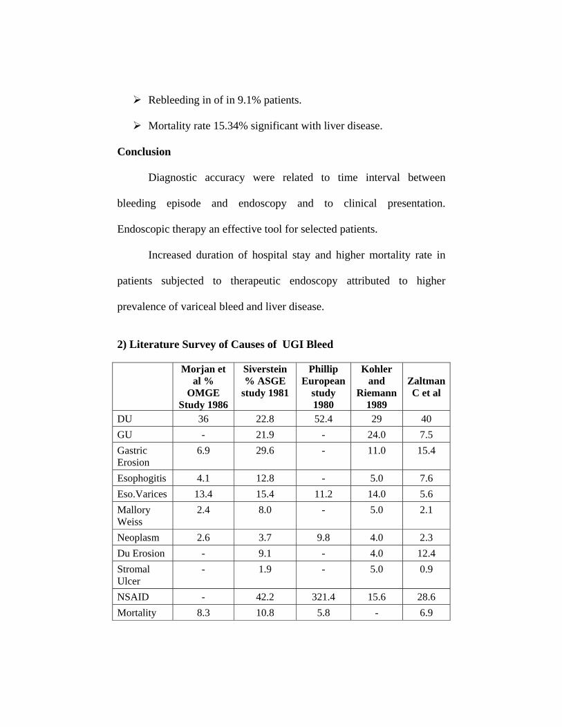
Rebleeding in of in 9.1% patients.
Mortality rate 15.34% significant with liver disease.
Conclusion
Diagnostic accuracy were related to time interval between
bleeding episode and endoscopy and to clinical presentation.
Endoscopic therapy an effective tool for selected patients.
Increased duration of hospital stay and higher mortality rate in
patients subjected to therapeutic endoscopy attributed to higher
prevalence of variceal bleed and liver disease.
2) Literature Survey of Causes of UGI Bleed Morjan et
al % OMGE
Study 1986
Siverstein % ASGE
study 1981
Phillip European
study 1980
Kohler and
Riemann 1989
Zaltman C et al
DU 36 22.8 52.4 29 40 GU - 21.9 - 24.0 7.5 Gastric Erosion
6.9 29.6 - 11.0 15.4
Esophogitis 4.1 12.8 - 5.0 7.6 Eso.Varices 13.4 15.4 11.2 14.0 5.6 Mallory Weiss
2.4 8.0 - 5.0 2.1
Neoplasm 2.6 3.7 9.8 4.0 2.3 Du Erosion - 9.1 - 4.0 12.4 Stromal Ulcer
- 1.9 - 5.0 0.9
NSAID - 42.2 321.4 15.6 28.6 Mortality 8.3 10.8 5.8 - 6.9

3) Etiology of UGI bleeding Jordanian patients, a prospective study.
Mustafa Mohennak MD, Department of Int. Medicine, GI &
Liver Unit, Jordan, University Hospital & A1 Bashir Hospital, Amman.
Conclusion :
1. Emergency endoscopy should be performed in all.
2. High risk patients transfer to special centre
3. DU disease more common.
4) Retrospective review of emergency department patients with non
variceal UGI bleeding for potential OP management.
Kum-ying Tham et al, Dept. of emergency medicine Tantock
Seng Hosp. Singapore. Dept. of Emergency and Critical Care Medicine
Keio University School of Medicine and Keio University Hospital,
Tokyo, Japan. Dept. of Emergency Medicine, Massachusetts General
Hospital, Boston, MA.
Objective : To determine number of ED patients with non variceal UGI bleeding who could have been managed as OP’s through previously developed clinical guidelines.
Results :
145 patients seen 128 admitted 111 (77%) underwent OGD, 21 (91%) had varices, 90 (81%) with non variceal UGI bleeding 18 of these 90 fulfilled guidelines for OP management and none of 18 had complication.
Conclusion :
In non HMO urban teaching hospital 18 patients with non
variceal UGI bleeding met criteria for OP management in 6 month
period and none developed complication during a mean in hospital –
stay of 21 day.
5) Acute UGI bleeding in Kuwait – 1995; Kuwait Medical Journal.
2001, 33(2) (44-14) Abdul Karrem. Y. Khajah et al, Gastroentrology
Department , Al-Amiri. Hospital – Retrospective study.
Results :
215 case admitted in 1995 overall incidence 28.3 per 100,000 .
Most common case – Peptic Ulcer Disease (19%) Esophageal
Varices (7%) 10 fatalities, due to 10- morbidities.
Conclusion :
UGI bleeding in Kuwait is most commonly due to peptic ulcer
disease and varices mortality rate lower than that from developed
countries.
6) UGI Bleeding in Medical Intensive Care Unit, Doha, Qatar-
Kamaha A et al, Dept. of Medicine, Medical Intensive Care Unit.
Hamad Medical Corporation, Doha, Qatar. QMJ, June 2003.
UGI Bleed is a common problem, and important cause of
morbidity and mortality study from June 1999 to May 2000, 860 patient
admitted 102 with UGIB. Common age group – 50 – 60 years (28.4%)
Frequent cause – Peptic ulcer disease 50 patients followed by variceal
bleeding.
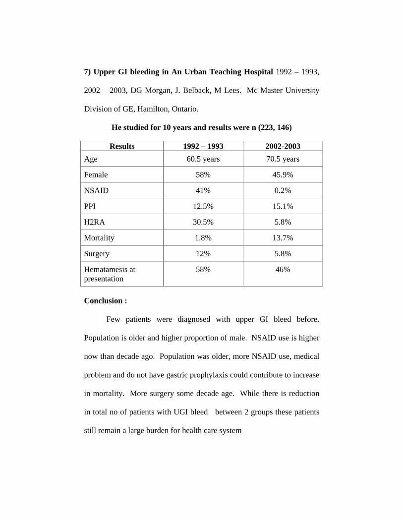
7) Upper GI bleeding in An Urban Teaching Hospital 1992 – 1993,
2002 – 2003, DG Morgan, J. Belback, M Lees. Mc Master University
Division of GE, Hamilton, Ontario.
He studied for 10 years and results were n (223, 146)
Results 1992 – 1993 2002-2003 Age 60.5 years 70.5 years
Female 58% 45.9%
NSAID 41% 0.2%
PPI 12.5% 15.1%
H2RA 30.5% 5.8%
Mortality 1.8% 13.7%
Surgery 12% 5.8%
Hematamesis at presentation
58% 46%
Conclusion :
Few patients were diagnosed with upper GI bleed before.
Population is older and higher proportion of male. NSAID use is higher
now than decade ago. Population was older, more NSAID use, medical
problem and do not have gastric prophylaxis could contribute to increase
in mortality. More surgery some decade age. While there is reduction
in total no of patients with UGI bleed between 2 groups these patients
still remain a large burden for health care system

8) Cooper et al studied effectiveness of performing an early endoscopy
with in first 24 hours of acute UGIB episode.
Early endoscopy associated with reduction in length of hospital
stay, rate of recurrent bleeding and need for surgical intervention.
9) Upper GI bleed an Etiological study of 552 cases Journal Pakistan
Institute of Medical Science, July 2004. 15 (1): 845 – 8. Study
conducted between 1992 – 2000. in Dept. of GE at PIMS.
Tashfeen et al.
Oesophageal varices accounted for majority of lesions causing
upper GI bleeding (44%). Peptic ulcer disease second commonest
(19.7%), Oesophageal lesions like oesophagitis and esophageal ulcers
6.6%, Tumors of UGI 1.1%, Gastric erosion 4.5. Age variation
maximum no of patient 50 – 59 years (22.6%).
It was concluded that causes of UGIB are similar to those in local
literature but different from causes described in Western literature.
10) Upper GI bleed in an urban hospital, etiology, recurrence and
prognosis. Ann. of surgery 1990, Oct, 212(4) 521 – 6.
Sugawa C et al. Dept. of Surgery, Wayne State University,
Detroit, M1 48201.

Acute UGIB common cause of hospital admission and morbidity
and mortality.
This study reviewed 469 patients.
Most common cause of bleeding, include gastric mucosal lesion
135 patients (24%) esophageal varices (221). Gastric ulcer 108 patient
s(19%). DU 78 (14%). Non operative treatment in 89.5%. Endoscopic
treatment in 144 cases. Operations performed in 58 cases (10.5%).
Emergency operation to control bleeding in 2.5% cases. There were 58
deaths to UGIB factors correlating with death include shock at
admission transfusion requirement more than 5 units.

MATERIALS AND METHODS
Present study made on 100 cases of UGI bleed admitted in our Stanley
Medical College from January 2004 to January 2006.
Proforma made and careful entry made for each case.
Analysis of case regarding age, sex, OGD finding and variety of
treatment opted and conclusion drawn.
All patients were admitted as emergency. Adequate laboratory
investigations, imaging studies, UGI scopy done as a routine for all
cases.
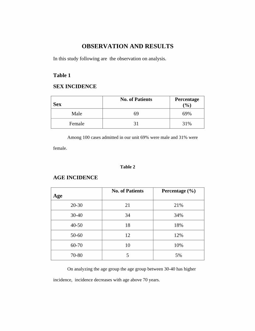
OBSERVATION AND RESULTS In this study following are the observation on analysis.
Table 1
SEX INCIDENCE
Sex No. of Patients Percentage
(%)
Male 69 69%
Female 31 31%
Among 100 cases admitted in our unit 69% were male and 31% were
female.
Table 2
AGE INCIDENCE
Age No. of Patients Percentage (%)
20-30 21 21%
30-40 34 34%
40-50 18 18%
50-60 12 12%
60-70 10 10%
70-80 5 5%
On analyzing the age group the age group between 30-40 has higher
incidence, incidence decreases with age above 70 years.


Table 3 DISEASE WISE INCIDENCE
Sl.No. Diseases No. of Cases
Peptic Ulcer Disease 32 Duodenal 23
1)
Gastric 9 2) Varices 19
Erosive Diseases 43 Gastric 37 Oesophageal 5
3)
Duodenal 1 4) Drug Induced gastritis 3 5) Mallory Weiss 1 6) Malignancy 2
On analyzing the causes it is found that erosive disease is more
common with 43% of patients and of these gastric erosion is more common.
Table 4 DISEASE WISE SEX INCIDENCE
Sl.No. Diseases No. of
Patients Male Female
Peptic Ulcer Diseases 33 23 10 Duodenal Ulcer 23 16 7
1)
Gastric Ulcer 9 5 4 2) Varices 19 15 4
Erosive disease 43 31 12 Gastric 37 27 10 Oesophageal 5 3 2
3)
Duodenal 1 1 0
On analyzing this it is found that erosive disease, peptic ulcer disease
are more common in male than female.
Table 5

MALIGNANCY
Sex No. of Cases Male 1
Female 1
Both patients were managed conservatively and taken up for
elective surgery.
Table 6
MASSIVE BLEEDING Defined as bleeding which needs 3 units or more for resuscitation.
Sl.No. No. of Transfusion No. of Cases
1) 1 Unit 43
2) 2 Units 38
3) 3 or more units 19
Most of patients who had massive bleeding were with oesophageal
varices and one patient was with duodenal ulcer and one with Mallory weiss
syndrome.

Table 7 TREATMENT
Sl.No. Treatment No. of Cases
1) Conservative 82
2) Endoscopic sclerotherapy 15
3) Surgery 3
Most of the 83% of cases were treated conservatively by medical
management.
Table 8 DISEASE WISE TREATMENT
Medical Surgical Erosive Disease Gastric 37 - Esophageal 5 - Duodenal 1 - Peptic Ulcer disease Duodenal 22 1 Gastric 9 - Oesophageal varices 12 2 Drug Induced Gastritis 3 -
In patients with erosive disease all patients (43) were managed
conservatively by medical treatment.
Peptic ulcer disease patients all gastric ulcer patients were
managed conservatively and out of 23 duodenal ulcer patients except 1
all were managed medically.

Out of 17 esophageal varices patients, 12 patients undergone
endoscopic sclerotherapy, 2 patients managed by surgical
devascularisation.
All patients with drug induced gastritis managed medically.
Table 9 MORTALITY
Sl.No. Diseases No. of Cases
1) Duodenal Ulcer 1
2) Varices 3
There were 4 deaths.
One case of bleeding duodenal ulcer expired following surgery.
Three cases of oesophageal varices died. All these patients
brought in a state of shock. We have revived and further they collapsed
following further episodes of bleeding.

DISCUSSION
Incidence of upper gastro intestinal bleeding is 220 per 100000
cases. Total number of cases admitted during the study period in our
hospital is 1,90,000 out of these UGI bleeding was 420 cases and out of
these 100 cases were selected for my study which were admitted in our
unit at Stanley Medical College.
ETIOLOGY
Among various etiologies enumerated alcoholism and smoking
induced and drug induced gastric erosions, peptic ulcer disease and
portal hypertension are most commonly noted.
EROSIVE DISEASE
This is the most common cause of upper GI bleeding in this study.
Gastric erosion more common than duodenal and esophageal
erosion.
Majority of them have significant risk factors like smoking, drug
intake, alchoholism, stress.
All had mild to moderate amount of hemetemesis.
All were treated medically with no mortality.
PEPTIC ULCER DISEASE
Majority of surgical textbooks and journal indicated that peptic
ulcer disease is most common cause of UGI bleeding. In my study it
occupies 2nd place. As in the books, DU is more common than GU.
Most duodenal ulcers were treated medically.
Majority gastric ulcer occur in incisura angularis and treated
medically. Emergency surgery for bleeding gastric ulcer is nil.
OESOPHAGEAL VARICES WITH PORTAL HYPERTENSION
This is third common cause, but many cases with available
investigations the cause was not detected.
Cirrhosis with portal hypertension is mainly admitted in medical
wards and managed conservatively. Endoscopic management
predominate than surgical intervention in cirrhosis with bleeding.
CLINICAL PRESENTATION
Severe bleeding and shock at initial presentation is more common
in portal hypertension. In other disease incidence of severe bleeding is
less than that of portal hypertension.
Presence of co morbid medical disease such as diabetes mellitus,
hypertension, Ischaemic heart disease, myocardial infarction, respiratory
disease are equal in all causes of UGI bleeding. They produce fatal

outcome particularly in portal hypertension probably due to massive
bleeding.
INVESTIGATIONS
Routine investigations along with platelet count done in all
patients.
Liver function tests abnormal with varying severity.
Most patient’s comes under child pugh’s criteria “B”.
They are the best candidates for surgical interventions.
UPPER GI ENDOSCOPY
Done in all cases
Majority show gastric erosion with gastritis.
Next common is duodenal ulcer in first part and gastric ulcer in
incisura angularis.
Of oesophageal varices , grade II varices more common
MALIGNANCY
Incidence is 2%
Not uncommon causes of bleeding
1 patient male and one female.
Over all mortality 4% and these were patients with portal
hypertension and with bleeding duodenal ulcer. All patients had
comorbid medical diseases such as myocardial infarction, hypertension,
diabetes.

CONCLUSION
Most patients were males (69%)
Erosive disease most common (43%) cause of UGI bleeding in our
hospital.
Of erosive disease gastric erosions with gastritis is more common.
All erosive disease patients were treated medically
Second common cause is peptic ulcer disease (33%).
In peptic ulcer disease, duodenal ulcer (23%) is more common than
gastric ulcer (10%).
Most duodenal ulcer patients were treated conservatively except one
with bleeding ulcer treated surgically by suturing ligation of bleeding
artery.
All gastric ulcer patients were managed conservatively.
Third common cause is oesophageal varices (19%).
Most patients were treated by endoscopic sclerotherapy(12%)

Patients with oesophageal varices presented with major bleeding
requiring more than 3 units.
Malignancy not uncommon cause for UGI bleeding.
Mortality nil in erosive disease and peptic ulcer disease.
Death more common in oesophageal varices with portal
hypertension, mainly due to massive bleeding and co-morbid
diseases.
BLEEDING OESOPHAGEAL VARICES
ENDOSCOPIC SCLEROTHERAPY FOR VARICES

DUODENAL ULCER
DUODENAL ULCER WITH VISIBLE VESSEL

BLEEDING GASTRIC ULCER
HEAT PROBE APPLICATION FOR BLEEDING ULCER

OESOPHAGEAL VARICES
OESOPHAGEAL VARICES
GRADE 2
OESOPHAGEAL VARICES GRADE 3

NSAID INDUCED
BENIGN GASTRIC ULCER
MALIGNANT GASTRIC ULCER WITH VISIBLE VESSEL