abnormal laborlibvolume7.xyz/nursing/bsc/4thyear/midwiferyand... · classification of abnormalities...
TRANSCRIPT
Abnormal labor
Li Ruzhi
Ob&Gy Hospital, Fudan University

Introduction
• Labor is a physiological process during
which a fetus is expelled.
• The mainly labor force is uterine
contracion.
• In the labor process, cervical effacement
and dilation and fetal delivery occur.
Normal labor
• Normal labor is divided into 3 stages by
Froedman.
• The first stage, the second stage and the
third stage.
• The first stage is subdivided into the latent
phase and the active phase.
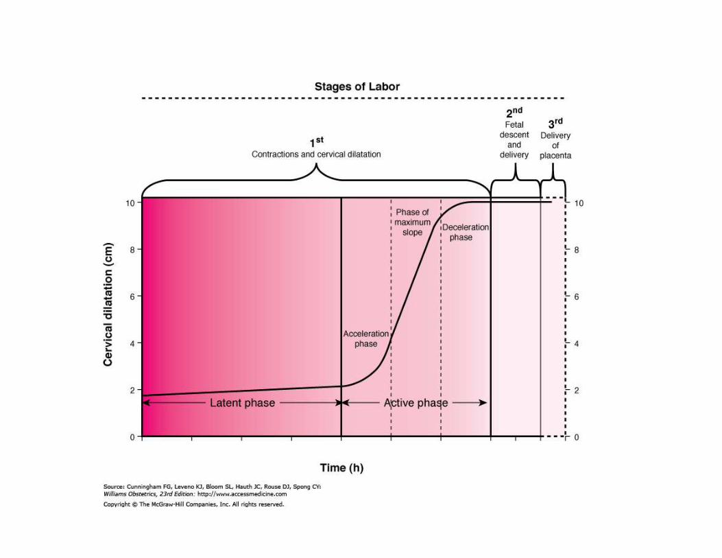

Normal labor staging
Labor period
The first stage
The latent phase
The active phase
From regular uterine contraction to complete cervical dilation
From regulation uterine contraction to 3cm cervical dilation
From 3cm cervical dilation to the full cervical dilation
The second stage From the full cervical dilation to delivery of baby
The third stage From delivery of baby to delivery of placenta
Abnormal labor
• Abnormal labor refers to difficult labor.
• Another name is dystocia.
• Clinical presentation is slow labor process.

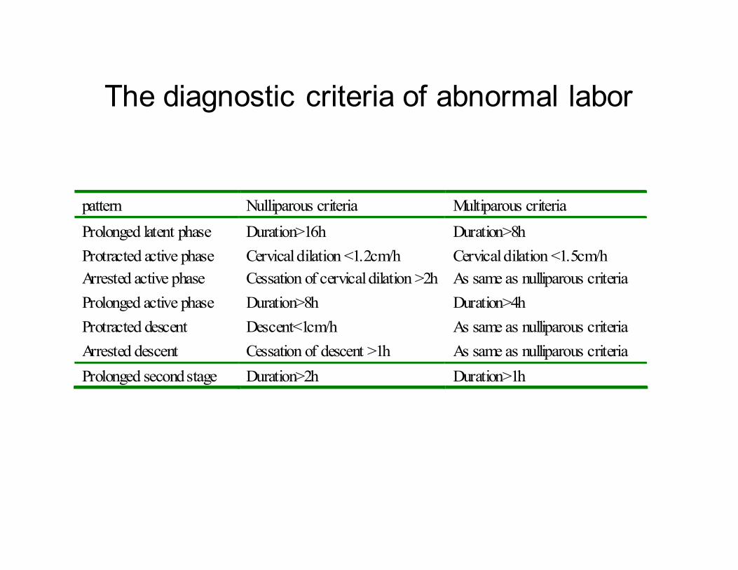
The diagnostic criteria of abnormal labor
pattern Nulliparous criteria Multiparous criteria
Prolonged latent phase Duration>16h Duration>8h
Protracted active phase Cervical dilation <1.2cm/h Cervical dilation <1.5cm/h
Arrested active phase Cessation of cervical dilation >2h As same as nulliparous criteria
Prolonged active phase Duration>8h Duration>4h
Protracted descent Descent<1cm/h As same as nulliparous criteria
Arrested descent Cessation of descent >1h As same as nulliparous criteria
Prolonged second stage Duration>2h Duration>1h
Causes of abnormal labor
• Abnormalities of expulsive forces
• Abnormalities of birth canal
• Abnormalities of presentation & position of
fetus

Abnormalities of birth canal
• The morphology and capacity are primary
causes of dystocia.
• Pelvic structure: pubis, sacrum and ischium.
• Pelvic plane: inlet, midpelvic and outlet
• Bony marker: ischial spine
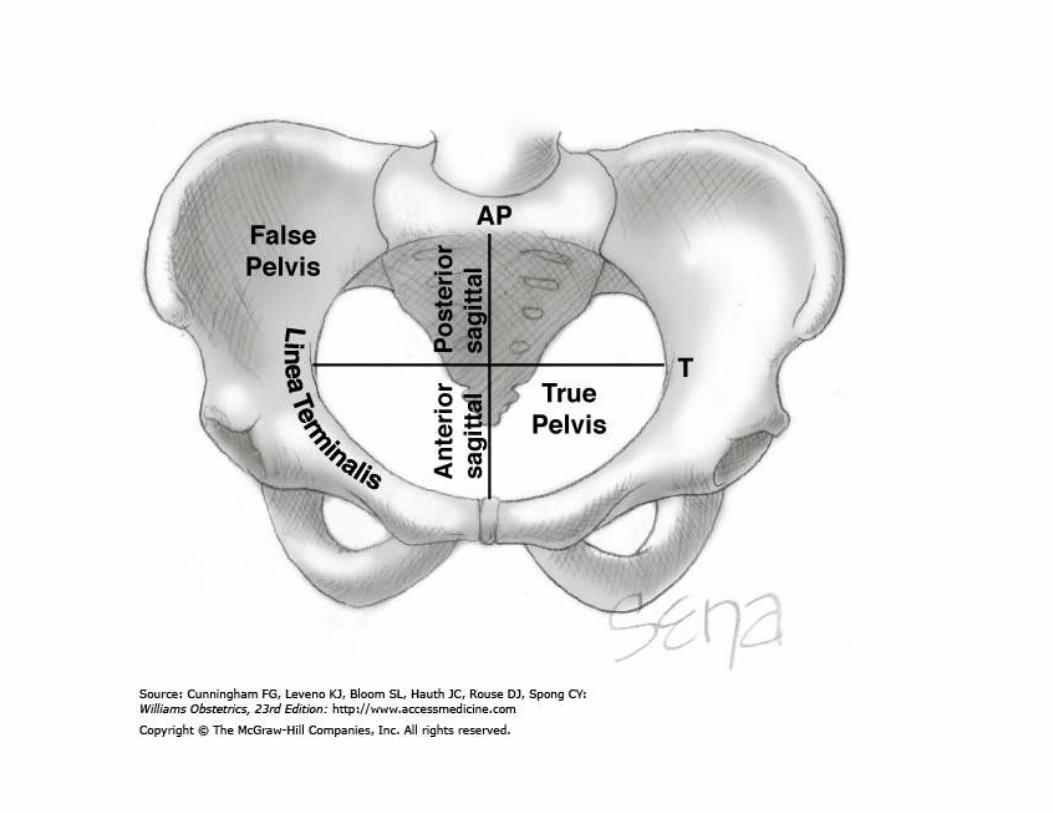
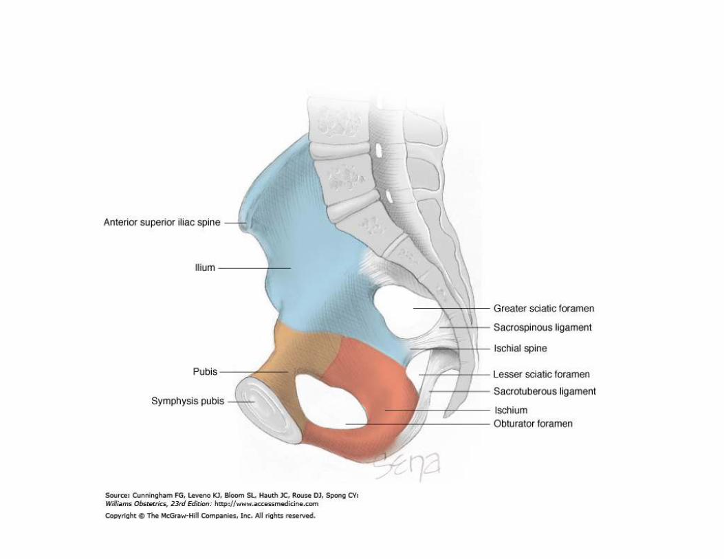

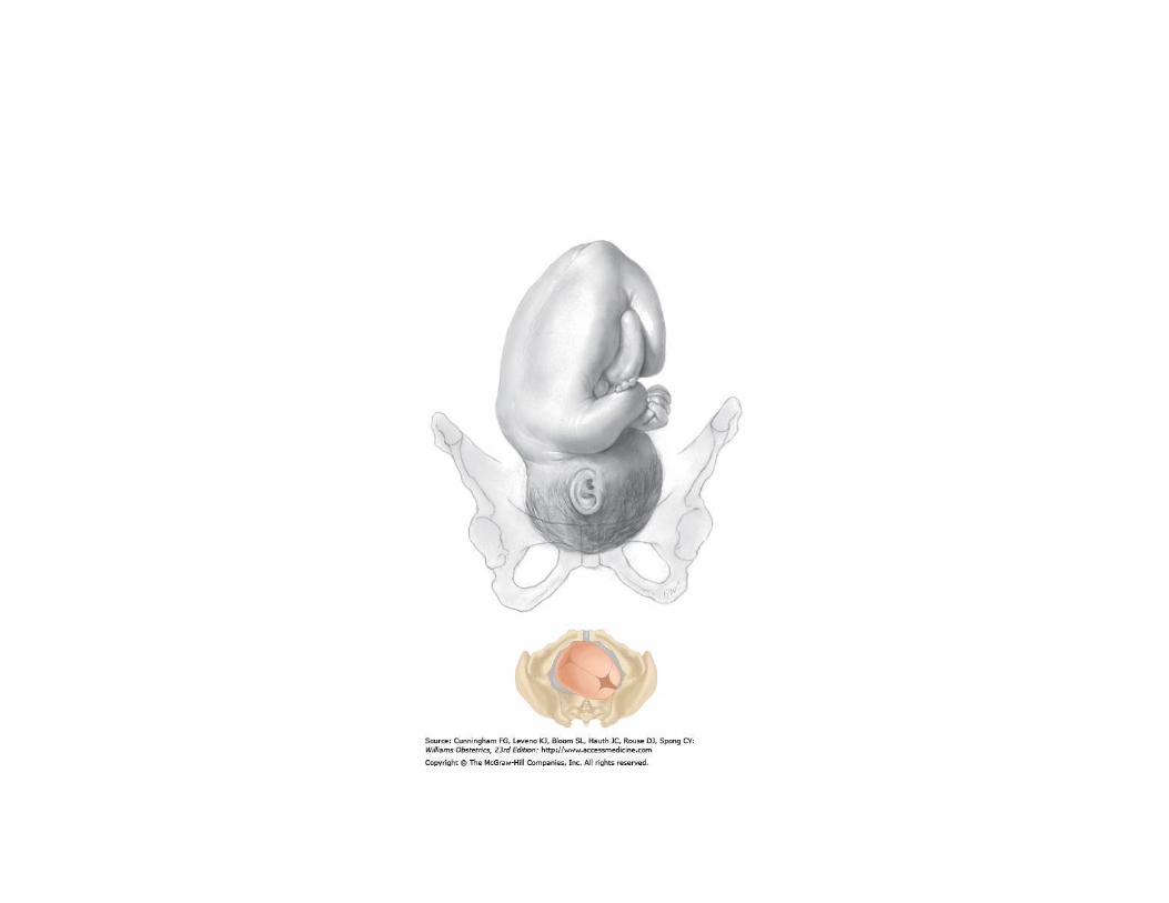
Ischial spine
• The Ischial spine is halfway of birth canal.
• Station of fetal presentation is discribed in relationship with the ischial spine.
• the axis of birth canal above and below the ischial spine is divided into fifth respectively.
• As the presenting part reaches the ischial spine, the designation is 0 station.
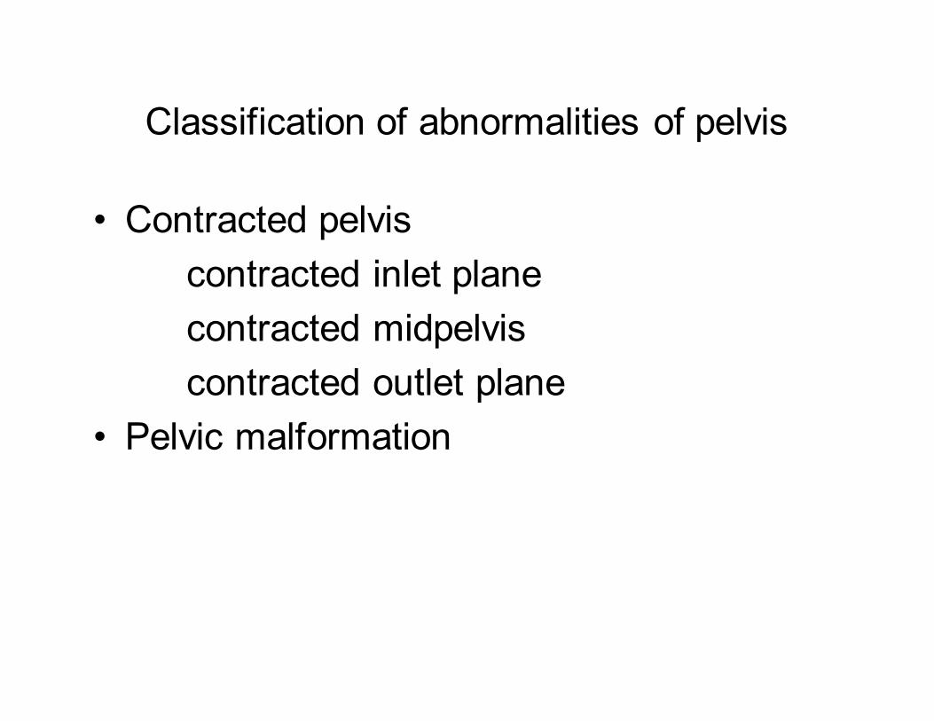
Classification of abnormalities of pelvis
• Contracted pelvis
contracted inlet plane
contracted midpelvis
contracted outlet plane
• Pelvic malformation
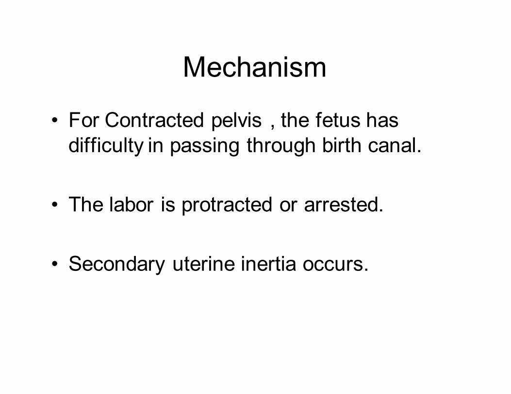
Mechanism
• For Contracted pelvis , the fetus has
difficulty in passing through birth canal.
• The labor is protracted or arrested.
• Secondary uterine inertia occurs.
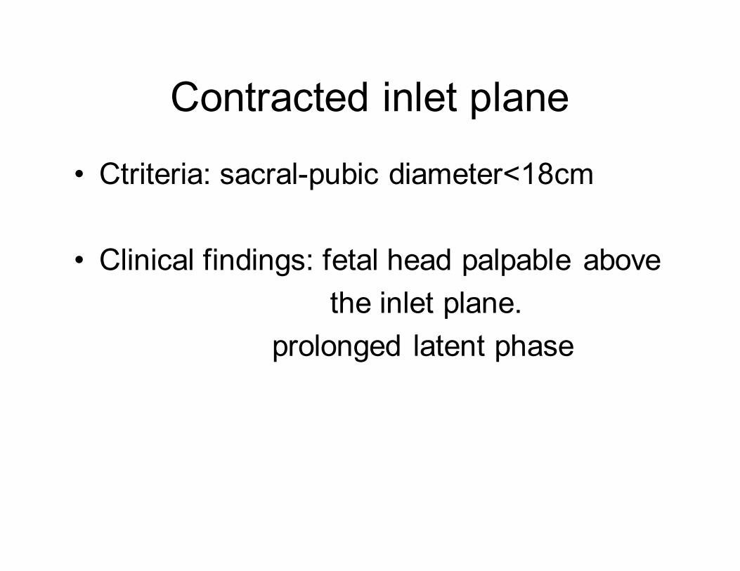
Contracted inlet plane
• Ctriteria: sacral-pubic diameter<18cm
• Clinical findings: fetal head palpable above
the inlet plane.
prolonged latent phase
Contracted midpelvis and outlet plane
• Bi-ischial spine diameter<10cm
• Bi-ischial tubercle diameter<8cm
• Clinical findings: disorders of active phase
and the second stage.
Management
• To assess cephalopelvic relationship by a
series of examination.
• Mild cephalopelvic disproportion: trial labor
• Obvious cephalopelvic disproportion:
cesarean section.
Abnormalities of fetus
• Abnormalities of fetal position
• Macrosomia
• Fetal malformation
Fetal status
• Fetal lie:The relation of the fetal long axis
to that of the mother is termed fetal lie and
is either longitudinal or transverse
• Fetal presentation: the foremost part in
birth canal.
• Cephalic, breech and should presentation.

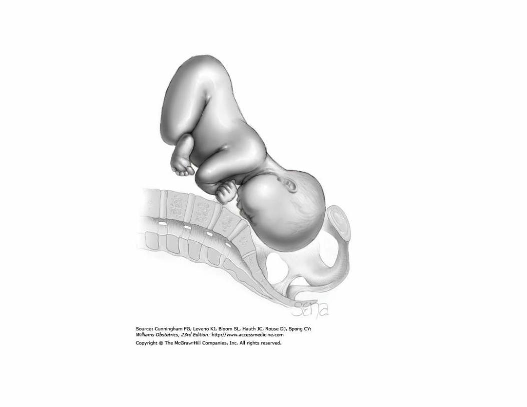
Cephalic presentation
• According to degree of fetal head flex,
cephalic presentation is divided into vertex,
brow and face presentation.
• Brow and face presentation result in
dystocia.

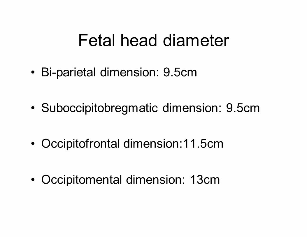
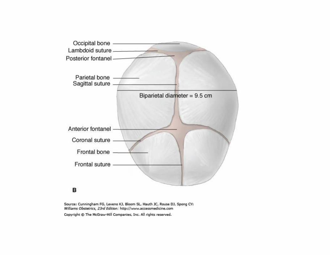
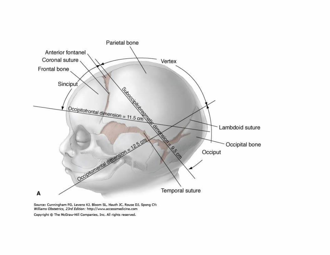
Fetal head diameter
• Bi-parietal dimension: 9.5cm
• Suboccipitobregmatic dimension: 9.5cm
• Occipitofrontal dimension:11.5cm
• Occipitomental dimension: 13cm
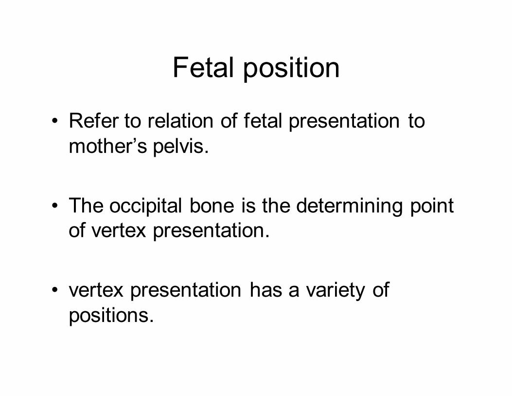
Fetal position
• Refer to relation of fetal presentation to
mother’s pelvis.
• The occipital bone is the determining point
of vertex presentation.
• vertex presentation has a variety of
positions.

• Definition of Persistent Occiput transverse
position : engagement and descent of fetal
head in Occiput transverse position.
• Definition of Persistent Occiput posterior
position : engagement and descent of fetal
head in Occiput posterior position.

Cephalic dystocia
• In cephalic presentation, when delivery
cannot be accomplished with occiput
anterior position, it is called cephalic
dystocia.
• Clinical findings: disorders of labor
process

Management
• To assess cephalopelvic relationship by a
series of examination.
• Mild cephalic distocia: trial labor
• Obvious cephalic distocia: cesarean
section.
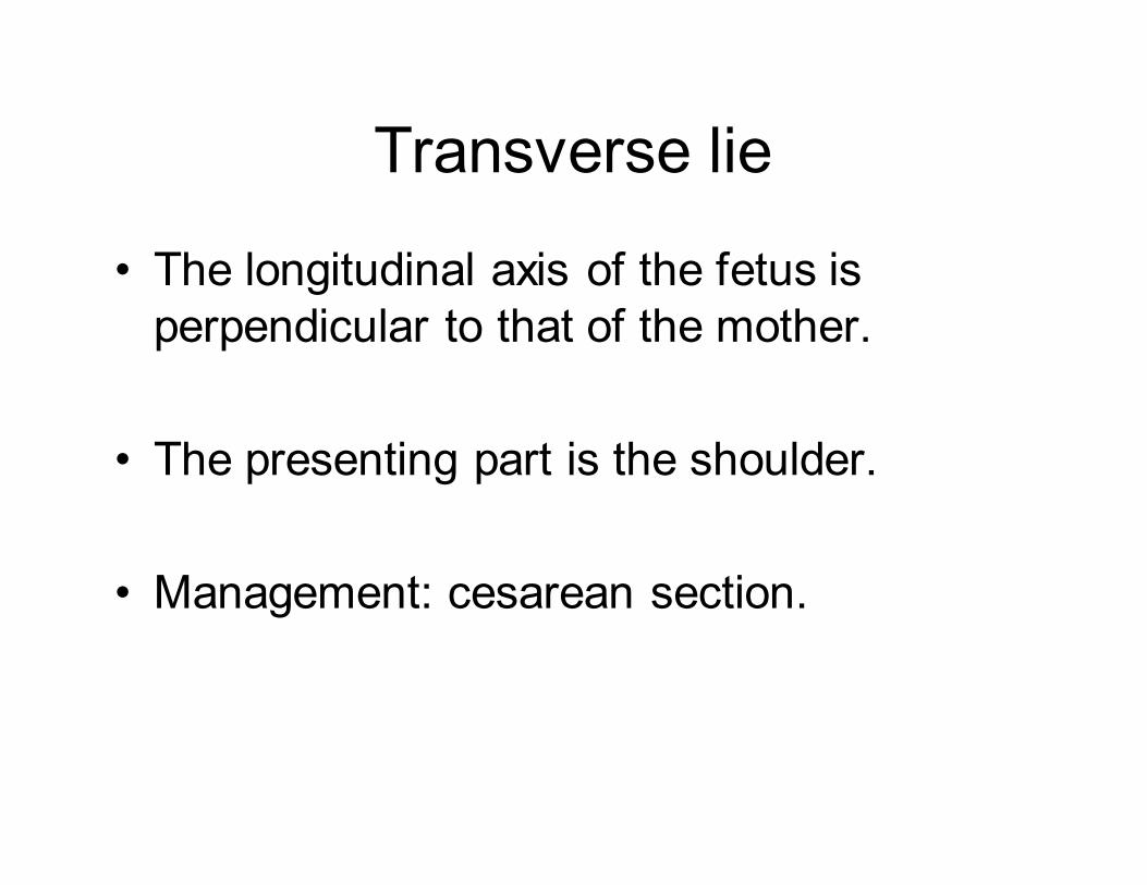
Transverse lie
• The longitudinal axis of the fetus is
perpendicular to that of the mother.
• The presenting part is the shoulder.
• Management: cesarean section.
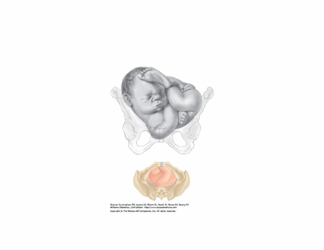
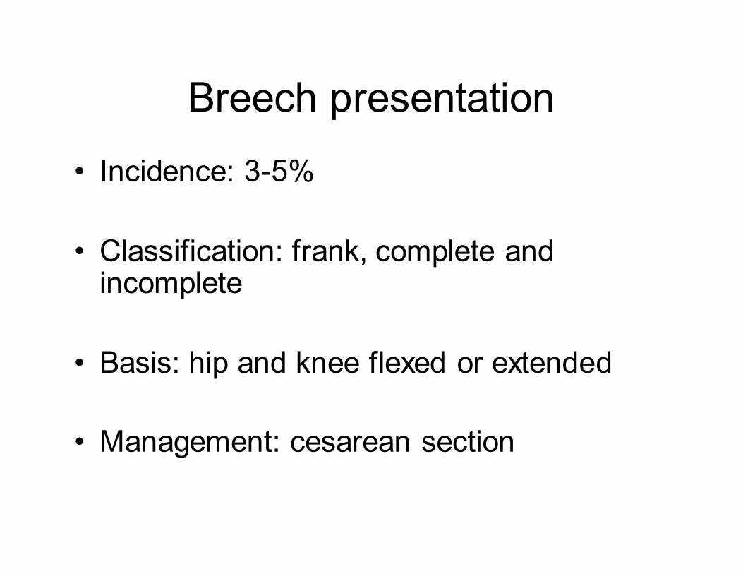
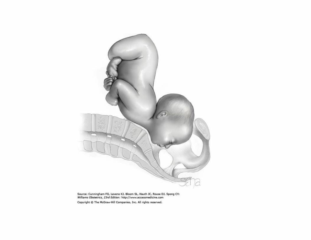
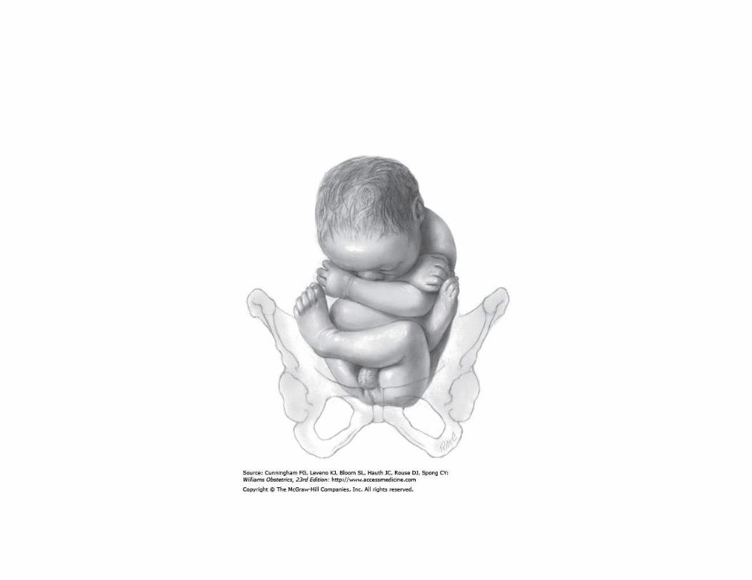
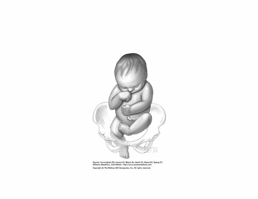
Breech presentation
• Incidence: 3-5%
• Classification: frank, complete and incomplete
• Basis: hip and knee flexed or extended
• Management: cesarean section

Abnormal uterine contractions
• The uterine contraction is the most
important expulsive force.
• Bring about dilation of cervix and
expulsion of fetus and placenta.
• Common causes of dystocia

Classification
• Hypotonic uterine dysfunction: another
name Uterine inertia.
Uterine contractions is less than normal.
• Hypertonic uterine dysfunction: uterine
tone elevated.
• Uterine inertia is more common.
Clinical presentation
• Abdominal palpation: uterine contraction is
weak, and intervals is prolonged.
• Abnormal labor course: the most important
clinical presentation.

The diagnostic criteria of abnormal labor
pattern Nulliparous criteria Multiparous criteria
Prolonged latent phase Duration>16h Duration>8h
Protracted active phase Cervical dilation <1.2cm/h Cervical dilation <1.5cm/h
Arrested active phase Cessation of cervical dilation >2h As same as nulliparous criteria
Prolonged active phase Duration>8h Duration>4h
Protracted descent Descent<1cm/h As same as nulliparous criteria
Arrested descent Cessation of descent >1h As same as nulliparous criteria
Prolonged second stage Duration>2h Duration>1h
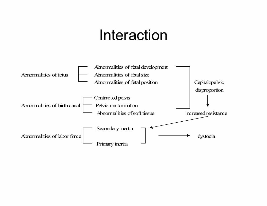
Interaction
Abnormalities of fetal development
Abnormalities of fetus Abnormalities of fetal size
Abnormalities of fetal position Cephalopelvic
disproportion
Contracted pelvis
Abnormalities of birth canal Pelvic malformation
Abnormalities of soft tissue increased resistance
Secondary inertia
Abnormalities of labor force dystocia
Primary inertia
Management
• Vaginal examination: rule out
cephalopelvic disproportion
• Supportive mangement
• augmentation
The Vaginal examination
• To determine fetal presentation, position
and station.
• To assess the cephalopelvic relation.
• To consider the route of delivery.

The supportive management
• Sufficient rest
• To relieve anxiety and fear.
• Fluid and food intake.
Augmentation
• Increase the frequency and force of the
existing uterine contractions.
• Methods: amniotomy
oxytocin administration
Amniotomy
• If the fetal head is engaged, amniotomy is
a choice to facilitate the uterine activity.
• After amnitomy the fetal head descends ,
pressing directly on cervix to enforce
uterine contraction. Accelerating labor.
oxytocin
• Capable of inducing uterine contracion in
the third trimester.
• Contraindiction: cephalopelvic
disproportion and severe fetal malposition.
questions
• To state The pattern of abnormal labor.
• To state the causes of abnormal labor.
• To state the classification of breech
presentation.

Thanks