aberrant expression of leukemia inhibitory factor receptor (lifr)...
TRANSCRIPT
214
http://journals.tubitak.gov.tr/medical/
Turkish Journal of Medical Sciences Turk J Med Sci(2015) 45: 214-220© TÜBİTAKdoi:10.3906/sag-1307-103
Aberrant expression of leukemia inhibitory factor receptor (LIFR) and leukemia inhibitory factor (LIF) is associated with tubal pregnancy occurrence
Yong LI1, Lizhou SUN2,*, Denmei ZHAO1, Jun OUYANG3, Mei XIANG1
1Department of Gynecology, Changzhou Maternal and Child Health Hospital Affiliated to Nangjing Medical University, Changzhou, China2Department of Obstetrics and Gynecology, First Affiliated Hospital of Nanjing Medical University, Nanjing, China
3Department of Pathology, Changzhou Maternal and Child Health Hospital Affiliated to Nangjing Medical University, Changzhou, China
* Correspondence: [email protected]
1. IntroductionEctopic pregnancies, with an occurrence of 1%–2% of pregnancies, usually occur in fallopian tubes and are a major cause of maternal death in the first trimester (1,2). Several risk factors, including smoking, infection of chlamydia trachomatis and Neisseria gonorrhea, surgery history, and in vitro fertilization, have been reported previously (3,4). However, the underlying molecular mechanisms responsible for the occurrence of tubal pregnancy remain largely elusive.
Embryo implantation is a complex physiological process and regulated by many factors. Leukemia inhibitory factor (LIF) is a polyfunctional cytokine of the interleukin-6 (IL-6) cytokine family; a cycle-dependent peak of LIF expression is observed in the mid and late secretory phase. This is also the timepoint when blastocyst implantation occurs and suggests an important role for LIF in human implantation (5). Some studies have shown that LIF plays
an important role in pregnancy maintenance. LIF can facilitate the implantation and promote the development of blastocyst in a time-/dose-dependent manner (6). Senturk proved that LIF is required for implantation in the mouse and monkey (7). Previous reports have revealed that loss of LIF expression in mice had no effect on fertility, but led to the failure of receiving either LIF-positive or LIF-negative embryos (5,8). From this observation, some may deduce that overexpression of LIF in the fallopian tubes may facilitate the implantation of embryos into oviduct tissues, thus leading to tubal pregnancy. However, the contribution of LIF expression to the pathogenesis of tubal pregnancy is still under debate (9–11). Ji and Keltz showed an increased expression of LIF in fallopian tubes from women with tubal pregnancy compared to nonpregnant women (9,10), while Kiran revealed that there was no significant difference in LIF expression between the tubal pregnancy group and early eutopic pregnancy group
Background/aim: Tubal pregnancy is a major cause of maternal death in the first trimester and exploration of its underlying molecular mechanism is of great importance. This study aimed to explore the association of tubal pregnancy with leukemia inhibitory factor (LIF) and leukemia inhibitory factor receptor (LIFR) expression in oviduct tissues.
Materials and methods: Immunohistochemistry was performed to probe the differential expression of LIF and LIFR in oviduct tissues among a control group (including NP group, n = 11; and IP group, n = 12), tubal pregnancy group (Ect-N group, n = 31; and Ect-A group, n = 40), and chronically inflamed group (including Inf-S group, n = 11; and Inf-P group, n = 9), followed by semiquantitative analysis.
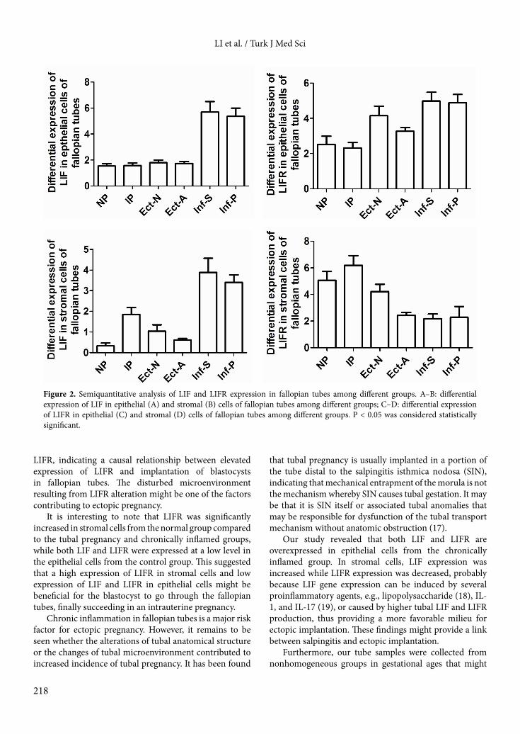
Results: Semiquantitative immunohistochemical analysis demonstrated that there was no significant difference in LIF expression in either the epithelial or stromal cells of oviduct tissues between the tubal pregnancy group and the control group (P < 0.05). However, LIF expression was remarkably elevated in the Inf-S and Inf-P group compared to the other groups (P < 0.05). In the epithelial cells of the fallopian tubes, LIFR expression was highest in the chronically inflamed group, followed by the tubal pregnancy group, outnumbering the control group (P < 0.05). More interestingly, an opposite expression trend of LIFR was observed in the stromal cells of the fallopian tubes among these groups (P < 0.05).
Conclusion: Aberrant expression of LIF and LIFR might be associated with the occurrence of tubal pregnancy.
Key words: LIF, LIFR, fallopian tube, tubal pregnancy, chronic salpingitis
Received: 22.07.2013 Accepted: 03.01.2014 Published Online: 12.01.2015 Printed: 09.02.2015
Research Article
215
LI et al. / Turk J Med Sci
(11). Although the results are contradictory, their control groups were different from each other, which may have had an effect on their conclusions.
Leukemia inhibitory factor receptor (LIFR) is a specific receptor of LIF and has been implicated in multiple physiological and pathological processes, including embryo implantation, orthodontic tooth movement, and cancer development (12–15). However, its role in tubal pregnancy, to the best of our knowledge, is still unknown. The present study aimed to explore the association of tubal pregnancy occurrence with the expression of LIF and LIFR in fallopian tubes from women.
2. Materials and methodsWe collected tubal samples from women in a nonpregnant group, who asked for tubal ligation (NP group, n = 11); an intrauterine pregnancy group, who underwent uterine cesarean delivery and tubal ligation, as well as 1 case of early pregnancy abortion and tubal ligation (IP group, n = 12); a contralateral normal side of tubal pregnancy group, who experienced an ectopic pregnancy resection of the fallopian tube and a contralateral tubal ligation (Ect-N group, n = 31); and an affected side of tubal pregnancy group (Ect-A group, n = 40). Since chronic salpingitis is a major contributor to tubal pregnancy, 2 additional groups were also included in our study, with the hope of providing new insights into the underlying mechanisms responsible for the occurrence of tubal pregnancy: a chronically inflamed secretory phase group (Inf-S group, n = 11) and a chronically inflamed proliferative phase group (Inf-P group, n = 9) who underwent tubal resection for
hydrosalpinx. Details and case data about the control group (NP and IP) are shown in Tables 1 and 2.2.1. Fallopian tube samplesAll clinical specimens were obtained from the Changzhou Maternal and Child Health Hospital of Nanjing Medical University from 2008 to 2012. This study was approved by the Research Ethics Committee of Changzhou Maternal and Child Health Hospital of Nanjing Medical University and patient consent was obtained before tissue collection.2.2. Immunohistochemistry analysisLIF antibody (Sc-1336) and LIFR antibody (Sc-659) were obtained from Santa Cruz Biotechnology (Santa Cruz, CA, USA). Immunohistochemical assay was performed using Histostain-Plus IHC Kit (ZSGR-Biotech, Beijing, China) according to the manufacturer’s instructions. Semiquantitative analysis was carried out as follows. The percentage of positive cells was divided into 5 groups: 0, 0%; 1, 1%–10%; 2, 11%–50%; 3, 51%–75%; and 4, >75%. Additionally, a scale from 0–3 (no staining to strong immunoreactivity) was assigned to staining intensity. Immunoreactive scores, ranging from 0 to 12, were calculated by multiplying the percentage score and the immunoreactivity score. Five visual fields of each clinical sample were randomly measured under a microscope using 10 × 40 lenses.2.3. Statistical analysisThe Kruskal–Wallis (KW) rank test was used to assess the statistical difference between groups. Our pairwise comparison of parameters was calculated with the Bonferoni-corrected Mann–Whitney test. The criterion P < 0.05 was considered statistically significant.
Table 1. The nonpregnant group (NP group, n = 11) patient data sheet.
No. Pathological examination no. Age Cause of fallopian tube operation Pregnancy
history IUD history EP history Hormonemedication history
1 20090280 16 Resection of rudimentary hornof the uterus and tube 0-0-0-0 N N N
2 20093144 44 Resection of ovarian teratomaand tubal ligation 1-0-2-1 Yes N N
3 20093861 28 Insist on sterilization for familyreasons 0-0-1-0 Yes N N
4 20091032 43 Resection of uterine fibroidsand tubal ligation 1-0-2-1 Yes N N
5 20091615 43 1-0-1-1 Yes N N6 20091680 44 1-0-0-1 Yes N N7 20091787 41 1-0-01 Yes N N8 20093669 45 1-0-1-1 Yes N N9 20094254 46 1-0-1-1 Yes N N10 20094576 37 3-0-5-3 N N N11 20098162 39 1-0-2-1 Yes N N

216
LI et al. / Turk J Med Sci
3. Results3.1. Detection of LIF and LIFR by immunohistochemistryWe probed the differential expression of LIF and LIFR in fallopian tubes among the NP group (n = 11), the IP group (n = 12), the Ect-N group (n = 31), the Ect-A group (n = 40), the Inf-S group (n = 11), and the Inf-P group (n = 9) as illustrated in Figures 1A–L.3.2. Semiquantitative analysis3.2.1. Differential expression of LIF in epithelial cells of fallopian tubesOur results showed that there was no significant difference in LIF expression in the epithelial cells of oviduct tissues between the control group (including the NP and IP groups) and the tubal pregnancy group (including the Ect-N and Ect-A groups) (Figure 2A, P > 0.05). However, interestingly, LIF expression was significantly elevated in chronically inflamed fallopian tubes (both Inf-S group and Inf-P group) compared to the other groups (Figure 2A, P < 0.05). 3.2.2. Differential expression of LIF in stromal cells of fallopian tubesIn stromal cells of fallopian tubes, LIF expression was remarkably increased in chronically inflamed fallopian tubes (both Inf-S group and Inf-P group) compared to the other groups (Figure 2B, P < 0.05), and modestly increased expression of LIF was observed in the IP group compared to the NP group, Ect-N group, and Ect-A group (Figure 2B).
3.2.3. Differential expression of LIFR in epithelial cells of fallopian tubesIn the epithelial cells of fallopian tubes, LIFR expression level was highest in the chronically inflamed group (including the Inf-S and Inf-P groups), followed by the tubal pregnancy group (including the Ect-N and Ect-A groups), while the control group (including the NP and IP groups) had the lowest (Figure 2C, P < 0.05).3.2.4. Differential expression of LIFR in stromal cells of fallopian tubesContrary to epithelial cells, LIFR expression in stromal cells was gradually decreased among the control group (including the NP and IP groups), the tubal pregnancy group (including the Ect-N and Ect-A groups), and the chronically inflamed group (including the Inf-S and Inf-P groups) (Figure 2D, P < 0.05).
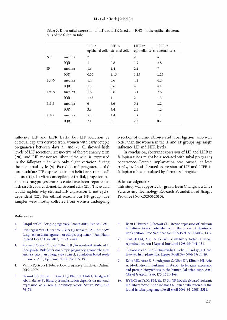
The differential expression of LIF and LIFR in the epithelial/stromal cells of fallopian tubes is shown in Table 3.
4. DiscussionPrevious studies have revealed that a number of cytokines, including LIF and LIFR, could influence the microenvironment of the fallopian tubes, thus facilitating the implantation of the embryos (16). We detected the differential expression and localization of LIF and its receptor LIFR in fallopian tubes from various physiological and pathological groups, including a nonpregnant secretory phase group, an intrauterine pregnancy group, a contralateral normal side of tubal pregnancy group,
Table 2. The intrauterine pregnancy group (IP group, n = 12) patient data sheet.
No. Pathological examination no. Age Pregnancy
gestational ageCause of fallopiantube operation
Pregnancy history
IUD history
EP history
Hormone medication history
1 20095585 40 44 day Abortion and tubal ligation 1-0-9-1 Yes N N
2 20090302 34 41w Uterine cesarean delivery and tubal ligation 1-0-3-1 N N N
3 20091148 27 36w 1-0-2-1 N N N
4 20091447 32 38w 1-0-0-1 N N N
5 20091700 38 37w 1-0-1-1 Yes N N
6 20091701 39 38w 1-0-8-1 Yes N N
7 20091702 28 38w 1-0-2-1 Yes N N
8 20093622 36 39w 1-0-1-1 Yes N N
9 20099859 36 39w 1-0-2-1 Yes N N
10 20084472 37 38w 1-0-3-1 N N N
11 20097330 34 40w 1-0-2-1 Yes N N
12 20098061 33 39w 1-0-1-1 Yes N N
217
LI et al. / Turk J Med Sci
an affected side of tubal pregnancy group, a chronically inflamed secretory phase group, and a chronically inflamed proliferative phase group.
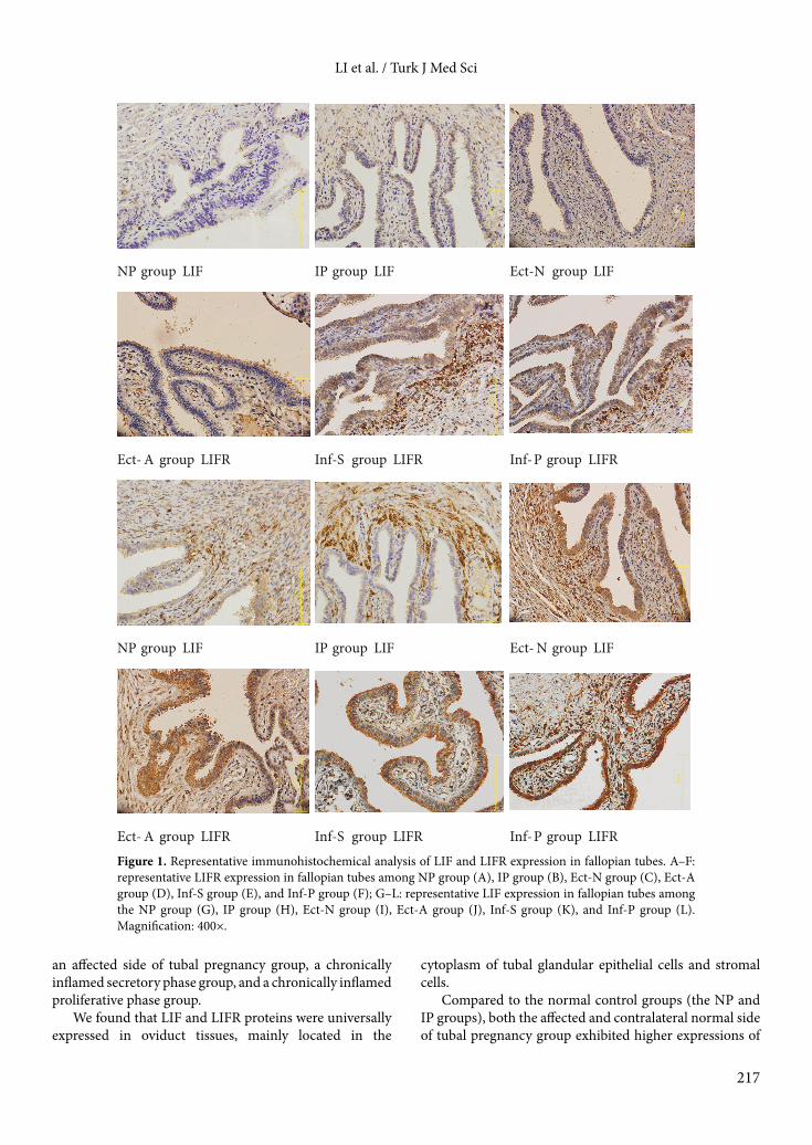
We found that LIF and LIFR proteins were universally expressed in oviduct tissues, mainly located in the
cytoplasm of tubal glandular epithelial cells and stromal cells.
Compared to the normal control groups (the NP and IP groups), both the affected and contralateral normal side of tubal pregnancy group exhibited higher expressions of
NP group LIF IP group LIF Ect-N group LIF
Ect-A group LIFR Inf-S group LIFR Inf-P group LIFR
NP group LIF IP group LIF Ect-N group LIF
Ect-A group LIFR Inf-S group LIFR Inf-P group LIFRFigure 1. Representative immunohistochemical analysis of LIF and LIFR expression in fallopian tubes. A–F: representative LIFR expression in fallopian tubes among NP group (A), IP group (B), Ect-N group (C), Ect-A group (D), Inf-S group (E), and Inf-P group (F); G–L: representative LIF expression in fallopian tubes among the NP group (G), IP group (H), Ect-N group (I), Ect-A group (J), Inf-S group (K), and Inf-P group (L). Magnification: 400×.
218
LI et al. / Turk J Med Sci
LIFR, indicating a causal relationship between elevated expression of LIFR and implantation of blastocysts in fallopian tubes. The disturbed microenvironment resulting from LIFR alteration might be one of the factors contributing to ectopic pregnancy.
It is interesting to note that LIFR was significantly increased in stromal cells from the normal group compared to the tubal pregnancy and chronically inflamed groups, while both LIF and LIFR were expressed at a low level in the epithelial cells from the control group. This suggested that a high expression of LIFR in stromal cells and low expression of LIF and LIFR in epithelial cells might be beneficial for the blastocyst to go through the fallopian tubes, finally succeeding in an intrauterine pregnancy.
Chronic inflammation in fallopian tubes is a major risk factor for ectopic pregnancy. However, it remains to be seen whether the alterations of tubal anatomical structure or the changes of tubal microenvironment contributed to increased incidence of tubal pregnancy. It has been found
that tubal pregnancy is usually implanted in a portion of the tube distal to the salpingitis isthmica nodosa (SIN), indicating that mechanical entrapment of the morula is not the mechanism whereby SIN causes tubal gestation. It may be that it is SIN itself or associated tubal anomalies that may be responsible for dysfunction of the tubal transport mechanism without anatomic obstruction (17).
Our study revealed that both LIF and LIFR are overexpressed in epithelial cells from the chronically inflamed group. In stromal cells, LIF expression was increased while LIFR expression was decreased, probably because LIF gene expression can be induced by several proinflammatory agents, e.g., lipopolysaccharide (18), IL-1, and IL-17 (19), or caused by higher tubal LIF and LIFR production, thus providing a more favorable milieu for ectopic implantation. These findings might provide a link between salpingitis and ectopic implantation.
Furthermore, our tube samples were collected from nonhomogeneous groups in gestational ages that might
Figure 2. Semiquantitative analysis of LIF and LIFR expression in fallopian tubes among different groups. A–B: differential expression of LIF in epithelial (A) and stromal (B) cells of fallopian tubes among different groups; C–D: differential expression of LIFR in epithelial (C) and stromal (D) cells of fallopian tubes among different groups. P < 0.05 was considered statistically significant.
219
LI et al. / Turk J Med Sci
influence LIF and LIFR levels, but LIF secretion by decidual explants derived from women with early ectopic pregnancies between days 35 and 76 all showed high levels of LIF secretion, irrespective of the pregnancy term (20), and LIF messenger ribonucleic acid is expressed in the fallopian tube with only slight variation during the menstrual cycle (9). Estradiol and progesterone did not modulate LIF expression in epithelial or stromal cell cultures (9). In vitro conception, estradiol, progesterone, and medroxyprogesterone acetate have been reported to lack an effect on endometrial stromal cells (21). These data would explain why stromal LIF expression is not cycle-dependent (22). For ethical reasons our NP group tube samples were mostly collected from women undergoing
resection of uterine fibroids and tubal ligation, who were older than the women in the IP and EP groups; age might influence LIF and LIFR levels.
In conclusion, aberrant expression of LIF and LIFR in fallopian tubes might be associated with tubal pregnancy occurrence. Ectopic implantation was caused, at least partly, by local elevated expression of LIF and LIFR in fallopian tubes stimulated by chronic salpingitis.
AcknowledgmentsThis study was supported by grants from Changzhou City’s Science and Technology Research Foundation of Jiangsu Province (No. CS20092013).
Table 3. Differential expression of LIF and LIFR (median (IQR)) in the epithelial/stromal cells of the fallopian tube.
LIF inepithelial cells
LIF instromal cells
LIFR inepithelial cells
LIFR instromal cells
NP median 2 0 2 6IQR 1 0.8 1.9 2.8
IP median 1.6 1.4 2.4 7IQR 0.35 1.15 1.25 2.25
Ect-N median 1.4 0.6 4.2 4.2IQR 1.5 0.6 4 4.1
Ect-A median 1.6 0.6 3.4 2.6IQR 1.45 1 2 1.3
Inf-S median 6 3.6 5.4 2.2IQR 3.3 3.4 2.1 1.2
Inf-P median 5.4 3.4 4.8 1.4IQR 2.1 0 2.7 0.2
References
1. Farquhar CM. Ectopic pregnancy. Lancet 2005; 366: 583–591.
2. Sivalingam VN, Duncan WC, Kirk E, Shephard LA, Horne AW. Diagnosis and management of ectopic pregnancy. J Fam Plann Reprod Health Care 2011; 37: 231–240.
3. Bouyer J, Coste J, Shojaei T, Pouly JL, Fernandez H, Gerbaud L, Job-Spira N. Risk factors for ectopic pregnancy: a comprehensive analysis based on a large case-control, population-based study in France. Am J Epidemiol 2003; 157: 185–194.
4. Varma R, Gupta J. Tubal ectopic pregnancy. Clin Evid (Online) 2009; 2009.
5. Stewart CL, Kaspar P, Brunet LJ, Bhatt H, Gadi I, Köntgen F, Abbondanzo SJ. Blastocyst implantation depends on maternal expression of leukemia inhibitory factor. Nature 1992; 359: 76–79.
6. Bhatt H, Brunet LJ, Stewart CL. Uterine expression of leukemia inhibitory factor coincides with the onset of blastocyst implantation. Proc Natl Acad Sci USA 1991; 88: 11408–11412.
7. Senturk LM, Arici A. Leukemia inhibitory factor in human reproduction. Am J Reprod Immunol 1998; 39: 144–151.
8. Salamonsen LA, Nie G, Dimitriadis E, Robb L, Findlay JK. Genes involved in implantation. Reprod Fertil Dev 2001; 13: 41–49.
9. Keltz MD, Attar E, Buradagunta S, Olive DL, Kliman HJ, Arici A. Modulation of leukemia inhibitory factor gene expression and protein biosynthesis in the human Fallopian tube. Am J Obstet Gynecol 1996; 175: 1611–169.
10. Ji YF, Chen LY, Xu KH, Yao JF, Shi YF. Locally elevated leukemia inhibitory factor in the inflamed fallopian tube resembles that found in tubal pregnancy. Fertil Steril 2009; 91: 2308–2314.

220
LI et al. / Turk J Med Sci
11. Kiran G, Kiran H, Ertopcu K, Kilinc M, Ekerbicer HC, Vardar MA. Tuba uterina leukemia inhibitory factor concentration does not increase in tubal pregnancy: a preliminary study. Fertil Steril 2005; 83: 484–486.
12. Wanggren K, Lalitkumar PG, Hambiliki F, Stabi B, Gemzell-Danielsson K, Stavreus-Evers A. Leukaemia inhibitory factor receptor and gp130 in the human Fallopian tube and endometrium before and after mifepristone treatment and in the human preimplantation embryo. Mol Hum Reprod 2007; 13: 391–397.
13. Liang Y, Zhou Y, Jiang T, Zhang Z, Wang S, Wang Y. Expression of LIF and LIFR in periodontal tissue during orthodontic tooth movement. Angle Orthod 2011; 81: 600–608.
14. Okamura Y, Nomoto S, Kanda M, Li Q, Nishikawa Y, Sugimoto H, Kanazumi N, Takeda S, Nakao A. Leukemia inhibitory factor receptor (LIFR) is detected as a novel suppressor gene of hepatocellular carcinoma using double-combination array. Cancer Lett 2010; 289: 170–177.
15. Chen D, Sun Y, Wei Y, Zhang P, Rezaeian AH, Teruya-Feldstein J, Gupta S, Liang H, Lin HK, Hung MC et al. LIFR is a breast cancer metastasis suppressor upstream of the Hippo-YAP pathway and a prognostic marker. Nat Med 2012; 18: 1511–1517.
16. Shaw JL, Dey SK, Critchley HO, Horne AW. Current knowledge of the aetiology of human tubal ectopic pregnancy. Hum Reprod Update 2010; 16: 432–444.
17. Hvid M, Baczynska A, Deleuran B, Fedder J, Knudsen HJ, Christiansen G, Birkelund S. Interleukin-1 is the initiator of Fallopian tube destruction during Chlamydia trachomatis infection. Cell Microbiol 2007; 9: 2795–2803.
18. Brown MA, Metcalf D, Gough NM. Leukemia inhibitory factor and interleukin 6 are expressed at very low levels in the normal adult mouse and are induced by inflammation. Cytokine 1994; 6: 300–309.
19. Derigs HG, Boswell HS. LIF mRNA expression is transcriptionally regulated in murine bone marrow stromal cells. Leukemia 1993; 7: 630–634.
20. Hambartsoumian E. Leukemia inhibitory factor (LIF) production by human decidua and its relationship with pregnancy hormones. Gynecol Endocrinol 1998; 12: 17–22.
21. Arici A, Engin O, Attar E, Olive DL. Modulation of leukemia inhibitory factor gene expression and protein biosynthesis in human endometrium. J Clin Endocrinol Metab 1995; 80: 1908–1915.
22. Vogiagis D, Marsh MM, Fry RC, Salamonsen LA. Leukemia inhibitory factor in human endometrium throughout the menstrual cycle. J Endocrinol 1996; 148: 95–102.