a young woman with odynophagia
TRANSCRIPT

A Young Woman with OdynophagiaNP Singh*, S Anuradha*, SK Jain*, SK Agarwal*, V Chowdhury**
A 36 year old woman presented with historyof retrosternal pain while swallowing, i.e.,odynophagia, and progressive dysphagia toso l ids , fo r two months. There was noassociated nausea, vomiting, haematemesis,heart-burn, fever, anorexia, or weight loss.Her genera l phys ica l and sys temicexaminat ion was non-cont r ibu tory.Investigations revealed- haemoglobin - 10.5g/dl, total leucocyte count - 6700/cumm,erythrocyte sedimentation rate - 62 mm in1st hour, chest skiagram - normal, and ELISAfor HIV 1 and 2 - negative.
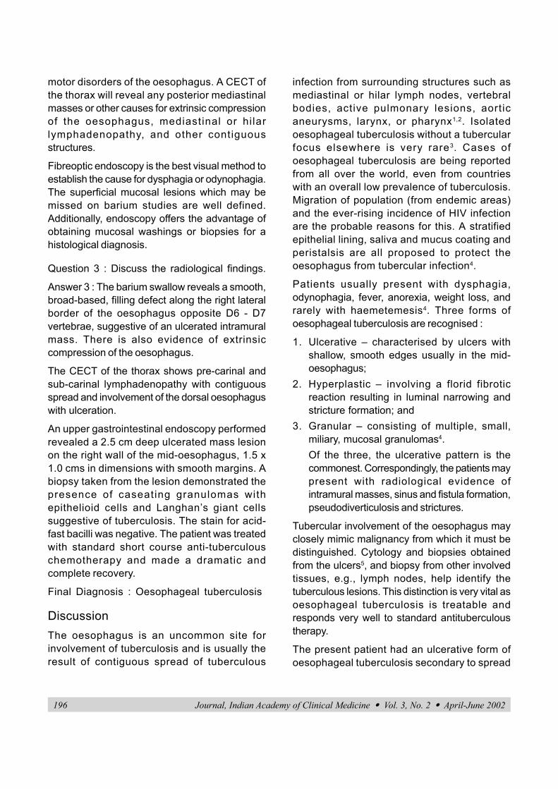
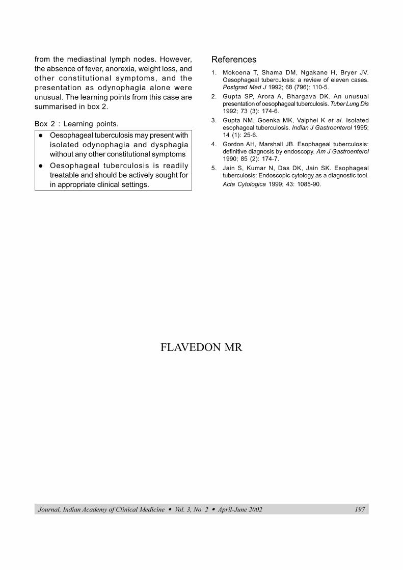
The barium swallowand contrastenhanced computedtomography (CECT)of the thorax of thispatient are depictedin figures 1 and 2.
Question 1 : Whatare the conditionscausingodynophagia?
Answer 1 :Odynophagia orpainful swallowingfrequently occurs withdysphagia and is acommon symptom ofoesophageal dis-orders. The conditionsresulting in odyno-phagia are summarised in box 1.
* Departments of Medicine and** RadiodiagnosisMaulana Azad Medical College andAssociated LN Hospital,New Delhi-110 002.
C A S E R E P O R T
Fig. 1 :
Fig. 2 :
JIACM 2002; 3(2): 195-7
Box 1 : Conditions causing Odynophagia1. Oesophagitis
a) Viral- Herpes simplex, Varicella - zoster,Cytomegalovirus, HIV
b) Fungal- Candidac) Bacterial - Lactobacillus, Streptococci
beta haemolytic, M. tuberculosisd) Pneumocystis cariniie) Radiation injuryf) Corrosive and caustic injuryg) Pill induced - NSAIDs, potassium
chlorideh) Associated with pemphigus
2. Oesophageal malignancy with peri-oesophageal involvement
3. Peptic ulcer of the oesophagus (Barrett’sulcer)
4. Reflux oesophagitis (rare).
Question 2 : How will you proceed toinvestigate this case?
Answer 2 : Radiological studies using bariumswallow and oesophagography are very usefulin delineating both structural disease as well as
196 Journal, Indian Academy of Clinical Medicine � Vol. 3, No. 2 � April-June 2002
motor disorders of the oesophagus. A CECT ofthe thorax will reveal any posterior mediastinalmasses or other causes for extrinsic compressionof the oesophagus, mediastinal or hi larlymphadenopathy, and other contiguousstructures.
Fibreoptic endoscopy is the best visual method toestablish the cause for dysphagia or odynophagia.The superficial mucosal lesions which may bemissed on barium studies are well defined.Additionally, endoscopy offers the advantage ofobtaining mucosal washings or biopsies for ahistological diagnosis.
Question 3 : Discuss the radiological findings.
Answer 3 : The barium swallow reveals a smooth,broad-based, filling defect along the right lateralborder of the oesophagus opposite D6 - D7vertebrae, suggestive of an ulcerated intramuralmass. There is also evidence of extrinsiccompression of the oesophagus.
The CECT of the thorax shows pre-carinal andsub-carinal lymphadenopathy with contiguousspread and involvement of the dorsal oesophaguswith ulceration.
An upper gastrointestinal endoscopy performedrevealed a 2.5 cm deep ulcerated mass lesionon the right wall of the mid-oesophagus, 1.5 x1.0 cms in dimensions with smooth margins. Abiopsy taken from the lesion demonstrated thepresence of caseating granulomas withepithelioid cells and Langhan’s giant cellssuggestive of tuberculosis. The stain for acid-fast bacilli was negative. The patient was treatedwith standard short course anti-tuberculouschemotherapy and made a dramatic andcomplete recovery.
Final Diagnosis : Oesophageal tuberculosis
DiscussionThe oesophagus is an uncommon site forinvolvement of tuberculosis and is usually theresult of contiguous spread of tuberculous
infection from surrounding structures such asmediastinal or hilar lymph nodes, vertebralbodies, active pulmonary lesions, aorticaneurysms, larynx, or pharynx1,2. Isolatedoesophageal tuberculosis without a tubercularfocus elsewhere is very rare3. Cases ofoesophageal tuberculosis are being reportedfrom all over the world, even from countrieswith an overall low prevalence of tuberculosis.Migration of population (from endemic areas)and the ever-rising incidence of HIV infectionare the probable reasons for this. A stratifiedepithelial lining, saliva and mucus coating andperistalsis are all proposed to protect theoesophagus from tubercular infection4.
Patients usually present with dysphagia,odynophagia, fever, anorexia, weight loss, andrarely with haemetemesis4. Three forms ofoesophageal tuberculosis are recognised :
1. Ulcerative – characterised by ulcers withshallow, smooth edges usually in the mid-oesophagus;
2. Hyperplastic – involving a florid fibroticreaction resulting in luminal narrowing andstricture formation; and
3. Granular – consisting of multiple, small,miliary, mucosal granulomas4.Of the three, the ulcerative pattern is thecommonest. Correspondingly, the patients maypresent with radiological evidence ofintramural masses, sinus and fistula formation,pseudodiverticulosis and strictures.
Tubercular involvement of the oesophagus mayclosely mimic malignancy from which it must bedistinguished. Cytology and biopsies obtainedfrom the ulcers5, and biopsy from other involvedtissues, e.g., lymph nodes, help identify thetuberculous lesions. This distinction is very vital asoesophageal tuberculosis is treatable andresponds very well to standard antituberculoustherapy.
The present patient had an ulcerative form ofoesophageal tuberculosis secondary to spread
Journal, Indian Academy of Clinical Medicine � Vol. 3, No. 2 � April-June 2002 197
from the mediastinal lymph nodes. However,the absence of fever, anorexia, weight loss, andother consti tut ional symptoms, and thepresentation as odynophagia alone wereunusual. The learning points from this case aresummarised in box 2.
Box 2 : Learning points.� Oesophageal tuberculosis may present with
isolated odynophagia and dysphagiawithout any other constitutional symptoms
� Oesophageal tuberculosis is readilytreatable and should be actively sought forin appropriate clinical settings.
References1. Mokoena T, Shama DM, Ngakane H, Bryer JV.
Oesophageal tuberculosis: a review of eleven cases.Postgrad Med J 1992; 68 (796): 110-5.
2. Gupta SP, Arora A, Bhargava DK. An unusualpresentation of oesophageal tuberculosis. Tuber Lung Dis1992; 73 (3): 174-6.
3. Gupta NM, Goenka MK, Vaiphei K et al. Isolatedesophageal tuberculosis. Indian J Gastroenterol 1995;14 (1): 25-6.
4. Gordon AH, Marshall JB. Esophageal tuberculosis:definitive diagnosis by endoscopy. Am J Gastroenterol1990; 85 (2): 174-7.
5. Jain S, Kumar N, Das DK, Jain SK. Esophagealtuberculosis: Endoscopic cytology as a diagnostic tool.Acta Cytologica 1999; 43: 1085-90.
FLAVEDON MR