a retrospective study of ulcerative keratitis in 32 dogs · a retrospective study of ulcerative...
TRANSCRIPT

Intern J Appl Res Vet Med • Vol. 7, No. 1, 2009. 27
KEY WORDS: ulcerative keratitis, corneal ulcer, conjunctival flap, dog
ABSTRACTThis study was performed to analyze risk factors for and treatment results of ulcerative keratitis. The mean age (±SD) of 32 dogs with ulcerative keratitis was 3.59 (±2.97) years and the most commonly affected breed was the Shih-tzu (50%). Keratoconjunctivi-tis sicca (31%) was the predominant cause of ulcerative keratitis. Superficial corneal ulcers (in 44% of animals) treated with medication alone required 5.1-13.4 days for healing and dogs recovered well, without complications. Deep corneal ulcers (56%) treated both by medication and conjunctival flap placement showed significantly better prognoses than did cases treated by medica-tion alone, although long healing periods of 28.4-40 days were required..
INTRODUCTIONUlcerative keratitis in dogs is the most commonly encountered ocular disease in veterinary ophthalmology.1 Fortunately, the condition is among the most treatable of various ophthalmic disorders that can threaten canine vision. Therefore, when
dogs presented with clinical signs of corneal ulcers, we were able to readily access useful treatment modalities.
Ulcerative keratitis can be classified into superficial keratitis, deep corneal ulcer keratitis, descemetocele keratitis, and per-foration keratitis, with reference to loss of corneal layers. Etiologies may be congenital, or result from infection, allergy, trichiasis, distichiasis, ectopic cilia, entropion, trauma, a foreign body, or lack of tears.1-3 Canine ocular disease characteristics have been extensively studied and the prevalence of corneal diseases in dogs has been recently reviewed.3-9 Most studies have reported breed or gender characteristics of individual ocular diseases associated with age or geo-graphic region. Few statistical relationships between ulcerative keratitis in dogs and other factors (age, breed, cause, location and type of corneal ulcer, treatment methods, and healing period) have been documented. Ulcerative keratitis may be treated by medi-cation or using various surgical procedures, depending on corneal stromal lesion sever-ity.5,6,10-16 Few reports on recovery rates or treatment-specific healing periods in dogs have appeared.
A Retrospective Study of Ulcerative Keratitis in 32 Dogs
Joon Young Kim, DVM, MSc, PhD,
Hye-Jung Won, DVM, MSc
Soon-wuk Jeong, DVM, MSc, PhD
Department of Veterinary Surgery, College of Veterinary Medicine,KonKuk University, Seoul, 143-701, Korea, Republic of
Intern J Appl Res Vet Med • Vol. 7, No. 1, 2009.28
In treating corneal ulceration, the most important step is to determine and eliminate the cause, followed by attempts to create an ideal environment for lesion repair, preven-tion of progression, and surgical treatment to prevent corneal rupture.
The purpose of the present study was to analyze the symptoms, causes, and clinical features of ulcerative keratitis in dogs, to ex-plore treatment methods, to review recovery data, to seek correlations between ulcerative keratitis and other factors and, especially, to determine the effect of conjunctival flap formation on healing of deep corneal ulcers.
MATERIALS AND METHODS
Animals and Ophthalmic ExaminationsClinical data on 32 dogs (36 eyes) were ret-rospectively reviewed. The animals present-ed with ulcerative keratitis to the Veterinary Medical Teaching Hospital (VMTH) of the College of Veterinary Medicine of KonKuk University between March 2002 and De-cember 2004. All dogs underwent general clinical examination (history taking, a physi-
cal examination, thoracic radiogra-phy, complete blood count [CBC], and serum chemistry) to determine if any systemic disease was present. No specific findings were found. In this work, we analyzed age and breed distribution of ulcerative keratitis.
All dogs received general ophthalmic examinations using direct ophthalmoscopy (Welch Allyn®, Skaneateles Falls, NY,), slit lamp biomicroscopy (SL-15; KOWA Inc., Tokyo, Japan), the Schirmer tear test (Color BarTM instrument; EagleVi-sion, Inc., Memphis, TN), applana-tion tonometry (Tono-Pen®VET; Medtronic SOLAN, North Jackson-ville, FL), and fluorescein dye (Fluo-rets®; Chauvin, France) staining.
Treatment Ofloxacin (OTRA eye drops; Binex., Seoul, Korea) were applied four times daily as a topical antibiotic. Clavamox (Clavamox®; 12.5 mg/kg;
Pfizer, Exton, PA) was used twice daily as a systemic antibiotic. Pharmacologic mydria-sis was treated with topical 1% (v/v) atro-pine (OcuTropine Eye Drops; Samil Pharm Co. Ltd., Seoul, Korea) in efforts to decrease pain. Topical flurbiprofen (OcufenTM Liquifilm® Eye Drops; Allergan, Westport, Ireland) and Carprofen (Rimadyl®; 2.2 mg/kg, bid po; Pfizer, Exton, PA) were used as anti-inflammatory agents. Acetylcysteine (5% w/v; Mucomist; BoRyrung Pharm, Seoul, Korea) was topically employed when necessary; the material has mucolytic and anticollagenic properties. Cyclosporine (2% w/v; CIPOL-N Soft Cap; Chong Kun Dang, Seoul, Korea) was used to stimulate tear production during treatment of corneal ulcers. Such ulcers were evaluated every 1-2 hr initially and rechecked within 72 hr. Fluorescein staining was employed during rechecking. Elizabethan collars were used to prevent eye self-trauma until the ulcers were healed. In the present study, debridement, tarsorrhaphy, and conjunctival flap construc-
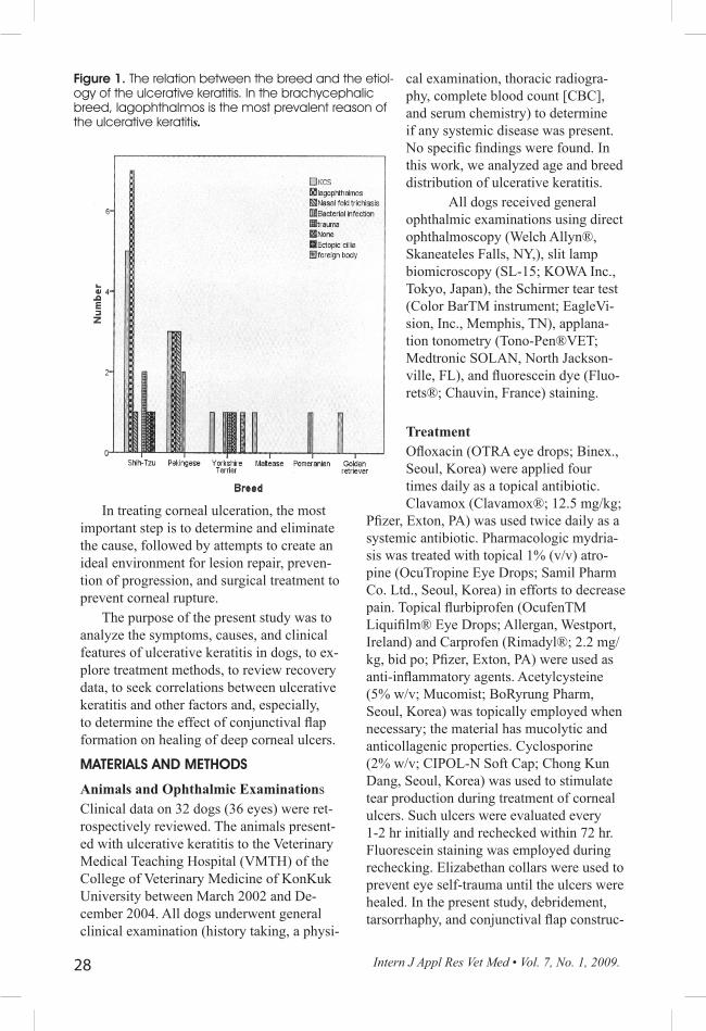
Figure 1. The relation between the breed and the etiol-ogy of the ulcerative keratitis. In the brachycephalic breed, lagophthalmos is the most prevalent reason of the ulcerative keratitis.

Intern J Appl Res Vet Med • Vol. 7, No. 1, 2009. 29
tion were used as surgical treatments of corneal ulceration. One case of ulceration with defined suppurative endophthalmitis was treated by enucleation. If a dog showed a deep corneal ulcer (loss of two-thirds of the stroma, descemetocele, or perforation), the conjunctival flap treatment method was usually employed.
Evaluation and Analysis of Medical Records Among medical record data, we analyzed age, breed, affected eye, etiology, healing rate, and duration of corneal ulceration, with reference to treatment methods. Corneal ul-cerations were divided into epithelial ulcers, superficial stromal corneal ulcers (loss of one-third of the stroma), deep stromal cor-neal ulcers (loss of two-thirds of the stroma), descemetocele, and corneal perforation. All comparisons were analyzed using the t-test and Fisher’s exact test. A P value <0.05 was considered significant. All analyses were performed using SPSS (SPSS 12.0 KO for Windows) (SPSS, Inc., Chicago, IL).
RESULTSThe mean age of dogs was 3.59±2.97 years (mean±SD). Ulcerative keratitis was seen mainly in dogs under 3 years of age (47%, 15 dogs); disease frequencies in animals aged 3-6 years, 6-9 years, and 9-12 years were 28% (9 dogs), 14% (5 dogs), and 9% (3 dogs), respectively among the total 32 dogs. Corneal ulceration thus decreases remarkably as dogs age. In the present study, six breeds (Shih-Tzu, Pekingese, Yorkshire Terrier, Maltese Terrier, Pomeranian, and Golden Retriever) were represented. Of these (total 32 dogs), the Shih-Tzu (50%, 16 dogs), Pekingese (25%, 8 dogs), and York-shire Terrier (16%, 5 dogs) showed the high-est ulcer frequencies. The Maltese Terrier, Pomeranian, and Golden Retriever frequen-cies were all low, at 3% (1 dog). Keratocon-junctivitis sicca (KCS) was the commonest cause of ulcerative keratitis (31%); other eti-ologies were lagophthalmos (28%), bacterial infection (11%), nasal fold trichiasis (11%), and trauma (8%) (P=0.002). In brachy-
cephalic breeds, however, lagophthalmos was the most prevalent cause of ulcerative keratitis and KCS was the next commonest etiology (Fig. 1). No other breed-specific etiological pattern was found (Fig. 1).
In the present study, superficial corneal ulcers including the epithelium only (22%) or with corneal stromal defects involving less than one-third of the cornea (22%) oc-curred in 44% of dogs. Deep corneal ulcers involving over two-thirds of the stroma (22%), descemetocele (26%), and corneal perforation (8 %), occurred in 56% of ani-mals.
Superficial corneal ulcers treated with medication took 5.1-13.4 days to heal and nine eyes with deep corneal ulcers, treated with conjunctival flap construction, took 28.4-40 days to heal. All eyes with super-ficial corneal ulcers recovered within 3 weeks; these were treated with medication alone. However, deep corneal ulcers did not heal properly with use of medication alone. Conjunctival flap construction was an effec-tive treatment for deep corneal ulcers. The recovery rate from superficial corneal ulcers was 100% and that from deep corneal ulcers 55% in the present study (Table 1). With medication alone, the recovery rate was 71%, although the recovery rate was 100% after surgical treatment (except for the single enucleation case) (Table 1). These recovery rates are statistically significant (P=0.002).
Superficial corneal ulcers healed rela-tively well, without complications. But most deep corneal ulcers treated without surgical intervention did not heal satisfactorily, and some animals lost vision (Table 1) because of severe corneal scarring. Ulcerative keratitis caused by KCS should be treated continuously. With use of the conjunctival flap method, all corneal ulcers healed well and prognoses were good even in dogs with descemetocele or corneal perforation; the re-sults were statistically significant (P=0.02).
DISCUSSION In this study, ulcerative keratitis oc-
curred most frequently in dogs under 3 years of age. Animals of this age are in their

Intern J Appl Res Vet Med • Vol. 7, No. 1, 2009.30
most active life period, so corneal traumatic injury is most prevalent. The frequencies of ulcerative keratitis in Shih-tzu and Peking-ese breeds were higher than in other breeds, consistent with previous findings.1,4 The Shih-tzu and Pekingese are the most popular breeds in Korea, and both suffer dispro-portionately from lagophthalmos. In fact, lagophthalmos is the most frequent cause of ulcerative keratitis in the brachycephalic breeds. Regular checking and control of brachycephalic eyes is important. Lagoph-thalmos in brachycephalic breeds must be properly treated to prevent ulcerative kera-titis. In the present study, the most common cause of ulcerative keratitis was KCS; this differs from previous findings that showed trauma and distichiasis to be of prime im-portance. In cases of superficial ulcerative keratitis, simple medication afforded good control. With ulcers affecting one-third of the corneal stroma, some affected eyes showed small corneal scars after healing, but these were insufficiently severe to affect vision. With deep corneal ulcers, many af-fected eyes lost vision because of complica-tions, even though the animals recovered in 4-6 weeks. In cases of deep ulcerative keratitis, we could not effectively control symptoms with medication alone. However, many owners did not want (expensive) sur-
gical intervention. In such cases, the medication-alone results were poor.
Conjunctival flap construction is effective for control of deep corneal ulcers, although a long healing period is required. The conjunctival flap permits healing after debridement of devitalized epithelium. For treatment of deep stromal corneal ulcers, a com-bination of medication and conjuncti-val flap construction is recommended.
In the present study, superficial corneal ulcers healed relatively well without complications. However, most deep corneal ulcers treated without surgical intervention healed poorly, and some animals lost vision. The results suggest that a combination of medication and conjunctival flap construction must be recommended for
treatment of deep stromal corneal ulcers.
ACKNOWLEGEMENTS
J. Y. Kim and H. J. Won contributed equally to this work as the co-first authors.
REFERENCES1. Gilger BC, Ollivier FJ, Bentley E. Diseases and Sur-gery of the Canine Cornea and Sclera. In: Gellatt KN, ed. Veterinary ophthalmology. 4th ed. Iowa: Blackwell, 2007:690-752.
2. Martin CL. Cornea and Sclera. In: Ophthalmic disease in veterinary medicine. London: Manson Pub-lishing Ltd, 2005:241-297.
3. Crispin S. The cornea. In: Petersen-Jones S, Crispin S, eds. BSAVA manual of small animal ophthalmology. 2nd ed. Gloucester: British Small Animal Veterinary Association, 2002:134-154.
4. Chae JM, Jeong MB, Yi NY, et al. Prevalence of cor-neal diseases of dogs in Korea. J Vet Clin 2007;24:557-562.
5. Choi YM, Kim JY, Park JI, Jeong SW. Evaluation of bovine amniotic membrane for the treatment of superfi-cial canine corneal ulcer. J Vet Clin 2007;24:358-366.
6. Kang MG, Choi YH, Kim JY, Jeong SW. Bovine amniotic membrane transplantation for the treatment of descemetocele in a dog. J Vet Clin 2006;23:334-336.
7. Kaswan R, Pappas C, Jr., Wall K, Hirsh SG. Survey of canine tear deficiency in veterinary practice. Adv Exp Med Biol 1998;438:931-939.
8. Whitley RD, McLaughlin SA, Gilger BC. Up-date on eye disorders among purebred dogs. Vet Med 1995;90:575-592.
Recovered, Corneal ulcer is healed and the eye preserve vision and; Non-recovered, Corneal ulcer is not healed and the eye looses vision.
*: Not conjunctival flap but tarsorraphy is performed.
Intern J Appl Res Vet Med • Vol. 7, No. 1, 2009. 31
9. Chavkin MJ, Roberts SM, Salman MD, Severin GA, Scholten NJ. Risk factors for development of chronic superficial keratitis in dogs. J Am Vet Med Assoc 1994;204:1630-1634.
10. Hedlund CS. Surgery of the Eye. In: Fossum TW, ed. Small animal surgery. 3rd ed. Missouri: Mosby, 2007:260-288.
11. Vanore M, Chahory S, Payen G, Clerc B. Surgi-cal repair of deep melting ulcers with porcine small intestinal submucosa (SIS) graft in dogs and cats. Vet Ophthalmol 2007;10:93-99.
12. Hale G. Treatment of corneal ulcers in dogs. Vet Rec 2006;158:108; author reply 108.
13. Yi NY, Park SA, Jeong MB, et al. Medial cantho-
plasty for epiphora in dogs: a retrospective study of 23 cases. J Am Anim Hosp Assoc 2006;42:435-439.
14. Hollingsworth SR. Corneal surgical techniques. Clin Tech Small Anim Pract 2003;18:161-167.
15. Stades FC, Gelatt KN. Diseases and Surgery of the Canine Eyelid. In: Gellatt KN, ed. Veterinary Ophthal-mology. 4th ed. Iowa: Blackwell, 2007:563-617.
16. Scagliotti RH. Tarsoconjunctival island graft for the treatment of deep corneal ulcers, desmetocoeles, and perforations in 35 dogs and 6 cats. Semin Vet Med Surg (Small Anim) 1988;3:69-