behçet’s disease: a case of peripheral ulcerative keratitis leading to corneal perforation
DESCRIPTION
Behçet’s Disease: A Case of Peripheral Ulcerative Keratitis Leading to Corneal Perforation. Selcuk Sizmaz, Aysel Pelit, Meltem Yagmur, Didem Arslan, Yonca Aydin Akova. None of the authors have any financial or proprietary interest in any material used in the study. A 27 year-old man - PowerPoint PPT PresentationTRANSCRIPT

Behçet’s Disease: A Case of Behçet’s Disease: A Case of Peripheral Ulcerative Keratitis Peripheral Ulcerative Keratitis Leading to Corneal PerforationLeading to Corneal Perforation
Selcuk Sizmaz, Aysel Pelit, Meltem Yagmur, Didem Arslan, Yonca Aydin Akova

• A 27 year-old man• Two day history of redness in the right eye• Corneal thinning involving the anterior
stroma and adjacent conjunctival hyperemia, in the corneal limbus, at the 2- to 3-o’clock quadrant
• No sign of dry eye• Topical ofloxacin and artificial tears
prescribed
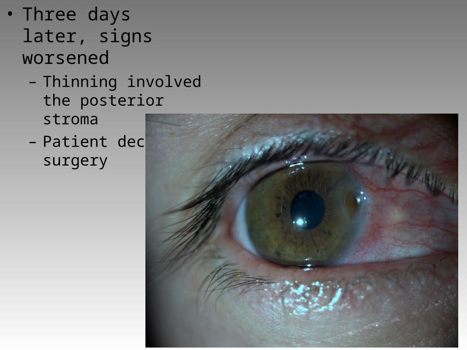
• Three days later, signs worsened– Thinning involved the
posterior stroma– Patient declined
surgery

• Two days later– Corneal perforation– İris prolapsed

• Corneal patch graft and amniotic membrane transplantation
Ophthalmic and systemic Ophthalmic and systemic evaluationevaluation
• No significant ocular history• Oral aphthous lesions that had occurred
once or twice a month for the previous 2 years
• Eight years history of lower back and buttock pain with morning stiffness for 30 minutes
• Arthralgia in the knee joints without swelling

• Arterial BP (mmHg)– 130/80 right arm– 100/70 left arm
• Weak pulse in left arm• Colour Doppler USG: left
subclavian artery narrowing
• MR angipgraphy: 4 cm-long diffuse stenosis in left subclavian artery
• Sacroiliac graphy: grade II-III sacroiliitis
• Pathergy test: (-)
Erithrocyte sedimantation rate
35 mm/h
C-reactive protein 4.5 mg/L (↑)
HBV (-)
HCV (-)
HIV (-)
ANA (-)
C-ANCA (-)
HLA-B27 (-)
HLA-B51 (-)

Diagnosis: Behçet’s diseaseDiagnosis: Behçet’s disease
• Oral aphthous lesions
• Ocular involvement
• Large vessel vasculitis
• Sacroiliitis

• Management:– Methylprednisolone, 32 mg/day, po; tapered– Azathioprine, 150 mg/day, po
• Prognosis (1 year follow-up):– No recurrence– Systemic symptoms improved

Differential diagnosisDifferential diagnosis• Takayasu arteritis
+ Aorta and branches involved– No aphthous lesions– Sacroiliitis uncommon– Ocular invovement rare
• Polyarteritis nodosa+ Peripheral ulcerative keratitis occurs– Predominantly females– Aneurisms in small-middle sized vessels– Constitutional symptoms