a formulation science perspective applied to …
TRANSCRIPT

Sede Amministrativa: Università degli Studi di Padova
Dipartimento di Biologia
SCUOLA DI DOTTORATO DI RICERCA IN BIOSCIENZE E BIOTECNOLOGIE
INDIRIZZO BIOLOGIA CELLULARE
CICLO XXVII
A FORMULATION SCIENCE PERSPECTIVE APPLIED TO COMBINATION VACCINES
Particles‐based multiplex immunoassays for improved antigens characterization
and rational design of adjuvants for tailored immunity to acellular pertussis vaccines
Direttore della Scuola: Ch.mo Prof. Giuseppe Zanotti
Coordinatore d’indirizzo: Ch.mo Prof. Paolo Bernardi
Supervisore: Ch.mo Prof. Cesare Montecucco
Co-supervisore: Dr. Barbara C. Baudner
Dottorando: Valentina Agnolon

2

i
Guarda lontano,
e quando pensi di aver guardato lontano,
guarda ancora più lontano.
‐ Robert Baden Powell


i


i
SUMMARY
Vaccines are complex multi-component products, in which an interdependent
relationship among constituents exists: the concomitant inclusion of antigens,
adjuvants, and excipients is essential to guarantee the efficacy of the final product.
To stimulate the immune system and raise immunogenicity against highly purified
vaccine components most likely adjuvants are required, thus becoming more and
more crucial elements of modern vaccine formulations. Physico-chemical and
functional attributes of all components and of the whole vaccine formulation have
to be controlled before injection. However, often characterization challenges are
experienced with the currently available analytical techniques, also because of
interferences with adjuvants.
In this thesis adjuvants were evaluated with the aim to improve quality and
immunogenicity of combination vaccines, in which the inclusion of multiple
antigenic components contributes to enhanced complexity of the final product.
In detail, three virulence factors of the Gram negative bacterium Bordetella
pertussis (pertussis toxin PT, filamentous heamagglutinin FHA, and pertactin 69K)
were chosen as model antigens. Currently licensed acellular pertussis (aP) vaccines
are combined with diphtheria and tetanus toxoids in the DTaP/TdaP vaccine, which
is considered a cornerstone of combination vaccines. This vaccine is typically
adjuvanted with aluminum salts, known to induce mainly a humoral immune
response which has been associated with recent pertussis outbreaks reported in
many countries. This suggests that existing vaccines may not be ideal to provide
protection against the disease. Thus, a pharmaceutical sciences approach focusing
on adjuvants was applied to investigate the potential improvement of acellular
pertussis vaccines through promotion of the analytical characterization and rational
polarization of the quality of immune responses.

ii
Improved in vitro characterization assays for combination vaccines adjuvanted
with aluminum hydroxide
Vaccines in vitro characterization is required to identify optimal formulation
conditions to ensure physical, chemical, and biological integrity of antigens and
adjuvants. Analytical methods currently in use are mostly suitable for the
characterization of unformulated antigens – requiring the complete desorption from
aluminum-based adjuvants - and are not always able to reveal individual antigens in
vaccine combinations. Here, the Luminex technology is proposed as proof of
concept for the development of an improved analytical method for vaccine
characterization, based on the use of specific antibodies bound to magnetic
microspheres presenting unique digital signatures for simultaneous recognition of
respective antigens in the entire formulation. TdaP combination vaccines were
chosen as a model to develop an assay able to quantify acellular pertussis antigens
and their levels of adsorption to adjuvant at the same time. This assay was directly
applicable on the commercial vaccine product, avoiding any laborious procedures
for separation of the antigens from the adjuvant. Accurate and reproducible
quantification of aP antigens in TdaP vaccine has been achieved in a range between
0.78 and 50 ng/mL, providing information on antigen identity, quantity, and degree
of adsorption to aluminum hydroxide. Importantly, the Luminex characterization
method has the potential to be further evaluated as correlate of in vitro potency
assays - ideally allowing reducing in vivo animal studies.
In vivo evaluation of novel adjuvants for improved aP-containing combination
vaccines
The successful approach of combining diphtheria, tetanus and pertussis antigens
into a single vaccine has become the cornerstone of pediatric and adults
immunization programs. Yet, even if vaccination coverage is high, a resurgence of
pertussis has been recently reported in many countries suggesting that current
vaccines may not provide adequate long-lasting protection.
In order to develop pertussis-containing vaccines able to induce a more durable and
better tailored immune response different approaches have been proposed,
including the use of novel adjuvants. Currently licensed pertussis vaccines contain
iii
aluminum salts, which are the most common adjuvants for human use. By inducing
mainly humoral immune responses aluminum salts might not be ideal for providing
protection against pathogens which require a more cellular immune response, such
as Bordetella pertussis.
Therefore, alternative adjuvants that induce more balanced T-helper profiles or even
Th1-prone responses might be more adequate. In this project, we tested three
different adjuvants: MF59 emulsion adjuvant – that induces a mixed Th1/2 response
- as well as the combination of a Toll-Like Receptor 4 agonist (TLR4a) or a Toll-
Like Receptor 7 agonist (TLR7a) with aluminum hydroxide to induce even further
Th1 polarization. The adjuvants were evaluated for their ability to improve immune
responses against a TdaP vaccine containing three B. pertussis antigens: genetically
detoxified pertussis toxin (PT-9K/129G), filamentous hemagglutinin (FHA) and
pertactin (69K).
The quicker onset of serum antibody titers and the changed quality of the antibody
responses induced by the adjuvants evaluated here fully support the potential
replacement of aluminum salts with alternative adjuvants to enhance pertussis
immunogenicity in aP-containing combination vaccines.
Conclusions
Overall, the collected results suggested the possibility to modulate quality and
efficacy of a complex vaccine product by carefully working on the adjuvant
component. Improving the analytical characterization of vaccines while addressing
a rational polarization of immune responses are proposed as effective strategies to
be pursued for the development of next generation combination vaccines.

iv

v
R IASSUNTO
I vaccini sono prodotti complessi costituiti da diversi elementi tra i quali esiste un
rapporto d’interdipendenza: la presenza concomitante di antigeni, adiuvanti, ed
eccipienti è essenziale per garantire l'efficacia del prodotto finale. Gli adiuvanti
sono spesso necessari per stimolare il sistema immunitario e aumentare
l'immunogenicità di un vaccino composto di antigeni molto purificati: diventano
perciò sempre più importanti nelle moderne formulazioni di vaccini. Le proprietà
fisico-chimiche di tutti gli elementi e dell'intera formulazione e devono essere
controllate prima dell'iniezione. Tuttavia, le tecniche analitiche ora disponibili
spesso interferiscono con gli adiuvanti, e problemi di caratterizzazione sono
incontrati.
In questa tesi gli adiuvanti sono stati sfruttati con l'obiettivo di indagare la
possibilità di migliorare la qualità e l'immunogenicità dei vaccini combinati, nei
quali l'inclusione di più elementi antigenici contribuisce alla maggiore complessità
del prodotto finale.
In particolare, tre fattori di virulenza del batterio Gram negativo Bordetella
pertussis (tossina della pertosse PT, emoagglutinina filamentosa FHA, e pertactina
69K) sono stati scelti come antigeni modello. I vaccini acellulari di pertosse (aP)
attualmente autorizzati sono combinati con le tossine inattivate di difterite e tetano
nel vaccino DTaP/TdaP, che è considerato la pietra miliare dei vaccini combinati.
Questo vaccino è tipicamente adiuvato con sali di alluminio, noti per indurre
prevalentemente una risposta immunitaria umorale che è stata associata con le
recenti epidemie di pertosse segnalate in molti paesi. Ciò suggerisce che il vaccino
esistente non sia ideale per fornire una protezione contro la malattia. Pertanto, un
approccio basato sulle scienze farmaceutiche e focalizzato sugli adiuvanti è stato
applicato per studiare il potenziale miglioramento dei vaccini acellulari di pertosse,
attraverso lo sviluppo della caratterizzazione analitica e la razionale polarizzazione
della qualità della risposta immunitaria.

vi
Sviluppo di saggi di caratterizzazione in vitro per vaccini combinati
adiuvantati con idrossido di alluminio
La caratterizzazione analitica in vitro è necessaria per individuare le condizioni di
formulazione ottimali che garantiscano l'integrità fisica, chimica e biologica di
antigeni e adiuvanti. I metodi analitici ora in uso sono soprattutto adatti per la
caratterizzazione di antigeni non formulati - richiedendo quindi il desorbimento
completo dalla superfice degli adiuvanti a base di alluminio - e non sempre sono in
grado di distinguere tra i singoli antigeni contenuti nei vaccini combinati. In questa
tesi la tecnologia Luminex è proposta come proof of concept per lo sviluppo di un
miglior metodo analitico per la caratterizzazione di vaccini. Questo metodo è basato
sull'utilizzo di anticorpi specifici legati a microsfere magnetiche che sono
univocamente riconoscibili tramite una firma digitale permettendo quindi
l’identificazione simultanea dei rispettivi antigeni formulati nel vaccino.
I vaccini combinati TdaP sono stati scelti come modello per sviluppare un saggio in
grado di quantificare simultaneamente gli antigeni acellulari della pertosse (aP) e il
loro grado di adsorbimento all’adiuvante. Questo saggio è direttamente applicabile
al vaccino commerciale, evitando qualsiasi procedura laboriosa per la separazione
degli antigeni dall’adiuvante. Una quantificazione accurata e riproducibile degli
antigeni aP nel vaccino TdaP è stata ottenuta in un intervallo di concentrazioni
compreso tra 0,78 e 50 ng/mL, fornendo informazioni sull'identità degli antigeni, la
loro quantità e il grado di assorbimento alla superficie dell’idrossido di alluminio. Il
metodo di caratterizzazione sviluppato sulla tecnologia Luminex ha la potenzialità
di essere valutato come test di in vitro potency - permettendo idealmente la
riduzione degli studi effettuati sugli animali.
Valutazione in vivo di nuovi adiuvanti per migliorare i vaccini combinati
contenenti aP
L'approccio di combinare gli antigeni di difterite, tetano e pertosse in un unico
vaccino è diventato il fondamento dei programmi d’immunizzazione pediatrici e per
adulti. Nonostante l’elevata copertura vaccinale, una riacutizzazione dei casi di
pertosse è stata recentemente registrata in molti paesi, suggerendo che i vaccini
attuali non possono fornire un’adeguata protezione di lunga durata.

vii
Diversi approcci sono stati proposti al fine di sviluppare vaccini in grado di indurre
una risposta immunitaria più durevole e più adeguata contro la pertosse, tra cui l'uso
di nuovi adiuvanti. I vaccini contro la pertosse ora autorizzati contengono sali di
alluminio, che sono gli adiuvanti più comuni per uso umano. Inducendo
principalmente risposte immunitarie umorali, i sali di alluminio non sono ideali per
fornire protezione contro gli agenti patogeni che richiedono una risposta
immunitaria cellulare, come ad esempio Bordetella pertussis.
Pertanto, altri adiuvanti che stimolino l’induzione di cellule Th1 potrebbero essere
più adeguati. In questo progetto, abbiamo provato tre diversi adiuvanti: l’emulsione
MF59 - che induce una risposta mista Th1/2 - oppure la combinazione di un
agonista del Toll-Like Receptor 4 (TLR4a) o del Toll-Like Receptor 7 (TLR7a) con
idrossido di alluminio – che polarizzano la risposta verso il profilo Th1. Gli
adiuvanti sono stati valutati per la loro capacità di migliorare le risposte
immunitarie contro il vaccino TdaP contenente tre antigeni di B. pertussis: tossina
della pertosse geneticamente detossificata (PT-9K/129G), emoagglutinina
filamentosa (FHA) e pertactina (69K).
La rapida induzione di titoli anticorpali e la polarizzazione della qualità della
risposta immunitaria indotte dagli adiuvanti qui esaminati sostengono pienamente la
potenzialità di sostituire i sali di alluminio con adiuvanti alternativi che migliorino
l’immunogenicità di vaccini combinati contenenti aP.
Conclusioni
Complessivamente, i risultati raccolti suggeriscono la possibilità di modulare la
qualità e l'efficacia di un vaccino complesso lavorando sulla componente adiuvante.
Migliorare la caratterizzazione analitica dei vaccini e indirizzare una polarizzazione
razionale della risposta immunitaria sono proposte come strategie efficaci da
perseguire per lo sviluppo dei vaccini combinati della prossima generazione.

viii

1
TABLEOF CONTENTS
Summary pag. i Riassunto pag. v Table of contents pag. 1 Preface
pag. 3
Chapter I. General Introduction and Objectives of the Thesis
pag. 5
Vaccines pag. 5 Combination vaccines pag. 6 Vaccine adjuvants pag. 9 Aluminum salts adjuvants pag. 12 Introduction of the model vaccine: acellular pertussis containing combination vaccines
pag. 16
Aim of the thesis
pag. 17
Chapter II. Improved in vitro characterization assays for aP combination vaccines
pag. 19
Luminex technology
pag. 19
Indirect Luminex assay for analysis of formulations
pag. 22
Direct Luminex assay for analysis of formulations
pag. 30
Multiplex immunoassay for in vitro characterization of acellular pertussis antigens in combination vaccines
pag. 31
Supplementary data
pag. 52
- Antigens precipitation upon centrifugation pag. 52

2
- Integrity of antigens
pag. 54
- Application on MF59 adjuvant
pag. 55
Chapter III. In vivo evaluation of novel adjuvants for improved aP combination vaccines
pag. 57
Bordetella pertussis
pag. 57
Evaluation of alternative adjuvants for improved immunity to Bordetella pertussis
pag. 61
The potential of adjuvants to improve immune responses against TdaP vaccines: a preliminary evaluation of MF59 and Monophosphoryl lipid A
pag. 64
Supplementary data
pag. 85
- Evaluation of a synthetic agonist of Toll-Like Receptor 7 pag. 85 Chapter IV. Conclusions pag. 93 References pag. 97 Ringraziamenti pag. 107

3
PREFACE
Vaccines, together with antibiotics, are among the most successful outcomes of
modern medicine. Regardless of this, people reveal a declining confidence and
compliance when facing vaccines, probably because these products are not
considered useful in preventing diseases which are mostly unknown and not
recognized as life-threatening. People are no longer aware of diseases that vaccines
protect from as they have never experienced them. As a consequence, vaccination
coverage decreases, resulting in large outbreaks of highly contagious diseases, as
recently seen e.g. for measles [1]. Additionally the complexity of vaccine products
contributes to concerns about vaccine ingredients and unexpected side-effects.
Vaccine research is a complex field, covering many areas ranging from the
discovery and evaluation of new antigenic candidates to the continuous
improvement and validation of products already available in the market.
During my PhD I have investigated the possibility to improve combination vaccines
through the application of pharmaceutical sciences approaches directed and
focusing on adjuvants. I have explored the potential of complete characterization
and rational design of adjuvants within complex vaccine formulations, with the
purpose of improving development and awareness of future vaccines - emphasizing
the importance of adjuvants.

4

5
CHAPTER I . GENERAL INTRODUCTIONANDOBJECTIVEOF THETHESIS
VACCINES
At the end of the nineteenth century Louis Pasteur identified microorganisms as
agents responsible for infectious diseases. Moreover, by accidentally leaving a
bacteria culture without nutrients, he observed that it was possible to artificially
obtain microorganisms showing attenuated virulence and able to confer immunity
against the disease. Pasteur referred to attenuated bacteria as “vaccines” in honor of
Edward Jenner’s discovery - dated back at the end of the eighteenth century - about
the possibility to protect people against smallpox through inoculation with cowpox,
a less dangerous disease which he denoted as Variolae Vaccinae (from the Latin
word vacca, cow).
More than a hundred years after Pasteur’s observation, vaccines are considered a
fundamental milestone for healthcare providing life-long protection against
numerous infections. At first the leading principle was the basic one proposed by
the French microbiologist, starting with the identification of the microorganism,
inactivation through heat or exsiccation and subsequent injection. A big step

6
forward was made during the 1920s by Gaston Léon Ramon who developed the
first two vaccines – against Corinebacterium diphtheriae and Clostridium tetanii -
constituted not by the whole bacterium but only by a part of it. Diphtheria and
tetanus toxins, identified as the causes of the respective diseases, were inactivated
with formaldehyde following a procedure similar to the one used in modern vaccine
manufacture [2].
A brief historical overview across vaccines discovery and development allow to
categorize vaccines in three main groups: live-attenuated whole microorganisms,
heat or chemically inactivated microorganisms, and microorganisms’ subunits [3].
This classification is still valid in modern vaccinations but technology
improvements and the continuous raising of safety concerns drive vaccine
development to limit the use of whole pathogens in favor to the identification and
isolation of well-characterized components responsible of conferring immunity to
the disease. Indeed, reversion of toxicity and high levels of reactogenicity with
subsequent adverse-effects are challenges that might be faced when working with
whole inactivated microorganisms. Significant improvements in terms of safety
were introduced by the advent of the recombinant DNA technology which allowed
the production of recombinant antigens [4].
COMBINATIONVACCINES
Continuous vaccine development (Figure 1 [5]) is pushing towards the constant
progress in the development of combination vaccines, which allow providing
immunity against more than one infectious disease. By incorporating separate
antigens or individual vaccines into a single product, combination vaccines
represent an important means of simplifying vaccination strategies. Their
development started over sixty years ago with the combination of diphtheria toxoid
(DT), tetanus toxoid (TT) and whole-inactivated pertussis bacteria (wP) into a
single DTwP vaccine that has become the cornerstone of pediatric and adult
immunization programs [6]. Over the years the wP component was replaced with
less reactogenic acellular pertussis (aP) antigens, and further routine vaccines were

7
FIGURE 1. Timeline of vaccine development showing the number of vaccines introducedin the market every decade (yellow) since the discovery of the first vaccine at the end ofthe eighteenth century. In orange the progressive cumulative number of vaccine products is recorded. 1790s: smallpox. 1880s: rabies, typhoid. 1890s: cholera, plague. 1920s: diphtheria, tetanus, pertussis, tuberculosis. 1930s: yellow fever, influenza, typhus. 1950s: polio (injected, inactivated). 1960s: polio (oral, live), measles, mumps, rubella. 1970s: anthrax, meningococcal disease (polysaccharide), pneumococcal disease(polysaccharide). 1980s: adenovirus infection, rabies (cell culture), tick-borne encephalitis, hepatitis B (plasma derived), Haemophilus influenza type B infection (polysaccharide), hepatitis B(yeast or baculovirus, surface antigen), Haemophilus influenza type B infection(conjugate), typhoid (other variants). 1990s: cholera (killed whole cell), Japanese encephalitis (inactivated), cholera (recombinant toxin B), cholera (live attenuated), typhoid (capsular polysaccharide),varicella zoster, acellular pertussis, hepatitis A (inactivated), lyme disease, rotavirusinfection, meningococcal disease (conjugate group C). 2000s: pneumococcal disease (7 valent, conjugate), cold-adapted influenza, meningococcal disease (4 valent, conjugate), varicella zoster (live), rotavirus infection (attenuated),human papilloma virus (4 valent, recombinant), cholera (whole cell), Japanese encephalitis(Vero cell colture), human papilloma virus (2 valent, recombinant). 2010s: (current decade): pneumococcal disease (13 valent, conjugate), meningococcaldisease (recombinant group B). Data collected from [5].

8
subsequently added to the DTaP combination following the growing number of
childhood diseases for which vaccination was recommended.
The latest advance in DTaP combination vaccine development is defined by
Infanrix™ -hexa from GSK, which protects against six different diseases by
carrying diphtheria, tetanus, and acellular pertussis antigens (DTaP) together with
inactivated polio vaccine (IPV), Haemophilus influenzae vaccine (Hib), and
hepatitis B vaccine (HepB) [7]. Other significant combination vaccines are the 23-
valent pneumococcal polysaccharide vaccine (Pneumovax 23™ from Merck) [8] or
the 4-valent meningococcal conjugate vaccine (Menveo™ from Novartis Vaccines)
[9], which respectively protect solely against pneumococcal and meningococcal
infections. It is likely that new combination vaccines will reach the market in the
coming years, combining DTaP-based with pneumococcal or meningococcal
vaccines. Promisingly, these products are already being co-administered at the same
vaccination visit, and no major negative effects have been reported [10].
Combination vaccines confer several advantages, starting with the simplification of
the immunization schedule and subsequent reduction of the number of injections
needed to confer protection against multiple diseases. Benefits can be identified on
various levels of society, with fever shots and improved compliance for recipients,
and lower costs, less medical visits needed, better coverage and the possibility to
more easily add new vaccines into the immunization program for national
healthcare systems [7].
Combining multiple vaccines into a single product potentially introduce some
challenges since fine-tuning respective immunization schedules which foresee a
different number of doses on different time points is not immediate (Figure 2).
Moreover adverse interactions among combined antigens have to be avoided while
maintaining the final product as safe and immunogenic as the individual vaccine
components. Improvements in this context may involve the inclusion of adjuvants,
which can potentially bring advantages, such as increased antigen stability and
reduced interference among vaccine components, as well as antigen dose reduction
and reduction of the number of scheduled immunizations [11].

9
VACCINEADJUVANTS
Adjuvants owe their name to the Latin word adjuvare, meaning “to help”. Ramon,
previously cited for his studies about diphtheria and tetanus toxins, was the first to
describe adjuvants as “substances used in combination with a specific antigen that
produced a more robust immune response than the antigen alone” [12]. In whole
live attenuated or inactivated vaccines the role of potentiation of the immune
response is carried out by components naturally present in respective
microorganisms, such as bacterial DNA or lipopolysaccharide, which are
recognized as non-self by the host thus generating an immune response. The
introduction of purified and recombinant antigens caused an evident increase in
safety profile but, despite that, antigens alone resulted to be less immunogenic and
not always protective. Therefore, vaccine development becomes a complex fine-
tuning between safety and efficacy of the final product. In this perspective,
adjuvants were introduced to balance the removal of natural immune-stimulatory
components of the pathogens with the aim to enhance an immune response similar
to the one triggered by the natural infection [11-13], while maintaining equilibrium
between vaccine safety and efficacy.
Birth 1 mo. 2 mo. 4 mo. 6 mo. 12 mo. 15 mo. 18 mo. 19‐23 mo. 2‐3 yr. 4‐6 yr.
Diphtheria,
Tetanus,
Pertussis
DTaP DTaP DTaP DTaP
Measles,
Mumps,
Rubella
MMR
Hepatitis B HepB
Heamophilus
influenzae
type B
Hib Hib Hib
Pneumococcal PCV PCV PCV
Rotavirus RV RV RV
Inactivated
PoliovirusIPV IPV IPV
Varicella Var.
Hepatitis A
Influenza
IPV
Flu (yearly)
MMR
Var.
HepA (2 doses)
VaccineAge
HepB HepB
DTaP
Hib
FIGURE 2. Recommended immunization schedule for children aged 0 through 6 years.Adapted from [7].

10
The inclusion of adjuvants in vaccines provides a number of advantages which
confer the potential to:
Increase antigens immunogenicity;
Lower the number of doses needed to confer protection – quicker response;
Lower antigen doses with reduced impact on production costs;
Modulate quality of the immune response to promote protection;
Modulate quantity of the immune response by extending duration and
potency of induced antibodies;
Enable complex vaccine combinations overcoming competition among
included antigens;
Overcome limited immune response in endangered population such as the
elderly, young children, and immunocompromised people.
For almost 80 years aluminum-based adjuvants - approved in the 1920s - were the
only adjuvants licensed for human use, followed by MF59 approval for inclusion in
an influenza vaccine in 1997 [13]. While the main feature of aluminum salts and
MF59 is the delivery of antigens to immune cells, “second-generation adjuvants”
have been developed combining a delivery system with an immune-potentiator
molecule [11]. Among these AS04 [14] combine the lipopolysaccharide analog
monophosphoryl lipid A (MPLA) with aluminum salts and was the first to be
approved for inclusion in a vaccine against Hepatitis B virus (Fendrix® from GSK)
in 2005 [15] and against human papilloma virus (Cervarix® from GSK) in 2007
[16]. In Table I a list of adjuvants licensed and evaluated in clinical studies is
presented. However those are only a subset among the variety of adjuvants
proposed and described in literature, indicating adjuvant development as a slow and
difficult process, which undergo many challenges mainly from a safety and
regulatory perspective [11, 17].

11
A growing number of research groups are directing their attention and investments
at deepening the knowledge about adjuvants mechanism of action and studying
adjuvants effect from an immunological point of view. Formulation science is
focusing on the complexity induced by the addition of adjuvants to the vaccine
formulation taking into consideration potential effects on product/antigen stability
and efficacy [18]. A complex set of analytical characterization techniques is usually
required for a complete characterization of formulated antigens and antigens-
adjuvants interactions further complicate their application [19]. In the next section
these challenges will be reviewed focusing on aluminum salts adjuvants, which are
the mostly used adjuvants for human use [20].
Adjuvant Description Status
Aluminum salts Insoluble aluminum salts (e.g. hydroxides and phosphates);
extensive safety record; antigens adsorbed to the surface.
Licensed in EU
and US
MF59 Squalene oil‐in‐water emulsion; extensive safety record;
antigens not associated with droplets.
Licensed in EU
AS03 Squalene oil‐in‐water emulsion added with the immune‐
potentiator α‐tocopherol; associated with narcolepsy.
Licensed in EU
and US
AS04 Combination of aluminum adjuvant with the TLR4 agonist
monophosphoryl lipid A (MPLA), co‐adsorbed.
Licensed in EU
and US
AS01 Liposomes composed added with MPLA and QS21. Clinical phase
CAF01 Cationic liposomes. Clinical phase
Poly I:C Synthetic double stranded oligonucleotide containing
repeating units of inosine and cytosine (TLR3 agonist).
Clinical phase
IC31 Antimicrobial peptide bound to a TLR9 agonist. Clinical phase
AS02 Co‐mixture of AS03 emulsion adjuvant with MPLA and QS21. Clinical phase
Imiquimod TLR7/8 agonist small molecules immune potentiators
(SMIP).
Clinical phase
CpG olugonucleotides TLR9 agonists based on bacterial DNA. Clinical phase
SE/SE‐GLA Squalene based emulsion added with the TLR4 agonist GLA. Clinical phase
ISCOMS/ISCOMATRIX Small (40 nm) lipid based adjuvants consisting of
phospholipids, cholesterol and saponins.
Clinical phase
TABLE I. List of adjuvants licensed or under clinical evaluation for use in humans. Adapted from [17].

12
ALUMINUMSALTSADJUVANTS
Aluminum-salts based adjuvants have become a reference standard for the
evaluation of new generation vaccines due to their long history of use. Formerly,
aluminum salts were commonly used for proteins purification by taking advantage
of their high absorbent capacity. The discovery of the adjuvant effect of this
material dates back to 1926 when Alexander Glenny and his colleagues described
the higher antigenic response of a diphtheria toxoid precipitated in presence of
aluminum potassium sulfate in comparison to the soluble diphtheria toxoid [21]; a
discovery that inspired Ramon’s adjuvant definition. During the twentieth century
aluminum adjuvants have been extensively studied and included in many human
vaccines against infectious diseases showing good safety profile (e.g. vaccines
against hepatitis B, Haemophilus influenzae and human papilloma virus). However,
aluminum salts mechanisms of action as vaccine adjuvants is not yet fully
understood, despite the role of many immunological components involved in the
response have been recently elucidated [22]. Among the key concepts to exploit
aluminum adjuvant potential there is the awareness for some antigens that
adsorption is of key importance to guarantee vaccines stability and adjuvanticity [18,
23].
Two main types of aluminum salts have been identified as adjuvants having
different chemical and physical compositions that strongly influence antigens
adsorption profile on their surfaces [22, 24]:
Aluminum hydroxide, chemically aluminum oxyhydroxide, has a crystalline
structure composed of needle-shaped nanoparticles which tends to aggregate
up to an average diameter of 10 µm. Aluminum hydroxide presents only
hydroxyl groups on its surface and have a point of zero charge (PZC) of
11.4 thus being positive charged at the neutral pH used for vaccine
formulations;
Aluminum phosphate, chemically aluminum hydroxyphosphate, has an
amorphous structure with a mean particles size of 50 nm which form
irregular aggregates of several microns. Both hydroxyl and phosphate

13
groups cover aluminum phosphate surface with a ratio between the two that
depends on manufacturing conditions. A PZC between 4.5 and 5.5 confer
aluminum phosphate a negative charge at neutral pH.
Characterization of aluminum salts chemico-physical properties is of key
importance for development of stable vaccines and induction of strong antigen-
specific immune responses. The diverse properties of the two mainly employed
aluminum salts adjuvants allow the rational choice of the one which most likely
favors antigens adsorption, according to isoelectric points and chemical-physical
characteristics. Indeed interactions between antigens and adjuvant surface are
mainly affected by electrostatic forces, hydrophobic interactions and ligand
exchange [25, 26]. Through antigen adsorption, aluminum salt adjuvant mainly act
as a delivery system and promotes retention of antigens at the injection site [27-29]
thus favoring uptake by immune cells [30, 31] while improving antigens physico-
chemical stability and immunogenicity. Antigen-adjuvant interactions have to be
optimized and characterized to ensure adsorption and favor the development of
stable and efficacious vaccines.
A complex set of analytical techniques is needed to characterize antigens and
antigen/adjuvant interactions. In particular, the adsorption of the antigen on the
adjuvant surface is an important parameter to be monitored to control the adjuvant
role of aluminum salts, along with total content and integrity of antigenic
components [26, 32]. Many analytical tools commonly applied to monitor proteins
(e.g. gel electrophoresis, Western blot, and liquid chromatography) cannot be
applied directly to the vaccine product due to interferences with the adjuvant and
other formulation excipients. Therefore, characterization of antigens in vaccines
currently requires their complete recovery from aluminum adjuvanted formulations
mainly through the use of high salt concentration and surfactants to dislocate
antigens from the adjuvant surface. This desorption procedure could result in
incomplete antigen recovery and could compromise the integrity of antigens [23,
33-35]. After centrifugation, antigens desorbed and released in the aqueous phase
are recovered and analyzed, nonetheless desorption buffer components could
interfere with the assays, too (e.g. surfactants impact HPLC methods, and histidine

14
buffers are not suitable for colorimetric assays) [19]. In addition, antigens are
usually formulated in vaccines at doses significantly lower than the quantification
limits of the analytical tools currently available, especially when considering
booster dosages [36].
Thus the variety of analytical tools currently available for the characterization of
soluble proteins is counteracted by adjuvant presence and antigen-adjuvant
interactions which cause the analysis of antigens in formulated vaccines to be an
enormous challenge [37]. In this perspective, the development of analytical assays
which allow the characterization of antigen-adjuvant interactions and the
investigation of their effect on antigens stability and immunogenicity is
continuously needed. It has been already observed that antigens undergo structural
changes due to interaction and adsorption to aluminum salts and the resulting
effects on antigens physico-chemical stability due to changes in their environment
have only recently started to be investigated [22, 32].
In the last years a variety of analytical techniques have been set up and improved
for vaccine characterization without requiring preliminary phase separation
procedures between antigens and aluminum salts adjuvant. The most commonly
employed techniques are presented in Table II along with the type of information
provided, the typical working range, and the specificity. The o-Phthalaldehyde
(OPA) fluorescent protein assay is extensively used to determine antigen
concentration while adsorbed to aluminum adjuvant and have a sensitivity (20 - 500
μg/ml) [38] similar to that of a chemo-luminescent method developed to quantify
formulated antigens through determination of total nitrogen content [39]. A
significant increase in terms of sensitivity is observed for assays which work on
antibody recognition (DAFIA, flow-cytometry, SPR) [40-42]. The availability of
antigen-specific and well-characterized monoclonal antibodies could allow gaining
information on antigen identity and integrity through analysis of epitopes critical for
vaccine immunogenicity, guaranteeing specificity to the assay. Spectroscopic
methods or thermo-analytical techniques could be instead applied to investigate
conformational and thermal stability of vaccine antigens while adsorbed on

15
aluminum adjuvants [43-48] and to screen different pH, formulation buffer
components and stabilizers [49].
In order to avoid diminished potency and to ensure the development of high quality
final products as required in modern vaccinology, a complex set of analytical
techniques is needed and should be ideally applied since the early stages of research
to characterize each antigen and antigen/adjuvant interaction. However, due to its
Method Type of information provided
Working range
Specificity References
OPA (o-Phthalaldehyde assay)
Antigen quantity 20 - 500 µg/mL - [38]
CLND (Chemiluminescent Nitrogen Detection)
Antigen quantity 6.75 - 400 µg/mL
- [39]
DAFIA (Direct Alum Formulation Immunoassay)
‐ Antigen-specific (antibody recognition)
‐ Antigen quantity, identity, integrity
‐ Direct analysis of immunological epitopes
0.16 - 10 µg/mL
+ [40]
Flow-cytometry ‐ Antigen specific (antibody recognition)
‐ Antigen quantity, identity, integrity
‐ Direct analysis of immunological epitopes
‐ Content uniformity
6.2 - 200 µg/mL
+ [41]
SPR - Biacore (Surface Plasmon Resonance)
‐ Antigen specific (antibody recognition)
‐ Direct analysis of immunological epitopes
‐ Antigen conformational and thermal stability
20 µg/mL + [42]
Fluorescence spectroscopy (Intrinsic/Extrinsic)
Antigen conformational and thermal stability
50 - 200 µg/mL - [43, 44, 46, 49]
Near IR transmittance spectroscopy
Antigen quantity 0 - 1.75 mg/mL - [48]
FTIR (Fourier Transform IR spectroscopy)
Antigen conformational and thermal stability
0.5 - 1 mg/mL - [47]
DSC (Differential Scanning Calorimetry)
Antigen conformational and thermal stability
0.5 - 1 mg/mL - [44, 45]
TABLE II. List and main features of the analytical methods currently available for characterization of vaccine antigens in presence of aluminum-based adjuvants.

16
complexity, current analytical panel results to be more suitable for proof of concept
studies and research purposes. In this perspective there is an increasing need for an
analytical method able to characterize formulated antigens with the sensitivity and
the specificity typical for antibody-based assays. In particular the specificity should
be considered as a necessary requirement to overcome the need of separate assays
for each different vaccine component, which is typically required when working
with multivalent formulations.
INTRODUCTIONOFTHEMODELVACCINE:ACELLULARPERTUSSIS‐CONTAININGCOMBINATIONVACCINES
The successful approach of combining diphtheria, tetanus and pertussis antigens
into a single vaccine has become a pillar in pediatric (DTaP) and adults (TdaP)
immunization programs. Even if vaccination coverage is high, the highly
contagious respiratory tract infection caused by the Gram negative bacterium
Bordetella pertussis is still endemic worldwide and the number of cases has been
increasing over the last years [50-54] suggesting that the current vaccine may not
provide adequate long-lasting protection against the disease.
Being among combination vaccines per excellence and jumping into the news for
worldwide resurgence were among the reasons why acellular pertussis vaccines
were chosen for the objectives of this thesis and will be discussed in more detail in
Chapter III.
17
AIMOFTHETHESIS
In this PhD work, formulation science approaches directed on adjuvants were
applied in order to improve current pertussis combination vaccines.
The aim was to put in the limelight vaccine characterization on one side and
rational use of adjuvants to shape the desired immune response on the other, as key
approaches for the development of improved acellular pertussis combination
vaccines. Accordingly, the structure of the thesis reflects this vision and is therefore
organized into two main chapters:
Chapter II - Improved in vitro characterization assays for aP combination
vaccines focusing on the development of improved in vitro characterization
methods which are required to identify optimal formulation conditions and
ensure physic-chemical integrity of antigens. Analytical methods currently
in use commonly require the complete desorption of antigens from
aluminum-based adjuvants, and are not always able to reveal individual
antigens in vaccine combinations. The potential of Luminex technology was
then exploited in an innovative way aiming to develop a multi-plex
sandwich immunoassay for simultaneous characterization of multiple
antigens in combination vaccines. The approach to directly analyze antigens
without physical separation from the adjuvant has been explored with the
potential to be widely applied to monitor integrity, quantity, and stability of
antigenic components in aluminum salts formulations. The assay was
developed for the simultaneous characterization of three acellular pertussis
antigens when formulated with tetanus and diphtheria toxoids in current
aluminum hydroxide adjuvanted TdaP vaccines.
Chapter III - In vivo evaluation of novel adjuvants for improved aP
combination vaccines pursue the approach to tailor immune responses to B.
pertussis antigens contained in TdaP combination vaccines. This chapter
takes its justification on the recent pertussis outbreaks reported in many
countries, which suggest that the existing vaccine may not provide adequate
long-lasting protection against the disease. The approach was designed

18
following the hypothesis that the humoral immune response induced by
aluminum salts – currently included as adjuvants in the vaccine - might not
be ideal for providing protection. In particular, MF59 emulsion adjuvant and
the combination of Toll-Like Receptor agonists (TLRa) with aluminum
hydroxide were rationally chosen as alternative adjuvants, characterized,
and compared in a mouse model to assess their potential to enhance and
fine-tune the immune response against TdaP antigens.

19
CHAPTER I I . IMPROVED IN VITRO CHARACTERIZATIONASSAYS
FORaP COMBINATIONVACCINES
The pharmaceutical industry has a little secret: even as it invents futuristic new drugs, its manufacturing techniques lag far behind
those of potato-chips and laundry-soap makers.
Wall Street Journal - Sep 3, 2003
LUMINEXTECHNOLOGY
A complex set of analytical characterization techniques is usually needed to both
qualitatively and quantitatively characterize antigens in vaccine formulations thus
guaranteeing high quality final products. In this perspective, Luminex technology
has been exploited with the potential to improve the analytical characterization of
antigens in vaccine formulations. Through the set-up of solution-phase bead-array
immunoassays the simultaneous analysis of identity, quantity, and stability of each
formulated antigen can be potentially achieved, differently from traditional
analytical methods which require a separate test for each immune response
constituent. Luminex technology is indeed mainly known for the possibility to
manage multiple analytes within a single assay run and has become of great impact
in many biological evaluations.
The Luminex approach relies on polystyrene micro-particles, commonly called
“beads”, internally dyed with different intensities of a red and an infrared
fluorophores. As a consequence it is possible to mix together differently dyed
beads which can still be identifiable via their unique spectral signature: hence

20
Luminex peculiarities of allowing multiple measurements simultaneously, in the so
called “multivalent approach”. Beads surface can be variously functionalized, e.g.
carboxylated, allowing covalent coupling of analytes and bio-analytical reactions
to take place over them. Inside the Luminex system, beads are lined up by the
fluidics in a single file and pass through a detection chamber where a red laser
excites their internal dyes allowing beads identification and a green laser quantifies
the bio-molecular interaction occurring at the surface of the beads. Software
analysis sort registered events by side scattering thus events larger or smaller than
a single microsphere are excluded, as well as aggregated microspheres. The
simultaneous analysis of multiple analytes offered by this technology appeared to
be valuable in various fields ranging from clinical research studies for recognition
and diagnosis of clinical markers [55, 56], to the vaccine field for the qualitative
and quantitative profiling of immunological responses [57-59].
In this study Luminex technology was exploited in an innovative way for the
simultaneous characterization of acellular pertussis (aP) antigens when formulated
with tetanus toxoid (TT) and diphtheria toxoid (DT) in aluminum hydroxide (AlOH)
adjuvanted TdaP vaccines. Luminex approach has been explored for the analysis of
quality, quantity and degree of adsorption of antigenic components in AlOH-based
formulations (Figure 3) avoiding physical separation procedures from the adjuvant.
The purpose was to develop an analytical method having the accuracy, sensitivity
and specificity typical of antibody-based immunoassays and able to fully
characterize vaccine antigenic components within a single analysis.
FIGURE 3. Characterization of vaccine antigens typically requires the application ofvarious analytical tools to gain information on antigens quality, quantity and degree ofadsorption to adjuvant surface. The approach based on Luminex technology has thepotential to provide a complete characterization within a single analysis.

21
In vitro vaccine characterization based on Luminex technology confers many
advantages:
Specificity of the assay – based on antigen-antibody recognition;
Simultaneous analysis of multiple antigens, - “multivalent approach”;
Potential application on the entire vaccine formulation without
separation between antigens and adjuvants.
The approach pursued in this PhD project foresees the application of Luminex
for vaccine formulation characterization following two opposite layouts
presented in Figure 4. The indirect assay approach relied on coupling of antigens
on the surface of beads, and owed its name to the fact that a reduction on the
fluorescence signal coming from beads through the green detection laser was
indirectly proportional to an increase of the amount of antigens in vaccine. The
direct approach rested on an opposite layout in which antibodies were coupled to
the beads and required to directly recognize antigens in vaccine formulation.
The two approaches will be reviewed in detail in the following pages.
FIGURE 4. Schematic representation of the two opposite layouts investigated for in vitro characterization of vaccine antigens using Luminex technology. Indirect assay: antigens are covalently coupled to the bead surface and the fluorescent (MFI) signal generated result indirectly proportional to the amount of formulated antigens. Direct assay: direct quantification of antigens in solution through a capture antibody linked to the bead surface, MFI signal result to be proportional to antigens amount.
Capture/detection
specific antibody
22
INDIRECTLUMINEXASSAYFORANALYSISOFFORMULATIONS
The first approach taken into account to investigate the possibility of developing an
analytical assay based on Luminex technology for vaccine formulations
characterization relied on coupling of antigens to the surface of magnetic
carboxylated beads. The underlying assumption (described in Figure 5) considered
the definition of a calibration curve for antigens quantification through the addition
of a fixed antibody dilution to known antigen concentrations. As a result only the
complementary amount of antibodies would not be saturated by the free antigens
thus being prone to be detected by the respective antigen-coupled beads. The
quantification of the fluorescence signal associated to beads would then be directly
proportional to the amount of not-saturated antibody and consequently indirectly
proportional to the amount of antigens in the sample. The obtained fluorescent
signal interpolated in the calibration curve provides a quantification of the antigen
in the analyzed sample.
Materials and Methods
Antigens and Adjuvants
Aluminum hydroxide (AlOH) adjuvant was obtained from Novartis Vaccines
(NVx, Marburg, Germany) manufacturing while the acellular pertussis antigens
genetically detoxified pertussis toxoid (PT-9K/129G) [60], filamentous
hemagglutinin (FHA), and pertactin (69K) were produced by Lonza Group (Basel,
Switzerland).
Preparation of adjuvanted formulations
TdaP antigens were subsequently adsorbed onto 2 mg/mL AlOH suspension
(correspondent to 0.45 mg/mL Al3+) [24] at the following final concentrations:
10 Lf/mL TT, 4 Lf/mL DT, 8 μg/mL PT, 8 μg/mL FHA, and 16 μg/mL 69K.
Formulations were then adjusted by the addition of 10 mM Histidine buffer and
9 g/l NaCl to the recommended physiological ranges of pH (6.5 ± 0.5) and
isotonicity (osmolality, 300 ± 60 mOsm/kg).

23
Characterization of antigens stability
Antigens integrity and stability when formulated with or without AlOH adjuvant in
formulation buffer or in formulation buffer plus 0.01% (v/v) Tween20 was
evaluated by Western Blot (WB) [61] and sandwich ELISA (according to internal
NVx procedures).
Size and melting temperatures of antigens formulated without AlOH were
determined with the particle size analyzer Zetasizer NanoZS (Malvern Instruments
Ltd., Malvern, UK).
Antibodies
Monoclonal antibodies (mAb) anti-69K were obtained from Novartis Vaccines
(NVx, Siena, Italy) manufacturing. Monoclonal antibodies (mAb) anti-PT or
anti-FHA were produced by Areta International (Varese, Italy). Polyclonal
FIGURE 5. Layout of the indirect assay: A. serial antigen dilution is incubated with B. afixed antibody concentration; C. only not-saturated antibodies will be recognized byantigen-coupled beads. A calibration curve is obtained (more antigen in solution, lessnot-saturated antibodies, less fluorescent signal from beads surface) and is used forinterpolation and quantification of antigens in formulation samples.

24
antibodies (pAb) anti-PT or anti-FHA were obtained through purification (Ab
SpinTrap, GE Healthcare, Little Chalfont, Buckinghamshire, UK) of sera of
mice immunized with monovalent antigens formulated with AlOH adjuvant as
described above. IgG concentration was measured by UV spectrum at 280 nm.
Phycoerythrin-conjugated anti-mouse secondary antibody (Jackson
ImmunoResearch Laboratories, Inc., West Grove, PA; 1:400) was used as
secondary detection reagent.
Luminex indirect assay procedure
Acellular pertussis antigens (PT, FHA, 69K, 20 µg each) were coupled to the
carboxyl groups of 2.5 x 10⁶ MagPlex microspheres (Luminex Corporation, Austin,
TX) following manufacturer's instructions. Each antigen was coupled to a
microsphere set, identifiable through its unique spectral signature.
Samples were diluted in formulation buffer (10 mM Histidine, 9 g/l NaCl)
containing 0.01% (v/v) Tween20. Diluted antigens and antibodies were added (10
µl/well and 50 µl/well respectively) to black, opaque, 96-well flat-bottom plates
(Bio-plex ProTM, Bio-Rad Laboratories, Inc., Hercules, CA). After a 30 minutes
incubation step antigen-coupled beads were mixed at 1:5 dilutions in 10 mM
phosphate buffer (PBS 1X) pH 7.2 containing 0.05% (v/v) Tween20 and were
added to the samples (10 µl/well). After 30 minutes, phycoerythrin-conjugated
anti-mouse secondary antibody (1:400) was used for detection in a 15 min
incubation step. The resulting immune-complexes were resuspended in formulation
buffer and analyzed on the Luminex LX-200 system.
After each incubation steps plates were washed with formulation buffer by an
automatized magnetic washer HydroSpeed 96i (Tecan, Männerdorf, Switzerland).
All reaction steps were carried out at room temperature, in the dark and under
agitation motion using a horizontal shaker.
Data analysis
The Bio-Plex Manager 5.0 software (Bio-plex ProTM, Bio-Rad Laboratories, Inc.,
Hercules, CA) was used to determine the concentration of antigens as median
fluorescence intensities (MFI) by using a not-adjuvanted aP formulation as

25
reference. MFI values were plotted against known concentration of the reference
standard and a five-parameters nonlinear regression (5PL) [62] fitting was used to
generate a curve and calculate the concentration of unknown samples through the
equation: Y=d+((a-d)/(1+(X/c)^b)^g) where a, b, c, d, g were the parameters
describing the curve, Y was the fluorescence reading and X the concentration to be
determined.
Results and Discussion
A Luminex single-plex immunoassay was initially developed to characterize 69K
antigen in mock formulations, i.e. model formulations in which all the conditions
(formulation buffer, pH, and ionic strength) are preserved except the presence of
adjuvant. The 69K antigen showed evidence of precipitation in its buffer what was
even more evident in mock formulation conditions and working in vaccines
concentration ranges (8-16 µg/ml). Various set of orthogonal techniques have been
applied to study this precipitation phenomenon and improved 69K stability was
achieved by the addition of 0.01% (v/v) Tween20 to the formulation buffer.
Western blot analysis
Western blot analysis was performed on 69K formulated with or without AlOH
adjuvant in formulation buffer or in formulation buffer plus 0.01% (v/v) Tween20.
FIGURE 6. Western blot analysis showing Tween20 0.01% (v/v) effect on 69K antigenformulated at 16 µg/ml with or without (Mock) AlOH adjuvant. TCA: formulationssupernatants (SN) treated with Trichloroacetic Acid to precipitate and reveal even lowamounts of not-adsorbed antigens. DES: formulations treated with desorption buffer (KPi300mM) to allow antigens separation from adjuvant surface. Mock formulations wereanalyzed directly or after centrifugation (both SN and resuspended pellet were loaded).The addition of Tween20 was shown to increase antigen stability.

26
The presence of the surfactant was shown to not impact antigen adsorption profile
onto AlOH surface. Moreover, Tween20 favored the recovery of 69K in vaccine
supernatant after centrifugation, suggesting improved antigen stability (Figure 6).
Sandwich ELISA
In parallel to Western Blot analysis, sandwich ELISA were performed to
investigate the effect of Tween20 on the stability of 69K antigen in AlOH or mock
formulations. Again, antigen adsorption onto AlOH resulted not to be impacted by
the presence of the surfactant in solution, which instead appeared essential for
accurate antigen quantification in mock formulations at both the tested
concentrations (Table III).
Dynamic Light Scattering
Dynamic Light Scattering (DLS) analysis revealed a remarkable increase in the
count rate over-time when 69K antigen were analyzed at 16 µg/ml in its buffer or
in mock formulations, suggesting antigen’s tendency to increasingly aggregate.
After 6 hours a drop in the count rate was registered, and protein precipitation
occurred. This trend was reversed by the addition of Tween20 to the formulation
buffer: when a stable count rate was recorded during the kinetic experiment, with
TABLE III. Sandwich ELISA results showing Tween20 0.01% (v/v) stabilizing effect on 69K antigen formulated at 8 or 16 µg/ml with or without AlOH adjuvant. As expected, 69K is adsorbed to AlOH surface thus no antigen is quantified in AlOH supernatants. Accurate antigen quantification in mock formulation is obtained only in presence of Tween20. N.d.: not defined. LOQ: limit of quantification of the method.

27
only a slight increase occurring after 6 hours. Moreover, a 4°C increase in the 69K
melting temperature was registered when including Tween20 in the formulation,
further proving the stabilizing effect of the surfactant (data not shown).
Gathering up the results provided by the various techniques applied, they all lead to
the conclusion to include Tween20 surfactant in the formulation buffer as a useful
tool to stabilize and better characterize antigens when formulated without AlOH.
Tween20 is a surfactant commonly used in vaccine products (e.g. vaccines against
Hepatitis A and B [63]) and was shown not to impact antigen adsorption profile
onto AlOH adjuvant.
The Luminex immunoassay was then developed and optimized preparing both the
calibration curve and the samples in formulation buffer containing 0.01% Tween20
(v/v). Analysis of TdaP vaccines formulated without AlOH adjuvant provided a
precise 69K quantification at 8 and 16 μg/ml with observed concentrations (Cobs) in
agreement with nominal amounts (Cexp) within a deviation of ± 10% (Table IV). In
addition to the previously evaluated techniques, also Luminex approach suggested
the need of Tween20 inclusion for accurate antigen quantification in mock
formulations. Moreover, analysis of TdaP vaccines supernatants after vaccine
centrifugation (10 min, 3500 x g) revealed that 69K adsorption rate onto AlOH
TABLE IV. Luminex indirect analysis of 69K antigen formulated at 8 or 16 µg/ml with or without AlOH adjuvant. Similarly to ELISA results, no antigen is quantified in AlOH supernatants, and accurate antigen quantification in mock formulation is obtained only in presence of Tween20. N.d.: not defined. LOQ: limit of quantification of the method.

28
adjuvant surface was almost complete at both the tested concentrations. Outcomes
obtained from the developed Luminex immunoassay resulted to be consistent and
fully comparable with those obtained by sandwich ELISA, which is considered a
reference assay for aP antigens characterization in AlOH adjuvanted vaccines.
Luminex single-plex indirect assays for PT and FHA were later developed and
optimized under the same conditions used for 69K antigen. Subsequently
interference among the three bead sets was investigated to evaluate the feasibility
to develop a tri-plex assay for the simultaneous detection of the three aP antigens.
No differences in assay sensitivity and no cross-reactivity were observed among
the single-plex assays thus confirming the feasibility to develop a unique tri-plex
assay (Figure 7).
The ability of the tri-plex assay to characterize acellular pertussis antigens in terms
of identity and quantity was evaluated on non-adjuvanted mock formulations, and
once established TdaP vaccines formulated in presence of AlOH were analyzed.
FIGURE 7. Evaluation of the cross-reactivity of the Luminex tri-plex assay. Differentdilutions of mAb (A) anti-PT, (B) anti-FHA, (C) anti-69K were incubated with a mix ofbeads coupled with PT, FHA or 69K antigens. A detectable MFI signal was generated byeach beads set in presence of the related antibody only, thus proving absence of cross-reactivity among analytes. Dotted lines represent mAbs working dilution, whichcorresponds to the concentration of detection antibody that yields 80% of the maximumobtainable signal (IC80).

29
The simultaneous characterization of the three aP antigens was performed on the
whole TdaP vaccine in presence of AlOH with or without a preliminary treatment
of the adjuvant with a saturating molecule to investigate if aspecific interactions of
assay components with AlOH surface occurred. As expected, the analysis of non-
saturated vaccine samples provided inaccurate quantification of antigens thus
confirming the aspecific binding hypothesis. On the other hand, AlOH saturation
with BSA or other saturating agents caused under-quantification of the antigens.
The assumption, supported also by the observation in Figure 8, relied on a steric
interference of the saturating molecule in the antigen-antibody recognition
happening over the AlOH surface.
In conclusion, the indirect Luminex approach for the characterization of antigens
was abandoned since in the presence of AlOH adjuvant antigen detection was not
feasible. Hence the decision to move on with the direct approach was taken,
relying on the direct antibody recognition, with the potential to work in a lower
concentration range - thus being less subjected to adjuvant presence.
FIGURE 8. Centrifuged AlOH pellet not-saturated compared to pellet saturated in 0.5%BSA. The effect was interpreted as indication of a steric interference of the saturatingmolecule in the antigen-antibody recognition happening over AlOH surface.

30
DIRECTLUMINEXASSAYFORANALYSISOFFORMULATIONS
The direct Luminex approach for analysis of vaccine formulations consisted of
coupling specific antibodies to magnetic carboxylated beads for the recognition of
related antigens in vaccine formulation (Figure 9). The quantification of the
fluorescence associated to each type of antibody-coupled bead led to the
determination of the amount of related antigen in solution. The developed method
was arranged like a sandwich ELISA but with two major differences:
The multiplexing approach, by which multiple antigens could be analyzed
simultaneously within the same sample by the use of uniquely identified
fluorescent beads;
The solution phase kinetic, which favors the detection of lower amounts of
analytes compared to planar kinetic immunoassays.
The investigation of the Luminex technology as an improved analytical method for
vaccine characterization through the application of the direct assay approach
resulted in a manuscript entitled “Multiplex immunoassay for in vitro
characterization of acellular pertussis antigens in combination vaccines” which was
submitted to the Analytical Biochemistry Journal. The manuscript is reported in the
following pages, describing in detail the procedures leading from assay
development to acellular pertussis antigens characterization in AlOH adjuvanted
TdaP vaccines.
FIGURE 9. Layout of the direct Luminex assay developed for characterization of aP antigens in TdaP vaccines adjuvanted with AlOH. Capture antibodies are coupled to beads and directly recognize formulated antigens.

31
MULTIPLEXIMMUNOASSAYFORINVITROCHARACTERIZATIONOF
ACELLULARPERTUSSISANTIGENSINCOMBINATIONVACCINES
Running title: “Luminex technology for vaccine characterization”
Valentina Agnolon1,2, Cristina Bruno1,3, Bruno Galletti1, Elena Mori1, Mildred
Ugozzoli4, Carlo Pergola1, Derek T. O’Hagan4, Barbara C. Baudner1
1 Novartis Vaccines Research, 53100 Siena, Via Fiorentina 1, Italy 2 Università degli Studi di Padova, 35129 Padova, Italy 3 Università di Siena, 53100 Siena, Italy 4 Novartis Vaccines Research, 02139 Cambridge MA, USA # Corresponding author - email address: [email protected]
Manuscript submitted to Analytical Biochemistry Journal.

32
Abstract
Vaccines in vitro characterization is required to identify optimal formulation
conditions to ensure physical, chemical, and biological integrity of antigens and
adjuvants. Analytical methods currently in use are mostly suitable for the
characterization of unformulated antigens – thus requiring the complete desorption
from aluminum-based adjuvants - and are not always able to reveal individual
antigens in vaccine combinations. Here, Luminex technology is proposed as proof
of concept for the development of improved analytical method for vaccine
characterization, based on the use of specific antibodies bound to magnetic
microspheres presenting unique digital signatures for simultaneous recognition of
respective antigens in the entire formulation. TdaP (tetanus, diphtheria and pertussis
antigens adjuvanted with aluminum hydroxide) combination vaccines were chosen
as model to develop an assay able to quantify acellular pertussis (aP) antigens and
their levels of adsorption to adjuvant at the same time. This assay was directly
applicable on the whole vaccine, avoiding any laborious procedure for separation of
the antigens from the adjuvant. Accurate and reproducible quantification of aP
antigens in TdaP vaccine has been achieved in a range between 0.78 and 50 ng/mL,
providing information on antigen identity, quantity, and degree of adsorption to
aluminum hydroxide. Luminex characterization method has the potential to be
evaluated as correlate of in vitro potency assays - ideally allowing reducing in vivo
animal studies.

33
1. Introduction
Vaccination is an effective and successful medical intervention and vaccines have
profoundly improved public health globally. Historically, progress in science and
modern technologies have allowed vaccinology to move from the use of whole
pathogen-based products to safer purified recombinant antigens, and adjuvants are
often required to induce proper immune responses and provide adequate protection.
Also, increasing interest has been shown in developing vaccines able to confer
immunity against more than one infection by means of a single injection. These
products are known as combination vaccines and consist of two or more
immunogens physically combined into a single preparation. The value of
combination vaccines is supported by the successful approach of combining
diphtheria, tetanus and pertussis antigens into a single product, which has become
the cornerstone of pediatric and adult immunization programs [6, 7]. Interestingly,
pertussis vaccines also exemplify how the replacement of whole-cell antigens (wP)
with the acellular antigens (aP) can significantly reduce reactogenicity and improve
safety, paving the way to even more complex combinations (e.g., with inactivated
polio, Haemophilus influenza and hepatitis B vaccine). In fact, it is expected that
the need for larger combinations will further increase in future vaccine development,
which may facilitate the introduction of new vaccines in current immunization
schedules and simplify vaccination strategies by reducing the number of injections.
It is evident that, as vaccines move toward advanced adjuvanted combination
strategies, an increase in product complexity is observed, which poses significant
challenges for vaccine characterization. In fact, the development of high quality
final products, as required by modern vaccinology, needs a complex set of
analytical techniques to characterize each antigen and antigen/adjuvant interaction,
which should be ideally applied since the early stages of research. For instance,
when working with insoluble aluminum salt (e.g., aluminum hydroxide, AlOH;
aluminum phosphate), antigens adsorption has to be monitored as an important
parameter related to the immune-potentiator role of these adjuvants [26, 32].
However, several biochemical and biophysical methods applied to monitor antigens
(e.g., gel electrophoresis, circular dichroism, liquid chromatography, colorimetric

34
assays) are yet mostly suitable for the characterization of unformulated antigens,
due to interference with the adjuvant and/or the excipients. For example the
turbidity caused by aluminum suspension inhibits direct antigen quantification by
colorimetric assays (e.g., bicinchonic acid or Lowry [64]). Therefore, the need for
antigens characterization in aluminum-adjuvanted vaccines is currently resolved by
adjuvant dissolution [65] or by laborious and time-consuming desorption
procedures, which are not always efficient and, more importantly, could alter
antigens structure [23, 34, 35]. In this respect, the use of immunoassays, as the
enzyme-linked immune-sorbent assay (ELISA), has represented a possible
alternative for antigen quantification. Despite being reproducible and specific,
ELISA methods require separate settings and analysis for each antigen included in a
combination vaccine, and may require [66] or not [67] antigen desorption from
aluminum salts prior quantitation, depending on the conditions. More recently
antibody-based immunoassay [40] and flow cytometry [41] were also used to
directly quantify antigens adsorbed onto aluminum adjuvant. However, there is an
obvious need for methods able to provide information about quantity, identity and
integrity of multiple formulated antigens along with the accuracy, sensitivity and
specificity of immunoassays [19].
In the present study we evaluate the potential of the Luminex technology for
simultaneous characterization of multiple antigens in a combination vaccine, as
regard to identity, quantity and degree of adsorption to aluminum hydroxide
adjuvant. Luminex is a solution-phase bead-array immunoassay, used for the
analysis of multiple analytes within a single assay run (multi-plex analyses). This
technology utilizes diverse sets of magnetic microsphere, each containing a
different color-code (digital signature) and coated with a specific reagent, thus
allowing the capture and detection of specific analytes from a sample. The analyzer
can then identify each microsphere particle through the digital signature, and
quantify the signal related to the captured analytes (e.g., as for classical sandwich
immunoassays). This method is widely applied in clinical diagnosis and proteomics
investigations [55, 56], as well as in the vaccine field for monitoring antibody titers
[57-59]. As model, we here exploit the multi-plex Luminex sandwich immunoassay

35
for characterization of three aP antigens (genetically detoxified pertussis toxin,
filamentous hemagglutinin, pertactin), when formulated with tetanus toxoid (TT)
and diphtheria toxoid (DT) in AlOH adjuvanted TdaP vaccines. In fact, Luminex
could combine ideally the specificity and sensitivity of immunoassays with the
multi-plex capability required for the characterization of combination vaccines and
the possibility to avoid laborious desorption procedure from AlOH, due to the use
of magnetic solid phase and to high sensitivity.
2. Materials and Methods
2.1 Antigens and Adjuvants
TT, DT, and AlOH adjuvant were obtained from Novartis Vaccines (NVx, Marburg,
Germany) manufacturing while the acellular pertussis antigens were produced by
Lonza Group (Basel, Switzerland): genetically detoxified pertussis toxoid (PT, PT-
9K/129G) [60], filamentous hemagglutinin (FHA) and pertactin (69K).
2.2 Preparation of adjuvanted formulations
TdaP antigens were subsequently adsorbed onto 2 mg/mL AlOH suspension
(correspondent to 0.45 mg/mL Al3+) [24] at the following final concentrations: 10
Lf/mL TT, 4 Lf/mL DT, 8 μg/mL PT, 8 μg/mL FHA, and 16 μg/mL 69K.
Formulations were then adjusted by the addition of 10 mM Histidine buffer and 9
g/l NaCl to the recommended physiological ranges of pH (6.5 ± 0.5) and isotonicity
(osmolality, 300 ± 60 mOsm/kg).
2.3 Antibodies
As capture reagents to be linked to Luminex beads polyclonal antibodies (pAb)
anti-PT, polyclonal antibodies anti-FHA and a monoclonal antibody (mAb) anti-
69K were used. Polyclonal antibodies anti-PT and anti-FHA were obtained through
purification (Ab SpinTrap, GE Healthcare, Little Chalfont, Buckinghamshire, UK)
of sera of mice immunized with monovalent antigens formulated with AlOH
adjuvant, as described above. IgG concentration was measured by UV spectrometry
at 280 nm. Anti-69K mAb clone E8D10D3 was obtained from Novartis Vaccines
(NVx, Siena, Italy) manufacturing. As primary detection reagents polyclonal sera

36
anti-PT, anti-FHA and anti-69K were used. Sera were obtained by immunization of
rabbits with monovalent antigens formulated with AlOH adjuvant. Animal studies
were performed in compliance with the Italian law, approved by the local Animal
Ethics Committee, and authorized by the Italian Ministry of Health.
Phycoerythrin-conjugated anti-rabbit secondary antibody (Jackson
ImmunoResearch Laboratories, Inc., West Grove, PA; 1:400) was then used as
secondary detection reagent.
2.4 Assay procedure
A Luminex tri-plex immunoassay was developed to characterize acellular pertussis
antigens in TdaP vaccines adjuvanted with AlOH. In detail, 40 µg of pAb anti-PT
or anti-FHA and 60 µg of mAb anti-69K were coupled to the carboxyl groups of 2.5
x 106 MagPlex microspheres (Luminex Corporation, Austin, TX), through
activation via EDC chemistry following manufacturer's instructions. Each antibody
was coupled to a microsphere set, identifiable through its unique spectral signature.
Samples were diluted in 40 mM phosphate buffer (4X PBS) pH 7.2 containing
0.01% v/v Tween20 and added (50 µl/well) to black, opaque, 96-well flat-bottom
plates (Bio-plex ProTM, Bio-Rad Laboratories, Inc., Hercules, CA). Antibody-
coupled beads were mixed at 1:5 dilution in 1X PBS + 0.05% v/v Tween20 and
were added to the samples (10 µl/well). Samples and beads were incubated for one
hour. Afterwards, the primary antibodies were diluted (rabbit pAb anti-PT 1:10000,
anti-FHA 1:6000, anti-69K 1:6000), mixed, and added to wells (50 µl/well).
Phycoerythrin-conjugated anti-rabbit secondary antibody (Jackson
ImmunoResearch Laboratories, Inc., West Grove, PA; 1:400) was then used for
detection in a 15 min incubation step. The resulting immune-complexes were
resuspended in 1X PBS pH 7.2 and analyzed on the Luminex LX-200 system. All
reaction steps were carried out at room temperature, in the dark and under agitation
motion using a horizontal shaker; after each incubation step, plates were washed in
1X PBS by an automatized magnetic washer HydroSpeed 96i (Tecan, Männerdorf,
Switzerland).

37
2.5 Data analysis
The Bio-Plex Manager 5.0 software (Bio-plex ProTM, Bio-Rad Laboratories, Inc.,
Hercules, CA) was used to determine the concentration of antigens from median
fluorescence intensities (MFI) by using a not-adjuvanted aP formulation as
reference. MFI values were plotted against known concentration of the reference
standard and a five-parameters nonlinear regression (5PL) [62] fitting was used to
generate the curve and calculate the concentration of unknown samples through the
equation: Y=d+((a-d)/(1+(X/c)^b)^g) where a, b, c, d, g were the parameters
describing the curve, Y was the fluorescence reading and X the concentration to be
determined.
2.6 Assay qualification
Repeatability of the assay was assessed by determination of intra-assay variation of
eleven tri-plex assays performed in different days under the same operating
conditions. Results were expressed as mean percentage coefficient of variation
(%CV). Indication on assay sensitivity was obtained through the analysis of
individual blank wells (n = 80) consisting of sample diluent. The detection limit
(DL) and the quantification limit (QL) were calculated for each analyte and
expressed respectively as DL = 3.3/S and QL = 10/S where is the standard
deviation of the MFI responses of blank samples and S is the slope of the
calibration curve.
Spike recovery studies were performed adding a known amount of aP antigens to
supernatants of centrifuged AlOH-containing vaccines (10 min, 3500 x g), thus
gaining indication on assay accuracy. Spike values were expressed as percentages
of aP antigens doses in TdaP vaccine: 1% spike corresponded to 80 ng/mL PT, 80
ng/mL FHA, 160 ng/mL 69K; 10% spike corresponded to 800 ng/mL PT, 800
ng/mL FHA, 1600 ng/mL 69K; and 100% spike corresponded to 8 µg/mL PT, 8
µg/mL FHA, 16 µg/mL 69K.
Specificity of the assay was evaluated through evaluation of the potential cross-
reactivity of the tri-plex assay, defined as the ability of an antibody to react with
similar antigenic sites in different proteins, thus providing non-specific responses.

38
Experiments were arranged to compare MFI generated by each specific assay in
presence of the relative antigen at 0.78 ng/mL or of the two non-related antigens in
combination (50 ng/mL each).
Inhibition experiments were performed to gain additional information on assay
specificity. In detail, individual aP antigens in their own buffer (bulk) were diluted
at concentrations ranging from 1.56 to 200 ng/mL and incubated in presence or in
absence (absence of inhibition) of fixed concentrations of capture antibodies anti-
PT (4 ng/mL), anti-FHA (2 ng/mL), and anti-69K (1.2 ng/mL) singularly or mixed
together. Remaining non saturated antigens were then quantified by the tri-plex
assay.
2.7 Assay application to TdaP vaccines
Assay application to TdaP vaccines followed three different procedures – illustrated
in Figure 5 - to gain a complete perspective of the state of antigens in the vaccine.
(A) After vaccine centrifugation (10 min, 3500 x g) formulation supernatants were
recovered and directly analyzed; (B) Entire TdaP vaccine formulation was linearly
diluted in PBS 4X + 0.01% v/v Tween20 to reach a concentration range for aP
antigens between 50 and 0.78 ng/mL, and directly analyzed in presence of AlOH
adjuvant; (C) TdaP vaccine was linearly diluted as described in procedure B,
samples from each dilution step were then centrifuged (10 min, 3500 x g) to allow
AlOH separation and supernatants were recovered for the analysis. The 80
< %(Cobs/Cexp) < 120 range for acceptability of antigens quantification was used.
2.8 Statistics
Statistical analysis was performed by the use of Graph Pad Prism 5.0 software
(GraphPad Software Inc, San Diego, CA). Calibration curves were interpolated with
a 5PL non-linear regression model. Comparison between single-plex and tri-plex
methods was based both on a Deming regression study followed by evaluation of
correlation coefficients using Pearson method. Significant differences (p ≤ 0.05)
among groups in cross reactivity experiments were ascertained using the two-tailed
t test at the 95% confidence interval, while inhibition experiments were evaluated
through ANOVA analysis followed by Bonferroni test of significance at the 95%

39
confidence interval. Linearity of the analytical procedure in the identified working
range was evaluated by linear regression among data points.
FIGURE 10. Calibration curves of a) mono-plex assays and b) tri-plex assay used to determine antigens concentration from median fluorescence intensities. Mean fluorescence values associated to each concentration are shown together with 95% confidence interval. Curves are fitted with a 5-PL nonlinear regression model (continuous connecting line), 95% confidence bands associated to the fitting are represented by the dots. Parameters describing 5-PL tri-plex curves: PT (d = 542.1, a = 23264.2, c = 0.57, g = 3.65, b = -0.61), FHA (d = 226.7, a = 25437.7, c = 109.2, g = 0.6, b = -1.5), 69K (d = 61.2, a = 25236.7, c = 54.7, g = 0.46, b = -2.1).

40
3. Results
The feasibility of Luminex technology for characterization of aP antigens was first
tested by creating individual single-plex sandwich immunoassays for each of the
three aP antigens. Three different digital signatures regions of magnetic beads (i.e.,
MC10033, MC10036, MC10039) were used for PT, FHA, and 69K, respectively.
The 5PL nonlinear regression models were chosen to generate calibration curves for
extrapolation of concentration values from MFI readings, as these models are
commonly used to fit immunoassay or bioassay data [62].
As capture reagents, mouse pAb anti-PT, pAb anti-FHA and mAb-anti69K were
selected and coupled to the respective Luminex beads, whereas rabbit polyclonal
sera anti-PT, anti-FHA and anti-69K were used as detection reagents. Bulk aP
antigens were then individually tested in the single-plex immunoassays at the
concentrations present in TdaP vaccine (i.e., 8 μg/mL PT, 8 μg/mL FHA, and 16
μg/mL 69K). Since the used bulk antigens may present physical instability when
formulated at the vaccine concentrations in absence of adjuvant (not shown), 0.01%
v/v Tween20 (a surfactant commonly used in vaccine products [63]) was added to
prevent precipitation events. Three independent calibration curves were generated
in the concentration range 0.1 - 800 ng/mL for each individual aP antigen (Figure
10a). The working range of each single-plex assay was identified between 0.78 and
50 ng/mL since in this range the analytical procedure provided appropriate accuracy
(deviation of ± 20% between Cobs and Cexp) and linearity (R2 > 0.89).
We thus analyzed if multiplex conditions, i.e. the simultaneous detection of the
three antigens (and thus of three bead sets), could lead to possible interference. To
this aim, a sample containing the three antigens in assay buffer at 800 ng/mL was
serially diluted 1:1 (v/v, with 4X PBS buffer) till 0.1 ng/mL and analyzed. As
shown in Figure 10b, curves obtained under the tri-plex assay configuration were
properly fitted into the 5-PL nonlinear regression model for each antigen (R2 =
0.877, 0.929, 0.962, for PT, FHA and 69K, respectively), indicating that no major
interference were occurring. Importantly, comparison of the MFI of the single-plex
assays with those of the tri-plex assay through Deming regression model and the
Pearson correlation coefficients (Figure 11) showed good correlation (PT = 0.9930,

41
FHA = 1.000, 69K = 0.9930). The confirmation that the two assay configurations
(single-plex and tri-plex) lead to comparable results prompted us to further evaluate
additional assay parameters.
3.1 Assay qualification
The repeatability of the tri-plex assay was determined through inter-assay variation
data, calculated in the 0.78 - 50 ng/mL working range of eleven independent
standard curves. Values of mean percentage coefficients of variation (Table V)
resulted below 15% for all the concentrations considered and for all three antigens,
thus showing repeatability of the multi-plex immunoassay [68].
The sensitivity of the tri-plex assay was assessed through the collection of MFI
values of 80 blank wells analyzed in independent experiments. Detection limits (DL)
and quantification limits (QL) were calculated as MFI values. QL were converted in
concentration through data interpolation in the relative 5-PL calibration curve,
FIGURE 11. MFI generated by single-plex and tri-plex assays were compared through the Deming regression model obtaining the following best fit values: PT slope 1.002 ± 0.04109, y-intercept when x=0 -1164 ± 854.7; FHA slope 0.9286 ± 0.02081, y-intercept when x=0 -509.1 ± 352.0; 69K slope 0.9322 ± 0.02494, y-intercept when x=0 -569.1 ± 470.1. Comparability of single-plex and tri-plex assays was further confirmed by Pearson correlation which provided these correlation coefficients: PT r = 0.9917, FHA r = 0.9975,

42
while DL (in MFI values) were lower than the lowest values of the respective
curves (Table VI). Spike recovery studies were performed by adding to AlOH-
adjuvanted vaccine supernatants known concentrations of aP antigens,
corresponding to 1%, 10% or 100% of the vaccine doses. Results are listed in Table
VII and showed a spike recovery equal or higher than 80% for all the condition
tested, thus establishing assay accuracy across the specified range.
To assess the specificity of the tri-plex assay, cross-reactivity experiments were
then conducted. To this aim, the MFI values were measured for each bead set in
presence of the lowest concentration (0.78 ng/mL) of the related antigen and
compared to the MFI signals measured in presence of the highest concentration (50
ng/mL) of both the other non-related antigens. For example, the signal obtained
TABLE VI. Detection limit (DL) and quantification limit (QL) for each aP antigen expressed as DL = 3.3/S and QL = 10/S, where S is the slope of the calibration curve and is the standard deviation of the responses of blank samples. (n.a., not available, MFI lower than the lowest point of calibration curve).
PT FHA 69K MFI ng/ml MFI ng/ml MFI ng/ml
DL 320.6 n.a. 162.2 n.a. 36.7 n.a. QL 971.5 0.188 491.5 0.691 111.3 0.087
TABLE V. Assay repeatability expressed as mean percentage coefficient of variation (% CV) of eleven tri-plex assays performed under the same operating conditions.
ng/ml % CV
PT FHA 69K 0.78 3 13 3 1.56 5 7 5 3.13 5 6 3 6.25 10 4 4 12.5 9 3 2 25 7 3 3 50 14 3 8
TABLE VII. Assay accuracy reported as percent spike recovery of known amounts of aP antigens added to AlOH-adjuvanted vaccine supernatants: % aP are referred to % of PT, FHA, or 69K antigens doses in TdaP vaccine (1% correspond to 80-80-160 ng/ml; 10% correspond to 800-800-1600 ng/ml; 100% correspond to 8-8-16 µg/ml).
% aP
% spike recovery PT FHA 69K
1 95.7 98.8 80.4 10 80.2 80.7 80.0
100 80.4 86.2 85.6

43
with beads recognizing PT in presence of 0.78 ng/mL PT was compared with the
signal generated in presence of 50 ng/mL of FHA plus 50 ng/mL 69K. As shown in
Figure 12, the non-specific signal with high concentrations of unrelated antigens
resulted in every case significantly lower than the signal generated by the lowest
concentration of related antigen (0.78 ng/mL). To further evaluate specificity,
inhibition experiments were also performed, meaning that the Luminex experiment
was started after preincubation of each aP antigen with the respective capture
antibody (as single or in mixture with the other two capture antibodies), resulting in
MFI inhibition, as expected. On the contrary, a response similar to the one obtained
without inhibition was found when using non-related antibodies, thus confirming
tri-plex assay ability to provide information on antigens identity (Figure 13).
Moreover, after exposure to a thermal stress (e.g., overnight incubation at a
temperature superior than antigens melting point) antigens were no more properly
recognized and quantified by the related antibody (data not shown).
3.2 Assay application to TdaP combination vaccine
As following step, tri-plex assay was applied to whole TdaP vaccine, to verify the
possibility to use it for characterization purposes. The TdaP vaccines were assayed
at linear dilutions from 50 to 0.78 ng/mL of aP antigens. To evaluate the accuracy
MF
I
PT (ng/ml) 0.78 0
FHA (ng/ml) 0 50
69K (ng/ml) 0 50
MF
I
PT (ng/ml) 0 50
FHA (ng/ml) 0.78 0
69K (ng/ml) 0 50
0
100
200
300
400
500
-69K assay
PT (ng/ml) 0 50
FHA (ng/ml) 0 50
69K (ng/ml) 0.78 0
***
FIGURE 12. Specificity of the tri-plex assay was assessed through analysis of cross-reactivity. MFI signals generated by each bead of the tri-plex assay in presence of the related antigen at 0.78 ng/ml - the lowest concentration in assay working range – were reported in black. Cross-reactivity fluorescence (grey bars) derived from beads which recognize the corresponding non-related antigens both at 50 ng/ml - the highest concentration at which antigens are analyzed. ** p< 0.01, *** p< 0.005

44
of calibration curves in quantifying antigens, control samples formulated at known
concentrations and in the absence of AlOH adjuvant were added to the experiments.
Samples containing AlOH only were also included in the analysis to explore the
risk of artificial fluorescence readouts. However, no evidence of indirect binding of
reagents used in the assay to AlOH surface was found (data not shown).
Assay application on TdaP vaccines followed three different procedures - illustrated
in Figure 14 - to gain a complete perspective of the state of antigens in the vaccine:
A. analysis of antigens in vaccine supernatant, to evaluate the amount of non-
adsorbed antigens; B. analysis of antigens in TdaP vaccine adjuvanted with AlOH,
to evaluate total antigen amount; C. analysis of vaccine supernatants after vaccine
dilution and AlOH separation, to evaluate antigen desorption from AlOH occurring
during dilution.
Interestingly, although aP antigens resulted almost completely adsorbed to AlOH
adjuvant, the high sensitivity of Luminex assay allowed the detection and the
quantification of very low amounts of not-adsorbed antigens. In fact, these values,
calculated as percent ratio between the amount of non-adsorbed proteins and the
total amount of formulated protein in several independent assays, were found to be
0.40 ± 0.10 % for PT, 0.18 ± 0.05 % for FHA and 0.16 ± 0.09 % for 69K. Efficient
FIGURE 13. Linear dilutions of individual aP antigens were incubated in presence or in absence (no inhibition) of fixed concentrations of capture antibodies pAb α-PT, pAb α-FHA, mAb α-69K singularly or mixed together. MFI reduction was observed for each aP antigen when incubated with the respective capture antibody or with a mixture of the three capture antibodies. On the contrary, a response similar to the one obtained without inhibition was found when using non-related antibodies, thus confirming specificity of the tri-plex assay. Statistical significance (*** p< 0.05) was calculated through ANOVA analysis followed by Bonferroni test using no inhibition as controls groups.

45
adsorption to AlOH was expected since the experimental isoelectric points of aP
antigens (PT = 5-6 or 8.6-9 depending on the subunit, FHA = 6.7-7.7, 69K = 5.9-6.1)
were compatible with the one of AlOH (pI = 11) when formulation pH was adjusted
to the physiological range. Moreover, the results obtained were in agreement with
the qualitative information obtained analyzing vaccine supernatants by Western blot
analysis in which no antigens were detected (data not shown).
Quantification of antigens in TdaP vaccines in presence of AlOH adjuvant was
performed by the analysis of six linear dilution steps of the whole vaccine, which
resulted in observed antigens concentrations (Cobs) in agreement with nominal
amounts (Cexp) with a deviation of ± 20%, indicating assay linearity (Figure 15).
The only measure showing a deviation higher than 20% was recorded when
analyzing PT at 25 ng/mL, likely related to the fact that this concentration is close
to the upper plateau of PT calibration curve, where linearity is poorly satisfied
(Figure 10). Assay performance for aP antigens quantification in TdaP vaccines
adjuvanted with AlOH was also compared to the quantification of not-adjuvanted
FIGURE 14. Assay application to TdaP vaccines followed three different procedures to gain a complete perspective of the state of antigens in the vaccine. A. analysis of non-adsorbed antigens in formulation supernatants after vaccine centrifugation; B. analysis of antigens in whole TdaP vaccine formulation linearly diluted and directly analyzed in presence of AlOH adjuvant; C. TdaP vaccine linearly diluted (as described in procedure B.) and centrifuged to allow AlOH separation and recovery of supernatants which were analyzed to evaluate the influence of experimental conditions on antigens adsorption to AlOH surface.

46
aP antigens in combination or not with TT and DT toxoids (mock formulations). As
shown in Figure 13, assay specificity was confirmed under these conditions
resulting in unequivocally assessment of the analytes, also in presence of potentially
interfering vaccine components, such as AlOH adjuvant or other antigens.
Finally, the influence of experimental conditions on antigens adsorption to AlOH
surface was evaluated by diluting TdaP vaccine in assay buffer, and analyzing
vaccine supernatants after centrifugation. As shown in Figure 16, data indicated that
aP antigens underwent a spontaneous desorption from AlOH surface during dilution,
FIGURE 15. Analysis of PT, FHA, and 69K antigens in whole TdaP vaccine in presence of AlOH adjuvant (dark red) performed at six linear dilution steps of the vaccine. Analysis of aP antigens in TdaP mock (orange) and in aP mock (yellow) formulated without adjuvant was added to evaluate assay ability to unequivocally quantify aP antigens also in presence of potentially interfering vaccine components. The highlighted region indicates the accepted range for quantification (80 < %(Cobs/Cexp) < 120). Quantification of PT at 25 ng/ml showed a deviation higher than 20% which is related to the fact that this concentration is close to the upper plateau of PT calibration curve.
Co
bs
(ng
/ml)
Co
bs
(ng
/ml)

47
probably due to the high concentration of the phosphate buffer used. Notably, while
the quantification of PT and 69K antigens in the centrifuged diluted samples was
complete, almost 40% of FHA resulted to be still bound to adjuvant particles
(Figure 16). Nevertheless, this did not impact the overall antigens quantification in
TdaP vaccine, which was achieved only by diluting TdaP vaccine in phosphate
buffer.
FIGURE 16. Analysis of PT, FHA, and 69K antigens recovered in supernatants after TdaP vaccine linear dilution and centrifugation. Supernatants analysis was performed to evaluate the influence of experimental conditions on antigens adsorption to AlOH surface: PT and 69K quantification in centrifuged diluted samples was complete while FHA appeared to be still partially bound to AlOH particles. The highlighted region indicates the accepted range for quantification (80 < %(Cobs/Cexp) < 120).
Co
bs
(ng
/ml)
0 10 20 300
10
20
30
Cexp (ng/ml)
FHA
0 20 400
20
40
60
Cexp (ng/ml)
Co
bs
(ng
/ml)
69K

48
4. Discussion
In this work the potential of Luminex technology to develop a multi-plex
microsphere-based immunoassay for simultaneous analysis of acellular pertussis
antigens (PT, FHA, 69K) in AlOH adjuvanted TdaP vaccines was exploited.
Luminex technology is currently applied in various fields ranging from genetic
disease diagnostic [69, 70] to bio surveillance [71, 72]. To the best of our
knowledge, this is the first reported application for multi-target characterization of
antigens in a complex vaccine formulation, such as the adjuvanted trivalent TdaP
vaccine.
Since the assay relies on antibody recognition and fluorescence detection, it
combines both the specificity and the sensitivity required for antigen
characterization in the final vaccine formulation. Particularly, the assay specifically
determines identity and quantity of antigens without laborious procedure for
physical separation from AlOH adjuvant, thus preventing protein loss and structure
alterations such as damage of critical epitopes by exposure to stringent experimental
conditions required for desorption processes [23, 34, 35]. In fact, the simultaneous
characterization of the three aP antigens was performed directly on the whole TdaP
vaccine containing AlOH without any preliminary treatment of the adjuvant.
Previous works reported that detection antibodies have the tendency to non-
specifically adsorb to AlOH [40, 41] mainly through ligand exchange mechanisms
and electrostatic interactions [73]. Despite that, no evidence of indirect binding to
AlOH of the reagents used leading to unspecific signals was found in the Luminex
assay, and analysis of adjuvant led to fluorescence readouts comparable to blank
thus confirming assay specificity.
The analytical procedure reported here was developed as proof of concept for
possible use of Luminex technology for vaccine characterization purposes, and thus
to identify possible advantages and/or drawbacks to support early R&D application.
In fact, the analysis of the current immunoassay for GLP-compliant pre-clinical
safety study or for clinical purposes would require a full-validation process and in
depth characterization of the reagent used [74]. For example, polyclonal antibodies
were used as capture reagents in this work and additional analyses should be

49
addressed to appropriately select monoclonal antibodies capable to recognize
antigen epitopes critical for vaccine immunogenicity. This would indeed have the
potential to test vaccine quality and consistency among batches, potentially
allowing correlations with the clinical efficacy of the product, which is commonly
evaluated through in vivo potency test in animal models. It is notably that
quantification of antigen content as a surrogate of potency has already been applied
for viral vaccines [75-79] with the aim to find a close correlation between antigen
amount, degree of adsorption to adjuvant and efficacy of the final product. The
current study could then be considered as a model to further set up in vitro potency
assays thus supporting the replacement of animal tests accordingly to the 3Rs
concept (Refinement, Reduction and Replacement of animal testing) [80].
Nevertheless, we addressed a number of typical steps and parameters required for
pre-validation analysis, in order to evaluate actual feasibility and possible
challenges. Importantly, the method has proved to work in the multi-plex
configuration with good specificity, accuracy and linearity, in a range between 0.78
and 50 ng/mL, which indeed allows simultaneous aP antigen characterization in a
single raw of linear dilution of vaccine. Moreover, the Luminex method presented
here provides a substantial improvement in terms of sensitivity as compared with
other commonly used immunoassays [81, 82], which is achieved through antibody
recognition, generation of a high resolution fluorescent signal and solution phase
kinetics. In fact, sensitivity is often a critical parameter for vaccine characterization
methods, since antigens are usually formulated in vaccines at significantly lower
concentrations than those measurable with currently available analytical tools,
especially when considering booster dosages [36]. For example, the o-
Phthalaldehyde (OPA) fluorescent protein assay is extensively used to determine
protein content in vaccine formulations with a moderate sensitivity (20 - 500
μg/mL), but it lacks specificity when multiple antigens are present in the samples
[38, 83]. Recently, a chemo-luminescent method with similar sensitivity and
specificity has been developed to quantify alum-adsorbed vaccine antigens through
determination of total nitrogen content [39]. Importantly, the high sensitivity of the
Luminex tri-plex assay allowed precise quantification of low amounts of antigens in

50
formulation supernatants after AlOH separation via vaccine centrifugation.
Antigens adsorption on AlOH adjuvant surface is yet considered a very important
parameter for the function of this adjuvant [26, 32] (e.g., WHO recommends
adsorption values equal or greater than 80% for DT and TT [84]).
Additional analysis performed on centrifuged aliquots of TdaP vaccine after
formulation dilution in assay buffer led to the conclusion that antigens undergo a
spontaneous desorption from AlOH surface during the experimental procedure.
Since antigens adsorption to AlOH is strongly influenced by pH, ionic strength, and
presence of anions such as phosphate [85], it is reasonable that phosphate ions
contained in assay buffer compete with proteins for AlOH binding [86]. While
complete desorption from AlOH was observed for PT and 69K antigens, only
partial recovery of FHA was possible in formulation supernatants, which could
probably be related to its filamentous and repetitive structure[87, 88] and thus to
stronger binding to AlOH. Nevertheless, quantification of antigen in the total
formulation was confirmed to be accurate, essentially showing that a common
characterization protocol including analysis of full vaccine formulation after linear
dilution and of supernatant would allow concomitant detection of total amount of
antigens and of percentage of adjuvant adsorption, respectively, through a simple
workflow (A and B in Fig. 12), and in an individual assay format.
Taking together, a novel vaccine characterization method through the application of
Luminex technology is here presented, with possible application in early research
and potential for consideration in future development and validation processes.
Importantly, this method could represent a significant improvement for the in vitro
characterization of complex AlOH-adjuvanted vaccines because providing - in a
single experiment - information on antigen identity, quantity, and degree of
adsorption to adjuvant surface of each immunogen included in a multivalent
combination. In principle, its use may also find application for larger vaccine
combinations and in presence of other adjuvants and delivery systems such as
emulsions and polymeric particles, and experiments are ongoing to evaluate this
possibility. In conclusion, this novel application of Luminex technology may
overcome certain challenges typically experienced with the available techniques for

51
antigens characterization, like sensitivity issues, laborious and potentially critical
procedures for antigen desorption from AlOH and simultaneous detection of
different antigens, with further potential for future development.
5. Acknowledgements
The authors are grateful to Francesco Berti, Michele Pallaoro, and Monica Casali
for scientific discussion and to Luca Moraschini for statistical advice. The authors
also acknowledge the financial support provided by Novartis Vaccines Research,
University of Padova, and University of Siena.

52
SUPPLEMENTARYDATA
ANTIGENSPRECIPITATIONUPONCENTRIFUGATION
As previously described, accurate and reproducible quantification of aP antigens in
TdaP vaccine has been achieved by the application of Luminex technology for the
characterization of TdaP vaccine formulations. Through direct antibody recognition
the developed assay provided information on antigen identity, quantity, and degree
of adsorption to adjuvant surface.
Among various scientific questions concerning in vitro vaccine characterization,
one of the major concerns for formulation scientists is the determination of protein
structure and stability in presence of an adjuvant. What happens to antigenic
structures when adsorbed to AlOH has been rarely characterized since this
information is not easily accessible due to the turbidity of the vaccine formulation
and adjuvant interference with many available analytical techniques. Another
challenge is the evaluation of the degree of adsorption of antigens to AlOH surface.
Usually if no antigen is recovered in the supernatant after vaccine centrifugation,
AlOH adjuvant adsorption is assumed. However, co-precipitation of antigens with
AlOH particles cannot be excluded. In this study, preliminary analysis of mock
FIGURE 17. Analysis of PT, FHA, and 69K antigens formulated in mock TdaP vaccines (without AlOH adjuvant). Analysis was performed on five linear dilution steps of the mock vaccine (left) or of the supernatant of the mock vaccine recovered after centrifugation. Supernatant analysis was performed to evaluate antigens stability in formulation conditions when not adsorbed to AlOH surface. The highlighted region indicates the accepted range for quantification (80 < %(Cobs/Cexp) < 120).

53
formulations subjected or not to a centrifugation step was performed to evaluate
protein precipitation in formulation conditions, with the conclusion that
centrifugation per se does not have an impact on the antigen stability when
formulated in absence of AlOH adjuvant (Figure 17). Despite that, phenomena
observed in mock experimental conditions may not represent a real case in presence
of the adjuvant, and antigens co-precipitation with AlOH upon centrifugation
cannot be excluded yet remaining an un-resolved technical challenge.
FIGURE 18. Analysis of PT, FHA, and 69K antigens after overnight exposure to different temperatures: 4, 25, 37, 60, 90 °C. Analysis was performed on six linear dilution steps of the stressed antigens mixed together to preliminary investigate the abilty of capture and detection antibodies used in the assay to recognize damaged antigen epitopes. The highlighted region indicates the accepted range for quantification (80 < %(Cobs/Cexp) < 120).
Co
bs
(ng
/ml)
54
INTEGRITYOFANTIGENS
Individual acellular pertussis bulk antigens (in their buffer) were exposed overnight
to different temperatures (4, 25, 37, 60 and 90 °C) and analyzed for the ability of
antibodies to still recognize antigen epitopes.
The analysis proved that the developed analytical method was suitable to provide
information about antigens integrity and stability. While increasing the temperature,
observed antigen concentrations (Cobs) resulted less in agreement with nominal
amounts (Cexp) (Figure 18). The highlighted regions in the graphs correspond to the
selected acceptable range for antigen quantification 80 < %(Cobs/Cexp) < 120. After
an overnight incubation at 60 or 90 °C, no points fit into respective regions any
longer, and none of the three antigens was properly quantified confirming that
antigens conformational changes could be assessed by this assay. Differential
scanning fluorescence (DSF) experiments performed by Novartis Vaccines
colleagues provided melting temperatures (Tm) of the antigens which resulted in
agreement with the qualitative information obtained using Luminex (data not
shown). In fact PT unfolding was identified as a multiple-transition process at 60,
72, and 85 °C, while FHA presented a single Tm at 45 °C. 69K was confirmed to be
the most stable antigen showing a slight transition at 65 °C followed by a more
marked structural rearrangement at 81 °C.
The ability of the antibodies used in this assay to recognize damaged antigen
epitopes could be exploited in forced degradation studies - an essential step in
vaccine formulation development - for the identification of potential vaccine
stabilizers [89]. Additional work should be addressed to replace capture antibodies
currently used in the assay with appropriately selected and characterized
monoclonal antibodies capable to recognize antigen epitopes critical for vaccine
immunogenicity.

55
APPLICATIONONMF59ADJUVANT
The potential of the developed Luminex-based assay to be applied for
characterization of vaccines in presence of different adjuvants was discussed in the
previously reported manuscript and is here further investigated for the MF59
adjuvant. The MF59 oil-in-water (o/w) emulsion was licensed in 1997, being the
first alternative adjuvant approved for human use after aluminum salts. The oil used
in MF59 is squalene which is emulsified - involving the non-ionic surfactants
Tween80 and Span85 - and then processed with a microfluidizer. The final o/w
emulsion is stable and composed of oil droplets uniformly distributed at 160 nm
[13].
In vitro characterization of vaccines adjuvanted with an emulsion normally requires
separation of oil phase from the aqueous phase by high speed centrifugation.
However oil droplets – even the little amount left after phase separation - might
interfere with routine analytical procedures causing light scattering phenomena. In
addition, the milky aspect of MF59 is not favorable when working with techniques
based on light absorbance [34].
Since Luminex technology conceives an event as “positive” only if recognized both
by the red and the green detection lasers, this investigation relied on the hypothesis
that oil droplets would not be registered and would pass undetected through the
FIGURE 19. Analysis of PT, FHA, and 69K antigens in TdaP vaccine adjuvanted with MF59. Analysis was performed at six linear dilution steps of the vaccine in presence of the emulsion adjuvant (left) or on the aqueous phase only after vaccine centrifugation (right). The highlighted region indicates the accepted range for quantification (80 < %(Cobs/Cexp) < 120).

56
detection chamber. Based on this interpretation, Luminex direct assay was applied
to TdaP antigens formulated in MF59. The quantification of aP antigens performed
in the whole MF59 formulation resulted comparable to the one performed in the
aqueous phase only (Figure 19) thus confirming the initial hypothesis.
It is known that antigens formulated in MF59 could encounter instability processes
due to interactions with oil and water interfaces [34]. Despite that, no modification
in quantification and identification of antigens were detected, suggesting that TdaP
antigens are compatible with MF59 emulsion adjuvant.

57
CHAPTER I I I . IN VIVO EVALUATIONOFNOVEL
ADJUVANTSFOR IMPROVED aP COMBINATIONVACCINES
“Messieurs, c'est les microbes qui auront le dernier mot" Gentlemen, it is the microbes who will have the last word
- Louis Pasteur
BORDETELLA PERTUSSIS
Bordetella pertussis is a Gram negative bacterium of the genus Bordetella, firstly
observed and isolated at the beginning of the twentieth century by Bordet and
Gengou [90]. B. pertussis is the etiologic agent of pertussis, a disease which results
in an acute respiratory tract infection and is usually known as whooping cough since
it causes severe and incontrollable cough which ends with a whoop when no more
air is present in the lungs. B. pertussis presents many virulence factors responsible
for infection and pathogenesis, which are mainly expressed over bacteria surface or
released in the surrounding environment (Figure 20) [91]. At first, bacteria interact
and adhere to the cilia of respiratory epithelia cells mainly through the action of
adhesins such as fimbriae, filamentous hemagglutinin (FHA), and pertactin (PRN or
69K). Then tracheal cytotoxin is produced to paralyze cilia causing inflammation of
the respiratory tract and making the cleaning of the infection harder, while pertussis
toxin (PT) is released to inhibit host immune system by inducing systemic
pathological effects.

58
Pertussis is a highly contagious disease and vaccination is recognized as the mean
to potentially eradicate the disease which is still endemic worldwide, despite
vaccines against pertussis are actually available and recommended. Indeed, the first
vaccine against pertussis was introduced during the 1940s and contained killed
whole-cell B. pertussis bacteria (wP) in combination with diphtheria toxoid (DT)
and tetanus toxoid (TT) inactivated with formaldehyde treatment [6]. The
introduction of the so-called DTwP vaccine resulted in a drastic decrease in the
number of reported cases of pertussis disease (Figure 21). However, reactogenicity
issues and frequent adverse events reduced the confidence in wP vaccines and
directed scientific efforts towards the development of safer vaccines which resulted
in the replacement of killed bacteria with purified protective antigens (acellular
pertussis, aP) [92]. DTaP vaccines were introduced during the 1990s and totally
replaced whole cell inactivated bacteria (wP) in the USA and in many developed
countries reaching a vaccination coverage superior than 95% [93, 94]. However,
FIGURE 20. Schematic representation of Bordetella pertussis bacteria and its main virulence factors. Adapted from [91].

59
following the DTaP vaccine introduction in immunization schedules, a widespread
pertussis outbreak has been reported in many countries [50-54], despite the
introduction of a booster vaccine (TdaP) formulated at different dosages for
adolescents and adults to control herd immunity in all age groups (Figure 21) [95].
The observation that the rate of infection was significantly higher among children
who have been vaccinated with only aP-containing vaccines led to the hypothesis
that acellular antigens might not provide adequate long lasting protection against
the disease. Other common hypothesis impute current vaccines to have imposed
selective pressure on protective antigens thus resulting sub-optimal for current
circulating strains which have for example evolved to express high levels of PT
(ptxP3) [96] or to be 69K deficient [97]. Moreover, an increase in the number of
reported cases could also be correlated with improved disease awareness,
surveillance, and diagnostic tools [98]. Despite this, the most reliable theory for
pertussis resurgence claims that aP-containing vaccines induce a waning immunity
FIGURE 21. Trend of reported cases of pertussis disease following the introduction of DTwP and DTaP vaccines. Adapted from: CDC, National Diseases Surveillance System and Supplemental Surveillance System and 1922-1949 passive reports to Public Health Service.

60
[99-102] which protects against severe disease but fails to prevent colonization and
transmission [103].
Each hypothesis comes along with different approaches to improve current
vaccination strategies, among which the selection and inclusion of new antigens
taken either from B. pertussis (e.g. adenylate cyclase toxin, [104]) or from other
pertussis strains. In this way antigenic divergence and resistance induced by current
vaccination would be overcome and the possibility to induce protective immunity
increased [105]. Locht et al [106] even foresee a revival of whole cell vaccines
attenuated by PT inactivation or by altering lipopolysaccharide (LPS) composition
thus being less reactogenic. A more straightforward approach considers the
optimization and improvement of existing aP-containing vaccines avoiding
extensive work on the discovery of new antigens. The rational design of an
improved formulation should depend on studies focusing on B. pertussis
immunogenicity.
FIGURE 22. Immunogenicity of Bordetella pertussis and immune responses induced by natural infection, immunization with whole-cell vaccines, or immunization with acellular pertussis antigens. Adapted from [107].

61
As clearly outlined in Figure 22, natural infections mediated by B. pertussis or
immunizations with whole cell vaccines are known to induce mainly a cellular
immune response mediated by Th1 and Th17 cells which is associated with an
efficient and rapid clearance of B. pertussis and confer high levels of protection
against subsequent infections. Using a mice infection model Kingston Mills’ group
showed that protection from B. pertussis colonization correlates with Th1/Th17
immune profile and subsequent induction of antibodies of the opsonizing IgG2a
subclass, with activation of macrophages and neutrophils as immune effector cells
for mediating intracellular killing of B. pertussis [107]. On the other hand acellular
pertussis vaccines are known to induce a strong humoral immunity with Th2/Th17
profile, production of toxin neutralizer IgG1 antibodies and activation of
eosinophils only, which have no known protective role in pertussis immunity [107].
This is a reflection of the presence of aluminum salts – known inducers of Th2
immunity - in aP-containing vaccines, which further polarize immunity towards the
Th2–type.
EVALUATIONOFALTERNATIVEADJUVANTSFORIMPROVEDIMMUNITYTOBORDETELLAPERTUSSIS
The approaches pursued here to enhance protective efficacy of acellular pertussis
vaccines are based on the crucial role of Th1-type immune response in clearing B.
pertussis infection [108, 109]. Thus the improvement of the current formulation
through the delivery of aP antigens with an appropriate Th1 promoting adjuvant
was explored. In particular, MF59 emulsion and the combination of a Toll-Like
Receptor 4 agonist (TLR4a) or of a Toll-Like Receptor 7 agonist (TLR7a) with
aluminum hydroxide (Alum) were investigated for their ability to enhance
immunogenicity of a TdaP combination vaccine containing genetically detoxified
PT toxin (PT-9K/129G) [60], filamentous hemagglutinin (FHA) and pertactin (69K)
as B. pertussis antigens.
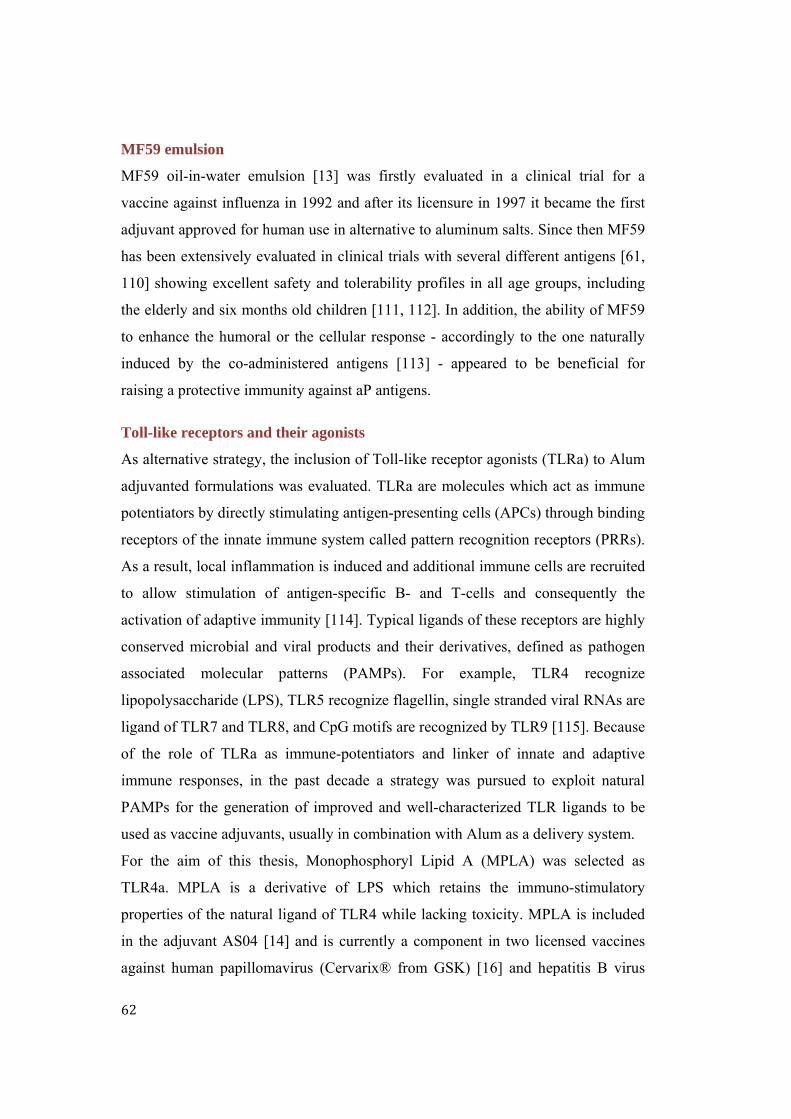
62
MF59 emulsion
MF59 oil-in-water emulsion [13] was firstly evaluated in a clinical trial for a
vaccine against influenza in 1992 and after its licensure in 1997 it became the first
adjuvant approved for human use in alternative to aluminum salts. Since then MF59
has been extensively evaluated in clinical trials with several different antigens [61,
110] showing excellent safety and tolerability profiles in all age groups, including
the elderly and six months old children [111, 112]. In addition, the ability of MF59
to enhance the humoral or the cellular response - accordingly to the one naturally
induced by the co-administered antigens [113] - appeared to be beneficial for
raising a protective immunity against aP antigens.
Toll-like receptors and their agonists
As alternative strategy, the inclusion of Toll-like receptor agonists (TLRa) to Alum
adjuvanted formulations was evaluated. TLRa are molecules which act as immune
potentiators by directly stimulating antigen-presenting cells (APCs) through binding
receptors of the innate immune system called pattern recognition receptors (PRRs).
As a result, local inflammation is induced and additional immune cells are recruited
to allow stimulation of antigen-specific B- and T-cells and consequently the
activation of adaptive immunity [114]. Typical ligands of these receptors are highly
conserved microbial and viral products and their derivatives, defined as pathogen
associated molecular patterns (PAMPs). For example, TLR4 recognize
lipopolysaccharide (LPS), TLR5 recognize flagellin, single stranded viral RNAs are
ligand of TLR7 and TLR8, and CpG motifs are recognized by TLR9 [115]. Because
of the role of TLRa as immune-potentiators and linker of innate and adaptive
immune responses, in the past decade a strategy was pursued to exploit natural
PAMPs for the generation of improved and well-characterized TLR ligands to be
used as vaccine adjuvants, usually in combination with Alum as a delivery system.
For the aim of this thesis, Monophosphoryl Lipid A (MPLA) was selected as
TLR4a. MPLA is a derivative of LPS which retains the immuno-stimulatory
properties of the natural ligand of TLR4 while lacking toxicity. MPLA is included
in the adjuvant AS04 [14] and is currently a component in two licensed vaccines
against human papillomavirus (Cervarix® from GSK) [16] and hepatitis B virus

63
(Fendrix® from GSK) [15]. A synthetic small molecule property of Novartis
Vaccines was instead used as agonist of TLR7 [116]. Despite differences in
expression and intra-cellular signaling, TLR agonists have been evaluated in
numerous preclinical models and are known to increase immunogenicity and
redirect the immune response induced by Alum toward the stimulation of the Th1-
type against a variety of antigens [117].
As mentioned MF59 and Alum+MPLA are the only two novel vaccine adjuvants
approved for human use after the approval of aluminum salts [118]. Therefore these
two adjuvants represent attractive options for quickly developing vaccines to fight
pertussis resurgence, reducing the risk to have the product rejected during clinical
evaluations. The results obtained with these two alternative adjuvants were gathered
in a manuscript entitled “The potential of adjuvants to improve immune responses
against TdaP vaccines: a preliminary evaluation of MF59 and Monophosphoryl
lipid A” which was submitted to the Clinical and Vaccine Immunology (CVI)
journal and is reported in the following pages.
Additional results generated with Alum+TLR7a are presented and discussed in a
supplementary data section.
64
THEPOTENTIALOFADJUVANTSTOIMPROVEIMMUNERESPONSESAGAINSTTdaPVACCINES:APRELIMINARYEVALUATIONOFMF59AND
MONOPHOSPHORYLLIPIDA
Running title: “The potential of adjuvants to improve TdaP vaccines”
Valentina Agnolon 1,2 *, Cristina Bruno 1,3 *, Rosanna Leuzzi 1, Bruno Galletti 1, Ugo D’Oro 1, Mariagrazia Pizza 1, Anja Seubert 1, Derek T. O’Hagan 4, Barbara C. Baudner 1 #
1 Novartis Vaccines Research, 53100 Siena, Via Fiorentina 1, Italy 2 Università degli Studi di Padova, 35129 Padova, Italy 3 Università di Siena, 53100 Siena, Italy 4 Novartis Vaccines Research, 02139 Cambridge MA, USA
*These authors contributed equally to this work
# corresponding author - mail address: [email protected]
Manuscript Submitted to Clinical and Vaccine Immunology.

65
Abstract
The successful approach of combining diphtheria, tetanus and pertussis antigens
into a single vaccine has become the cornerstone of pediatric and adults
immunization programs. Yet, even if vaccination coverage is high, a resurgence of
pertussis has been reported recently in many countries suggesting that current
vaccines may not provide adequate long-lasting protection.
In order to develop pertussis-containing vaccines able to induce a more durable and
better tailored immune response different approaches have been proposed,
including the use of novel adjuvants. Currently licensed pertussis vaccines contain
aluminum salts, which are the most common adjuvants for human use. By inducing
mainly humoral immune responses aluminum salts might not be ideal for providing
protection against pathogens which require a cellular immune response, such as
Bordetella pertussis.
Other adjuvants that induce more balanced T-helper profiles or even Th1-prone
responses might be more adequate. In this study, we tested two different adjuvants:
MF59 emulsion adjuvant – that induces a mixed Th1/2 response - as well as the
combination of a Toll-Like Receptor 4 agonist (TLR4a) with aluminum hydroxide
to induce even further Th1 polarization. The adjuvants were evaluated for their
ability to improve immune responses against a TdaP vaccine containing three B.
pertussis antigens: genetically detoxified pertussis toxin (PT-9K/129G),
filamentous hemagglutinin (FHA) and pertactin (69K).
The quicker onset of serum titers and the changed quality of the antibody responses
induced by the adjuvants evaluated here fully support the potential replacement of
aluminum salts with alternative adjuvants to enhance pertussis immunogenicity in
aP-containing combination vaccines.

66
Introduction
The highly contagious respiratory tract infection caused by the Gram negative
bacterium Bordetella pertussis is still endemic worldwide, despite the fact that
vaccines are available and recommended. The first pertussis vaccines based on
whole, killed bacteria (wP) were developed in the 1930s and were later combined
with diphtheria and tetanus toxoids. The derived DTwP vaccines have been a
cornerstone of childhood immunization since the 1940s [6]. Effectiveness of DTwP
vaccination has been documented in numerous clinical trials, but because of
occurring adverse events the confidence in wP vaccines and vaccination compliance
decreased with declining case numbers [106]. The efforts to develop safer vaccines
have resulted in the replacement of inactivated bacteria with purified protective
antigens of B. pertussis (aP) [7]. These DTaP vaccines have been introduced for
both primary and booster immunizations [95] and depending on the manufacturer
contain one to five Bordetella proteins.
Thanks to the reduced reactogenicity, aP vaccines have totally replaced wP in the
USA and in many developed countries and vaccination coverage is in excess of
95% [93, 94].
However, the number of whooping cough cases started to increase soon after the
introduction of aP vaccines. Attempts have been made to counter this with
additional booster vaccines suitable for adolescents and adults (TdaP) since 2005.
Yet, these just postponed the medium age of pertussis cases from infant to older
children. Moreover total case numbers have still been increasing over the last years
[50-54].
Common hypotheses for the resurgence of pertussis claim that aP vaccines are less
effective than wP vaccines and that aP-induced immunity wanes more quickly [99-
102]. Also changes in the genetic makeup of circulating pertussis strains have been
observed [105, 119]. Another plausible explanation has been suggested by a recent
study showing that aP vaccines protect against severe disease but do not prevent
infection and transmission of B. pertussis in a nonhuman primate model [103].
Therefore little herd immunity should be conferred by aP vaccines on a population
basis, in contrast to wP vaccines that were shown to shorten B. pertussis

67
colonization in the primate model [103]. An immunological explanation for this
finding has been extensively assessed in a mouse aerosol challenge model [120].
Studying the protection from colonization with different vaccine formulations and
in various knockout mice this group could show that protection from colonization
correlates with a Th1/ Th17 immune profile and the induction of antibodies of the
IgG2a subclass. IgG2a antibodies in mice are known for their better opsonizing
capacities, in contrast to IgG1 antibodies. Classical aP vaccines induce mainly Th2
and IgG1 responses and work presumably mostly via toxin-neutralization activity
[107]. Analysis of the pertussis-induced helper T-cell immune response
demonstrated that aP vaccination promotes mostly Th2 responses, whereas wP and
natural infection induce Th1 and Th17 responses [121-123].
In addition, the currently licensed DTaP vaccines are administered with insoluble
aluminum salts, which preferentially induce Th2 response [23]. Based on the crucial
role Th1 response has been found to play in clearing the infection [108, 109] one of
the proposed approaches to enhance the protective efficacy of aP vaccines considers
the inclusion of novel Th1-promoting adjuvants in the formulation [106, 124, 125].
Several studies have shown that alternative adjuvants to aluminum salts, such as
micro and nanoparticles, reduced the bacterial loads in comparison to not-
adjuvanted formulations [126] and induced enhanced and persistent responses in
serum antibody titers against aP antigens [127, 128].
Here, the emulsion MF59 and the combination of the Toll-Like Receptor 4 agonist
Monophosphoryl Lipid A (MPLA) with aluminum hydroxide (Alum), in a
formulation similar to AS04, were evaluated for their ability to adjuvant a TdaP
trivalent vaccine containing the genetically detoxified PT toxin (PT-9K/129G) [60],
filamentous hemagglutinin (FHA) and pertactin (69K) as B. pertussis antigens.
These adjuvants were chosen since they are the only two novel adjuvants already
included in approved vaccines for human use [118] and represent attractive options
for fast development of vaccines to counter pertussis resurgence quickly. MF59 is
an oil-in-water sub-micron emulsion [13, 129] which has been extensively
evaluated in clinical trials with several different antigens including recombinant
proteins [110] and bacterial toxoids [61]. The excellent safety and tolerability MF59

68
has demonstrated even in toddlers, infants, and newborns [111, 112] encouraged its
evaluation in this work whose focus are combination vaccines for children and
adolescents. In addition, MF59 adjuvant is able to enhance the humoral or the
cellular response accordingly to the one naturally induced by the co-administered
antigens, without biasing the immunological profile [113].
As alternative strategy, the inclusion of the MPLA [130] into an Alum-based TdaP
formulation has been exploited. Acting as PAMPs, the TLRa have been shown to be
strong vaccine adjuvants improving the immunogenicity of numerous antigens in
preclinical models [117]. Similar adjuvants are currently licensed for adolescent and
adult populations but the development for infants might be more challenging as
compared to MF59. Yet TLRa in association with Alum, in particular the TLR9a
CpG oligodeoxynucleotide, were found to be highly effective to stimulate Th1 type
immune response against B. pertussis antigens, including PT and FHA and to
enhance protective capacity [120, 123]. Therefore, the combination of Alum and
TLRa might constitute the ideal adjuvant to prime the most adequate responses
against pertussis.
The different TdaP-adjuvanted formulations were characterized and compared in a
mouse model to assess the potential of MF59 and Alum+MPLA respectively to
enhance and better tailor the immune response against TdaP antigens.
Materials and Methods
Antigens and Adjuvants
Tetanus toxoid (TT) and diphtheria toxoid (DT) were obtained from Novartis
Vaccines (NV) manufacturing (Marburg, Germany). Genetically detoxified
pertussis toxin (PT-9K/129G), filamentous hemagglutinin (FHA) and pertactin
(69K) antigens were produced by Lonza Group (Basel, Switzerland).
Aluminum hydroxide (Alum) and MF59 adjuvants were obtained from NV
manufacturing (Marburg, Germany). Toll-Like Receptor 4 agonist (MPLA, 699800)
was commercially obtained by Avanti Polar Lipids, Inc. (Alabaster, AL).

69
Preparation of adjuvanted formulations
TdaP antigens 10 Lf TT, 4 Lf DT, 8 μg PT-9K/129G, 8 μg FHA, and 16 μg 69K in
a volume of 1 ml were adsorbed onto 2 mg/ml Alum suspension [24]. When
required, MPLA powder was resuspended in 0.5% v/v triethanolamine (TEoA) [131]
and added to the formulation [132] at the final dose of 10 μg.
Formulations were adjusted by the addition of 10 mM Histidine buffer and 9 g/l
NaCl to the recommended physiological ranges of pH and isotonicity.
For MF59-adjuvanted formulations the TdaP antigens mixture was diluted 1:1 (v:v)
with MF59 and 10X phosphate buffered saline (PBS) was added to adjust
formulation pH and isotonicity.
Adjuvanted formulations characterization
Antigens identity and integrity was evaluated by Western Blot (WB) analysis.
Briefly, Alum-based formulations were centrifuged (3500 g for 10 min at 25°C) and
the supernatant removed [61]. The pellet was then resuspended in SDS-extraction
buffer, boiled and centrifuged. The resulting supernatant was loaded on NuPAGE®
Novex 4-12% Bis-Tris gels (Life Technologies, Carlsbad, CA). The aqueous phase
of MF59 emulsion was separated via Airfuge® Air-Driven (Beckman Coulter Inc.,
Pasadena, CA; 70000 rpm for 20 min) and loaded for SDS-PAGE. Gels were run
for 1 hour at 150 V and then blotted to a nitrocellulose membrane via iBlot® (Life
Technologies, Carlsbad, CA). Membranes were blocked for 1 hour in 5% w/v
milk/TPBS (PBS 1X + Tween20 0.05% v/v) and later incubated with optimized
dilutions of monovalent polyclonal mouse anti-sera generated at NV. Horseradish
peroxidase-conjugated goat anti-mouse IgG (Southern Biotech, Birmingham, AL)
was used as secondary antibody (1:5000). After 1-hour incubation at room
temperature, membranes were washed and Opti-4CN™ Substrate Kit (Bio-Rad,
Hercules, CA) was used for bands revelation.
Antigens adsorption onto Alum was evaluated via micro BCA analysis [133] on
formulation supernatants. A 5-points standard curve was prepared and a formulation
containing TdaP antigens and the components of the vaccine, except for the
adjuvants, was included as positive control.

70
The size distribution of Alum adjuvanted formulations and MF59 emulsion was
measured by laser light scattering on the LS 13 320 SW (Beckman Coulter Inc,
Pasadena, CA) and on the Zetasizer Nano ZS (Malvern Instruments Ltd., Malvern,
UK) instruments respectively. The zeta potential of the formulations was measured
using the Zetasizer Nano ZS after sample dilution in water.
Animal experiments
Animal studies were performed in compliance with the Italian law, approved by the
local Animal Ethics Committee, and authorized by the Italian Ministry of Health.
Two similar mouse studies with TdaP antigens formulated with Alum,
Alum+TLR4a or MF59 respectively were carried out one month a part from each
other. Groups of 8 female, 6-week-old, specific-pathogen-free BALB/c mice
(Charles River Laboratories International Inc., Wilmington, MA) were used for the
studies. Each animal received two intramuscular (i.m.) immunizations at days 1 and
21. Injections were performed in quadriceps of hind legs for a total dose volume of
100 μl (50 μl/leg).
In all studies control groups with non-immunized mice were included. Animals
were bled two weeks after each immunization and serum samples were analyzed by
Luminex immunoassay for circulating antibodies titers. Toxin neutralizing antibody
titers, inhibition of FHA binding activity and IgG subclasses were analyzed after the
second bleeding.
Specific IgG measurement using protein-coupled microspheres
A Luminex penta-plex immunoassay was developed to determine antibody titers in
mouse sera. In detail, 20 μg of each antigen were coupled to the carboxyl groups of
1.25 million MagPlex microspheres (Luminex Corporation, Austin, TX) following
manufacturer's instructions.
IgG titers were determined in individual animals after 30 min incubation of antigen-
coupled beads with the sera diluted in PBS 1X 1:10000 (post-1) or 1:20000 (post-2)
into 96-well Multiscreen HTS filter plates (Merck Millipore, Darmstadt, Germany).
A phycoerythrin-conjugated secondary antibody was then used for detection
(Jackson ImmunoResearch Laboratories Inc., West Grove, PA; 1:400; 15 min). The

71
obtained immune-complexes were resuspended in PBS 1X and analyzed on the
Luminex system.
For IgG subclasses analysis a 30 min incubation step with biotin-labeled antibodies
specific for IgG1 or IgG2a subclasses (Jackson ImmunoResearch Laboratories Inc.,
West Grove, PA; 1:200) was performed after the first reaction between beads and
diluted sera (1:20000 post-2). Following the incubation with R-Phycoeritrin
conjugated to extravidin (Sigma-Aldrich, St. Louis, MO; 1:200; 15 min) the
obtained immune-complexes were resuspended in PBS 1X and analyzed on the
Luminex system.
All reaction steps were carried out in agitation motion using a horizontal shaker in
the dark at room temperature. Among each incubation step, plates are washed three
times in PBS 1X by a multiscreen vacuum manifold and a Chemical Duty Pump
(Merck Millipore, Darmstadt, Germany).
IgG measurements were determined as median fluorescence intensities (MFI) on a
Luminex FLEXMAP 3D analyzer (Luminex Corporation, Austin, TX) using the
Bio-Plex Manager 5.0 software (Bio-Rad, Hercules, CA). A hyper-immune
antiserum specific for each antigen was used as reference to convert MFI values in
RLU/ml (Relative Luminex Units) for total IgG quantification. The limit of
quantification (LOQ) of the assay was determined for each antigen and was
considered as threshold for positive results.
DT neutralization assay
Vero cell line (monkey kidney epithelial cells) were obtained from American Type
Culture Collection (ATCC, Rockville, MD) and maintained in Eagle's Minimum
Essential Medium (EMEM, Sigma-Aldrich, St. Louis, MO) supplemented with 10%
heat-inactivated fetal bovine serum (FBS, Gibco, Life Technologies, Carlsbad, CA)
and antibiotics. Cells were grown at 37°C with 5% CO2.
The DT-induced cell death was tested by the CellTiter-Glo Luminescent Cell
Viability Assay (Promega, Madison, WI), based on the measurement of cytoplasmic
ATP in metabolically active cells. To test the minimal dose of DT needed to cause
cell death in 16 hours (1 CTU100), cells were plated at 2x10⁴/well in 96-well plates

72
in presence of serially diluted DT (Sigma-Aldrich, St. Louis, MO). The CTU100 was
defined as 10 ng/ml in the tested experimental conditions.
Post-2 mouse sera were two-fold serially diluted in cell medium, pre-incubated with
4 CTU100 of DT toxin and then incubated for 90 min at 37°C. Subsequently, 50 µl
of Vero cells at density of 2x10⁵cells/ml were incubated with 50 µl of sera and
toxin mixtures for 16 hours at 37°C. Endpoint titers were defined as the reciprocal
of the highest dilution able to inhibit cell death. Pre-immune sera were used as
negative controls.
PT neutralization assay
CHO-K1 cell line (Chinese hamster ovary cells) were obtained from American
Type Culture Collection (ATCC, Rockville, MD) and maintained in F12-K medium
supplemented with 10% heat-inactivated fetal bovine serum (FBS, Gibco, Life
Technologies, Carlsbad, CA) and antibiotics.
In vitro neutralization assay was optimized following the principles described
previously [134, 135]. To test the minimal dose of PT needed to cause 100% cell
clustering in 16 hours (1 CTU100), cells were plated at 2x10⁴/well in 96-well plates
in the presence of serially diluted PT (Sigma-Aldrich, St. Louis, MO). The CTU100
was defined as 8 ng/ml in the tested experimental conditions.
Post-2 mouse sera were diluted and incubated with PT as described for DT
neutralization assay. Subsequently, 50 µl of CHO-K1 cells at density of
4x10⁵cells/ml were incubated with 50 µl of sera and toxin mixtures for 16 hours at
37°C. Cells were observed by light microscopy to evaluate morphological
alterations (clustered phenotype). Endpoint titers were defined as the reciprocal of
the highest dilution able to inhibit cell clustering. Pre-immune sera were used as
negative controls. Sera of mice immunized with commercially available TdaP
vaccine (Boostrix®, antigens doses 10 Lf TT, 5 Lf DT, 16 μg PT, 16 μg FHA, and
5 μg 69K in a volume of 1 ml) were included in the assay to evaluate the
neutralizing effect induced by a chemically detoxified PT toxoid (PTd) included in
Boostrix®.

73
FHA binding inhibition assay
A549 cells (Human lung epithelium) were obtained from American Type Culture
Collection (ATCC, Rockville, MD) and maintained in F12-K medium
supplemented with 10% heat-inactivated fetal bovine serum (FBS, Gibco, Life
Technologies, Carlsbad, CA) and antibiotics. Cells were seeded on 96-well plate
(2.5x10⁴/ well) and cultured for one day. To perform the serum-mediated inhibition
assay, pooled post-2 mouse sera were two-fold serially diluted in F12-K medium
containing 1% naïve mouse serum and pre-incubated with 2.5 µg/ml FHA for 1
hour at 37°C. The mixtures were then added to cells and incubated for 30 min at
37°C. After extensive washing to remove unbound protein, cells were fixed with
3.7% paraformaldehyde and incubated with sheep anti-FHA serum (1:500) (NIBSC,
National Institute for Biological Standards and Control) for 1 hour at room
temperature. After washings, samples were incubated with Alexa Fluor 488 donkey
anti-sheep IgG (1:500) (Life Technologies, Carlsbad, CA) and fluorescence
associated to cell surface was measured at excitation/emission 485/535 nm by
Tecan Infinite F200PRO microplate reader.
Statistical analysis
Graph Pad Prism 5.0 software was used for statistical analysis. Serum antibody
titers were reported as median while neutralizing titers were reported as mean and
significant differences (p ≤ 0.05) among groups were ascertained using the two-
tailed Mann-Whitney test at the 95% confidence interval.
Results
Adjuvanted formulations characterization
Vaccines were prepared and characterized to guarantee the quality of the final
formulations as specified in Materials and Methods. The average particle size of
Alum suspension was determined to be around 10 µm before and after TdaP
antigens adsorption (Table VII). When adding MPLA to the vaccine Alum particles
size doubled and an evident broadening of size distribution was observed.
Significant reduction of the zeta potential was registered during adsorption of

74
antigens and MPLA onto Alum suspension. MF59 emulsion showed comparable
particles size, polydispersity and zeta potential at every stage of the formulation
process (Table VII). TdaP antigens identity and integrity were confirmed by
Western Blot analysis in Alum as well as in MF59-based formulations.
Characterization of freshly prepared vaccines gave comparable results to vaccines
stored up to three months at 4°C (data not shown). To discriminate between Alum-
adsorbed and not-adsorbed antigens, adjuvant was separated by centrifugation. The
micro BCA performed on the supernatants showed that the adsorption of TdaP
antigens onto Alum was higher than 95% and that the inclusion of MPLA in the
formulation did not alter antigens adsorption rate (Table VIII).
Adjuvant TdaP antigens Particle size
(µm± SD)
Zeta potential
(mV ± SD)
Antigen
adsorption (%)
Alum
- 10.8 ± 0.17 25.6 ± 0.87 n.a.
+ 11.1 ± 0.23 9.50 ± 0.61 > 95%
Alum+MPLA + 21.6 ± 0.75 -2.50 ± 0.81 > 95%
MF59
- 0.15 ± 0.30 -39.0 ± 0.40 n.a.
+ 0.15 ± 1.00 -38.1 ± 0.81 n.a.
In vivo adjuvant effect
To assess in vivo potency of the formulations, groups of Balb/c mice were
vaccinated twice (day 0, 21) and serum antibody titers were measured two weeks
after each immunization. The analysis of total serum IgG indicated that adjuvants
were needed to elicit potent immune responses (Figure 22). When antigens were
formulated in absence of any adjuvant, no response was detected after one
immunization against TT, FHA and 69K, or in the case of DT neither after the
second immunization. The only exception was constituted by PT, which was readily
detected even without any adjuvant after the first immunization. Yet, this might be
due to the specific properties of the genetically inactivated PT toxin, which is
TABLE VIII. Physical characterization of various vaccine formulations. N.a. not applicable.

75
FIGURE 23. Serum (a) anti-TT, (b) anti-DT, (c) anti-FHA, (d) anti-69K, (e) anti-PT antibody titers in Balb/C mice after one and two immunizations at days 1 and 21. Averaged results of the two mouse studies are shown as median with interquartile range presented for each group. • serum IgG titers below 1 RLU/ml; LLOQ ≤ 0.9 RLU/ml; * p < 0.05, ** p < 0.01, *** p < 0.001

76
known to be more immunogenic as compared to its chemically detoxified
counterpart included in commercially available aP vaccines [60, 136]. Further
explanation could be associated with the intrinsic adjuvant activity that the
genetically detoxified PT was shown to harbor [137, 138]. Serum IgG analysis
revealed that two immunizations with Alum-adjuvanted formulations were required
to induce detectable IgG titers against DT and FHA antigens.
Similar results were also observed when including MF59 adjuvant in the vaccine.
For the other antigens (TT, PT, 69K) MF59 was instead shown to be superior to
Alum after a single immunization and the trend was confirmed after the second
immunization. Serological analysis performed on the first bleeding for DT and FHA
antigens indicated Alum+MPLA as the only adjuvant able to raise detectable
responses. Overall, a single immunization with Alum+MPLA adjuvant induced IgG
titers higher than the ones induced by Alum. As previously stated, PT-9K/129G was
the only antigen to be immunogenic after a single immunization without any
adjuvantsand resulted as effective as Alum. On the contrary, the alternative
adjuvants tested were shown to have comparable adjuvant activity while raising
significantly higher IgG responses in comparison to Alum. For the other TdaP
antigens Alum+MPLA adjuvanticity was superior to MF59. As a whole, post-1 data
depicted Alum+MPLA as the most potent adjuvant inducing quicker onset of the
immune response and higher antibody titers against TdaP antigens, while MF59
performed at least as effectively as Alum.
After the second dose Alum+MPLA confirmed its stronger adjuvanticity as
compared to Alum. In addition, Alum+MLPA was also more effective than MF59
for DT antigen confirming Alum as a successful adjuvant for this bacterial toxoid.
On the contrary, formulations adjuvanted with Alum+MLPA or MF59 resulted in
comparable responses against PT and FHA and MF59 was shown to be the most
effective adjuvant for TT and 69K antigens.
On the basis of the serological responses at both vaccine doses, both the approaches
investigated could be considered as promising alternatives to Alum in TdaP
vaccines.

77
DT and PT toxin neutralizing activity
To evaluate the ability of the induced antibodies to recognize antigenic functional
epitopes, different toxin neutralization assays were performed after the second
bleeding. The neutralization assay against DT indicated that Alum+MPLA was able
to stimulate significantly higher neutralizing titers in comparison to Alum (Figure
24a).
In contrast, pertussis toxin neutralizing assay showed that the use of adjuvants in
the formulation did not lead to significant enhancement of the neutralizing activity
of anti-PT antibodies (Figure 24b).
As previously noticed for total serum antibody titers, this might be explained by the
strong immunogenic properties of this antigen. In fact, evident differences in
FIGURE 24. Post-2 in vitro neutralization titers (a) anti-DT and (b) anti-PT. (c) comparison of the neutralizing activity of genetically detoxified PT-9K/129G and chemically detoxified PT (PTd) included in Boostrix vaccine. Mean with standard deviation presented for each group. • titers below detection limit. * p <0.05, *** p <0.001

78
neutralizing activity were observed when comparing TdaP formulations containing
the genetically detoxified PT to Boostrix® vaccine in which a chemically detoxified
PT (PTd) is used (Figure 24c). Notably, PT-9K/129G induced significantly higher
neutralizing titers despite being formulated at half of the dose of PTd, further
supporting the potential of PT-9K/129G as candidate antigen to develop improved
vaccines against pertussis.
FHA binding inhibition
FHA is an important adhesion factor of Bordetella pertussis and the inhibition of its
binding to ciliary epithelial cells is of significant relevance to prevent pathogen
adherence to the host respiratory tract. Achieving higher adhesion blocking
antibodies should therefore contribute to greater protection from bacterial
colonization and so from contracting the disease [139].
The adjuvant effect on the induction of antibodies able to block FHA binding
activity was evaluated on pooled sera after the second immunization and indicated
that Alum+MPLA and MF59 equally improved the inhibition activity of anti-FHA
antibodies in comparison to Alum (Figure 25).
Shift in the IgG1/ IgG2a profile
Alternative adjuvants to Alum were investigated for their ability to induce a more
Th1 oriented immune response against aP antigens using TdaP as a model vaccine.
FIGURE 25. FHA binding activity of pooled post-2 sera of mice immunized with different formulations. Mean with standard deviation presented for each group. FI: fluorescence intensity at excitation/emission 485/535 nm.

79
FIGURE 26. Median fluorescence intensities (MFI) relative to specific IgG1 and IgG2a responses (a) anti-TT, (b) anti-DT, (c) anti-FHA, (d) anti-69K, (e) anti-PT in Balb/C mice after two immunizations. Median with interquartile range presented for each group. • values below 1 MFI.
(b)
(d)
(a)
(e)
(c)

80
In the mouse model, a Th2 response is correlated to IgG1 antibodies, while Th1
responses are characterized by the subclass IgG2a, b or c (depending on the
individual mouse strain). Classical aP vaccines induce mainly Th2 and IgG1
responses and work presumably mostly via toxin-neutralization activity [107]. In
contrast, IgG2a, b or c antibodies are known for their better opsonizing capacities
and should account, together with the phagocyte-activating-cytokine-secreting Th1
cells, for the improved bacterial clearance shown in the mouse model [120, 140].
To assess a potential shift in the IgG1/IgG2a balance and hence quality of the
immune response, antibody subclasses were analyzed after the second
immunization. As expected Alum induces a more Th2-prone response with reduced
IgG2a and enhanced IgG1 titers in comparison to the not-adjuvanted formulation,
as can be readily observed for FHA and PT. For the other antigens the overlaying
adjuvant effect of Alum, enhancing all titers, make a comparison of a potential shift
less assessable. Importantly a shift to a more IgG2a/Th1 prone profile was observed
as compared to Alum when combining Alum and MPLA or using MF59 emulsion.
Both adjuvants clearly enhanced IgG2a titers against the five TdaP antigens and, as
expected, MF59 was shown to maintain the natural immunological profile of the
antigens (Figure 26). On the contrary, Alum adjuvant completely reset the intrinsic
Th1 inducing effect showed by FHA and PT antigens. The ability of MPLA to
redirect the immune response against these antigens appeared evident through the
reversion of the quality of the immune response when the immune-potentiator was
combined with Alum.
Discussion
Despite a generally high vaccination coverage there is a recent resurgence of
pertussis in many countries worldwide. Several hypotheses have been formulated
but most experts agree that waning immunity and inadequate immune profiles
induced by acellular vaccines are to blame, next to changes in the genetic makeup
of circulating pertussis strains. Induction of a Th1 profile – as induced by wP
vaccines or actual infection - is seen as beneficial to protect from bacterial

81
colonization and to enhance herd immunity. In contrast, currently available
acellular vaccines mainly induce a Th2-prone response.
Future pertussis vaccines should have a safety profile similar to current acellular
pertussis-containing vaccines, but ideally induce longer lasting immunity and more
adequate T-helper response profiles. Therefore in this study the potential of
alternative adjuvants to aluminum salts to enhance the efficacy of aP vaccines was
assessed, and two different approaches that have already been successfully used in
human vaccinations were chosen.
On the basis of the well-established safety of MF59 emulsion [141] and of the
strong immunological stimulation induced by TLRa [142], TdaP formulations
adjuvanted with MF59 or with the TLR4 agonist Monophosphoryl lipid A (MPLA)
in combination with Alum have been studied. The possibility to develop stable and
defined formulations has been evaluated through the physical characterization of
TdaP formulations and the investigation of their in vivo immunogenicity and
individual immune profile.
With respect to quantity, quality and functionality of the induced immune responses
both alternative approaches created a promising scenario for the development of
next generation aP containing vaccines.
The adjuvant effect of MF59 resulted in immune responses against TdaP antigens
higher or at least comparable to Alum. This finding, along with the demonstrated
enhancement of antibody responses against other antigens routinely included in the
pediatric immunization schedules, such as those from Haemophilus influenzae type
b (34) and hepatitis B virus (35), show the strong potential of MF59-adjuvanted
TdaP vaccines to be readily further developed to improve current infant-pertussis
combination vaccines. As revealed from the unchanged results of size distribution
and zeta potential pre- and post-formulation, TdaP antigens remained mostly free in
the aqueous phase of MF59 emulsion. Despite this condition could possibly induce
instability processes [34], no modification in the antigenic electrophoretic profiles
were detected suggesting TdaP antigens are compatible with MF59 adjuvant.
Antigens adsorption on adjuvant surface is instead one of the main feature of
formulations based on aluminum salts and it is considered a very important

82
parameter for their function [26, 32], such that WHO recommended adsorption
values equal or greater than 80% for diphtheria and tetanus toxoids [84]. The
reduction of the zeta potential registered during formulation procedure confirmed
TdaP antigens are adsorbed onto Alum adjuvant, condition known to ensure
antigens stability in liquid vaccine formulations. Further proof came from micro
BCA analysis, which measured in supernatants of formulation less than 5% of the
total antigenic amount formulated, thus indicating only a minor part of antigens
remained not-adsorbed to the adjuvant. In addition, TdaP antigens adsorption rate
onto Alum was not modified when including MPLA in the formulation. Aluminum
salts have been previously shown to induce enhanced and better modulated immune
responses when combined with TLR agonists through the co-administration of both
antigens and agonists to the same antigen presenting cell [14, 143, 144]. In addition
Geurtsen et al. [145] demonstrated that aP vaccine adjuvanted only with MPLA
were more protective than aP vaccines containing Alum. Yet, safety concerns have
been raised for formulations where MPLA is not adsorbed to a delivery system like
Alum or nanoparticles. Moreover, several other studies demonstrated that MPLA in
combination with Alum adjuvant resulted in more potent immune responses and
recommended MPLA adsorption onto Alum surface to prevent potential systemic
toxicity [22]. In this context, the adjuvanticity of Alum in combination with the
immune-potentiator MPLA for a TdaP trivalent vaccine has been exploited. Beyond
the overall higher serum titers raised by the inclusion of MPLA in the formulation
in comparison to Alum, Alum+MPLA was also shown to accelerate the onset of the
immune responses suggesting that the addition of a TLR agonist to Alum-based
formulations could potentially reduce the number of injections required to ensure aP
vaccine immunogenicity.
A crucial parameter to develop a highly effective vaccine against pertussis is to
induce high antibody titers against PT, one of the major virulence factors of B.
pertussis. In this perspective, the inclusion of PT 9K/129G in future pertussis
combination vaccines appears promising also considering that the genetically
modified toxin has already been shown to retain higher immunogenicity than the
chemically detoxified PT (PTd) included in the currently licensed vaccines [60,

83
146]. Furthermore, PT 9K/129G has been identified as ideal vaccine candidate since
it cannot revert to toxicity while conserving the native B and T cell epitopes which
may be lost during a chemical detoxification [147] and has been reported to have an
intrinsic adjuvant activity in mice [137, 138]. The adjuvant effect for PT 9K/129G
was less evident in comparison to what was observed for the other antigens of the
combination, however significant improvement in terms of total IgG antibody titers
was detected in presence of MPLA. Dose titration studies of PT antigen are planned
to provide more detailed insights on the direct effect of TLRa and MF59 adjuvants
on its immunogenicity.
To assess if the alternative adjuvants tested in the current work were effective in
inducing Th1 response, the induction of opsonizing IgG2a was evaluated and taken
as indirect evidence of enhanced cell mediated responses. In literature those
responses are associated with an efficient and rapid clearance of B. pertussis [120]
and with the superior long term protection of wP vaccination [148]. As expected,
Alum+MPLA strongly potentiated the Th1 immune response leading to increased
IgG2a antibody titers. Interestingly, an appreciable enhancement of IgG2a titers
against TT and B. pertussis surface adhesins was also observed with MF59
emulsion thus confirming the peculiarity of this adjuvant to strengthen the immune
responses naturally induced by the co-administered antigens [113]. The observed
stimulation of Th1-type immune response against B. pertussis surface adhesins is
encouraging for the improvement of aP-containing vaccines efficacy since these
antigens are reported amongst the main mediators of pathogen colonization and
transmission [103].
Considering that both Alum+MPLA and MF59 adjuvants have been proven to be
valuable approaches in the mouse model, it would be interesting to further
investigate if the adjuvant effect on pathogen colonization and immune response
persistence could be answered in the baboon model which has emerged as an
appropriate animal model to reproduce all aspects of pertussis clinical disease [103].
Overall, the high antibody titers against PT and the promotion of a Th1-oriented
response against B. pertussis surface adhesins obtained with the alternative
formulations developed are initial encouraging data fully supporting the potential of

84
adjuvants to raise better tailored immunological responses against aP-containing
combination vaccines and to effectively control pertussis spread and overcome
waning immunity issues. The critical adjuvant selection relying on adjuvants
already licensed in vaccines for infants and toddlers, such as MF59, or on the
addition of a TLR agonist to existing formulations, emerged as a simpler and faster
approach for better vaccine development than the identification of entirely new
vaccine candidates.
Acknowledgments
The authors are grateful to Marco Tortoli and the animal facility personnel for the
precious help with the in vivo studies.
Contributions
V.A. actively contributed to perform IgG measurement experiments through the
development of the Luminex penta-plex immunoassay. V.A. and C.B. analyzed and
interpreted the results and drafted the manuscript.

85
SUPPLEMENTARYDATA
EVALUATIONOFASYNTHETICAGONISTOFTOLL‐LIKERECEPTOR7
Pursuing Novartis Vaccines’ strategy of using SMIP (small molecule immune-
potentiators) as agonist to activate immune cells through the targeting of Toll-like
Receptor 7 (TLR7), a synthetically obtained TLR7 agonist (TLR7a) was evaluated
for its adjuvant effect when included in Alum adjuvanted vaccine formulations. A
soluble agonist was designed and functionalized with PEG linker and a terminal
phosphonate group to allow adsorption to aluminum hydroxide via ligand exchange
with the aim of limiting SMIP’s systemic exposure while ensuring co-localization
of antigens and adjuvants and favoring retention time in the muscle for interaction
with immune cells [116].
Materials and Methods
Only deviations from the procedures described in the previously reported
manuscript are included in this section.
Antigens and Adjuvants
Toll-Like Receptor 7 agonist was internally synthetized by the Genomic Institute of
the Novartis Research Foundation (GNF).
Preparation of adjuvanted formulations
TdaP antigens (10 Lf TT, 4 Lf DT, 8 μg PT, 8 μg FHA, and 16 μg 69K) were
adsorbed onto 2 mg/ml Alum suspension [24] to a final volume of 1 ml. When
required, TLR7a was added to the formulations at 50 μg per dose. Animals were
immunized intramuscularly with a total volume of 100 μl (50 μl/leg).
TLR7a identity and degree of adsorption onto Alum adjuvant
TLR7a identity and adsorption rate onto Alum were assessed via reverse phase
ultra-performance liquid chromatography (RP-UPLC) analysis. Briefly, after
spinning down the vaccine, the supernatant was removed and aliquots of 7.5 µl
were injected in the UPLC system. The pellet was resuspended in 500mM

86
potassium phosphate buffer (P3786 Sigma Aldrich) pH 9.0 and incubated at 95°C
for 10 min. After Alum separation via centrifugation, the resulting supernatant was
appropriately diluted and 7.5 µl were used for UPLC analysis. TLR7a separation
was achieved by using a 50mm x 2.1mm, 1.7µm particle size, Acquity UPLC®
BEH-C18 column (Waters). The mobile phase was a 10 to 90% acetonitrile gradient
prepared in water added of 0.1% trifluoroacetic acid (TFA) at a flow rate of 1.2
mL/min. 268 nm UV was used for TLR7a detection. Detection and quantification
limits of the method were found to be 0.39 µg/ml and 0.78 µg/ml respectively.
Results and Discussion
Similarly to what observed with Alum+MPLA and MF59 adjuvants, Alum+TLR7a
proved to be a valuable approach for improving pertussis immunogenicity in the
mouse model, too. Antigens adsorption onto Alum was estimated to be higher than
90% by WB analysis: none of TdaP antigens was in fact detectable in the
supernatants of Alum or Alum+TLR7a formulations even after protein
concentration via TCA (data not shown). Overall the inclusion of TLR agonists in
Alum formulations was shown not to modify TdaP-antigens adsorption profile.
Adsorption rate of TLR7a onto Alum was calculated by using a five-point
calibration curve and was found to be higher than 95%. TLR7a identity and
integrity was confirmed after desorption from Alum. In addition, RP-UPLC
analysis of the desorbed TLR7a allowed a recovery higher than 95% of the agonist
(data not shown). The agonist was always tested in combination with Alum
adjuvant to prevent systemic adverse events. Moreover, previous studies showed
that SMIP alone was inefficient in the induction of protective antibodies against
tested antigens [116].
In vivo adjuvant effect
To assess in vivo potency of Alum+TLR7a, groups of Balb/c mice were vaccinated
twice (day 0, 21) and serum antibody titers were measured before and two weeks
after each immunization. Observations made for Alum+MPLA resulted to be valid
also for Alum+TLR7a and serological analysis for DT and FHA antigens performed

87
FIGURE 27. Serum (a) anti-TT, (b) anti-DT, (c) anti-FHA, (d) anti-69K, (e) anti-PT antibody titers in Balb/C mice after one and two immunizations at days 1 and 21. (f) anti-PT antibody titers in Balb/C mice immunized at days 1 and 28. Results are shown as median with interquartile range presented for each group. • serum IgG titers below 1 RLU/ml; LLOQ ≤ 0.9 RLU/ml; * p < 0.05, ** p < 0.01, *** p < 0.001

88
on serum samples taken after the first immunization revealed that the combination
of Alum with the agonist was the only strategy able to induce detectable antibody
responses. Overall, a single immunization with Alum+TLR7a adjuvant raised IgG
titers higher than the ones induced by Alum. The immune potentiator role of the
TLR7 agonist was particularly evident for aP surface adhesins leading to titers
comparable to those raised by two immunizations with the non-adjuvanted antigens.
After the second dose Alum+TLR7a confirmed its stronger adjuvanticity in
comparison to Alum for all the tested antigens with the exception of PT. The
observation about the less pronounced adjuvant effect for PT was already described
in the manuscript above and justified by the strong immunogenic properties of this
antigen. Follow-up studies performed with Alum+TLR7a adjuvant increasing the
number of immunized animals and extending the injection schedule from day 0, 21
to day 0, 28 made the adjuvant effect more evident and significantly higher than the
on induced by Alum (Figure 27).
Neutralizing activity
To evaluate the ability of the induced antibodies to recognize functional epitopes of
the antigens, toxin neutralization assays were performed after the second bleeding.
Again, the combination of TLR7 agonist with Alum adjuvant stimulated
significantly higher neutralizing titers against DT in comparison to Alum, while not
providing an enhancement of neutralizing activity against PT. As previously noticed
for total serum antibody titers, increasing the number of immunized animals and the
interval between the two injections allowed the observation of an adjuvant effect for
PT antigen (data not shown).
Shift in the IgG1/ IgG2a profile
Alum+TLR7a was investigated as alternative adjuvant to Alum for its ability to
induce a more Th1 oriented immune response against aP antigens in TdaP vaccine.
Classical Alum adjuvanted aP vaccines induce mainly Th2 responses with
antibodies of the IgG1 subclass and work presumably mostly via toxin-
neutralization activity [107]. In contrast, Th1 responses are characterized by the
subclass IgG2a, b or c (depending on the individual mouse strain) which are known
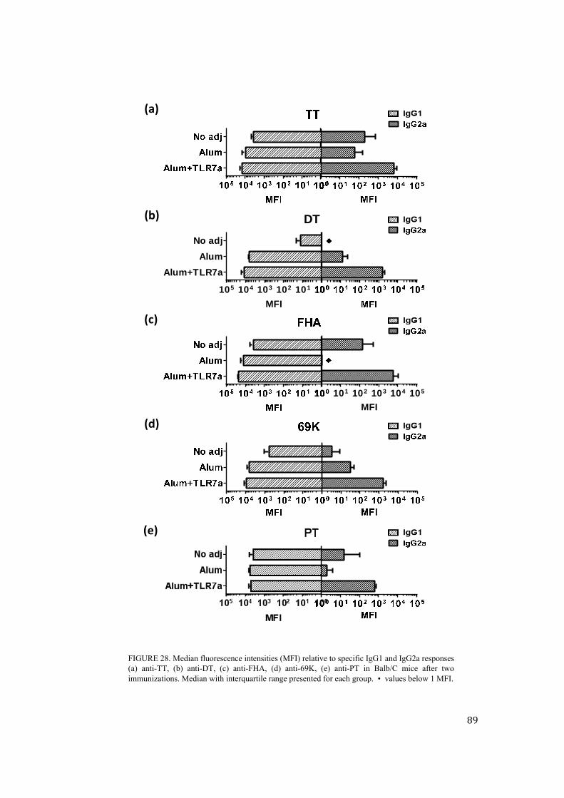
89
FIGURE 28. Median fluorescence intensities (MFI) relative to specific IgG1 and IgG2a responses (a) anti-TT, (b) anti-DT, (c) anti-FHA, (d) anti-69K, (e) anti-PT in Balb/C mice after two immunizations. Median with interquartile range presented for each group. • values below 1 MFI.
100 101 102 103 104 105
MFI
100101102103104105
Alum+TLR7a
Alum
No adj
MFI
DT(b)
(d)
(a)
(e)
(c)

90
for their better opsonizing capacities and should improve bacterial clearance [120,
140]. To assess a potential shift in the IgG1/IgG2a balance and hence quality of
serum antibody responses, subclasses were analyzed after the second immunization.
The immune-potentiator role of TLR4a in inducing a shift to a more protective Th1
(IgG2a) profile was observed also for TLR7a when combined to Alum (Figure 28).
As already stated, an enhancement of the Th1-type immune response is associated
with a more efficient clearance of B. pertussis and with an extended protection like
the one induced by wP vaccines or natural infection [120, 148].
In addition, preliminary evaluation of the stimulation of memory B cells was
performed. However despite a visible trend indicating the immune-stimulatory
effect of TLR7a the assay needs to be further improved – data not shown.
Dose-titration and antibody duration studies
Alum+TLR7a was selected as internal strategy for the development of improved
acellular pertussis combination vaccines thus being used as main alternative
adjuvant in follow-up studies. Immunization schemes were arranged vaccinating
Balb/c mice twice (day 0, 28) with different doses of TdaP antigens and of TLR7
agonist in combination with Alum. Dose-titration studies revealed that similar
results could be obtained using ten times less antigen doses with respect to the
doses used in the previous studies (see paragraph Preparation of adjuvanted
formulations). Also the dose of the immune-potentiator could be reduced to 10
µg/injection while maintaining comparable results. Importantly, together with an
enhancement in quantity, total antibody titers as well as IgG2a-specific antibodies
were found to persist up to ten months after the last immunization (data not shown).
Evaluation of larger TdaP-based combination vaccines
The feasibility to develop larger TdaP-based combination vaccines was evaluated
through the co-administration of TdaP antigens together with three recombinant
antigens conferring protection against Neisseria meninigitidis type B. Analysis of in
vivo immune responses conveyed that the presence of additional antigens in the
vaccine combination had no negative interference on the quantity of antibody titers
raised against TdaP antigens (Figure 29).

91
Conclusions
Results obtained using Alum+TLR7a as improved adjuvants for TdaP vaccine
provided encouraging prospective of raising enhanced and better tailored
immunological responses against aP-containing combination vaccines. TLR7a
molecule is chemically and structurally defined and was specifically designed to be
recognized by specific receptors thus stimulating the immune response. In
comparison to MPLA, the synthetically obtained molecule TLR7a has the
advantage of being structurally simpler thus potentially leading to more
reproducible results, both in terms of vaccine formulations and induced immune
responses.
FIGURE 29. Serum anti-FHA and anti-69K antibody titers are taken as representative of all the five TdaP antigens. Results were obtained in Balb/C mice after two immunizations with TdaP or TdaP+MenB antigens. Median with interquartile range is shown for each group.

92

93
CHAPTER IV . CONCLUSIONS
The objective of this PhD work was to investigate the possibility of improving
quality and immunogenicity of current acellular pertussis combination vaccines,
which are combined with diphtheria and tetanus toxoids in the keystone of
combination vaccines. Vaccines and combination vaccines in particular, are
complex multi-component products whose efficacy relies on the concomitant
inclusion of antigens, adjuvants, and excipients. Currently licensed aP vaccines are
typically adjuvanted with aluminum salts, known to induce mainly a humoral
immune response which has been associated with the recent pertussis outbreaks
reported in many countries. Therefore, existing vaccines are perceived as not ideal
to provide protection against the disease. A formulation science approach was here
proposed and developed to investigate the potential improvement of acellular
pertussis vaccines by working on the adjuvant component. In particular, the
promotion of the vaccine analytical characterization and the rational polarization of
the quality of immune responses were suggested as effective strategies to be
pursued for the development of improved combination vaccines.

94
Improved in vitro characterization assays for combination vaccines adjuvanted
with aluminum hydroxide
In Chapter II significant progresses for the in vitro characterization of complex
aluminum salts-adjuvanted vaccines were introduced. In particular, the possible use
of Luminex technology for vaccine characterization purposes was exploited and
developed in a multi-plex microsphere-based immunoassay for simultaneous
analysis of aP antigens in TdaP vaccines. In a single experiment, the proposed assay
provided - information on antigen identity, quantity, and degree of adsorption to
adjuvant surface of each immunogen included in a multivalent combination. The
simultaneous characterization of the three aP antigens was performed directly on
the whole TdaP vaccine without any preliminary treatment of the adjuvant or
laborious procedures for adjuvant separation, as commonly required for available
analytical methods. The assay relied on antibody recognition and fluorescence
detection, thus demonstrating specificity and providing a substantial improvement
in terms of sensitivity as compared with other commonly used immunoassays.
Importantly, the high sensitivity of the Luminex assay allowed precise
quantification of low amounts of antigens not-adsorbed to adjuvant surface.
Polyclonal antibodies were used as proof of concept in this work. Their replacement
with appropriately selected monoclonal antibodies could allow the qualitative
analysis of antigenic epitopes critical for vaccine immunogenicity. In this
perspective, the possibility to correlate quality of antigenic components with the
efficacy of the product could lead to the set-up of an in vitro potency assays
accordingly to the 3Rs concept. Moreover, the Luminex-based approach has the
potential to be implemented for larger vaccine combinations and to be applied in
presence of other adjuvants and delivery systems, such as emulsions and polymeric
particles.
In vivo evaluation of novel adjuvants for improved aP-containing combination
vaccines
Chapter III was dedicated to study aP vaccine efficacy and the possibility of
tailoring immune responses by the modulation of the vaccine formulation. Despite
generally high vaccination coverage a pertussis outbreaks are registered in many

95
countries worldwide. The most reliable hypothesis for pertussis resurgence claims
that waning immunity and inadequate immune profiles induced by acellular
vaccines are to blame. Induction of a Th1-type immune response - as induced by
wP vaccines or natural infection - is seen as beneficial to protect from bacterial
colonization and to enhance herd immunity. In contrast, currently available
acellular vaccines mainly induce a Th2-type response. In this perspective, MF59
emulsion and two different TLR agonists in combination with aluminum salts were
critically selected as alternative adjuvants for acellular pertussis antigens.
Alternative approaches tested created a promising scenario for the development of
next generation aP containing vaccines with respect to quantity, quality and
functionality of the induced immune responses. Importantly, high antibody titers
against PT and the promotion of a Th1-oriented response against B. pertussis
surface adhesins were obtained with the alternative formulations developed.
Moreover, the addition of a TLR agonist to Alum-based formulations was shown to
accelerate the onset of the immune responses suggesting the potential to reduce the
number of injections required to ensure aP vaccine immunogenicity. Encouraging
data were obtained supporting the potential of adjuvants to raise better tailored
immunological responses against aP-containing vaccine combinations and to
effectively control pertussis spread and overcome waning immunity issues. The
modulation of vaccine formulation through careful selection of adjuvants has thus
emerged as a straight forward approach for the development of effective vaccines.
Overall, advancing the analytical characterization of vaccines on one side and
inducing rational polarization of immune responses on the other were presented as
suitable approaches to improve both quality and efficacy of future vaccines.
Accordingly, the optimization of complex vaccine products through the rational use
of adjuvants was proposed as a promising strategy for the development of next
generation vaccines.

96

97
REFERENCES
1. Mortamet,G., etal., [MeaslesinFrance].Archivesdepediatrie:organeofficiel
delaSocietefrancaisedepediatrie,2012.19(11):p.1269‐1272.2. Rappuoli,R.andL.Vozza,Ivaccinidell'eraglobale‐Comesiprogettano,dache
cosacidifendono,perchésonosicuri.ChiavidiLettura,ed.Z.Editore.2009.3. Montomoli, E., et al., Current adjuvants and new perspectives in vaccine
formulation.2011.4. Plotkin,S.A.,Historyofvaccinedevelopment.2011:Springer.5. Plotkin, S.A. and S.L. Plotkin,Thedevelopmentofvaccines:howthepastledto
thefuture.NatureReviewsMicrobiology,2011.9(12):p.889‐893.6. Shapiro, E.D., Acellular vaccines and resurgence of pertussis. JAMA, 2012.
308(20):p.2149‐2150.7. Skibinski, D.A., et al., Combination vaccines. Journal of global infectious
diseases,2011.3(1):p.63.8. Moffitt, K.L. and R. Malley, Next generation pneumococcal vaccines. Current
opinioninimmunology,2011.23(3):p.407‐413.9. Cooper, B., L. DeTora, and J. Stoddard, Menveo®: a novel quadrivalent
meningococcalCRM197conjugatevaccineagainstserogroupsA,C,W‐135andY.Expertreviewofvaccines,2011.10(1):p.21‐33.
10. Tozzi,A.E., et al.,Canhexavalentvaccinesbesimultaneouslyadministeredwithpneumococcal ormeningococcal conjugate vaccines? Human vaccines, 2007.3(6):p.252.
11. O’Hagan,D.T.andE.DeGregorio,Thepathtoasuccessfulvaccineadjuvant–‘thelongandwindingroad’.Drugdiscoverytoday,2009.14(11):p.541‐551.
12. Dey, A.K. and I.K. Srivastava, Novel adjuvants and delivery systems forenhancing immune responses induced by immunogens. Expert review ofvaccines,2011.10(2):p.227‐251.
13. O'Hagan,D.T.,etal.,ThehistoryofMF59®adjuvant:aphoenixthatarosefromtheashes.2013.
14. Didierlaurent, A.M., et al., AS04, an aluminum salt‐and TLR4 agonist‐basedadjuvantsystem, inducesatransient localized innate immuneresponse leadingtoenhancedadaptiveimmunity.TheJournalofimmunology,2009.183(10):p.6186‐6197.
15. Kundi, M., New hepatitis B vaccine formulated with an improved adjuvantsystem.2007.
16. Harper, D.M., et al., Efficacy of a bivalent L1 virus‐like particle vaccine inprevention of infectionwith human papillomavirus types 16 and 18 in youngwomen:arandomisedcontrolledtrial.TheLancet,2004.364(9447):p.1757‐1765.
17. Brito, L.A. and D.T. O'Hagan, Designing and building the next generation ofimprovedvaccineadjuvants. JournalofControlledRelease,2014.190:p.563‐579.
18. Fox, C.B., et al.,Working together: interactionsbetweenvaccineantigensandadjuvants.Therapeuticadvancesinvaccines,2013.1(1):p.7‐20.

98
19. Dey, A.K., P. Malyala, and M. Singh, Physicochemical and functionalcharacterizationofvaccineantigensandadjuvants. Expert reviewof vaccines,2014.13(5):p.671‐685.
20. Singh,M.andD.O'Hagan,Advancesinvaccineadjuvants.Naturebiotechnology,1999.17(11):p.1075‐1081.
21. Glenny,A.,etal.,Immunologicalnotes.XVII–XXIV.TheJournalofPathologyandBacteriology,1926.29(1):p.31‐40.
22. HogenEsch, H., Mechanism of immunopotentiation and safety of aluminumadjuvants.Frontiersinimmunology,2012.3.
23. Gupta, R.K., Aluminum compounds as vaccine adjuvants. Advanced drugdeliveryreviews,1998.32(3):p.155‐172.
24. Vecchi, S., et al.,Aluminumadjuvantdoseguidelinesinvaccineformulationforpreclinicalevaluations. Journal of pharmaceutical sciences, 2012. 101(1): p.17‐20.
25. Levesque, P.M. and U. de Alwis, ResearchPaperMechanismofAdsorptionofThree Recombinant Streptococcus Pneumoniae (Sp) Vaccine Antigens by anAluminumAdjuvant.Humanvaccines,2005.1(2):p.70‐73.
26. Hem, S.L. and H. HogenEsch, Relationship between physical and chemicalpropertiesofaluminum‐containingadjuvantsandimmunopotentiation.2007.
27. Leeling, J.L., et al.,RatesofReleaseofSubcutaneouslyInjectedAntigens intheRat: Comparison of an Aqueous Preparation with Two Alum‐PrecipitatedPreparations.Allergy,1979.34(5):p.339‐344.
28. Weissburg,R.P.,etal.,CharacterizationoftheMNgp120HIV‐1vaccine:antigenbindingtoalum.Pharmaceuticalresearch,1995.12(10):p.1439‐1446.
29. Noe, S.M., et al., Mechanism of immunopotentiation by aluminum‐containingadjuvants elucidated by the relationship between antigen retention at theinoculationsiteandtheimmuneresponse.Vaccine,2010.28(20):p.3588‐3594.
30. Mannhalter, J., et al.,Modulationof thehuman immune responseby thenon‐toxic and non‐pyrogenic adjuvant aluminium hydroxide: effect on antigenuptakeandantigenpresentation.Clinicalandexperimentalimmunology,1985.61(1):p.143.
31. Morefield, G.L., et al., Role of aluminum‐containing adjuvants in antigeninternalizationbydendriticcellsinvitro.Vaccine,2005.23(13):p.1588‐1595.
32. Clapp, T., et al., Vaccines with aluminum‐containing adjuvants: Optimizingvaccineefficacyandthermalstability.Journalofpharmaceuticalsciences,2011.100(2):p.388‐401.
33. Hem, S.L., et al., Preformulation studies—the next advance in aluminumadjuvant‐containingvaccines.Vaccine,2010.28(31):p.4868‐4870.
34. Brito, L.A., P. Malyala, and D.T. O’Hagan. Vaccine adjuvant formulations: Apharmaceuticalperspective.inSeminarsinimmunology.2013.Elsevier.
35. Hutcheon,C.J.,etal.,Physiochemicalandfunctionalcharacterizationofantigenproteinseluted fromaluminumhydroxideadjuvant. Vaccine, 2006.24(49): p.7214‐7225.
36. Metz,B.,etal.,Thepotentialofphysicochemicalandimmunochemicalassaystoreplace animal tests in the quality control of toxoid vaccines.The report andrecommendationsofECVAMworkshop61. Alternatives to laboratory animals:ATLA,2007.35(3):p.323.
37. Zhu,D.,etal.,Efficientextractionofvaccinesformulatedinaluminumhydroxidegelby including surfactants in theextractionbuffer. Vaccine, 2012. 30(2): p.189‐194.

99
38. Zhu, D., et al.,Useofo‐phthalaldehydeassay todetermineproteincontentsofAlhydrogel‐basedvaccines.Vaccine,2009.27(43):p.6054‐6059.
39. Amari,J.V.,etal.,Concentrationdeterminationofarecombinantvaccineantigenadsorbed onto an alum adjuvant by chemiluminescent nitrogen detection.Pharmaceuticalresearch,2005.22(1):p.33‐37.
40. Zhu, D., et al., Development of a direct alhydrogel formulation immunoassay(DAFIA).Journalofimmunologicalmethods,2009.344(1):p.73‐78.
41. Ugozzoli, M., et al., Flow cytometry: an alternative method for directquantificationofantigensadsorbedtoaluminumhydroxideadjuvant.Analyticalbiochemistry,2011.418(2):p.224‐230.
42. Hansen,B.,etal.,EffectofthestrengthofadsorptionofHIV1SF162dV2gp140toaluminum‐containing adjuvants on the immune response. Journal ofpharmaceuticalsciences,2011.100(8):p.3245‐3250.
43. Wagner, L., et al., Structural and immunological analysis of anthraxrecombinant protective antigen adsorbed to aluminum hydroxide adjuvant.ClinicalandVaccineImmunology,2012.19(9):p.1465‐1473.
44. Peek, L.J., et al., Effects of stabilizers on the destabilization of proteins uponadsorption to aluminum salt adjuvants. Journal of pharmaceutical sciences,2007.96(3):p.547‐557.
45. Jones, L.S., et al., Effects of adsorption to aluminum salt adjuvants on thestructure and stability of model protein antigens. Journal of BiologicalChemistry,2005.280(14):p.13406‐13414.
46. Régnier,M.,etal.,Structuralperturbationofdiphtheriatoxoiduponadsorptiontoaluminiumhydroxideadjuvant.Vaccine,2012.30(48):p.6783‐6788.
47. Dong, A., et al., Secondary structures of proteins adsorbed onto aluminumhydroxide: infrared spectroscopic analysis of proteins from low solutionconcentrations.Analyticalbiochemistry,2006.351(2):p.282‐289.
48. Lai, X., et al., Determination of adsorbed protein concentration in aluminumhydroxide suspensions by near‐infrared transmittance Spectroscopy. Appliedspectroscopy,2008.62(7):p.784‐790.
49. Ausar,S.F.,etal.,Applicationofextrinsicfluorescencespectroscopyforthehighthroughputformulationscreeningofaluminum‐adjuvantedvaccines. Journal ofpharmaceuticalsciences,2011.100(2):p.431‐440.
50. Control,C.f.D.,Pertussisepidemic—Washington,2012.MorbidityandMortalityWeeklyReport(MMWR),2012.61(28):p.517‐522.
51. Celentano, L.P., et al., Resurgence of pertussis in Europe. The Pediatricinfectiousdiseasejournal,2005.24(9):p.761‐765.
52. Tan, T., E. Trindade, and D. Skowronski, Epidemiology of pertussis. ThePediatricinfectiousdiseasejournal,2005.24(5):p.S10‐S18.
53. Baron,S.,etal.,EpidemiologyofpertussisinFrenchhospitalsin1993and1994:thirtyyearsafteraroutineuseofvaccination. ThePediatric infectiousdiseasejournal,1998.17(5):p.412‐418.
54. Cherry, J.D., Epidemic pertussis in 2012—the resurgence of a vaccine‐preventabledisease.NewEngland Journal ofMedicine, 2012.367(9):p.785‐787.
55. Liu, M.Y., et al.,Multiplexedanalysisofbiomarkers related toobesityand themetabolic syndrome inhumanplasma,using theLuminex‐100 system. ClinicalChemistry,2005.51(7):p.1102‐1109.

100
56. Zhang, J., et al., Detection of serum VEGF and MMP? 9 levels by Luminexmultiplexed assays in patients with breast infiltrative ductal carcinoma.ExperimentalandTherapeuticMedicine,2014.8(1):p.175‐180.
57. Andersen, N.J., et al., Detection of immunoglobulin isotypes from dried bloodspots.Journalofimmunologicalmethods,2014.404:p.24‐32.
58. Britze, A., et al., 44‐plex cytokine profile of cholesteatoma. Acta oto‐laryngologica,2014.134(1):p.41‐50.
59. van Gageldonk, P.G., et al., Development and validation of a multipleximmunoassay for the simultaneous determination of serum antibodies toBordetellapertussis,diphtheriaandtetanus.Journalofimmunologicalmethods,2008.335(1):p.79‐89.
60. Pizza, M., et al., Mutants of pertussis toxin suitable for vaccine development.Science,1989.246(4929):p.497‐500.
61. Singh,M.,etal.,Apreliminaryevaluationofalternativeadjuvantstoalumusinga range of established and new generation vaccine antigens. Vaccine, 2006.24(10):p.1680‐1686.
62. Findlay, J.W. and R.F. Dillard, Appropriate calibration curve fitting in ligandbindingassays.TheAAPSjournal,2007.9(2):p.E260‐E267.
63. Control,C.f.D.,Vaccineexcipientandmediasummary.2012,February.64. Noble, J.E. and M.J. Bailey, Quantitationofprotein. Methods in enzymology,
2009.463:p.73‐95.65. Seeber, S.J., J.L. White, and S.L. Hem, Solubilization of aluminum‐containing
adjuvantsby constituentsof interstitial fluid. PDA Journal of PharmaceuticalScienceandTechnology,1991.45(3):p.156‐159.
66. Katz, J., Desorption of porcine parvovirus from aluminum hydroxide adjuvantwith subsequent viral immunoassay or hemagglutination assay. Veterinaryresearchcommunications,1987.11(1):p.83‐92.
67. Katz, J.B., et al., In vitro assessment of viral antigen content in inactivatedaluminumhydroxideadjuvantedvaccines.Journalofvirologicalmethods,1989.25(1):p.101‐108.
68. Angeloni,P.D.,S.,Cordes,B.A.,R.,Dunbar,Ph.D.,S.,Garcia,B.A.,C.,Gibson,Ph.D.,G.,Martin,Ph.D.,C.,&Stone,M.S.,C.T.(A.S.C.P),Vajdy,Michael.xMAPCookbook:AcollectionofmethodsandprotocolsfordevelopingmultiplexassayswithxMAPTechnology. 2013 [cited 2014 04/07/2014]; 1st edition:[Available from:http://info2.luminexcorp.com/download‐the‐xmap‐cookbook.
69. Lacbawan,F.L., et al.,VerificationofPerformanceSpecificationsofaMolecularTest: Cystic Fibrosis Carrier Testing Using the Luminex Liquid Bead Array.Archivesofpathology&laboratorymedicine,2012.136(1):p.14‐19.
70. Iijima,M.,etal.,NewmoleculardiagnostickittoassessY‐chromosomedeletionsintheJapanesepopulation.InternationalJournalofUrology,2014.
71. Hindson, B.J., et al.,Developmentofanautomatedsamplepreparationmoduleforenvironmentalmonitoringofbiowarfareagents.Analyticalchemistry,2004.76(13):p.3492‐3497.
72. Biagini, R.E., et al., Comparison of a multiplexed fluorescent covalentmicrosphere immunoassay and an enzyme‐linked immunosorbent assay formeasurementofhumanimmunoglobulinGantibodiestoanthraxtoxins.Clinicalanddiagnosticlaboratoryimmunology,2004.11(1):p.50‐55.
73. Wolff, L., et al., Forces determining theadsorption ofamonoclonalantibodyontoanaluminiumhydroxideadjuvant:Influenceofinterstitialfluidcomponents.Vaccine,2009.27(12):p.1834‐1840.

101
74. Findlay, J., et al.,Validationofimmunoassaysforbioanalysis:apharmaceuticalindustryperspective.JournalofPharmaceuticalandBiomedicalAnalysis,2000.21(6):p.1249‐1273.
75. Maas,P.,etal.,Antigenquantificationasinvitroalternativeforpotencytestingofinactivatedviralpoultryvaccines.VeterinaryQuarterly,2000.22(4):p.223‐227.
76. Hendriksen,C.F.,Replacement,reductionandrefinementalternativestoanimaluseinvaccinepotencymeasurement.2009.
77. Maas,R.,etal.,Correlationofhaemagglutinin‐neuraminidaseandfusionproteincontentwithprotectiveantibodyresponseafter immunisationwith inactivatedNewcastlediseasevaccines.Vaccine,2003.21(23):p.3137‐3142.
78. Claassen,I.,etal.,Feasibilitystudytoevaluatethecorrelationbetweenresultsofa candidate in vitro assay and established in vivo assays for potencydeterminationofNewcastlediseasevaccines. Pharmeuropa bio/the BiologicalStandardisationProgramme,EDQM,2003.2003(1):p.51‐66.
79. Claassen,I.,etal.,Validationstudytoevaluatethereproducibilityofacandidatein vitro potency assay of newcastle disease vaccines and to establish thesuitability of a candidate biological reference preparation. Pharmeuropabio/theBiologicalStandardisationProgramme,EDQM,2004.2004(1):p.1‐15.
80. Russell, W.M.S., R.L. Burch, and C.W. Hume, The principles of humaneexperimentaltechnique.1959.
81. Vignali, D.A., Multiplexed particle‐based flow cytometric assays. Journal ofimmunologicalmethods,2000.243(1):p.243‐255.
82. Pickering, J.W., et al., Comparison of amultiplex flow cytometric assaywithenzyme‐linked immunosorbentassay forquantitationofantibodies to tetanus,diphtheria, and Haemophilus influenzae type b. Clinical and diagnosticlaboratoryimmunology,2002.9(4):p.872‐876.
83. Church, F.C., et al., An o‐phthalaldehyde spectrophotometric assay forproteinases.Analyticalbiochemistry,1985.146(2):p.343‐348.
84. Organization,W.H.,Manualfortheproductionandcontrolofvaccines:tetanustoxoid.1977.
85. Hansen,B.,etal.,Relationshipbetweenthestrengthofantigenadsorptiontoanaluminum‐containing adjuvant and the immune response. Vaccine, 2007.25(36):p.6618‐6624.
86. Iyer, S., H. HogenEsch, and S.L. Hem, Effect of the degree of phosphatesubstitution in aluminum hydroxide adjuvant on the adsorption ofphosphorylatedproteins. Pharmaceutical development and technology, 2003.8(1):p.81‐86.
87. Kajava,A.V.,etal.,Beta‐helixmodelforthefilamentoushaemagglutininadhesinof Bordetella pertussis and related bacterial secretory proteins. Molecularmicrobiology,2001.42(2):p.279‐292.
88. Clantin, B., et al.,Thecrystalstructureof filamentoushemagglutininsecretiondomainanditsimplicationsforthetwo‐partnersecretionpathway.Proceedingsof the National Academy of Sciences of the United States of America, 2004.101(16):p.6194‐6199.
89. Hasija, M., et al., Forced degradation studies: an essential tool for theformulationdevelopmentofvaccines.Vaccine:Dev.Ther,2013.3:p.11‐33.
90. Guiso, N., Bordetella pertussis and pertussis vaccines. Clinical infectiousdiseases,2009.49(10):p.1565‐1569.

102
91. Weiss, A.,Mucosal ImmuneDefensesand theResponseofBordetellaPertussis.ASMNews,1997.63(1):p.22‐28.
92. Poolman, J.T. and H.O. Hallander, Acellularpertussisvaccinesand the roleofpertactinandfimbriae.2007.
93. Dalby, T., et al.,Antibodyresponsestopertussistoxindisplaydifferentkineticsafter clinical Bordetella pertussis infection than after vaccination with anacellularpertussis vaccine. Journal of medical microbiology, 2010. 59(9): p.1029‐1036.
94. Friedrich,M.,Researchaimstoboostpertussiscontrol. JAMA,2011.306(1):p.27‐29.
95. Control, C.f.D. and Prevention, Updated recommendations for use of tetanustoxoid, reduced diphtheria toxoid and acellular pertussis (Tdap) vaccine fromtheAdvisory Committee on ImmunizationPractices, 2010. MMWR. Morbidityandmortalityweeklyreport,2011.60(1):p.13.
96. Bart,M.J.,etal.,ComparativegenomicsofprevaccinationandmodernBordetellapertussisstrains.BMCgenomics,2010.11(1):p.627.
97. Martin,S.W.,etal.,Pertactin‐NegativeBordetellapertussisStrains:Evidencefora Possible Selective Advantage. Clinical Infectious Diseases, 2015. 60(2): p.223‐227.
98. He,Q.andJ.Mertsola,Factorscontributingtopertussisresurgence.2008.99. Heininger, U.,Updateonpertussis inchildren. Expert review of anti‐infective
therapy,2010.8(2):p.163‐173.100. Hewlett, E., et al., Adenylate cyclase toxin from Bordetella pertussis: current
conceptsandproblems inthestudyoftoxin functions. International journal ofmedicalmicrobiology,2000.290(4):p.333‐335.
101. Hozbor,D., et al.,PertussisepidemiologyinArgentina:trendsover2004–2007.JournalofInfection,2009.59(4):p.225‐231.
102. Huang, C.C., et al., Experimentalwhooping cough. New England Journal ofMedicine,1962.266(3):p.105‐111.
103. Warfel, J.M., et al., Nonhuman primate model of pertussis. Infection andimmunity,2012.80(4):p.1530‐1536.
104. Sebo,P.,R.Osicka,andJ.Masin,Adenylatecyclasetoxin‐hemolysinrelevanceforpertussisvaccines.Expertreviewofvaccines,2014.13(10):p.1215‐1227.
105. Mooi, F.R., et al., Bordetellapertussis strainswith increased toxinproductionassociatedwithpertussisresurgence.Emerginginfectiousdiseases,2009.15(8):p.1206.
106. Locht, C. andN.Mielcarek,Newpertussisvaccinationapproaches:enroutetoprotectnewborns?FEMSImmunology&MedicalMicrobiology,2012.66(2):p.121‐133.
107. Mills, K.H., et al., Dowe need a new vaccine to control the re‐emergence ofpertussis?Trendsinmicrobiology,2014.22(2):p.49‐52.
108. Mills,K., et al.,Cell‐mediatedimmunitytoBordetellapertussis:roleofTh1cellsin bacterial clearance in amurine respiratory infectionmodel. Infection andimmunity,1993.61(2):p.399‐410.
109. Higgs, R., et al., Immunity to the respiratory pathogen Bordetella pertussis.Mucosalimmunology,2012.
110. Giuliani, M.M., et al., A universal vaccine for serogroup B meningococcus.Proceedings of the National Academy of Sciences, 2006.103(29): p. 10834‐10839.

103
111. Podda, A. and G. Del Giudice, MF59‐adjuvanted vaccines: increasedimmunogenicitywithanoptimalsafetyprofile.2003.
112. Vesikari, T., et al., Immunogenicity and safety of MF59‐adjuvanted H5N1influenza vaccine from infancy to adolescence. Pediatrics, 2010. 126(4): p.e762‐e770.
113. O'Hagan,D.,A.Wack,andA.Podda,MF59isasafeandpotentvaccineadjuvantfor fluvaccines inhumans:whatdidwe learnduring itsdevelopment? ClinicalPharmacology&Therapeutics,2007.82(6):p.740‐744.
114. Stanley, M., Imiquimod and the imidazoquinolones:mechanism of action andtherapeuticpotential. Clinical and experimental dermatology,2002.27(7):p.571‐577.
115. Hood, J.D., et al., Immunoprofiling toll‐like receptor ligands Comparison ofimmunostimulatoryandproinflammatory.Humanvaccines,2010.6(4):p.322‐335.
116. Wu, T.Y.‐H., et al., Rational design of smallmolecules as vaccine adjuvants.Sciencetranslationalmedicine,2014.6(263):p.263ra160‐263ra160.
117. Van Duin, D., R. Medzhitov, and A.C. Shaw, Triggering TLR signaling invaccination.Trendsinimmunology,2006.27(1):p.49‐55.
118. Garçon,N.,P.Chomez,andM.VanMechelen,GlaxoSmithKlineAdjuvantSystemsinvaccines:concepts,achievementsandperspectives.2007.
119. Zeddeman, A., et al., Investigations into the emergence of pertactin‐deficientBordetella pertussis isolates in six European countries, 1996 to 2012. Eurosurveillance: bulletin Européen sur les maladies transmissibles= Europeancommunicablediseasebulletin,2014.19(33).
120. Ross, P.J., et al., Relative contribution of Th1 and Th17 cells in adaptiveimmunity toBordetellapertussis: towards the rationaldesign ofan improvedacellularpertussisvaccine.PLoSpathogens,2013.9(4):p.e1003264.
121. Gustafsson, L., et al., A controlled trial of a two‐component acellular, a five‐componentacellular,andawhole‐cellpertussisvaccine.NewEnglandJournalofMedicine,1996.334(6):p.349‐356.
122. Steed, L.L., E. Akporiaye, and R.L. Friedman, Bordetella pertussis inducesrespiratory burst activity in human polymorphonuclear leukocytes. Infectionandimmunity,1992.60(5):p.2101‐2105.
123. Asokanathan,C.,M.Corbel,andD.Xing,ACpG‐containingoligodeoxynucleotideadjuvantforacellularpertussisvaccineimprovestheprotectiveresponseagainstBordetellapertussis.HumVaccinImmunother,2013.9.
124. Libster,R.andK.M.Edwards,Re‐emergenceofpertussis:whatarethesolutions?Expertreviewofvaccines,2012.11(11):p.1331‐1346.
125. E Tefon, B., Pertussis Vaccines: State‐of‐the‐Art and Future Trends. Currenttopicsinmedicinalchemistry,2013.13(20):p.2581‐2596.
126. Garlapati,S.,etal.,ImmunizationwithPCEPmicroparticlescontainingpertussistoxoid, CpG ODN and a synthetic innate defense regulator peptide inducesprotectiveimmunityagainstpertussis.Vaccine,2011.29(38):p.6540‐6548.
127. Sharma, S., et al., Enhanced immuneresponseagainstpertussis toxoidby IgA‐loaded chitosan–dextran sulfate nanoparticles. Journal of pharmaceuticalsciences,2012.101(1):p.233‐244.
128. Polewicz, M., et al., Novel vaccine formulationsagainstpertussisoffer earlieronsetofimmunityandprovideprotectioninthepresenceofmaternalantibodies.Vaccine,2013.31(31):p.3148‐3155.
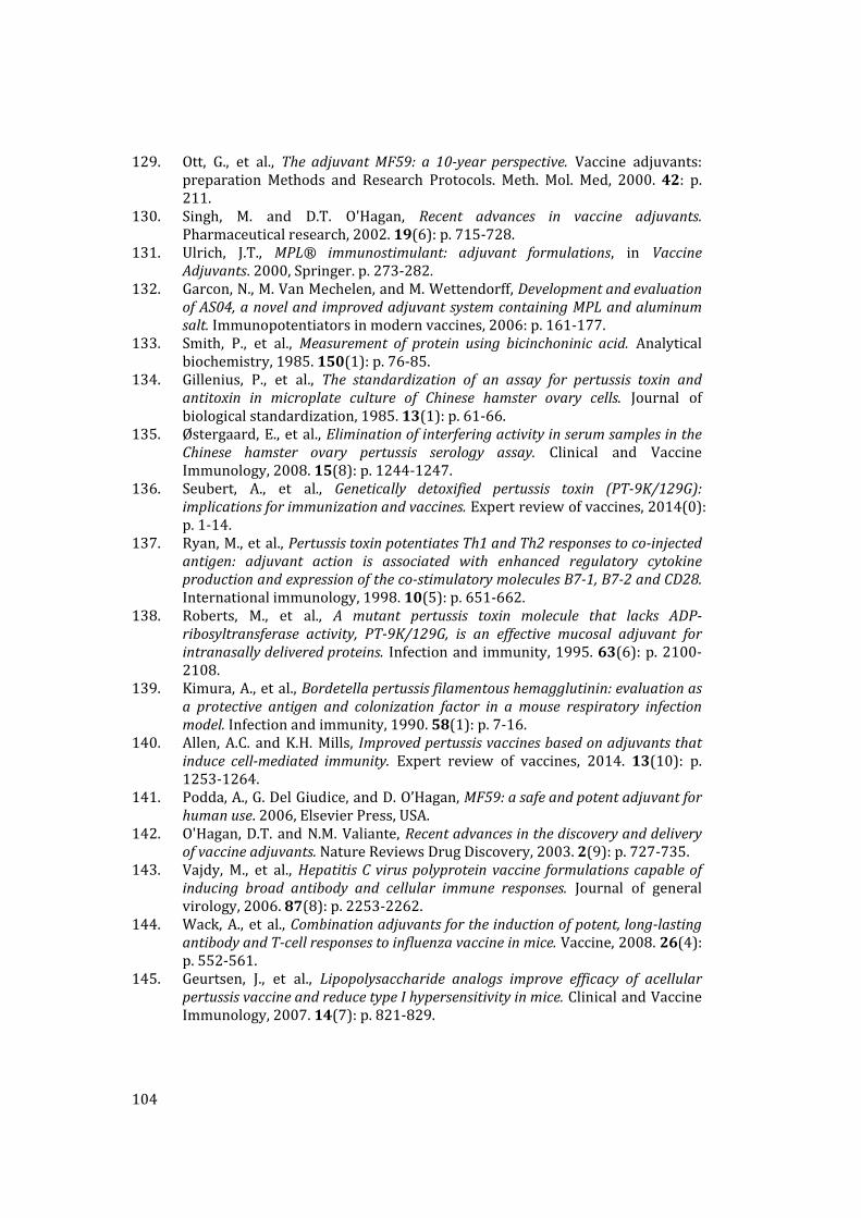
104
129. Ott, G., et al., The adjuvantMF59: a 10‐year perspective. Vaccine adjuvants:preparation Methods and Research Protocols. Meth. Mol. Med, 2000. 42: p.211.
130. Singh, M. and D.T. O'Hagan, Recent advances in vaccine adjuvants.Pharmaceuticalresearch,2002.19(6):p.715‐728.
131. Ulrich, J.T., MPL® immunostimulant: adjuvant formulations, in VaccineAdjuvants.2000,Springer.p.273‐282.
132. Garcon,N.,M.VanMechelen,andM.Wettendorff,DevelopmentandevaluationofAS04,anoveland improvedadjuvantsystemcontainingMPLandaluminumsalt.Immunopotentiatorsinmodernvaccines,2006:p.161‐177.
133. Smith, P., et al., Measurement of protein using bicinchoninic acid. Analyticalbiochemistry,1985.150(1):p.76‐85.
134. Gillenius, P., et al., The standardization of an assay for pertussis toxin andantitoxin in microplate culture of Chinese hamster ovary cells. Journal ofbiologicalstandardization,1985.13(1):p.61‐66.
135. Østergaard,E., etal.,EliminationofinterferingactivityinserumsamplesintheChinese hamster ovary pertussis serology assay. Clinical and VaccineImmunology,2008.15(8):p.1244‐1247.
136. Seubert, A., et al., Genetically detoxified pertussis toxin (PT‐9K/129G):implicationsforimmunizationandvaccines.Expertreviewofvaccines,2014(0):p.1‐14.
137. Ryan,M.,etal.,PertussistoxinpotentiatesTh1andTh2responsestoco‐injectedantigen: adjuvant action is associated with enhanced regulatory cytokineproductionandexpressionoftheco‐stimulatorymoleculesB7‐1,B7‐2andCD28.Internationalimmunology,1998.10(5):p.651‐662.
138. Roberts, M., et al., A mutant pertussis toxin molecule that lacks ADP‐ribosyltransferase activity, PT‐9K/129G, is an effective mucosal adjuvant forintranasallydeliveredproteins. Infectionand immunity,1995.63(6):p.2100‐2108.
139. Kimura,A.,etal.,Bordetellapertussisfilamentoushemagglutinin:evaluationasa protective antigen and colonization factor in amouse respiratory infectionmodel.Infectionandimmunity,1990.58(1):p.7‐16.
140. Allen,A.C. andK.H.Mills, Improvedpertussisvaccinesbasedonadjuvantsthatinduce cell‐mediated immunity. Expert review of vaccines, 2014. 13(10): p.1253‐1264.
141. Podda,A.,G.DelGiudice,andD.O’Hagan,MF59:asafeandpotentadjuvantforhumanuse.2006,ElsevierPress,USA.
142. O'Hagan,D.T. andN.M.Valiante,Recentadvancesinthediscoveryanddeliveryofvaccineadjuvants.NatureReviewsDrugDiscovery,2003.2(9):p.727‐735.
143. Vajdy, M., et al.,HepatitisCviruspolyproteinvaccine formulationscapableofinducing broad antibody and cellular immune responses. Journal of generalvirology,2006.87(8):p.2253‐2262.
144. Wack,A., et al.,Combinationadjuvantsfortheinductionofpotent,long‐lastingantibodyandT‐cellresponsestoinfluenzavaccineinmice.Vaccine,2008.26(4):p.552‐561.
145. Geurtsen, J., et al., Lipopolysaccharide analogs improve efficacy of acellularpertussisvaccineandreducetypeIhypersensitivityinmice.ClinicalandVaccineImmunology,2007.14(7):p.821‐829.

105
146. Petre,J.,etal.,Thereactionofbacterialtoxinswithformaldehydeanditsuseforantigenstabilization.Developmentsinbiologicalstandardization,1995.87:p.125‐134.
147. Podda,A., et al.,Metabolic,humoral,andcellularresponsesinadultvolunteersimmunizedwiththegeneticallyinactivatedpertussistoxinmutantPT‐9K/129G.TheJournalofexperimentalmedicine,1990.172(3):p.861‐868.
148. Mahon,B.P.,M.T.Brady,andK.H.Mills,ProtectionagainstBovdetellapertussisinMiceintheAbsenceofDetectableCirculatingAntibody:ImplicationsforLong‐Term Immunity inChildren. Journal of Infectious Diseases, 2000. 181(6): p.2087‐2091.