5 materials and methods - uni-halle.de
TRANSCRIPT
5 Materials and Methods
Materials and Methods
80
5.1 Materials
5.1.1 Chemicals and Reagents
Acetic acid Merck (Darmstadt, Germany) 30% Acrylamide/bisacrylamide solution, 37.5:1 Biorad (München, Germany) Adenosine 5’-triphosphate (ATP) Sigma-Aldrich (Steinheim, Germany) Agar-agar Serva (Heidelberg, Germany) Agarose Invitrogen (Karlsruhe, Germany) Alexa Fluor® 546 goat anti-mouse/rabbit IgG Invitrogen (Karlsruhe, Germany) Alexa Fluor® 488 goat anti-mouse/rabbit IgG Invitrogen (Karlsruhe, Germany) Ampicillin Grünenthal (Aachen, Germany) Ammonium chloride Merck (Darmstadt, Germany) Ammonium sulfate Gibco BRL (Eggstein, Germany) Ammonium peroxodisulfate Sigma-Aldrich (Steinheim, Germany) Bacto tryptone Difco (Detroit, USA) Bacto yeast extract Difco (Detroit, USA) BAPTA 1,2-bis(2-aminophenoxy) ethane-N,N,N’,N’-tetraacetic acid Invitrogen (Karlsruhe, Germany) BCATM Protein Assay Kit Pierce (Rockford, USA) Bench MarkTM Prestained Protein Ladder Invitrogen (Karlsruhe, Germany) Bovine serum albumin (BSA) Fluka (Steinheim, Germany) Bromophenol blue (3’,3’’,5’,5’’-tetrabromophenolsulfonephthalein) Sigma-Aldrich (Steinheim, Germany) Calcium chlorid Merck (Darmstadt, Germany) CNBr-activated SepharoseTM 4B Amersham Biosciences (Braunschweig,
Germany) CompleteTM EDTA-free Protease Inhibitor Cocktail Roche (Mannheim, Germany) Coomassie brilliant blue G-250 Sigma-Aldrich (Steinheim, Germany) Creatine phosphate Roche (Mannheim, Germany) Cycloheximid (3-(2-(3,5-dimethyl 2-oxocyclohexyl) 2-hydroxyethyl)glutarimide) Simar-Aldrich (Steinheim, Germany) L-Cysteine Merck (Darmstadt, Germany) Cytochalasin B Sigma-Aldrich (Steinheim, Germany) 4’,6-Diamidino 2-phenyindole (DAPI) Sigma-Aldrich (Steinheim, Germany) 1-(4,5-Dimethoxy 2-nitrophenyl)ethyl ester (A23187) Invitrogen (Karlsruhe, Germany) Dimethyl sulfoxid (DMSO) Merck (Darmstadt, Germany) DNA oligos MWG Biotech (Ebersberg, Germany) 1,1'-Dioctadecyl-3,3,3',3'-tetramethylindocarbocyanine perchlorate (DiIC18 )
Invitrogen (Karlsruhe, Germany) 1,4-Dithio L-threitol (DTT) Merck (Darmstadt, Germany) Dulbecco modified Eagle’s medium (DMEM) Invitrogen (Karlsruhe, Germany) ECLTM anti-rabbit IgG Horseradishperoxidase Linked whole antibody (from donkey) Amersham Bioscience (Braunschweig,
Germany) ECLTM anti-mouse IgG, Horseradishperoxidase Linked whole antibody (from sheep) Amersham Bioscience (Braunschweig,
Germany) Ethanol Merck (Darmstadt, Germany) Ethidium bromide Serva (Heidelberg, Germany) Ethylenediamine-N,N,N',N'-tetraacetic acid (EDTA) Sigma-Alrich (Steinheim, Germany) Ethylene glycol-bis(2-aminoethylether)-N,N,N’,N’-tetraacetic acid (EGTA) Sima-Aldrich (Steinheim, Germany) Fetal calf serum (FCS) PAA Laboratories (Cölbe, Germany) Gene RacerTM Kit Invitrogen (Karlsruhe, Germany) L-Glutamine Invitrogen (Karlsruhe, Germany) Glutaraldehyde (50% aqueous solution) Sigma-Aldrich (Steinheim, Germany) Glutathione Sepharose 4 Fast Flow Amersham Bioscience (Braunschweig,
Germany)
Materials and Methods
81
Glycerol (87% aqueous solution) Merck (Darmstadt, Germany) Glycine Merck (Darmstadt, Germany) Glycogen (source: oyster) Amersham Bioscience (Braunschweig,
Germany) Guanosine 5’-diphosphate (GDP) Sigma-Aldrich (Steinheim, Germany) Guanosine 5’-triphosphate (GTP) Merck (Darmstadt, Germany) Hydrochloric acid, 37% (HCl) Merck (Darmstadt, Germany) HeLa CCL-2 (human) cell line LGC Promochem GmbH (Wesel, Germany) Human chorionic gonadotropin (hCG) Sigma-Aldreich (Steinheim, Germany) 4-(2-Hydroxyethyl)piperazine-1-ethansulfonic acid (HEPES) Sigma-Aldrich (Steinheim, Germany) Igepal CA-630 (NP-40) Sigma-Aldrich (Steinheim, Germany) Imidazole Sigma-Aldrich (Steinheim, Germany) Immersion oil Leica (Heidelberg, Germany) Intergonan (PMSG) Intervet (Unterschleißheim, Germany) Isopropyl β-D-thiogalactopyranoside (IPTG) Sigma-Aldrich (Steinheim, Germany) Kalium chloride Merck (Darmstadt, Germany) Kanamycin Serva (Heidelberg, Germany) Lysolecithin Sigma-Aldrich (Steinheim, Germany) Magnesium chloride Merck (Darmstadt, Germany) Magnesium acetate Merck (Darmstadt, Germany) 2-Mercaptoethanol Sigma-Aldrich (Steinheim, Germany) Methanol Merck (Darmstadt, Germany) Milk powder Frema Reform (Lüneburg, Germany) MiniEluteTM Gel Extraction Kit QIAgen (Hilden, Germany) Ni-NTA QIAgen (Hilden, Germany) Nucleotides (dNTP) PEQlab (Erlangen, Germany) Oligofectamine Invitrogen (Karlsruhe, Germany) OptiMEM Invitrogen (Karlsruhe, Germany) pEGFP Clontech (Carlsbad, USA) pET28a vector Novagen (Darmstadt, Germany) pETM90 vector EMBL, Protein Expression and
Purification Facility pET60 vector EMBL, Protein Expression and
Purification Facility pGEX-KG vector EMBL, Protein Expression and
Purification Facility Paraformaldehyde Sigma-Aldrich (Steinheim, Germany) Penicillin/Streptomycin Invitrogen (Karlsruhe, Germany) PFU polymerase Stratagene (La Jolla, USA) Piperazine 1,4-bis(2-ethanesulfonic acid) (PIPES) Sigma-Aldrich (Steinheim, Germany) Phenylmethylsulfonyl fluoride (PMSF) Sigma-Aldrich (Steinheim, Germany) Poly-L-Lysine solution Sigma-Aldrich (Steinheim, Germany) Potassium acetate Merck (Darmstadt, Germany) Protein A Sepharose TM CL-4B Amersham Bioscience (Braunschweig,
Germany) QIAquick® PCR Purification Kit QIAgen (Hilden, Germany) QIAquick Gel Extraction Kit QIAgen (Hilden, Germany) Restriction enzymes New England Biolabs (Ipswich, USA) Reverse transcriptase (RT AMV) Stratagene (La Jolla, USA) RNAsin Promega (Mannheim, Germany) RNAseH Invitrogen (Karlsruhe, Germany) Shrimp alkaline phosphatase (SAP) Amersham Bioscience (Braunschweig,
Germany) Silver nitrate Sigma-Aldrich (Steinheim, Germany) Sodium borate Merck (Darmstadt, Germany) Sodium borohydride Sigma-Aldrich (Steinheim, Germany) Sodium cloride Merck (Darmstadt, Germany) Sodium dodecylsulfate Serva (Heidelberg, Germany) Sodium thiosulfat Merck (Darmstadt, Germany)
Materials and Methods
82
Spermidine tetrachloride Sigma-Aldrich (Steinheim, Germany) Spermine Sigma-Aldrich (Steinheim, Germany) Sucrose Merck (Darmstadt, Germany) T4-DNA ligase Fermentas (St. Leon-Roth, Germany) Taq polymerase Roche (Mannheim, Germany) N,N,N′,N′-Tetramethylethylenediamine (TEMED) Sigma-Aldrich (Steinheim, Germany) Thrombin Novagen (Darmstadt, Germany) Titer Max Gold adjuvant Sigma-Aldrich (Steinheim, Germany) Tris(hydroxymethyl)amino methane (Tris) Sigma-Aldrich (Steinheim, Germany) Triton X-100 Sigma-Aldrich (Steinheim, Germany) Trypan blue Invitrogen (Karlsruhe, Germany) Trypsin EDTA 1x GibcoTM Invitrogen (Karlsruhe, Germany) Tween®-20, Sigma-Ultra Sigma-Aldrich (Steinheim, Germany) Urea Merck (Darmstadt, Germany) Vectashield® mounting medium H-1000 Vector Laboratories (Grünberg, Germany) Western LightningTM Chemiluminiscence reagent PerkinElmer Life Science (Boston, USA)
5.1.2 Commonly used buffers, solutions and media
AB (acetate buffer) 20 mM HEPES, pH 7.4 100 mM KOAc 3 mM KCl 50 mM EGTA 150 mM Sucrose
Coomassie staining for SDS gels 0.2% (w/v) Coomassie brilliant blue 40% (v/v) Methanol 10% (v/v) Acetic acid
Cysteine buffer 2% (w/v) Cysteine in 0.25x MMR adjusted to pH 7.8 with NaOH
Coomassie destain solution 40% (v/v) Methanol 10% (v/v) Acetic acid
DMEM + Glucose DMEM (Sigma) was prepared according to manufacturers instructions and supplemented with : 44 mM NaHCO3
17 mM Glucose
Laemmli sample buffer 10% (v/v) Glycerol 50 mM Tris, pH 6.8 10% (w/v) SDS 1 mM DTT 0.1% (w/v) Bromphenol blue
10x Energy mix 100 mM Creatine phosphate 5 mM GTP 5 mM ATP 0.5 mg/ml Creatine kinase
6x DNA loading buffer 0.25% (w/v) Bromphenol blue 0.25% (w/v) Xylene cyanol FF 40% (w/v) Sucrose in H2O

Materials and Methods
83
LB-agar (autoclaved) 1.5% (w/v) Bacto agar in LB medium
LB-media (autoclaved) 1% (w/v) Bacto tryptone 0.5% (w/v) Bacto yeast extract 170 mM NaCl adjusted to pH 7.6 with NaOH
MMR buffer 50 mM HEPES, pH 7.9 100 mM NaCl 10 mM MgCl2 20 mM CaCl2 1 mM EDTA
Paraformaldehyde fix 4% (w/v) Paraformaldehyde in H2O, pH 7.1
PBS (phosphate buffered saline) 130 mM NaCl 100 mM Na2HPO4, pH 7.0
PBS blocking solution for immunofluorescence
PBS 3% (w/v) BSA 0.1% (v/v) Triton X-100
PBS washing buffer for immunofluorescence
PBS 0.1% (v/v) Triton X-100
PBS washing buffer for Western blots PBS 0.05% (v/v) Tween20
PBS blocking buffer PBS 0.1% (v/v) Triton X-100 5% (w/v) Milk powder
S250 buffer 20 mM HEPES, pH 7.5 250 mM Sucrose 50 mM KCl 2.5 mM MgCl2
S500 buffer 500 mM Sucrose 20 mM HEPES, pH 7.5 50 mM KCl 2.5 mM MgCl2
S250+/S500+ buffer S250/S500 buffer supplemented with: 1 mM DTT 1x CompleteTM EDTA-free Protinase Inhibitor Cocktail
Standard protein purification buffer 20 mM HEPES, pH 7.5 300 mM NaCl 5% (v/v) Glycerol 2 mM MgCl2 2 mM β-Mercaptoethanol 1x CompleteTM EDTA-free Protinase Inhibitor Cocktail (10 mM Imidazole, only for His-fusions)
Materials and Methods
84
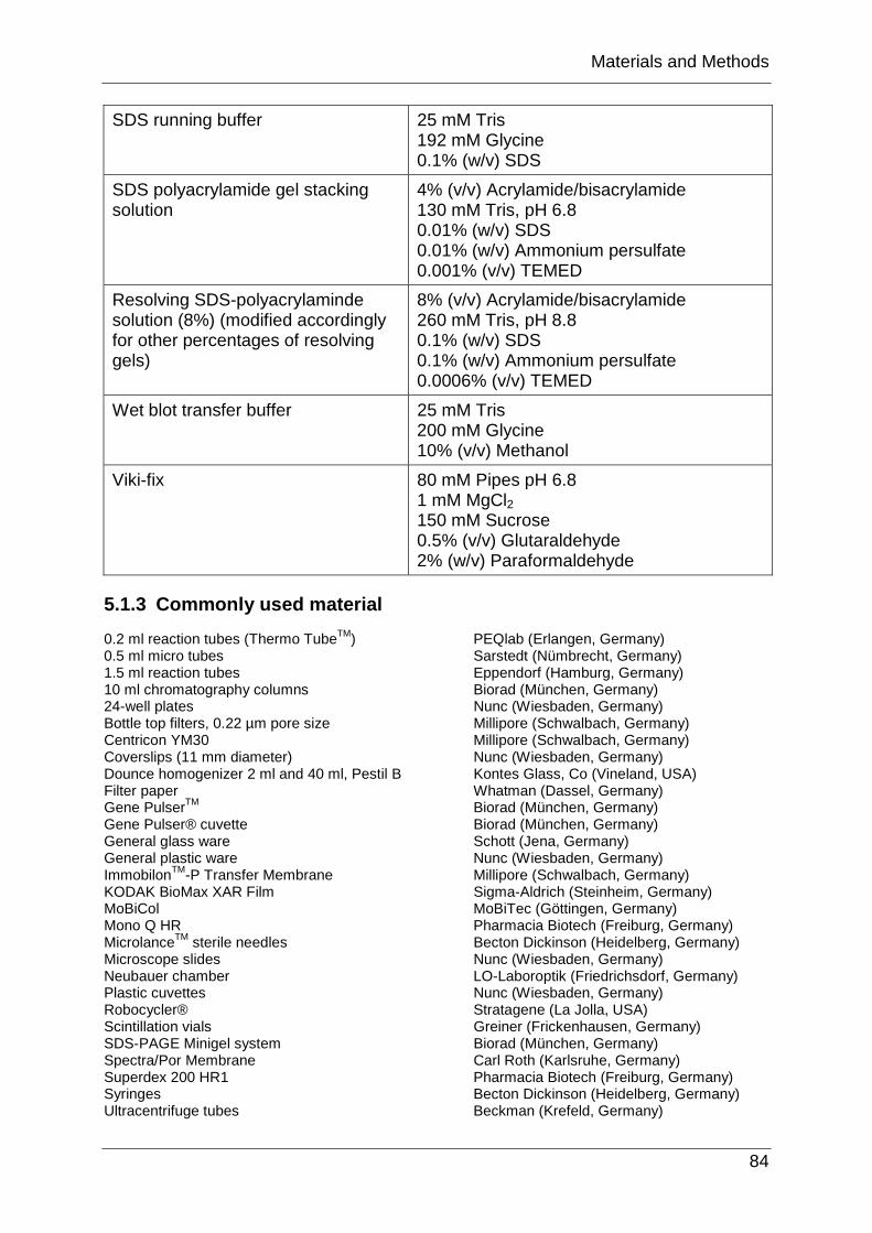
SDS running buffer 25 mM Tris 192 mM Glycine 0.1% (w/v) SDS
SDS polyacrylamide gel stacking solution
4% (v/v) Acrylamide/bisacrylamide 130 mM Tris, pH 6.8 0.01% (w/v) SDS 0.01% (w/v) Ammonium persulfate 0.001% (v/v) TEMED
Resolving SDS-polyacrylaminde solution (8%) (modified accordingly for other percentages of resolving gels)
8% (v/v) Acrylamide/bisacrylamide 260 mM Tris, pH 8.8 0.1% (w/v) SDS 0.1% (w/v) Ammonium persulfate 0.0006% (v/v) TEMED
Wet blot transfer buffer 25 mM Tris 200 mM Glycine 10% (v/v) Methanol
Viki-fix 80 mM Pipes pH 6.8 1 mM MgCl2 150 mM Sucrose 0.5% (v/v) Glutaraldehyde 2% (w/v) Paraformaldehyde
5.1.3 Commonly used material
0.2 ml reaction tubes (Thermo TubeTM) PEQlab (Erlangen, Germany) 0.5 ml micro tubes Sarstedt (Nümbrecht, Germany) 1.5 ml reaction tubes Eppendorf (Hamburg, Germany) 10 ml chromatography columns Biorad (München, Germany) 24-well plates Nunc (Wiesbaden, Germany) Bottle top filters, 0.22 µm pore size Millipore (Schwalbach, Germany) Centricon YM30 Millipore (Schwalbach, Germany) Coverslips (11 mm diameter) Nunc (Wiesbaden, Germany) Dounce homogenizer 2 ml and 40 ml, Pestil B Kontes Glass, Co (Vineland, USA) Filter paper Whatman (Dassel, Germany) Gene PulserTM Biorad (München, Germany) Gene Pulser® cuvette Biorad (München, Germany) General glass ware Schott (Jena, Germany) General plastic ware Nunc (Wiesbaden, Germany) ImmobilonTM-P Transfer Membrane Millipore (Schwalbach, Germany) KODAK BioMax XAR Film Sigma-Aldrich (Steinheim, Germany) MoBiCol MoBiTec (Göttingen, Germany) Mono Q HR Pharmacia Biotech (Freiburg, Germany) MicrolanceTM sterile needles Becton Dickinson (Heidelberg, Germany) Microscope slides Nunc (Wiesbaden, Germany) Neubauer chamber LO-Laboroptik (Friedrichsdorf, Germany) Plastic cuvettes Nunc (Wiesbaden, Germany) Robocycler® Stratagene (La Jolla, USA) Scintillation vials Greiner (Frickenhausen, Germany) SDS-PAGE Minigel system Biorad (München, Germany) Spectra/Por Membrane Carl Roth (Karlsruhe, Germany) Superdex 200 HR1 Pharmacia Biotech (Freiburg, Germany) Syringes Becton Dickinson (Heidelberg, Germany) Ultracentrifuge tubes Beckman (Krefeld, Germany)
Materials and Methods
85
5.1.4 Instrumental equipment
5.1.4.1 Centrifuges and rotors
Centrifuges: Rotors: Heraeus Megafuge 1.0 Beckman SW55Ti Heraeus BiofugeA Beckman SW40 Beckman L8-70M Beckman JLA-8.1000 Beckman Coulter Optima TLX Beckman TLA100.4 Beckman Coulter Avanti J-20 XP Sorvall SS34 Du Pont Instruments Sorvall RC-5B Sorvall HB6
5.1.4.2 Others
Air liquide ESPACE 331 GAZ (Liquid nitrogen tank) Amersham Biosciences Ultraspec 2100 pro (Spectrometer) Avestin EmulsiFlex-C5 (Homogeniser) B. Braun, Thermomic Bu (Waterbath) Biorad Power Pac 300 (Power supply) Biorad gelelectrophoresis system Biorad Transfer Cell (Western blot wet blot chamber) Biorad Gene PulserTM Branson Sonifier B15, Tip: Branson Converter BSB7 (Sonicator) FPLC LKB (Pharmacia Biotech) Hera cell 240 (Incubator) Infors AG HT (Shaking incubators) Leica DMIRE2, Leica confocal software TCS SP2 Philips CM120 BioTwin (electron microscope) Pharmacia Biotech Controller LCC-501 Plus (FPLC System) Radiometer Copenhagen pH M82 Standard pH Meter Snjiders Scientific test tube rotator Model 34528
5.1.5 Nucleotide sequences
5.1.5.1 cDNA sequences
X. laevis Mel28 expressed sequence tags (ESTs): CA790829 BU913218 BG363745 BU911025 BJ039820 BG486972 BU911216 BU911644 AW642642 BU916589 CA792499 BI477619 BU913754 BI477619
X. laevis Nup155 full length cDNA from RZPD: Clone ID: IMAGp998J1010564Q3 X. laevis Mel28 full length cDNA from RZPD: Clone ID: IMAGE6637561
Materials and Methods
86
5.1.5.2 Primers
Primers used to sequence and clone xMel28: P585: GGAGGATCCATGCAAAACCTTGAAGCTCAGGZC P586: GTAAGAGTCACATTATTCCACGTCCAC P587: TAACAGGTGTCTTGTCGCTGGCCTGCT P588: GCAGTCATGCATTCCAGTGCATTTGGA P589: TGCAGAGCTGGCACTCACAGAAATGCA P590: CGGAAGTCATCAAAGCCTGCTGAC P547: GCTGTCAACGATACGCTACGTAACG PS: TGCTCTAGATCATCTCATCTTTCGCCGCGTGACAGG PDS: TGCTCTAGAGTCATTTTTAACTTT P1: ATGTTGGACATGTACTTCTTATT P2: TCTGGCATTTGTGCTTGATACC P3: CACAGGAAAATACCCACCTGC P4: CATGGAAAGAGATTTTTGTTTCTT P5: CCCCTTTGAATGGTCCAAATGCAC P6: CCGGTACCAAGCTTGATGCATAGC P7: CACCTTCTGTGGAAGATTTACAAG P8: GTTCTTGTAGATGGACCAGAG P9: TGAAAATTGTGAAACTACTGAG P10: CATGATCACTGTCCACAAGAGGGG P11: GACCCTTAGTTGGAGTAGAAGGC P12: TAACATCAGATGACATCAGAGAG GAP1: ATGAAGTCACATCGCCTGATA Gap3: ATATACTAGTATCACTCCTCAGTC GAP2: AAAATCAGGAGCTGCTATTGGTGC P6n: ACTGAGAAAATACCTGCGAATATGT P7n: CACCTTCTGGGGAAGATTTACAAG Pa: AAGATCTAGCGTGCGCACTAC Pb: CAGTTACAGAAACTGCGCTGC Pc: ATCTTGAAAGCAAGATCCTGG Pd: GATCTTGTAATTCTGGTAGGA
Primers to clone xMel28 fragments for protein expression: MelP1NX: CGGGATCCATGCAAAACCTCAAAGCTCAG MelP1NP: GGAATTCCATATGCAAAACCTCAAAGCTCAG MelP195CXP: CCGCTCGAGTCAAGCTGGGATTCCACTCATGAC MelP189NX: CGGGATCCATGGTCATGAGTGGAATCCCAGCT MelP189NP: CTAGCTAGCATGGTCATGAGTGGAATCCCAGCT MelP1026CX: CATGCCATGGTCATAACGGTTGTGGTCTAGCAAC MelP1026CP: CGGGATCCTCATAACGGTTGTGGTCTAGCAAC MelP1918NX: CGGGATCCATGAATCAAATAATTAATGCAACT MelP1918NP: CTAGCTAGCATGAATCAAATAATTAATGCAACT MelPstopXP: CCGCTCGAGTCATATCATCTTTCGGCGCAT MelP1661NX: CGGGATCCATGAATGCCATGATGTCTTTAGAC MelP1661NP: GGAATTCCATATGAATGCCATGATGTCTTTAGAC MelP1924CXP: CCGCTCGAGTCAAGTTGCATTAATTATTTGATT MelP1388NX: CGGGATCCATGTCGAATGGTCCAAGTGCACTC MelP1388NP: GGAATTCCATATGTCGAATGGTCCAAGTGCACTC MelP1667CXP: CCGCTCGAGTCAGTCTAAAGACATCATGGCATT
Materials and Methods
87
MelP1602NX: CGGGATCCATGGCAAATGTGTCCCCTCTTGTG MelP1602NP: CTAGCTAGCATGGCAAATGTGTCCCCTCTTGTG MelP2120CXP: CCGCTCGAGTCACCCAACTTCAGGAACCTCTGT
Primers to clone full length xNup155 for expression: N155p28N: CTAGCTAGCATGCCCAGTGCAACACCT N155p28C: CCCAAGCTTGCTAGCTCAACCTCTCCAATT N155pM90N: CATGCCATGGGACCCAGTGCAACACCTGGGG N155pM90C: TCCCCGCGGTCTAGCTCAACCTCTCCAATT N155pM60C: CGGCCGCCCTAGCTCAACCTCTCCAATT
Primers to clone xNup155 fragments for protein expression: N155N1C: TCCGGCCGCTAGTTTGAGGAAGCAGAAAATCC N155N2N: CTAGCTAGCATGGCTTCCTCAAACGTGGAGAAG N155N2C: TCCGGCCGCTATGCATGTAGTGCTGACTGTAC N155C2N: CTAGCTAGCATGGGGGAGGCTCAGTTGCGTGTA N155C2C: TCCGGCCGCTAGACAGGTGGGCCTGGTTTTTT N155C1N: CTAGCTAGCATGGCACCACAATCACCCAGTGTT N155C1C: TCCGGCCGCTAGCTCAACCTCTCCAATTT
5.1.5.3 siRNAi sequences
Firefly Luciferase: AATCGAAGTATTCCGCGTACG Mel28: AATATCTACATAATTGCTCTT Nup155: AACAAGGATCATATTCCAATA
5.1.5.4 Plasmids
pET28a-xNup155: The complete ORF of X. laevis Nup155 was obtained from RZPD (clone ID: IMAGp998J1010564Q3). Nup155 cDNA in a pCMV-SPORT6 vector was amplified by PCR (chapter 5.2.1.3) using the primers N155p28N/C listed above (chapter 5.1.5.2). pET28a vector (chapter 5.1.5.4) and PCR product were digested (chapter 5.2.1.6) with restriction enzymes NheI and HindIII and Nup155 cDNA was ligated with the vector (chapter 5.2.1.8). The construct was then used for xNup155 expression in bacteria (chapter 5.2.2.1.2).
The following xNup155 fragments were cloned into pET28a for expression (for primer sequences see chapter 5.1.5.2): xNup155-N1(1-395aa) using primers N155p28N and N155N1C; xNup155-N2(392-584aa), primers N155N2N/C; xNup155-N1+N2(1-584aa) primers N155p28N and N155N2C; xNup155-C2(572-1000aa) primers N155C2N/C; xNup155-C1(986-1388aa) primers N155C1N/C. All constructs were cloned using NheI (NEB) and EagI restriction sites. pETM90-xNup155: generated as described for pET28a-xNup155. PCR was performed with the primers N155pM90N/C (chapter 5.1.5.2). Restriction sites NcoI and SacII were used for cloning. pETM60-xNup155: generated as described for pET28a-xNup155. Primers N155pM90N and N155pM60C (chapter 5.1.5.2) were used for PCR amplification and restriction enzymes NcoI and EagI were employed for cloning.
The following xMel28 fragments were cloned into pGEX-KG (chapter 5.1.5.4) for protein expression (for primer sequences see chapter 5.1.5.2): xMel(1-195aa) primers MelP1X and MelP1195CX; xMel(189-1026aa) primers MelP189NX and MelP1026CX; xMel(1918aa-stop) primers MelP1918NX and

Materials and Methods
88
MelPstopXP; xMel(1661-1924aa) primers MelP1661NX and MelP11924CX; xMel(1388-1667aa) primers MelP1388NX and MelP1667CX; xMel(1661aa-stop) primers MelP1667CX and MelPstopXP; xMel(1602-2120aa) primers MelP1602 and MelP2120CX. BamHI and XhoI restriction sites were used to clone all fragments, except xMel(189-1026aa) was cut with BamHI and NcoI.
Three xMel28 fragments were cloned into pET28a for protein expression: xMel(189-1026aa) primers MelP189NP and MelP1026CP; xMel(1918aa-stop) primers MelP1918NP and MelPstopXP; xMel(1602-2120aa) primers MelP1602NP and MelP2120CXP. For all fragments restriction enzymes NheI and XhoI were used except for xMel(189-1026aa) BamHI was employed instead of XhoI.
5.1.6 Bacteria strains for cloning and protein expression
XL1Blue: genotype: recA1 endA1 gyrA96 thi-1 hsdR17 supE44 relA1 lac (F’proAB lacIqZDM15 Tn 10 (Tetr)) (Stratagene). BL21(DE3): genotype: F- ompT hsdSB (rB
-mB-) gal dcm (DE3), a lambda prophage
carrying the T7 RNA polymerase gene (Novagene). RosettaTM(DE3): genotype: F- ompT hsdSB (rB
-mB-) gal dcm (DE3) pRARE2 (CamR)
(Novagene).
5.1.7 Antibodies
5.1.7.1 Primary antibodies
Anti-Nup107 antiserum: polyclonal antibodies were directed against aa76-171 of rat Nup107. The fragment was expressed as a His-tagged fusion protein in the vector pQE30 in BL21 (pRep4). Antibodies were raised in rabbits. ODCT 3rd bleed antiserum used in this study was available in the lab.189 Anti-Pom121 antibodies: a fragment of X. laevis Pom121 (aa381-622) was cloned into the vector pETM60 and expressed in the E. coli strain BL21(DE3) and purified on Ni-NTA agarose. The purified fragment was injected into rabbits for polyclonal antibody generation as described in Antonin et al., (2005).173 The antiserum was affinity purified and kindly provided by Dr. W. Antonin. Anti-gp210 antiserum: anti-X. laevis gp210 polyclonal antiserum was generated against a C-terminal gp210 fragment as described in Antonin et al., (2005) and kindly provided by the author.173 Anti-Schnickschnack antibodies: a fragment of an uncharacterized human INM protein carrying a LEM domain was used for rabbit immunization (Dr. W. Antonin, unpublished data). The antiserum was affinity purified and kindly provided by Dr. W. Antonin. Anti-importinα antiserum: polyclonal antibodies against full length X. laevis importin α were generated in rabbits as described in Hachet et al., (2004) and kindly provided.181
Anti-Nup93 and anti-Nup205 antisera were generated against recombinant fragments of the respective X. laevis proteins and were raised in rabbits (unpublished). The antisera were kindly provided by Dr. Wolfram Antonin. Anti-Nup153 antiserum was generated by Dr. Tobias Walther against an N-terminal fragment (aa1-149) of X. laevis Nup153 in rabbits.129 Anti-Nup35 antiserum: antibodies were raised against full length recombinant X. laevis Nup35 as described in Hawryluk-Gara et al., (2005).118 Antiserum was
Materials and Methods
89
kindly provided by Richard W. Wozniak. Anti-Nup155 antiserum: X. laevis full length Nup155 was expressed in pET28a in BL21(DE), purified from inclusion bodies (chapter 5.2.2.1.4) and injected for rabbit immunization (this study). Anti-N-Mel28 antiserum: an N-terminal fragment of X. laevis Mel28(1-195aa) was expressed in pGEX-KG in RossettaTM(DE) and purified on glutathione sepharose (chapter 5.2.2.1.1) for injection into rabbits (this study). Anti-C-Mel28 antiserum: a fragment covering the C-terminal aa1602-2120 of X. laevis Mel28 was expressed in pET28a in BL21(DE) and purified on Ni-NTA agarose. Eluates were used for rabbit immunization (chapter 5.2.2.4.1)). Antiserum and affinity purified anti-C-Mel antibodies were used (this study). Purified mouse monoclonal Antibody 414 (mAb414) was purchased from Babco.224
Anti-αtubulin mouse monoclonal antibody was purchased from Sigma.
5.1.7.2 Secondary antibodies
ECLTM anti-rabbit IgG horseradishperoxidase linked whole antibody from donkey (Amersham Bioscience) was used for antigen detection on Western blots by enchanced chemo luminescence (ECL). ECLTM anti-mouse IgG horseradishperoxidase linked whole antibody from sheep (Amersham Bioscience) was used to detect mouse monoclonal antibodies on a Western blot by ECL. Alexa Fluor® 546 goat anti-mouse/rabbit IgG (Invitrogen (Karlsruhe, Germany)) and Alexa Fluor® 488 goat anti-mouse/rabbit IgG (Invitrogen (Karlsruhe, Germany)) were employed for immunofluorescence.
5.2 Methods
All molecular biology methods were conducted as described in Sambrook et al., (1989), unless otherwise stated.241
5.2.1 Molecular biological methods
5.2.1.1 Isolation of DNA and RNA from X. laevis oocytes
150 µl oocytes were incubated with 1.5 ml homomix buffer (50 mM Tris, pH 7.4; 5 mM EDTA; 1.5% (w/v) SDS; 300 mM NaCl; 1.5 mg/ml proteinase K) for 1 h at 50 °C under rotation. SDS in the homomix buffer denatured proteins and proteinase K destroyed nucleases. An equal volume of phenol was added and the reaction was vortexed for 1 min. The oocyte mixture was spun for 4 min at 3000 rpm at 4°C in a Heraeus BiofugeA. Phenol extraction of the supernatant was repeated until it appeared clear after centrifugation. Then, extraction was carried out with half a volume phenol and half a volume chloroform relative to the volume of the extracted oocytes. After vortexing and centrifugation, the final extraction was performed with one volume chloroform (vortexing and centrifugation as described above). The isolated DNA and RNA was precipitated with 2.5 volumes ethanol and frozen at -20°C overnight. Next, the precipitate was pelleted at 7000 rpm for 20 min at 4 °C. The pellet was washed with 70% ethanol, dried and resuspended in 200 µl TE-buffer (10 mM Tris, pH 7.5; 0.5 mM EDTA).

Materials and Methods
90
5.2.1.2 RT-PCR to generate Mel28 cDNA fragments
A cDNA sequence of X. laevis Mel28 was not available. Therefore, primers for generation of Mel28 cDNA fragments by reverse transcription and subsequent PCR reactions for subcloning were designed according to X. laevis ESTs (chapter 5.1.5.1). These ESTs were selected due to their homology to the human Mel28 cDNA sequence. The 5’-end of X. laevis Mel28 cDNA was amplified employing “GeneRacerTM Kit” according to manufacturer’s instructions. Truncated mRNA and non-mRNA was eliminated and only capped, full length mRNA remained for the reaction. A GeneRacerTM RNA oligo was fused to the 5’-end from where the amplification by RT-PCR occured via a GeneRacerTM 5’-primer specific for the added oligo. ESTs covering the 3’-end of the X. laevis Mel28 sequence were identified and specific revers primers employed.
For RT-PCR 5 µg RNA purified from X. laevis oocytes was annealed with 0.5 µM (f.c.) of a sequence specific reverse primer and 2 µM (f.c.) of the GeneRacerTM 5’-primer for 5 min at 65 °C. Next, the reaction was cooled on ice and 10x AMV RT buffer, dNTP (0.2 mM each), 1 µl of the reverse transcriptase AMV RT (25 U/µl), 2 µl RNAsin and 25 mM DTT were added. The elongation reaction took place at 42 °C for 1 h. The reverse transcriptase was inactivated for 15 min at 85 °C. RNA bound to DNA was digested by RNAse H (0.32 U/µl (f.c.)) at 37°C for 20 min.
Isolated fragments were purified and subcloned and sequenced by “Genomics Core Facility” EMBL, Heidelberg.
To obtain the full length X. laevis Mel28 sequence, contigs derived from one expressed allele were assembled in “The cap EST assembler” (http://bio.ifom-firc.it/ASSEMBLY/assemble.html). New primers were designed to amplify and subclone further regions of the fragments. Annotation of xMel28 cDNA, revealed that one of the obtained IMAGE clones (IMAGE6637561) contained the full length ORF. The clone was entirely sequenced but contained two stop codons. The complete sequence was assembled by ligation of the in frame sequence of the IMAGE clone with one of the cloned fragments derived from the same allele.
5.2.1.3 PCR reactions
PCR reactions to generate DNA fragments for subcloning were routinely set up in a total reaction volume of 50 µl containing: 20 ng of DNA template, dNTPs 250 µM (f.c.) each, forward and reverse primers at 0.4 µM (f.c.), 10x PFU reaction buffer and PFU Polymerase (2.5 U/µl). A typical PCR termocycle was comprised of 25 cycles. DNA strands were separated for 2 min at 95 °C in the first cycle and 1 min in all subsequent ones. Annealing temperature of the primers was calculated according to the equation (2(AT) + 4(GC)-4) and set for 30 sec. Product elongation by PFU occurred at 72 °C for 1 min per kilobase template. The final extension step of a PCR cycle was continued for 10 min. All PCR reactions were carried out in a Robocycler®. The amplified PCR product was either purified with a PCR purification kit (chapter 5.2.1.4) after ensuring the correct fragment size on an agarose gel (chapter 5.2.2.5.4), or completely loaded on an agarose gel and purified by gel extraction (chapter 5.2.1.5).
Materials and Methods
91
5.2.1.4 Purification of PCR products
A commercial kit (QIAquick® PCR Purification Kit) was used to purify PCR products according to manufactuer’s instructions (QIAgen).
5.2.1.5 DNA purification from agarose gels
A commericial kit from QIAgen was employed to purify DNA from agarose gels (MiniEluteTM Gel Extraction Kit).
5.2.1.6 Restriction digest
1 µg vector DNA or 3 µg of a DNA insert were routinely applied in a 50 µl reaction volume for 1 h at 37 °C for digestion. 10-20 units of restriction enzymes and 1x BSA (NEB) were added if necessary. DNA was purified after digestion by electrophoresis on an agarose gel and gel extraction, or precipitated with sodium acetate and ethanol.241
5.2.1.7 DNA extraction from agarose gels
DNA was extracted from agarose gels with a commercial kit (QIAquick Gel Extraction Kit) according to manufacturer’s instructions.
5.2.1.8 Ligation of DNA fragments
To diminish self ligation of the cut vector ends, it was treated with shrimp alkaline phosphatase (SAP) (1 U/µl) for 30 min at 37 °C in a 30 µl reaction volume. The reaction was stopped by heat inactivation of SAP at 65 °C.
For ligation, vector and insert were added in a molar ratio of 1:3. A standard reaction contained ~100 ng vector DNA and respective amounts of the DNA insert, T4 DNA ligase and ligase reaction buffer according to manufacturer’s instructions. Ligation reactions were incubated for 1 h at room temperature or overnight at 16 °C and transformed into competent E. coli bacteria (chapter 5.2.1.9-12).
5.2.1.9 Preparation of chemically competent cells
For uptake of DNA plasmids by heat shock bacteria were prepared according to the following standard procedure. E. coli strains stored at -80 °C as glycerol stocks were applied to a LB-agar plate and incubated at 37 °C. The following day, a single colony was picked and grown to saturation in LB-media overnight. 5 ml of the overnight culture was used to inoculate 1 l of LB-media and grown to an optical density of 0.3 at 600 nm wavelength. The culture was chilled on ice for 15 min and the bacteria spun down by centrifugation at 4 °C by 4000 rpm in a Sorvall RC-5B centrifuge. The cell pellet was resuspended in 500 ml ice cold 50 mM CaCl2. The centrifugation step was repeated and the pellet resuspended in 80 ml 50 mM CaCl2, 15% (v/v) glycerol. Cells were aliquoted and frozen in liquid nitrogen.
5.2.1.10 Transformation of plasmid DNA into chemically competent cells
10-20 µl of a standard ligation reaction (chapter 5.2.1.8) was transformed into chemically competent E. coli cells by heat shock. Cells were thawed quickly and
Materials and Methods
92
incubated with plasmid DNA for 30 min on ice. Bacteria were heat shocked for 45 sec at 42 °C and next cooled on ice for 5 min. 1 ml LB-media without antibiotics was added and incubated for 30 min at 37 °C under rotation to allow for the expression of the antibiotic resistence. 200 µl of the bacteria suspension was plated on LB agar plates containing 100 µg/ml Ampicillin and/or 30 µg/ml Kanamycin corresponding to the resistence gene of the vector. After incubation overnight at 37 °C, colonies were picked, diluted in 50 µl water and subjected to PCR to confirm insertion of desired constructs.
5.2.1.11 Preparation of electro-competent cells
Cells were prepared according to standard procedures similarly as described above for chemically competent cells (chapter 5.2.1.9) but resuspension of the cell pellet was done in ice cold water, supplemented with 10% (v/v) glycerol.
5.2.1.12 Transformation of plasmid DNA into electro-competent cells
Electro-competent cells were thawed on ice. Ligation reactions were mixed with the cells and transferred to a chilled Gene Pulser® cuvette. An electric pulse was applied with a Gene PulserTM according to manufacturer´s instructions and cells were immediately recoverd with 1 ml of prewarmed LB-media. Cells were grown for 1 h at 37 °C and plated on LB-Agar plates with respective antibiotics.
5.2.1.13 Preparation of DNA from E. coli
Bacteria were transformed with the respective DNA plasmid and grown to saturation under standard conditions. Plasmids were purified from bacteria using a commercial kit (QIAprep Miniprep spin Kit®) according to manufacturer’s instructions.
5.2.2 Biochemical standard methods
5.2.2.1 Protein expression and purification
5.2.2.1.1 Expression and purification of GST-fusion proteins
pGEX-KG-xMel constructs were transfected into the E. coli strain RosettaTM (DE) (chapter 5.1.6) for expression. Rosetta host strains are BL21 (Novagen) derivatives designed to enhance the expression of eukaryotic proteins that contain codons rarely used in E. coli. Therefore, tRNAs for these codons are supplied on an additional plasmid. Transformants were plated on LB-agar plates containing 100 µg/ml Ampicillin, 0.33 µg/ml Chloramphenicol and incubated overnight at 37 °C. A single colony was picked to inoculate LB culture that was grown overnight to saturation. From this culture 1 l LB-medium was inoculated for protein expression and cultures were grown to an OD of 0.6 at 600 nm wavelength. Protein expression was induced by addition of 0.2 mM IPTG (f.c.) for 4-5 h at 21 °C. Cells were harvested by centrifugation at 4000 rpm for 30 min at 4 °C in a Sorvall RC-5B centrifuge. The bacteria pellet corresponding to 1 l culture was resuspended in 10 ml purification buffer (20 mM HEPES, pH 7.5; 300 mM NaCl; 5% (v/v) glycerol; 2 mM MgCl2; 2 mM β-mercaptoethanol and 1 mM PMSF). Cells were lysed in an Avestin EmulsiFlex-C5 homogenizer and Triton-X100 was added to 0.1% (v/v) final

Materials and Methods
93
concentration and incubated at room temperature (RT) for 30 min under rotation. The lysate was cleared by centrifugation at 12000 rpm for 20 min at 4 °C in a Sorvall RC-5B centrifuge using an SS34 rotor. The supernatant was applied to 0.5 ml equilibrated glutathione sepharose per 1 l culture and incubated for 1 h at 4 °C under rotation. The resin was transferred to a plastic affinity chromatography column and was washed twice with 10 bed volumes purification buffer. Further, one wash was performed with purification buffer supplemented with 500 mM NaCl, 2 mM ATP, and 5 mM MgCl2. The resin was equilibrated with purification buffer before elution of the GST-fusion proteins with 20 mM glutathione. It was tried to remove the GST-tag by cleavage with Thrombin according to manufacturer’s instructions, but xMel(1-195aa) was instable without the fusion tag. GST-xMel(1-195aa) was used for polyclonal antibody generation in rabbits (chapter 5.2.2.4.1).
5.2.2.1.2 Expression and purification of His-fusion proteins
Expression of His-fusion proteins in pET28a were performed as described above with the following modifications: vectors were transformed into BL21(DE) for expression. The purification buffer contained: 20 mM HEPES, pH 7.5; 300 mM NaCl; 5% (v/v) glycerol; 2 mM MgCl2; 2 mM β-mercaptoethanol; 10 mM imidazole and 1x CompleteTM EDTA-free Protease Inhibitor Cocktail. The bacteria suspension was lysed in an Avestin EmulsiFlex-C5 homogenizer and cleared by centrifugation. The supernatant was applied to Ni-NTA agarose equilibrated in purification buffer (0.3 ml resin per 1 l bacteria culture). His-tagged proteins were incubated with affinity resin for 1 h at 4 °C on a rotating weel. After extensive washes, fusion proteins were eluted with 300 mM imidazole in purification buffer.
For purification of Nup155, eluted fractions containing Nup155 were pooled and concentrated in YM-30 centricons by centrifugation in a Heraeus Megafuge 1.0 at 4000 rpm at 4 °C. At the same time purification buffer was exchanged with S250 buffer.
Removal of the His-tag of xMel(1602-2120aa) was assessed by cleavage with thrombin according to manufacturer’s instructions but the protein was degraded by the protease.
5.2.2.1.3 Protein purification under denaturing conditions
E. coli cell pellet was resuspended in 70 ml of the respective purification buffer per 1 l expression culture. Urea was added to a final concentration of 4 M and the solution was homogenized with a hand blender. The lysate was cleared by centrifugation and bound to an affinity resin, washed and eluted as described above. If proteins did not elute in the standard purification buffer supplemented with imidazole, urea elutions with 50 mM NaH2PO4, 8 M urea at pH 5.8 and pH 4.5 were performed.
5.2.2.1.4 Purification of inclusion bodies containing expressed protein
After protein expression E. coli cells were pelleted and resuspended in 13 ml of 50 mM Tris-Cl, pH 8.0; 25% sucrose, and 10 mM DTT per 1 l culture. Cells were lysed by sonication and 100 µl lysozyme (50 mg/ml in H2O), 250 µl DNaseI (1 mg/ml in 50% glycerol, 75 mM NaCl), and 50 µl MgCl2 (1 M) was added. The solution was vortexed, 12.5 ml lysis buffer (50 mM Tris-Cl, pH 8.9; 1% (v/v) Triton X-100; 1% (w/v) sodium deoxycholate; 100 mM NaCl; 10 mM DTT) were added and vortexed. The
Materials and Methods
94
suspension was incubated for 1 h at RT, frozen in liquid nitrogen and thawed again at 37 °C for 30 min. 200 µl MgCl2 (1 M) was added and incubated until the viscosity decreased. 350 ml EDTA (0.5 M in 50 mM Tris-Cl, pH 8.9) was adjusted to chelate Mg2+. The E. coli lysate was pelleted at 11000 g for 20 min at 4 °C in a Sorvall RC-5B centrifuge and the pellet resuspended in 10 ml washing buffer (50 mM Tris-Cl, pH 8.0; 100 mM NaCl; 1 mM DTT; 1 mM EDTA), sonicated and centrifuged as before. The pellet was then resuspended in washing buffer with Triton X-100 (50 mM Tris-Cl, pH 8.0; 1% Triton X-100; 100 mM NaCl; 1 mM DTT), sonicated and centrifuged. The pellet consisted mainly of purified inclusion bodies that were dissolved in 8 M urea by incubation at RT, aliquoted, frozen in liquid nitrogen, and stored at -80 °C. Inclusion bodies were purified by the “Protein Expression and Purification Core Facility”, EMBL Heidelberg.
5.2.2.2 Purification of proteins by fast protein liquid chromatography
Further purification of xMel(1-195aa) and xMel(1602-2120aa) fusion proteins was tested by ion exchange chromatography on a MonoQ column on a FPLC LKB apparatus. Proteins were dialysed into 20 mM HEPES, pH 7.5; 50 mM NaCl; 5% (v/v) glycerol, 2 mM β-mercaptoethanol, and 1 mM MgCl2 and loaded onto the column according to manufacturer’s instructions. However, during elution with increasing salt concentrations, both fusion proteins precipitated on the column. Alternatively, xMel(1602-2120aa) was loaded on a Superdex 200 gelfiltration column to separate contaminating proteins by size but it was not possible to remove copurifying proteins efficiently.
His-xNup155 precipitated during every attempt to exchange it into a different buffer required for FPLC purification and therefore, could not be purified any further.
5.2.2.3 Identification of proteins by mass spectrometric analysis
Malgorzata Schelder did mass spectrometric analysis of proteins that co-immunoprecipitated with anti-Nup155 antibodies and such that copurified with full length Nup155 under native conditions.
5.2.2.4 Antibodies
5.2.2.4.1 Polyclonal antibody generation in rabbits
All antigens used for rabbit immunization were recombinant proteins, purified either under native conditions (chapter 5.2.2.1.1/2) or from inclusion bodies (chapter 5.2.2.1.4). Per boost and rabbit, 0.5 mg recombinant protein was diluted with PBS to 300 µl and mixed with 300 µl Titer Max Gold adjuvant. The emulsion was sonicated with a Branson Sonifier B15, tip B5B7 until it became white and viscous. 500 µl of the emulsion were injected per rabbit. After the primary immunization, rabbits were boosted three times in a 14 day rhythm. Subsequent boosts were repeated every four weeks. Rabbit bleeds were heated to 37 °C for 1 h and stored overnight at 4 °C. The following day, the coagulated blood was spun down at 4000 rpm for 30 min in a Heraeus Megafuge 1.0, the serum was collected, frozen in N2 and stored at -80 °C. Rabbit immunizations were performed by the “Laboratory Animal Resources” EMBL, Heidelberg. Antibodies for this study were generated against His-xNup155 purified with inclusion bodies, GST-xMel(1-195aa) and His-xMel(1602-2120aa).
Materials and Methods
95
5.2.2.4.2 Affinity purification of polyclonal antibody serum
His-xMel(1602-2120aa), a C-terminal fragment of Mel28, was expressed and purified as described (chapter 5.2.2.1.2). The protein solution was dialyzed against buffer A (100 mM NaHCO3, pH 8.3; 500 mM NaCl; 2 mM MgCl2). CNBr-activated SepharoseTM 4B was swollen in ice cold 1 mM HCl, pH 2-3, and equilibrated in buffer A. 10 ml protein solution at a concentration of 3.5 mg/ml protein was incubated with 1 ml CNBr SepharoseTM 4B for 1 h at RT under rotation. Excess ligand was washed off with 5x bed volume buffer A. Remaining, free binding sites of CNBr SepharoseTM 4B were blocked with 1 M ethanolamine, pH 8 in buffer A for 2 h at RT. The resin was washed in two cycles, twice with buffer A and twice with buffer B (100 mM acetate, pH 4.2; 500 mM NaCl; 2 mM MgCl2). The sepharose was equilibrated in PBS and pre-eluted with 200 mM glycine, pH 2.2 and 150 mM NaCl. 30 ml rabbit antiserum was incubated with the affinity resin overnight at 4 °C under rotation. The resin was sedimented by low speed centrifugation at 1000 rpm in a Heraeus, Megafuge 1.0. The serum was removed and the beads were washed with PBS, 0.05% (v/v) Tween®-20. Antibodies bound to recombinant His-xMel(aa1602-2120) were eluted by applying 10x 1.5 ml 200 mM glycine, pH 2.7; 150 mM NaCl. Eluates were immediately neutralized by addition of 120 µl 1.5 M Tris, pH 8.8. This procedure was repeated with 200 mM glycine, pH 2.2, neutralized with 200 µl 1.5 M Tris, pH 8.8. Purified antibodies were precipitated with 60% (w/v) ammonium sulfate on ice overnight. The precipitate was sedimented by centrifugation at 4000 rpm for 30 min at 4 °C in a Sorvall RC-5B, HB6 rotor and the pellet was resuspended in PBS, 5% (v/v) glycerol, dialysed against the same buffer and aliquots were frozen in liquid nitrogen.
5.2.2.5 Electrophoretic Methods
5.2.2.5.1 Sodium dodecyl sulfate polyacrylamide gel electrophoresis
The standard method of Laemmli for discontinuous gel electrophoresis under denaturing conditions was used in this study.242
The glass-plate sandwich of the electrophoresis apparatus was assembled according to manufacturer’s instructions. Separation gel was induced to polymerize and poured between the two glass plates. To guarantee a straight surface, the gel solution was overlaid with water-saturated 1-butanol. After gel polymerization 1-butanol was decanted and the gel surface was rinsed with water. The stacking gel solution was added onto the polymerized gel and a comb was inserted, providing gel pockets. The gel polymerized and the comb was removed. The gel sandwich was attached to the upper and lower buffer chambers and buffer was filled into both containers.
Protein samples containing 1x sample buffer at final concentration were denatured at 95 °C or 65 °C for 10 min and pipetted into the gel wells. Molecular weight markers (5 µl on minigels and 12 µl on large gels) were included at all times. Minigels were run at 130 V through the stacking gel and were separated under 200 V in the resolving gel until the dye front had entered the buffer in the lower buffer chamber.
After the separation by SDS-PAGE proteins were either stained in the gel (chapter 5.2.2.5.2/3) or transferred to membranes for immunodetection (chapter 5.2.2.6).
Materials and Methods
96
5.2.2.5.2 Staining of proteins with Coomassie Brilliant Blue
After electrophoresis, gels were removed from the glass plate and incubated for 1 h in Coomassie Brilliant Blue staining solution (40% (v/v) methanol, 10% (v/v) acetic acid and 0.2% (w/v) Coomassie Brilliant Blue) on a rocking tabel at RT. Next, the dye solution was removed and gels were incubated in destain solution (40% (v/v) methanol, 10% (v/v) acetic acid) on a shaker until the background of the gel became clear. The destain solution was renewed several times.
5.2.2.5.3 Silver staining of proteins
For silver staining, proteins in SDS-gels were fixed with 50% (v/v) methanol, 12% (v/v) acetic acid, 0.05% (v/v) formaldehyde for 30 min shaking at RT. The gel was washed twice for 10 min in 50% ethanol on a shaker. The alcohol was removed and the gel incubated for 1 min in 0.02% (w/v) Na2S2O3-solution followed by three washes with water 10 seconds each. The gel was incubated for 15 min with 0.2% (w/v) silver nitrate in water. The gel was washed several times with water and was developed in 6% (w/v) Na2CO3, 0.05% (v/v) formaldehyde, 0.0004% (w/v) Na2S2O3-solution, placed on an illuminator to control the staining. Rection was stopped with 50 mM EDTA before the background became dark.
5.2.2.5.4 Agarose gel electrophoresis
Agarose was dissolved to the desired concentration (w/v) in 1x TBE and heated to boiling in a microwave. When the agarose was completely dissolved, ethidium bromide was added 1:40000 from the stock solution (10 mg/ml). The gel solution was poured into a horizontal gel carrier and a comb for forming wells was inserted. When the gel was polymerized, the comb was removed and the carrier was placed into the gel container. The gel and both gel chambers were filled with TBE buffer. The DNA samples and DNA molecular weight markers were loaded into gel wells. Agarose gels were run at 100 V until the dye front reached the last quarter of a gel. DNA was visualized by the fluorophor emitting light when placed on an UV illuminator.
5.2.2.5.5 Wet Blotting
The SDS gel was soaked in transfer buffer (25 mM Tris, 200 mM glycine, 10% methanol) for 15 min at RT. A polyvinylidendifluorid (PVDF) membrane was activated in 100% methanol for 1 min and was soaked in water for 5 min afterwards. Before the gel/membrane sandwich was assembled, the membrane was washed briefly in transfer buffer. The gel and membrane were placed between two pieces of Whatman paper and covered by two sponges. These layers were held by a lattice cassette and placed into a Biorad Transfer Cell according to manufacturer’s instrucions. The sandwich was placed upright into the container filled with transfer buffer. The transfer of proteins took place at 300 mA for 4 h at 4 °C and was controlled by monitoring the transfer of prestained molecular weight standards present in the gel.

Materials and Methods
97
5.2.2.6 ECL detection
Following transfer PVDF membranes were incubated in 5% milk powder, PBS, 0.05% Tween®-20 (PBS-T) shaking at RT for 1 h or at 4°C overnight to saturate non-specific protein background on the membrane. The blocking reagent was removed and the immobilized proteins were incubated in primary antibody at RT for 1 h while shaking or at 4°C overnight.
The antibody solution was retained for repeated use and the membrane was washed three times in PBS-T for 5 min on a shaker to remove remaining primary antibody that was not tightly bound. The secondary antibodies, ECLTM anti-rabbit or anti-mouse IgG linked to horseradishperoxidase, were applied and incubated at RT for 1 h on a shaker. The blot was washed ten times in PBS-T. To localize the bound antibody, the membrane was covered with Western LightningTM Chemiluminiscence reagent according to manufacturer’s instructions. After 1 min incubation on the membrane, the reagent was removed and the membrane was covered with Saran Wrap. The membrane was exposed to an X-ray film for 1-5 min in a dark room and the film was developed mechanically.
5.2.2.7 Determination of protein concentration
The protein concentration of a solution was determined using BCATM Protein Assay Kit (Pierce) following manufacturer’s instructions. Briefly, bovine serum albumin (BSA) protein standard dilutions were made in duplicates ranging from 2 µg-50 µg protein and several dilutions of the protein solution in 100 µl H2O. 900 µl BCA working solution (49 units buffer A and 1 unit buffer B supplied with the kit) were added and samples incubated for 30 min at 37 °C. Optical density was determined at a wavelength of 562 nm and the protein concentration was calculated via a standard curve.
5.2.3 Biochemical methods related to the X. laevis egg extract system
5.2.3.1 Preparation of X. laevis egg extract
The protocol was modified from Newmever and Wilson, (1991) and Hartl et al., (1994).208, 243
Female X. laevis frogs were primed by injection with pregnant mare serum gonadotropin (PMSG) (1000 U in 0.5 ml per frog) that triggered eggs to complete meiosis I and arrest in metaphase of meiosis II. Ovulation of mature eggs was stimulated with human chorionic gonadotropin (hCG, 1000 U/frog). Ten days later, frogs were transferred into plastic boxes, covered with ~0.5 l 1x MMR buffer (50 mM HEPES, pH 7.9; 100 mM NaCl; 10 mM MgCl2; 20 mM CaCl2; 1 mM EDTA) and incubated at 16 °C for 16 h in the dark. The following day, eggs laid in MMR buffer were collected and frogs were returned to normal tanks. Damaged eggs (white) were selected and discarded. Dirt and debris was washed away with MMR buffer. The jelly coat of the eggs was removed by incubation in a freshly prepared 0.25x MMR supplemented with 2% cystein. As soon as eggs started to become closely packed (~10 min), dejellying solution was discarded and eggs were extensively washed with MMR. Following dejellying, exposure to air was avoided since the eggs were very fragile and lysed easily after removal of the jelly coat. Eggs were released from cell cycle arrest at metaphase of meiosis II by addition of the calcium ionophore A23187
Materials and Methods
98
(8 µl, (2 mg/ml in ethanol) per 100 ml MMR) that mimicks fertilization. Animal cap contraction was visible after 3 min, as judged by an upturn of the vegetal, black pool. Activation was stopped after 7-10 min by thorough washing with MMR buffer which removed the ionophore. Eggs were incubated at RT for 15 min and subsequently washed 3x with S250 buffer (20 mM HEPES, pH 7.5; 250 mM sucrose; 50 mM KCl; 2.5 mM MgCl2). Necrotic eggs were removed with a pasteur pipette. Remaining eggs were transferred with an edgeless plastic Pasteur pipette to SW55 plastic centrifuge tubes and supplemented with 1 mM DTT, 44 µg/ml cycloheximide, 5 µg/ml cytochalasin B, and 1x CompleteTM EDTA-free Proteinase Inhibitor Cocktail. Cycloheximide, a protein synthesis inhibitor, was included to prevent further progression of the extracts into mitosis. Cytochalasin B inhibited actin polymerization and prevented the extract from coagulation. Low speed centrifugation for 30 sec at 800 rpm followed by a 30 sec spin at 1600 rpm (Heraeus, Megafuge 1.0 R), was used to pack eggs and remove additional buffer. Centrifuge tubes were filled to the top with additional eggs and subjected to a “low speed” centrifugation for 20 min at 25000g (4 °C) in a Beckman L8-70M ultracentrifuge employing a SW55Ti rotor. Tubes were broached with a syringe and nuclear material, organelles and glycogen were removed, leaving lipids, yolk and pigment behind. DTT, cycloheximide, cytochalasin B and CompleteTM EDTA-free Proteinase Inhibitor Cocktail were added to this low speed extract as described above and subjected to a high speed centrifugation at 225000g for 40 min using the same centrifuge and rotor as in the previous step. Repeatedly, cytosol, membranes and mitochondria were removed with a syringe, diluted with 0.3 fold AB-buffer (20 mM HEPES, pH 7.4; 100 mM KOAc; 3 mM KCl; 50 mM EGTA; 150 mM sucrose) and spun again at the same centrifugation settings. Cytosol was removed with a Gilson P1000 pipette, glycerol was added to 3% (v/v) and aliquots of 50 µl were snap frozen in liquid nitrogen and stored at -160 °C. Cytosol for immunodepletion was immediatedly processed as described in chapter 5.2.3.4. Membranes were further purified as described (chapter 5.2.3.3).
5.2.3.2 Preparation of sperm chromatin
This protocol was modified from Gurdon et al., (1976).244 Testis were removed from male X. laevis frogs and separated into smaller pieces with forceps in HPS buffer (15 mM HEPES, pH 7.4; 250 mM sucrose; 0.5 mM spermidine tetrachloride; 0.2 mM spermine). Resuspended organ fragments were further dispersed by pressing the solution through a 1 ml syringe onto a glass plate. To remove debris, the solution was filtered through cheese cloth. Material was pelleted by 2000 rpm for 10 min at 4 ° C in a Heraeus Megafuge 1.0R. The cell pellet was resuspended in 1 ml HSP supplemented with 0.3 mM PMSF and 50 µl 10 mg/ml lysolecithin. Plasma membranes were permeabilized for 5 min at RT and the reaction was stopped by addition of 10 ml chilled HSP buffer containing 0.3 mM PMSF and 3% BSA. Cells were pelleted repeatedly as described above and washed with 3 ml HSP, 0.3 mM PMSF, 0.3% (w/v) BSA and sedimented again. Sperm heads were resuspended in 2.5 ml HSP buffer, 0.3% (w/v) BSA, and 30% (v/v) glycerol, diluted and counted on a Neubauer chamber according to manufacturer’s instructions. Staining with trypan blue showed whether cells had been properly permeabilized. The preparation was diluted to 3000 sperm heads/µl, aliquoted, snap frozen, and stored at -80 °C.

Materials and Methods
99
5.2.3.3 Preparation of the total membrane fraction from X. laevis egg
extracts
For preparation of total membranes, the membrane fraction was diluted in 20 volumes S250+ buffer (1mM DTT and CompleteTM EDTA-free Proteinase Inhibitor Cocktail), homogenized in a 40 ml dounce homogenizer (pestil B) and laid on top of a 800 µl S500+ buffer cushion. Membranes were sedimented through the cushion at 28000g for 15 min (4° C) in a Beckman L8-70M ultracentrifuge employing a SW40 rotor. Membranes were resuspended and homogenized in 1/10th of the corresponding cytosol volume in S250+ buffer in a 2 ml dounce homogenizer (pestil B), snap frozen and stored at -160 °C.178
5.2.3.4 Preparation of floated membranes
To generate a discontinuous sucrose step gradient, the following sucrose solutions were prepared in S250 buffer and supplemented with 1mM DTT and 0.1x CompleteTM EDTA-free Proteinase Inhibitor Cocktail: 2.1 M, 1.4 M, 1.3 M, 1.1 M, 0.9 M and 0.7 M. Membranes were homogenized with 2 ml 2.1 M sucrose solution per ml membranes in a chilled 40 ml dounce homogenizer. 2 ml of the membrane solution were added into a Beckman centrifuge tube suitable for SW40Ti rotor and overlaid with 1.6 ml of each sucrose solution with decreasing density. On top of the gradient S250 buffer was added to fill the tube. Membranes were floated for 4 h at 250000g at 4 °C with slowed deceleration. The membrane fraction with lowest density was collected, diluted 1:2.5 in S250 buffer containing 1 mM DTT and CompleteTM EDTA-free Proteinase Inhibitor Cocktail and pelleted in a Beckman Coulter Optima TLX, TLA100.4 rotor at 420000g for 30 min at 4 °C. The pellet was resuspended in 1/30th of the cytosol volume in S250 buffer, homogenized in a 2 ml dounce homogenizer (pestil B) and frozen in liquid nitrogen. Membranes were stored at -160 °C.245
5.2.3.5 Immunodepletion of proteins form X. laevis egg cytosol
For immunodepletion of proteins from X. laevis egg cytosol respective antibodies were crosslinked to protein A sepharose to generate antibody columns. 1 ml protein A sepharose resin was equilibrated in PBS and incubated with 3 ml of antiserum over night at 4 °C. The resin was pelleted by centrifugation at low speed, and the serum was removed. Beads were washed twice in coupling buffer (200 mM NaHCO3, pH 9.3; 100 mM NaCl). Antibodies were crosslinked to protein A sepharose with 10 mM dimethylpimelimidate (DMP) in coupling buffer for 10 min at RT on a rotating wheel. The resin was pelleted, washed with coupling buffer, and the crosslinking step was repeated. Subsequently, antibody-resin was washed twice alternating in buffer A (100 mM NaOAc, pH 4.2; 500 mM NaCl) and buffer B (100 mM NaHCO3, pH 8.3; 500 mM NaCl). The antibody column was then washed in S250 buffer and blocked for 1 h at 4 °C on a rotator in S250 buffer supplemented with 3% BSA and CompleteTM EDTA free Protienase Inhibitor Cocktail. Beads were stored in this blocking solution containing 0.02% NaN3.
Before use, beads were washed in blocking solution and transferred to Mobicols columns. For depletion of Nup155 from cytosol, beads were preblocked with inactive Nup155-depleted cytosol for 30 min at 4 °C to reduce loss of unspecific proteins from the cytosol. Freshly prepared cytosol was mixed in a 1.3:1 ratio with the
Materials and Methods
100
antibody column, and incubated for 30 min at 4 °C under rotation. The flow through was collected and the depletion step repeated on fresh, preblocked antibody resin. Depleted cytosol was recovered and immediatedly used for in vitro nuclear reconstitution reactions (chapter 5.2.3.6).
5.2.3.6 In vitro nuclear envelope assembly
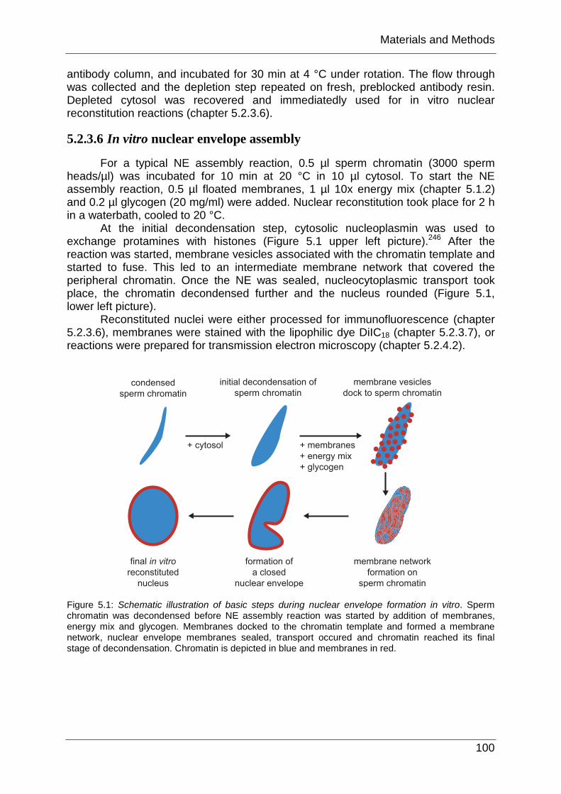
For a typical NE assembly reaction, 0.5 µl sperm chromatin (3000 sperm heads/µl) was incubated for 10 min at 20 °C in 10 µl cytosol. To start the NE assembly reaction, 0.5 µl floated membranes, 1 µl 10x energy mix (chapter 5.1.2) and 0.2 µl glycogen (20 mg/ml) were added. Nuclear reconstitution took place for 2 h in a waterbath, cooled to 20 °C.
At the initial decondensation step, cytosolic nucleoplasmin was used to exchange protamines with histones (Figure 5.1 upper left picture).246 After the reaction was started, membrane vesicles associated with the chromatin template and started to fuse. This led to an intermediate membrane network that covered the peripheral chromatin. Once the NE was sealed, nucleocytoplasmic transport took place, the chromatin decondensed further and the nucleus rounded (Figure 5.1, lower left picture).
Reconstituted nuclei were either processed for immunofluorescence (chapter 5.2.3.6), membranes were stained with the lipophilic dye DiIC18 (chapter 5.2.3.7), or reactions were prepared for transmission electron microscopy (chapter 5.2.4.2).
Figure 5.1: Schematic illustration of basic steps during nuclear envelope formation in vitro. Sperm chromatin was decondensed before NE assembly reaction was started by addition of membranes, energy mix and glycogen. Membranes docked to the chromatin template and formed a membrane network, nuclear envelope membranes sealed, transport occured and chromatin reached its final stage of decondensation. Chromatin is depicted in blue and membranes in red.

Materials and Methods
101
5.2.3.7 Immunofluorescence of in vitro reconstituted nuclei
An NE assembly reaction was fixed in 0.5 ml 4% PFA in PBS and incubated for 30 min on ice. Cover slips (round, 11 mm in diameter) were coated with poly-L-lysine solution for 5 min at RT. Poly-L-lysine solution was removed and cover slips dried and transferred to scintillation vials. Fixed NE assembly reactions were applied to 0.7 ml 30% sucrose in S250 buffer. The reaction was spun through the cushion onto cover slips at 4000 rpm for 15 min (4 °C) in a Heraeus Megafuge 1.0R. Cover slips were lifted by punctoring the vials from the bottom with a syringe needle and then removed with forceps. Immunofluorescence staining was performed in a 24-well plate. The fixative was quenched with 50 mM NH4Cl in PBS for 5 min at RT. Blocking solution contained 3% BSA in 0.1% Triton-X100 in PBS and was applied to the cover slips for 30 min at RT. Antibodies were diluted in blocking solution. Primary antibodies were incubated with the nuclei on the cover slips for 1.5 h, followed by 5 washes with 0.1% Triton-X100 in PBS. Secondary antibodies coupled to fluorophors (Alexa Fluor® 488/546 goat anti-mouse/rabbit IgG were diluted 1:2000 in blocking solution and applied for 45 min. After labeling with secondary antibodies, cover slips were washed 10 times with 0.1% Triton-X100 in PBS and 1x with PBS. To stain the chromatin, DAPI (1 mg/ml) was diluted 1:10000 in PBS and reactions on cover slips were stained for 5 min at RT. After two additional washes with PBS and H2O, cover slips were removed from solutions and remaining liquid was removed. Samples were mounted on a drop of Vectarshield® mounting media on glass slides. Liquid was removed from the rim of the cover slips with a tissue and the preparation was sealed with nail polish at the perimeter. Reactions were evaluated by confocal microscopy.247
A modified protocol was used when membranes were investigated. Viki-fix (80 mM Pipes, pH 6.8; 1 mM MgCl2; 150 mM sucrose; 0.5% glutaraldehyde; 2% paraformaldehyde) yielded better conservation of nuclear membranes due to the presence of glutaraldehyde. The fixation step was quenched with 1mg/ml NaBH4 in PBS for 10 min at RT. The cross linking potential for glutaraldehyde was higher than for formaldehyde. Aldehyde groups that remained free could unspecifically bind proteinaceous reagents, for example, antibodies. However, mAb414 was employed on reactions fixed in the presence of glutaraldehyde, leading to well preserved nuclear membranes in which a subset of nucleoporins were stained by mAb414.
5.2.3.8 DilC18 labeling of membranes
Membranes in an NE assembly reaction were stained before fixation with 0.2 µl of the lipophilic dye DiIC18 (0.1 mg/ml in DMSO) for 5 min at RT. For better preservation of the membranes Viki-fix was used (80 mM Pipes pH 6.8; 1 mM MgCl2; 150 mM sucrose; 0.5% glutaraldehyde; 2% paraformaldehyde; 0.1 µg/ml DAPI). Samples were spun as described (chapter 5.2.3.7), mounted and processed for confocal microscopy.173
5.2.3.9 Immunoprecipitation of proteins from X. laevis egg cytosol
Egg cytosol was diluted 1:1 with PBS supplemented with CompleteTM EDTA-free Proteinase Inhibitor Cocktail and spun for 20 min at 55000g in a TLA100.4 rotor. 50 µl antiserum were added to 800 µl cytosol after centrifugation and incubated for 2 h at 4 °C. 100 µl Protein A sepharose was added as 50% slurry and incubation
Materials and Methods
102
continued for 1 h. Beads were sedimented, supernatant removed, and the resin was washed 10x with PBS. Bound proteins were eluted in 40 µl 1x SDS-sample buffer supplemented with 100 mM DTT. 10 µl of the eluates were separated on 8% polyacrylamide gels and analyzed by immunostaining on Western blots (chapter 5.2.2.5.5).
5.2.4 Microscopy
5.2.4.1 Light microscopy
A Leica AOBS Sirius DMIRE2 confocal microscope was used for immunofluorescence. Laser lines at 405 nm, 488 nm and 532 nm were employed to excite respective fluorophores, which were predominantly DAPI, Alexa 488 and Alexa 546. Settings were adjusted to exclude cross-talk between different channels. For image processing “ImageJ, Version 1.34k” and “Adobe Photoshop 7.0” were used.
5.2.4.2 Transmission electron microscopy
NE assembly reactions (chapter 5.2.3.5) were scaled up to 60 µl and fixed in 1 ml Viki-fix (chapter 5.1.2) for 30 min on ice. Reactions were spun through a cushion of 0.8 ml 30% sucrose in S250 at 3500 rpm for 15 min at 4 °C in a Heraeus Megafuge 1.0 onto poly-L-lysine coated coverslips. Coverslips were washed in 1x PBS and fixed for 1 h on ice in 1% (v/v) glutaraldehyde in fix-buffer (25 mM HEPES, pH 7.5; 25 mM Pipes; 1 mM EGTA; 50 mM KCl; 2 mM MgAc; 5% sucrose). Reactions on coverslips were subsequently subjected to a second fixation for 2 h in 2.5% glutaraldehyde and 10 mg/ml tannic acid in fix-buffer. Samples were handed over for OsO4 treatment and epon embedding to the Electron Microscopy core facility, EMBL Heidelberg.
5.2.5 Cell culture
HeLa “Kyoto” (received from Shuh Narumiya Department of Pharmacology, Kyoto University Graduate School of Medicine) and HeLa CCL2 cell strains were maintained in Dulbecco’s modified Eagle medium (DMEM), high glucose, provided by EMBL’s media kitchen, supplemented with 10% fetal calf serum, 2 mM glutamine, 100 µg/ml penicillin and 100 U/ml streptomycin. Cells were grown in a Hera cell 240 incubator at 37 °C under 5% CO2.
Xl177 were maintained by Dr. Wolfram Antonin and propagated according to published procedures.248
5.2.6 siRNA knock down of gene expression in HeLa cells
6x10⁵ cells were seeded on round coverslips (11 mm in diameter) per 6-well 19 h prior transfection. siRNA oligos were designed and purchased from MWG-Biotech. siRNA duplexes were transfected using oligofectamine according to manufacturer’s instructions, yielding 200 nM siRNA oligos. Cells were overlaid with the transfection reaction in DMEM, high glucose, supplemented with 2 mM glutamine and incubated for 4 h at 37 °C, 5% CO2 in a Hera cell 240 incubator. Transfection was stopped by addition of DMEM high glucose containing 2 mM glutamine, 30% fetal calf serum, 100 µg/ml penicillin and 100 U/ml streptomycin. Samples were taken

Materials and Methods
103
after 24 h, 48 h, and 72 h by removing cover slips and subjecting them to fixation in 4% PFA and immunofluorescence as described (chapter 5.2.3.6) with the only exception that for detection of Nup155 cells were preextracted before fixation in PBS, 0.1% TritonX-100 for 2 min at RT. To assess the knock down efficiency of proteins by Western blot, cells were harvested and lysed by resuspension in 1% NP-40, 0.5% sodium deoxycholate, 0.1% CompleteTM EDTA free Protease Inhibitor Cocktail in PBS. Protein concentration was determined by BCA (chapter 5.2.2.7) and equal amounts of total protein loaded per lane on a SDS-PAGE (chapter 5.2.2.5.1).