3d mr neurography of the lumbosacral plexus: obtaining ... · plexus nerve roots and sciatic and...
TRANSCRIPT

ORIGINAL RESEARCHPERIPHERAL NERVOUS SYSTEM
3D MR Neurography of the Lumbosacral Plexus: ObtainingOptimal Images for Selective Longitudinal Nerve Depiction
X G. Cho Sims, X E. Boothe, X R. Joodi, and X A. Chhabra
ABSTRACT
BACKGROUND AND PURPOSE: The number of centers currently performing 3D fat-suppressed isotropic imaging is limited. If the angularorientations of the major lumbosacral plexus nerves on 3D isotropic MR neurography could be determined, similar planes could beprescribed during acquisition of 2D or 3D nonisotropic techniques for optimal depiction of various nerves. Our aim was to determineoblique sagittal and coronal angular measurements for longitudinal depiction of lumbosacral plexus nerves. Interobserver and intraob-server performance and mean calibers of sciatic and femoral nerves were also determined.
MATERIALS AND METHODS: A consecutive series of lumbosacral plexus MR neurography examinations with 3D nerve-selective imagingperformed during a 10-month period on a 3T scanner were evaluated. Two observers performed reconstructions and angular measure-ments. Sciatic and femoral nerve diameters were measured. Descriptive statistics and intraclass correlation coefficient correlations wereused.
RESULTS: There were 52 subjects, 11 men and 41 women. Mean sagittal thecal sac angles for coronal demonstration of lumbosacral plexusnerve roots from L1 to S1 for 2 independent observers measured 13.58° � 2.87° and 13.61° � 2.18°. Mean sagittal femoral nerve angles were27.78° � 4.81° and 28.94° � 4.49°, and mean sagittal sciatic nerve angles were �10.7° � 3.75° and �11.82° � 2.87°. Coronal angularmeasurements of the femoral and sciatic nerves were similar. The intraclass correlation coefficient was moderate (0.582– 0.671) forinterobserver performance. For intraobserver performance among various angular measurements, the intraclass correlation coefficientwas moderate to good (0.586 – 0.788). Femoral nerve caliber on MR imaging was almost half that of the sciatic nerve. Mean right femoralnerve thickness was 4.52 � 1.11 mm and 4.85 � 0.64 mm for the 2 observers, and mean left femoral nerve thickness was 4.48 � 0.97 mm and4.94 � 0.57 mm. Mean right sciatic nerve thickness was 9.71 � 1.76 mm and 9.94 � 0.83 mm, and mean left sciatic nerve thickness was10.03 � 1.71 mm and 9.98 � 0.99 mm.
CONCLUSIONS: Angular lumbosacral plexus measurements aid in the prescription of different planes on MR imaging for the optimallongitudinal demonstration of nerves.
ABBREVIATIONS: ICC � intraclass correlation coefficient; LS � lumbosacral; SHINKEI � nerve-SHeath signal increased with INKed rest-tissue rarE Imaging
The lumbosacral (LS) plexus is a complex network of nerves,
which provides both motor and sensory innervation to
most structures of the pelvis and lower extremities. Most anat-
omy illustrations in the text books and Internet Web sources
depict coronal views of the LS plexus and its branch nerves to
demonstrate their longitudinal extent. Some surgeons and
physicians prefer to visualize the LS plexus in the coronal
plane, and this plane may be beneficial for patient consulta-
tions. MR imaging provides the best soft-tissue contrast for the
evaluation of deep soft-tissue structures, including the nerves.1
With increasing frequency, MR neurography is playing aprominent role in the diagnosis, characterization, localization,and determination of the extent of pathology in patients withsymptoms of lumbar plexopathy.2,3 Due to the complex anat-omy and oblique course of the plexus branches, however, thelongitudinal extent of pathology is difficult to determine ondirect coronal or sagittal MR images. 3D isotropic MR imagescan be reconstructed in various arbitrary planes to depict theperipheral nerves in their entirety.4 However, the number ofcenters currently performing 3D fat-suppressed isotropic im-aging is limited due to various hardware and software limita-tions. If the angular orientations of the major LS plexus nerves
Received April 13, 2016; accepted after revision May 25.
From the Department of Musculoskeletal Radiology, University of Texas South-western Medical Center, Dallas, Texas.
Please address correspondence to Avneesh Chhabra, MD, Department of Musculosk-eletal Radiology, UT Southwestern Medical Center, 5323 Harry Hines Blvd, Dallas, TX75390-9178; e-mail: [email protected]
http://dx.doi.org/10.3174/ajnr.A4879
2158 Cho Sims Nov 2016 www.ajnr.org
on 3D isotropic MR neurography could be determined, similarplanes could be prescribed during acquisition of 2D or 3Dnonisotropic techniques for optimal depiction of variousnerves, depending on the clinical suspicion of different neu-ropathies. This added oblique imaging plane/sequence wouldhelp the radiologists and referring physicians precisely assessthe location and extent of the pathology while facilitating mul-tidisciplinary communication.
The primary aim of the study was to determine oblique sagittaland coronal angular measurements for the optimal longitudinal de-piction of lumbosacral plexus nerve roots and sciatic and femoral
nerves from their origin to the most distalextent without a break in continuity. Assecondary aims, interobserver and intrao-bserver performance was assessed and theaverage calibers of sciatic and femoralnerves were measured to generate norma-tive data.
MATERIALS AND METHODSInformed consent for this retrospective
evaluation was waived, and anonymized
data were evaluated. A consecutive series
of LS plexus MR neurography examina-
tions for pelvic pain performed during a
10-month period (November 2013 to
August 2014) on a 3T scanner (Achieva;
Philips Healthcare, Best, the Nether-
lands) were evaluated. The inclusion cri-
teria were consecutive examinations
with no distortion of images by metal,
motion, or poor fat suppression. The ex-
clusion criteria were incomplete imag-
ing or evidence of any major nerve pa-
thology, which could cause abnormal
thickening in the evaluated nerves. All
the MR imaging examinations were
performed on nerve-SHeath signal in-
creased with INKed rest-tissue rarE
Imaging (SHINKEI; Philips Healthcare)
acquisitions, which were obtained as
part of the LS plexus MR neurography
examination. This sequence uses adia-
batic inversion recovery fat suppression
for uniform fat saturation and a motion-
sensitive driven equilibrium pulse for
vascular signal suppression for selective
demonstration of the LS plexus nerves.5
The SENSE XL Torso coil (Philips
Healthcare) combined with spine ele-
ments was used for imaging. The param-
eters of the sequence included the fol-
lowing: TR, 2000 ms; TE, 78 ms; voxels,
1.5 mm isotropic; acquisition time, 7– 8
minutes; fat suppression, spectral adia-
batic inversion recovery. The source
data were manipulated on independent
Aquarius iNtuition software (TeraRe-
con, San Mateo, California). Two observers (E.B. and R.J., sec-
ond- and third-year radiology residents) performed the recon-
structions and measurements independently, following training
on an initial set of 10 cases. Sagittal and coronal oblique 20-mm-
thick-slab maximum-intensity reconstructions were performed,
which allowed maximum longitudinal visualization of the LS
plexus nerve roots and sciatic and femoral nerves from their ori-
gin to termination without a break in continuity (ie, the distal-
to-inguinal ligament in the case of femoral nerves). Thinner
slab MIP reconstructions did not show the maximum longitu-
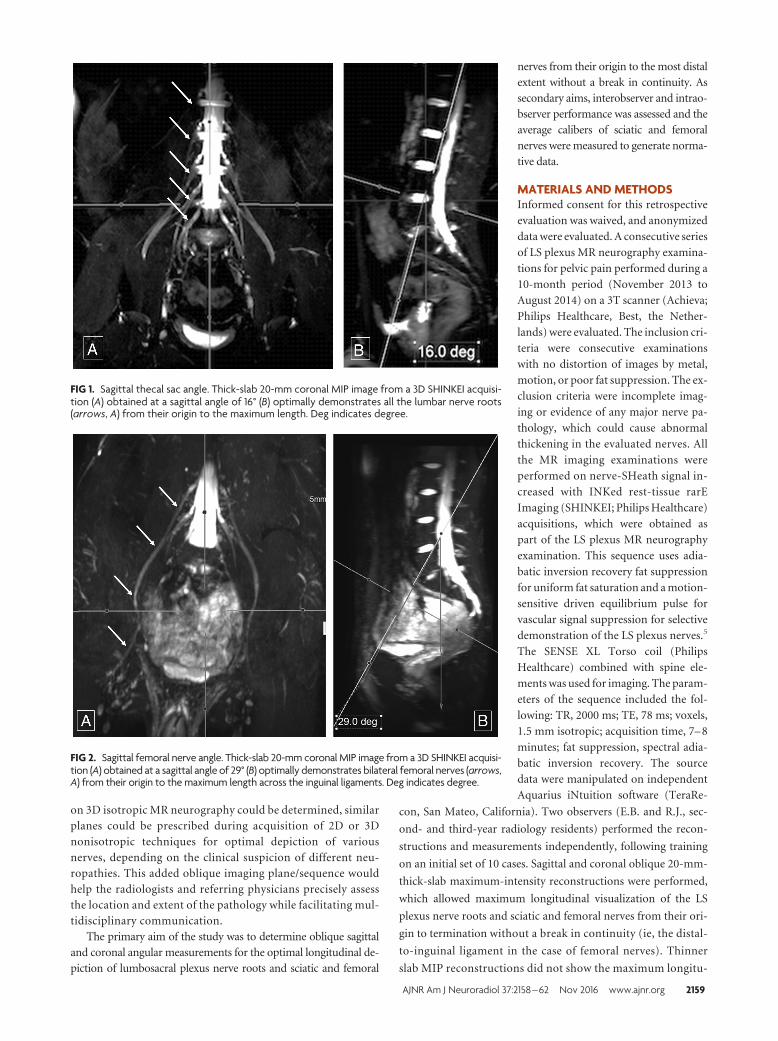
FIG 1. Sagittal thecal sac angle. Thick-slab 20-mm coronal MIP image from a 3D SHINKEI acquisi-tion (A) obtained at a sagittal angle of 16° (B) optimally demonstrates all the lumbar nerve roots(arrows, A) from their origin to the maximum length. Deg indicates degree.
FIG 2. Sagittal femoral nerve angle. Thick-slab 20-mm coronal MIP image from a 3D SHINKEI acquisi-tion (A) obtained at a sagittal angle of 29° (B) optimally demonstrates bilateral femoral nerves (arrows,A) from their origin to the maximum length across the inguinal ligaments. Deg indicates degree.
AJNR Am J Neuroradiol 37:2158 – 62 Nov 2016 www.ajnr.org 2159
dinal course of the nerves, while thicker slab MIP reconstruc-
tions resulted in blurring of the nerves due to overlapping of
adjacent structures. In our initial testing, 20-mm-thick slabs
provided the optimal longitudinal depiction of the nerves; MIP
parameters would not be expected to change substantially in
patients with conditions such as chronic inflammatory demy-elinating polyneuropathy, or neurofibromatosis, though pa-rameters could be optimized for each patient.
The sagittal plane measurements for the angle ventral to thethecal sac were denoted by positive numbers and dorsal to thethecal sac, by negative numbers. Femoral and sciatic nerve cali-bers were measured in their midportions in the abdomen and
pelvis, respectively, at the site of optimalnerve visualization and maximumthickness (ie, immediately below thepiriformis muscle and sciatic notch forthe sciatic nerve and adjacent to the ili-opsoas crotch for the femoral nerve).Descriptive statistics were performed,and data were expressed as mean � SD.Interobserver variance was evaluated byusing the intraclass correlation coeffi-cient (ICC), with values of �0.65 con-sidered good; �0.65 to �0.50, moder-ate; �0.50 to �0.40, fair; and �0.40,poor correlations. Intraobserver perfor-mance was also assessed by using theICC for the reader E.B., who obtainedrepeat measurements approximately 25weeks following the initial measurements.
RESULTSThere were 62 examinations with full
abdomen and pelvis MR neurography
imaging, though 10 were excluded due
to hardware artifacts (n � 3) and mo-
tion and suboptimal fat suppression and
incomplete imaging (n � 7). Final sam-
ples included 52 subjects, 11 men (mean
age, 45.73 � 18.4 years) and 41 women
(mean age, 48.85 � 12.65 years). All re-
constructions were successfully ob-
tained in �7 minutes.
Mean sagittal thecal sac angles for
the coronal demonstration of LS
plexus nerve roots from L1 to S1 for
the 2 independent observers measured
13.58° � 2.87° and 13.61° � 2.18° (Fig
1). For complete longitudinal nerve
course depiction bilaterally in the cor-
onal plane, the mean sagittal angular
measurements of the femoral nerve
were 27.78° � 4.81° and 28.94° � 4.49°
(Fig 2). Similarly, mean sagittal sciatic
nerve angle measurements for coronal
depiction were �10.7° � 3.75° and
�11.82° � 2.87° (Fig 3).
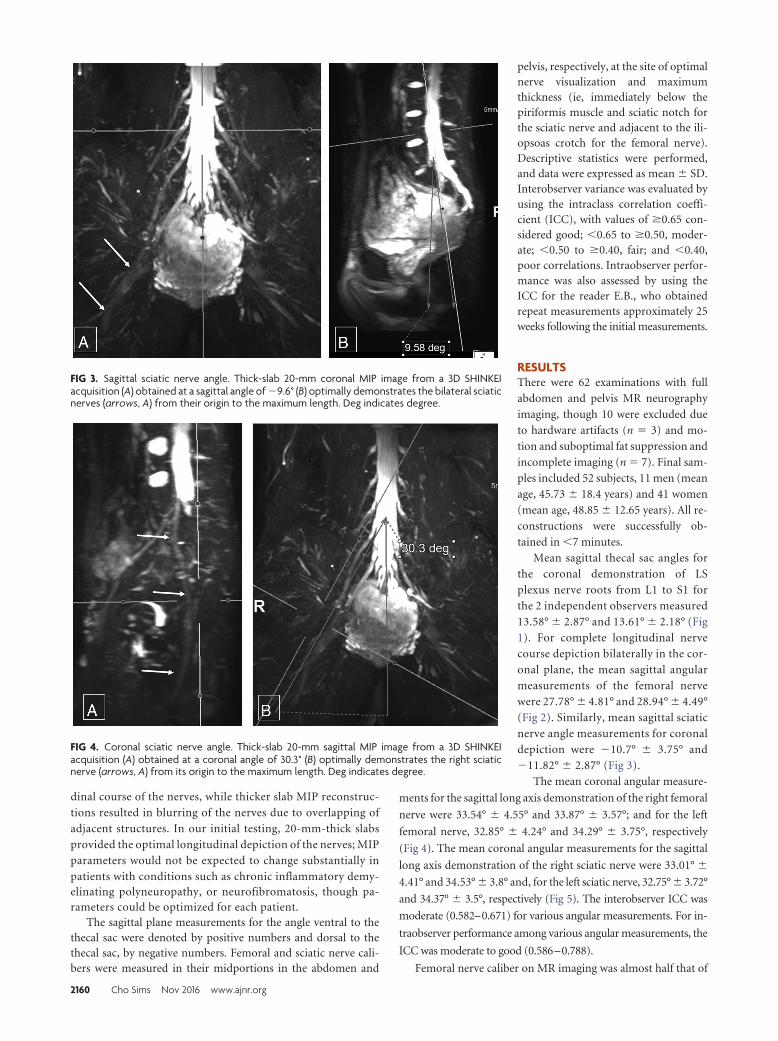
The mean coronal angular measure-
ments for the sagittal long axis demonstration of the right femoral
nerve were 33.54° � 4.55° and 33.87° � 3.57°; and for the left
femoral nerve, 32.85° � 4.24° and 34.29° � 3.75°, respectively
(Fig 4). The mean coronal angular measurements for the sagittal
long axis demonstration of the right sciatic nerve were 33.01° �
4.41° and 34.53° � 3.8° and, for the left sciatic nerve, 32.75° � 3.72°
and 34.37° � 3.5°, respectively (Fig 5). The interobserver ICC was
moderate (0.582–0.671) for various angular measurements. For in-
traobserver performance among various angular measurements, the
ICC was moderate to good (0.586–0.788).
Femoral nerve caliber on MR imaging was almost half that of
FIG 3. Sagittal sciatic nerve angle. Thick-slab 20-mm coronal MIP image from a 3D SHINKEIacquisition (A) obtained at a sagittal angle of �9.6° (B) optimally demonstrates the bilateral sciaticnerves (arrows, A) from their origin to the maximum length. Deg indicates degree.
FIG 4. Coronal sciatic nerve angle. Thick-slab 20-mm sagittal MIP image from a 3D SHINKEIacquisition (A) obtained at a coronal angle of 30.3° (B) optimally demonstrates the right sciaticnerve (arrows, A) from its origin to the maximum length. Deg indicates degree.
2160 Cho Sims Nov 2016 www.ajnr.org

the sciatic nerve. Mean right femoral
nerve thickness was 4.52 � 1.11 mm and
4.85 � 0.64 mm for the 2 observers, and
mean left femoral nerve thickness was
4.48 � 0.97 mm and 4.94 � 0.57 mm,
respectively. Mean right sciatic nerve
thickness was 9.71 � 1.76 mm and
9.94 � 0.83 mm, and mean left sciatic
nerve thickness was 10.03 � 1.71 mm
and 9.98 � 0.99 mm, respectively (Fig
6). The interobserver ICC was moderate
to good (0.557– 0.737) for the above
thicknesses except for the right femoral
nerve thickness, which was poor (0.343).
Intraobserver performance for nerve
thickness was moderate to good (0.646 –
0.756), except for the left sciatic nerve
thickness, which was poor (0.392)
(Table).
DISCUSSION3D SHINKEI allows excellent vascular
and fat signal suppression, leading to se-
lective nerve depiction.6,7 This sequence
uses adiabatic inversion recovery for
uniform fat suppression and a motion-
sensitive driven equilibrium pulse for
vascular signal suppression, thereby en-
abling selective demonstration of the LS
plexus nerves.5 The variable turbo spin-
echo component limits the acquisition
time to 7– 8 minutes.
This imaging technique allows ready
depiction of the nerves in various arbi-
trary planes. Thus, the angular prescrip-
tions could be easily and confidently
measured8,9; these results are reflected
in nearly good interobserver and
intraobserver performance in angular
measurements.
The femoral nerve calibers were
�4 –5 mm, the sciatic nerve calibers
were �8 –11 mm, and the interobserver
performance was moderate to good ex-
cept for the right femoral nerve, due to the relatively smaller size
of the femoral nerve. The normative data can be used for identi-
fication of pathologies that frequently cause abnormal nerve
thickening, such as chronic demyelinating polyneuropathy, dia-
betic polyneuropathy, perineurioma, or Charcot-Marie-Tooth
disease and so forth.10-12 The nerve-caliber measurements are
also reproducible, as shown by moderate-to-good ICC results for
intraobserver performance, except for the left sciatic nerve cali-
ber, which was poor (0.392).
Although this is the first study depicting angular and nerve-
caliber measurements, there are some limitations. We did not
obtain surgical or cadaveric correlation because it would not be
practical to obtain such correlations for a large sample. In addi-
FIG 5. Coronal femoral nerve angle. Thick-slab 20-mm sagittal MIP image from a 3D SHINKEIacquisition (A) obtained at a coronal angle of 31° (B) optimally demonstrates the right femoralnerve (arrows, A) from its origin to the maximum length. Deg indicates degree.
FIG 6. Femoral and sciatic nerve calibers. Thick-slab 20-mm sagittal MIP images from a 3DSHINKEI acquisition through the abdomen (A) and pelvis (B) show the femoral nerve diameter (4.8mm), which is almost one-half of sciatic nerve diameter (9.7 mm).
ICC: intraobserver performanceVariable Intraobserver ICC
Fem cor Lt 0.75146Fem cor Rt 0.69077Fem sag 0.78773Fem width Lt 0.64571Fem width Rt 0.75638L plexus sag 0.67175Sciatic width Lt 0.39228Sciatic width Rt 0.73182Sciatic cor Lt 0.58572Sciatic cor Rt 0.61695Sciatic sag 0.72101
Note:—Fem indicates femoral; cor, coronal; Lt, left; Rt, right; L, lumbrosacral; sag,sagittal.
AJNR Am J Neuroradiol 37:2158 – 62 Nov 2016 www.ajnr.org 2161
tion, no correlations with diffusion imaging were obtained, which
is also shown to produce nerve-selective images. The observers
felt confident in nerve identification on 3D SHINKEI during the
evaluation of the initial training set due to excellent vascular signal
suppression and nerve depiction by this technique. It was there-
fore decided that diffusion imaging evaluation can be avoided and
left as the subject of another article. Another limitation is that we
evaluated only patients with normal nerves. Patients with signifi-
cant nerve pathology/known mass lesions were excluded from
our patient population. Patients with significant nerve pathology
may require different angles to optimize the longitudinal display
of the nerves, due to distortion of nerve course. Nerve caliber in
these patients would also be expected to differ from that of pa-
tients with normal nerves. Finally, we did not measure obturator
nerve angles. Because this nerve has been shown to travel in a
straight coronal plane, it was thought that further study would not
yield a significant angle.13
CONCLUSIONSThe study fills a gap in the literature regarding angular LS plexus
nerve measurements for their maximum length depiction and nerve
thickness. It will aid in prescription of predictable nerve planes on
MR imaging for their optimal longitudinal demonstration.
ACKNOWLEDGMENTSThe authors would like to acknowledge Dr Vibhor Wadhwa for
help with image formatting.
Disclosures: Avneesh Chhabra—UNRELATED: Consultancy: Siemens (CAD Consult-ing); Royalties: Jaypee, Wolters Kluwer, Elsevier, Comments: book royalties; Paymentfor Development of Educational Presentations: educational symposia; OTHER: A.Chhabra has received research grants from GE-Radiology Research Academic Fel-lowship, Siemens, Gatewood Fellowship Award, and Integra Life Sciences.
REFERENCES1. Neufeld EA, Shen PY, Nidecker AE, et al. MR imaging of the lumbo-
sacral plexus: a review of techniques and pathologies. J Neuroimag-ing 2015;25:691–703 CrossRef Medline
2. Soldatos T, Andreisek G, Thawait GK, et al. High-resolution 3-T MRneurography of the lumbosacral plexus. Radiographics 2013;33:967– 87 CrossRef Medline
3. Delaney H, Bencardino J, Rosenberg ZS. Magnetic resonance neu-rography of the pelvis and lumbosacral plexus. Neuroimaging Clin NAm 2014;24:127–50 CrossRef Medline
4. Vargas MI, Gariani J, Delattre BA, et al. Three-dimensional MR im-aging of the brachial plexus. Semin Musculoskelet Radiol 2015;19:137– 48 CrossRef Medline
5. Kasper JM, Wadhwa V, Scott KM, et al. SHINKEI–a novel 3D isotro-pic MR neurography technique: technical advantages over3DIRTSE-based imaging. Eur Radiol 2015;25:1672–77 CrossRefMedline
6. Yoneyama M, Takahara T, Kwee TC, et al. Rapid high resolution MRneurography with a diffusion-weighted pre-pulse. Magn Reson MedSci 2013;12:111–19 CrossRef Medline
7. Huisman M, Staruch RM, Ladouceur-Wodzak M, et al. Non-invasivetargeted peripheral nerve ablation using 3D MR neurography andMRI-guided high-intensity focused ultrasound (MR-HIFU): pilotstudy in a swine model. PLoS One 2015:14;10:e0144742 CrossRefMedline
8. Burge AJ, Gold SL, Kuong S, et al. High-resolution magnetic reso-nance imaging of the lower extremity nerves. Neuroimaging Clin NAm 2014;24:151–70 CrossRef Medline
9. Chhabra A, Rozen S, Scott K. Three-dimensional MR neurographyof the lumbosacral plexus. Semin Musculoskelet Radiol 2015;19:149 –59 CrossRef Medline
10. Ellegala DB, Monteith SJ, Haynor D, et al. Characterization of genet-ically defined types of Charcot-Marie-Tooth neuropathies by usingmagnetic resonance neurography. J Neurosurg 2005;102:242– 45CrossRef Medline
11. Mauermann ML, Amrami KK, Kuntz NL, et al. Longitudinalstudy of intraneural perineurioma: a benign, focal hypertrophicneuropathy of youth. Brain 2009;132(pt 8):2265–76 CrossRefMedline
12. Lozeron P, Lacour MC, Vandendries C, et al. Contribution of plexusMRI in the diagnosis of atypical chronic inflammatory demyelinat-ing polyneuropathies. J Neurol Sci 2016;360:170 –75 CrossRefMedline
13. Petchprapa CN, Rosenberg ZS, Sconfienza LM, et al. MR imaging ofentrapment neuropathies of the lower extremity, Part 1: the pelvisand hip. Radiographics 2010;30:983–1000 CrossRef Medline
2162 Cho Sims Nov 2016 www.ajnr.org