research article open access in lumbosacral plexus ... fileresults: lumbosacral plexus injuries...
TRANSCRIPT

JOURNAL OF BRACHIAL PLEXUS AND PERIPHERAL NERVE INJURY
Garozzo et al. Journal of Brachial Plexus and Peripheral Nerve Injury 2014, 9:1http://www.jbppni.com/content/9/1/1
RESEARCH ARTICLE Open Access
In lumbosacral plexus injuries can we identifyindicators that predict spontaneous recovery orthe need for surgical treatment? Results from aclinical study on 72 patientsDebora Garozzo*, Gianluca Zollino and Stefano Ferraresi
Abstract
Background: Post-traumatic lumbosacral plexus injuries seem to be rare events, spontaneously recovering in highpercentage: as surgery is often challenging and results in poor outcome, many Authors have advocated conservativetreatment only. Nevertheless surgery should not be ruled out: in invalidating injuries, it can restore basic function in thelower extremities.Therefore, it might be necessary to establish guidelines for the management and the indication to surgery in such cases.This study aims to identify indicators predicting spontaneous recovery or the need for surgery.
Method: The clinical and radiological data of 72 patients with a post-traumatic lumbosacral plexus injury werereviewed. A follow up equal or superior to 3 years is available in 42 cases.
Results: Lumbosacral plexus injuries mostly occurred during road accidents. The incidence of associated lesions wasrelevant: bone injuries were found in 85% of patients, internal lesions in 30% and vascular injuries in 8%.Lumbosacral trunk and sacral plexus palsies were the most frequent injury patterns.Root avulsions were revealed in 23% of cases and only in sacral plexus and complete lumbosacral plexus injuries: L5 andS1 were the roots more prone to avulsions.About 70% of cases recovered spontaneously, mostly in 18 months. Spontaneous recovery was the rule in lumbar plexusand lumbosacral trunk injuries (where root avulsions never occurred) or in sacral and complete lumbosacral plexuspalsies due to compression injuries.The causative mechanism correlated with the injury pattern, the associated bone injury being often predictive of theseverity of the nerve injury.Lumbosacral plexus injuries occurred in car crashes were generally associated with fractures causing compression on thenerves, thus resulting in injuries often amenable of spontaneous recovery.Motorcycle accidents implied high kinetic energy traumas where traction played an important role, as the highpercentage of sacroiliac joint separations demonstrated (found in more than 50% of cases and always associatedto root avulsions).Loss of sphincteral control and excruciating leg pain were also invariably associated with avulsions.
Conclusions: Clinical and radiological data can help to predict the occurrence of spontaneous recovery or theneed for surgery in post-traumatic lumbosacral plexus injuries.
Keywords: Lumbosacral plexus, Nerve injury, Root avulsions, Pelvic trauma, Sacroiliac joint separation,Sacral fracture
* Correspondence: [email protected] of Neurosurgery, Ospedale S. Maria della Misericordia, Viale TreMartiri 140, 45100 Rovigo, Italy
© 2014 Garozzo et al.; licensee BioMed CentraCommons Attribution License (http://creativecreproduction in any medium, provided the or
l Ltd. This is an open access article distributed under the terms of the Creativeommons.org/licenses/by/2.0), which permits unrestricted use, distribution, andiginal work is properly cited.

Garozzo et al. Journal of Brachial Plexus and Peripheral Nerve Injury 2014, 9:1 Page 2 of 9http://www.jbppni.com/content/9/1/1
BackgroundThe lumbosacral plexus was illustrated by the Italiananatomist Giulio Casserio in 1632 [1] but the firstdescription of a post-traumatic injury only dates backto approximately 50 years ago [2].Lumbosacral plexus (LSP) injuries are considered rare
events and their spontaneous recovery occurs in highpercentage [3-6]: on the other hand, surgery is oftendemanding and implies multidisciplinary cooperation,whereas outcome is frequently poor. Therefore, someAuthors advocate only conservative treatment [5].Yet, in cases of severe, devastating injuries with no
chances of spontaneous recovery, the role of surgery shouldnot be denied as it could offer undoubted advantages topatients otherwise destined to permanent disability [7].It can be clearly inferred that there is a need to establish
precise guidelines concerning the management and timingfor surgery in these lesions, ruling out those patientsamenable of a spontaneous recovery.The aim of this study was to identify indicators that
could help to predict the occurrence of spontaneousrecovery or the need for surgical treatment.
MethodsThe present study was a retrospective review of a seriesof 72 patients with a post-traumatic LSP injury. Theywere referred to the Authors’ observation several monthsor even years after the injury in the period betweenJanuary 1991 and May 2009.The Authors collected and analysed the data regarding:
a) Gender and age incidenceb) Causative eventsc) Timing of diagnosisd) Association with other injuriese) Incidence of bilateral injuriesf ) Injury patternsg) Incidence of root avulsionsh) Natural history
a) Gender and age incidence
The series consist of 60 males and 12 females,including two children (a 4 year old boy and a 18month old girl). Patients’ age ranged from 18months to 68 years, with no peak of age.b) Causative events
LSP injuries mostly occurred in road accidents: carcrashes in 59% of cases and motorcycle accidents inabout 20%.A detailed description of the causative events isgiven on Table 1.c) Timing of diagnosis
The nerve injury was diagnosed during the earlyassessment after the trauma in 30% of cases, aboutone month later in 20%, within 3 months in 25%,within 6–8 months in 10% of cases. In 5% ofpatients the nerve injury was diagnosed one yearafter the traumatic event.d) Associated injuries
The patients described in this series were oftenmulti-traumatized but for “associated injuries” here wedeal only with the injuries strictly correlated to the LPSinjury: bone injuries of the pelvic ring and proximalfemoral fractures, internal and vascular injuries.Bone injuries were found in 83% of cases.Every type of pelvic fracture could be found(including comminuted fractures of the pelvic ring);iliac-ischiopubic fractures, acetabular fractures(Figure 1), sacroiliac joint separations (Figure 2)and sacral fractures were the most frequent injuries.Internal injuries were present in about 30% of patients.Extraperitoneal bladder ruptures with a clinicalpresentation of hematuria, abdominal pain and/orabdominal swelling and requiring urgent surgery werethe most common internal injuries (found in morethan 50% of cases): they could be caused by directpenetration of a bone fragment in ischiopubic orsinphysis fractures or due to rupture or stretchinginjury of the pubo-prosthatic ligament. Bladder injurieswere more likely to occur when the organ was full.Intestinal perforation generally occurred in theterminal part of the sigmoid colon or rectum.Vascular injuries were found in about 8% of cases:in these patients a retroperitoneal haemorrhagedue to bleeding of the gluteal (2 cases) or iliacvein (2 cases) or artery (2 cases) required urgentembolization. In 30% of cases nerve injurieswere associated with both bone and internalor vascular injuries.e) Incidence of bilateral injuries
A bilateral palsy of the lumbosacral plexus occurredin 5,5% of cases (4 patients): 1 bilateral lumbarplexus injury, 2 bilateral sacral palsies, 1 completelumbosacral palsy associated with a sacral palsy.f ) Clinical patterns
Patients were always evaluated clinically and byelectrodiagnostic and radiological studies.Clinical assessment was focused to reveal musclesweakness and sensory disturbances as well as the
Table 1 Causative mechanisms in lumbosacral plexus injuries
Car crash Motorcycle crash Other roadaccidents
Work accident(different from fallingfrom on high)
Sport accident Accidental fallingfrom above (includingwork accident)
Other
Detaileddescriptionof the causalmechanisms
Frontal impact againstanother veicle (car, van,track, motorcycle): 9 cases
Frontal impactagainst anotherveicle (car): 6 cases
3 patients (2 smallchildren) run downby a car
1 patient° crushed by asilo falling from on highon a ship
1 ski accident (hitting apole during downhill)
1 patient falling fromthe fourth floor
1 patient run down byan earthwork and hitby a slab of asphalt
Frontal impact against theguardrail: 2 cases or a wall:1 case or a gate: 2 cases
Bus/trackoverriding themotorcycle: 2cases
1 patient crushedby a car againsta gate
1 patient whose footwas trapped and pulledby a reaper machine
1 watermotorcycleaccident
1 Pt. fell off a ladder, lied on thefloor on his left side for several hrsbefore rescue (nervous ischemia?)
Frontal impact againsta tree lining the road:4 cases
1 patient slipping andfalling down during abasketball match
2 patients falling from a scaffold(work accident)
Driving off the road:8 cases
No clear car accidentdynamics:17 cases
No clear accidentdynamics: 7 cases
Total numberof patients
43* 15* 4 2 3 4* 1
*patient with associated brachial plexus palsy °patient with bilateral lumbosacral plexus palsy.
Garozzo
etal.Journalof
BrachialPlexusand
PeripheralNerve
Injury2014,9:1
Page3of
9http://w
ww.jbppni.com
/content/9/1/1

Fig
Figure 1 Acetabular fracture.
Garozzo et al. Journal of Brachial Plexus and Peripheral Nerve Injury 2014, 9:1 Page 4 of 9http://www.jbppni.com/content/9/1/1
presence of pain. The Medical Research CouncilGrading system was used to classify muscle strength [8].A thorough examination of the hip girdle and legenabled to classify the injury pattern. The superiorgluteal nerve and the posterior cutaneous nerve ofthe thigh are two nerves whose function wascarefully assessed to determine the level of theinjury. The superior gluteal nerve emerges from theL5 spinal nerve or the lumbosacral trunk and fromthe S1 spinal nerve in the proximal part of thelumbosacral plexus: abolished activity in gluteimuscles impairs pelvic stability resulting in a positiveTrendelemburg test. The posterior cutaneous nerveof the thigh forms at the lateral part of the sacralplexus as the sacral spinal nerves merge to form thesciatic nerve: its intact function (sensation on the
ure 2 Sacroiliac joint separation.
dorsal aspect of the thigh) with abolished activity inharmstrings and muscles distal to the knee indicatesa proximal sciatic nerve lesion rather than a sacralplexus injury.Concerning electrodiagnostic tools, studies withH-reflex and F-response as well electromyography(EMG) were usually performed: EMG could berepeated at regular intervals to reveal possible signsof progressive reinnervation.Radiological investigations included X-rays,angiography, CT and MRI imaging: theseexaminations were generally performed duringthe early assessment after the trauma whereas.CT myelography and 3 dimensional MRI (3DMRI)were prescribed after avulsive injuries weresuspected (mostly after referral to the Authors).
Garozzo et al. Journal of Brachial Plexus and Peripheral Nerve Injury 2014, 9:1 Page 5 of 9http://www.jbppni.com/content/9/1/1
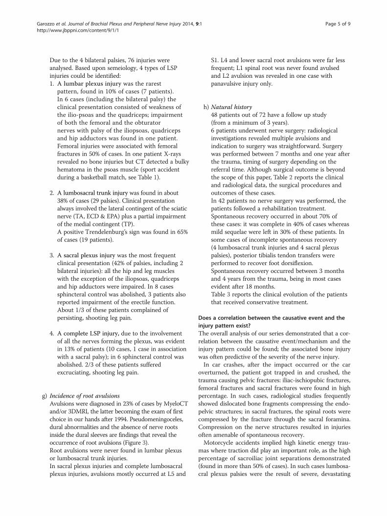
Due to the 4 bilateral palsies, 76 injuries wereanalysed. Based upon semeiology, 4 types of LSPinjuries could be identified:1. A lumbar plexus injury was the rarest
pattern, found in 10% of cases (7 patients).In 6 cases (including the bilateral palsy) theclinical presentation consisted of weakness ofthe ilio-psoas and the quadriceps; impairmentof both the femoral and the obturatornerves with palsy of the iliopsoas, quadricepsand hip adductors was found in one patient.Femoral injuries were associated with femoralfractures in 50% of cases. In one patient X-raysrevealed no bone injuries but CT detected a bulkyhematoma in the psoas muscle (sport accidentduring a basketball match, see Table 1).
2. A lumbosacral trunk injury was found in about38% of cases (29 palsies). Clinical presentationalways involved the lateral contingent of the sciaticnerve (TA, ECD & EPA) plus a partial impairmentof the medial contingent (TP).A positive Trendelenburg’s sign was found in 65%of cases (19 patients).
3. A sacral plexus injury was the most frequentclinical presentation (42% of palsies, including 2bilateral injuries): all the hip and leg muscleswith the exception of the iliopsoas, quadricepsand hip adductors were impaired. In 8 casessphincteral control was abolished, 3 patients alsoreported impairment of the erectile function.About 1/3 of these patients complained ofpersisting, shooting leg pain.
4. A complete LSP injury, due to the involvementof all the nerves forming the plexus, was evidentin 13% of patients (10 cases, 1 case in associationwith a sacral palsy); in 6 sphincteral control wasabolished. 2/3 of these patients sufferedexcruciating, shooting leg pain.
g) Incidence of root avulsions
Avulsions were diagnosed in 23% of cases by MyeloCTand/or 3DMRI, the latter becoming the exam of firstchoice in our hands after 1994. Pseudomeningoceles,dural abnormalities and the absence of nerve rootsinside the dural sleeves are findings that reveal theoccurrence of root avulsions (Figure 3).Root avulsions were never found in lumbar plexusor lumbosacral trunk injuries.In sacral plexus injuries and complete lumbosacralplexus injuries, avulsions mostly occurred at L5 andS1. L4 and lower sacral root avulsions were far lessfrequent; L1 spinal root was never found avulsedand L2 avulsion was revealed in one case withpanavulsive injury only.
h) Natural history
48 patients out of 72 have a follow up study(from a minimum of 3 years).6 patients underwent nerve surgery: radiologicalinvestigations revealed multiple avulsions andindication to surgery was straightforward. Surgerywas performed between 7 months and one year afterthe trauma, timing of surgery depending on thereferral time. Although surgical outcome is beyondthe scope of this paper, Table 2 reports the clinicaland radiological data, the surgical procedures andoutcomes of these cases.In 42 patients no nerve surgery was performed, thepatients followed a rehabilitation treatment.Spontaneous recovery occurred in about 70% ofthese cases: it was complete in 40% of cases whereasmild sequelae were left in 30% of these patients. Insome cases of incomplete spontaneous recovery(4 lumbosacral trunk injuries and 4 sacral plexuspalsies), posterior tibialis tendon transfers wereperformed to recover foot dorsiflexion.Spontaneous recovery occurred between 3 monthsand 4 years from the trauma, being in most casesevident after 18 months.Table 3 reports the clinical evolution of the patientsthat received conservative treatment.Does a correlation between the causative event and theinjury pattern exist?The overall analysis of our series demonstrated that a cor-relation between the causative event/mechanism and theinjury pattern could be found; the associated bone injurywas often predictive of the severity of the nerve injury.In car crashes, after the impact occurred or the car
overturned, the patient got trapped in and crushed, thetrauma causing pelvic fractures: iliac-ischiopubic fractures,femoral fractures and sacral fractures were found in highpercentage. In such cases, radiological studies frequentlyshowed dislocated bone fragments compressing the endo-pelvic structures; in sacral fractures, the spinal roots werecompressed by the fracture through the sacral foramina.Compression on the nerve structures resulted in injuriesoften amenable of spontaneous recovery.Motorcycle accidents implied high kinetic energy trau-
mas where traction did play an important role, as the highpercentage of sacroiliac joint separations demonstrated(found in more than 50% of cases). In such cases lumbosa-cral plexus palsies were the result of severe, devastating

Figure 3 MRI study of complete LSP injury with multiple avulsions.
Garozzo et al. Journal of Brachial Plexus and Peripheral Nerve Injury 2014, 9:1 Page 6 of 9http://www.jbppni.com/content/9/1/1
stretch injuries: radiological studies always revealed rootavulsions in patients with sacroiliac joint separations.Concerning the clinical presentation, abolished sphinc-
teral control was a bad prognostic factor as it was almostinvariably associated with avulsions.
DiscussionIn recent times, reports have been published in theliterature stating that LSP injuries might be significantlymore frequent than it was thought in the past: Lindahland Hirvensalo [9] report an incidence of LSP injuriesequal to 40% in type-C pelvic ring injuries, Tonetti et al.[6] find out that about 52% of posterior osteo-ligamentaryinjuries were associated with neurological symptoms. Thepresumed rare incidence of these injuries might be dueto the fact that they might go unrecognized in many cases[6,7,10]. Neurological complications are easily underesti-mated during the early assessment of these patients afterthe trauma. Patients are often multitraumatized andthe life-treating injuries captivate the attention of thephysicians; on the other hand the patient may beunconscious or uncooperative and thus unable to bringthe palsy to notice. A careful, complete neurologicalexamination is often difficult or impossible: Tonetti [6]
reports that it could not be performed at admission in 10out of 44 patients included in their series of posteriorosteoligamentary lesions of the pelvic girdle.Functional impairment of the lower limbs can also
initially be put down to bone injuries. Weis [11] ana-lysed 28 patients with a fracture of the acetabulum,pelvis or sacrum and found that in 11 cases there wereelectromyographic changes due to a lumbosacral plexusinjury: clinical manifestations seemed very subtle butfollow up demonstrated that even mild, subclinicalfunctional impairment did interfere with the rehabili-tation process.In our series, only 30% of the nerve injuries were
recognized during the early diagnostic assessment afterthe trauma, in many of the remaining cases there was asignificant delay in diagnosis that resulted in late referralto the Authors. Moreover, the vast majority of thesepatients had been offered conservative treatment only. Aspreviously pointed out, LSP injuries do recover spon-taneously in high percentage whereas on the other handsurgery is known to be demanding and often resulting inpoor outcomes: these facts nourish scepticism in manyphysicians that therefore advocate conservative treatment,ruling surgery out.

Table 2 Nerve surgery and outcomes
Patients’ age (years) andgender, causative mechanism
Injury pattern Associated injuries Root avulsions Timing of surgery(months after injury)
Surgical procedure Outcome
33, M, motorcycle accident Complete lumbosacral injury Sacroiliac joint separation,bladder injury
L5, S1 9 Laminotomy + Henry’s approach:from the contralateral plexus suralgrafts to the gluteal nerves
No result
13, M, sport accident Complete lumbosacral injury Sacroiliac joint separation,retroperitoneal hematoma
L3,L4,L5 12 Anterolateral extraperitoneal approach:from the contralateral obturator nervetwo grafts to the femoral nerve
No result
20, F, car crash Complete lumbosacral injury Multiple fractures of thepelvic ring, femoral fracture
L4, L5, S1 10 Laminotomy + Henry’s approach:from the contralateral plexus suralgrafts to the gluteal nerves
No result
18, M, motorcycle accident Complete lumbosacralplexus injury
Sacroiliac joint separation,retroperitoneal hematoma
L2,L3,L4, L5, S1,S2 9 Laminotomy + anterior approach:from the contralateral plexus 2sural grafts to the femoral nerve
No result
25, M, motorcycle accident Complete lumbosacral palsy Sacroiliac joint separation L4,L5,S1 7 Laminectomy + Henry’s approach:from the contralateral plexus nervegrafts to the gluteal and sciatic nerves
No result
24, M, car crash Sacral plexus injury Iliac-ischiopubic fractures,sacral fracture
L5 10 Hemilaminectomy: neurolysis anddecompression of S1 from abone fragment
Global improvementof the leg
Garozzo
etal.Journalof
BrachialPlexusand
PeripheralNerve
Injury2014,9:1
Page7of
9http://w
ww.jbppni.com
/content/9/1/1

Table 3 Clinical evolution after conservative treatment
N° of cases per injury patter Root avulsions Complete spontaneousrecovery
Incomplete spontaneousrecovery
No spontaneousrecovery
Lumbar plexus injuries: 5 patients* None 5
Lumbosacral plexus injuries: 13 patients None 7 6^
Sacral plexus injuries: 18 patients Avulsions were detectedin 7 cases
5 4° 9
Complete LSP injuries: 6 patients§ Avulsions were detectedin 3 cases
3 3§
Total number of patients: 42 17 13 12
*1 bilateral lumbar plexus palsy.^no recovery of foot dorsiflexion, in 4 cases posterior tibialis muscle scored M4 and these patients underwent a tibialis transfer.°no recovery of foot dorsiflexion, tibialis tendon transfer was perfomed.§1patient with an association of complete LSP injury and sacral plexus palsy, the latter presenting complete recovery.
Garozzo et al. Journal of Brachial Plexus and Peripheral Nerve Injury 2014, 9:1 Page 8 of 9http://www.jbppni.com/content/9/1/1
Yet the role of surgery should not be denied: althoughfunction in the foot cannot be restored after severeinjuries, if reinnervation occurs in a few proximal keymuscles of the leg the situation of the patient can dra-matically improve, changing from being in a wheelchairto being able to stand and walk independently [7]. Sur-gery can also contribute in reducing pain, remarkablyimproving the quality of life and allowing to resumerehabilitation [7].Better surgical outcomes could be obtained if early
diagnostic assessment and a correct surgical timing werethe rule: therefore it would be useful to establish preciseguidelines of management and treatment.Even if it might be objected that the number of patients
in this series cannot account to statistically draw relevantconclusions, the present study clearly demonstrated corre-lations between the causative mechanism of the nerveinjuries and their clinical presentation, pointing out pos-sible indicators that seemed to distinguish the caseseligible for surgical treatment from those amenable ofspontaneous recovery.In plexus injuries, the first step of the diagnostic assess-
ment is to detect the presence of root avulsions. Whenmultiple avulsions are revealed, indication to surgery isundoubtedly straightforward: preganglionic injuries haveno chances to recover spontaneously.In this series root avulsions were never detected in lum-
bar plexus. These palsies were associated in high percent-age to femoral fractures: the femoral nerve was injured bycompression due to a peri-fracture hematoma and recov-ery was the rule, as it was previously found by Tonetti [6].No root avulsions were revealed in lumbosacral trunk
injuries; at follow up spontaneous recovery (complete orpartial) always occurred, as it was also reported in theliterature [6].It might be therefore inferred that nerve surgery could
be ruled out in these two injury patterns and rehabili-tation is the treatment of choice (plus palliative surgeryin case of incomplete recovery in lumbosacral trunkinjuries).
Radiological studies revealed multiple root avulsions insacral plexus and complete lumbosacral plexus injuries.In our series root avulsions were invariably detected in
patients with sacroiliac joint separations (found in highpercentage in injuries sustained during motorcycle acci-dents), in agreement with findings previously reported inthe literature [6,12].Sacral fractures seem to mostly be associated to crush
injuries in our series; in this kind of bone injury, a com-pression of the spinal roots by the fracture through thesacral foramina is reported between 22 and 56% of casesin the literature [6,13-16]. Although these cases are amen-able of spontaneous recovery, experience has proved thatearly surgery with reduction of the fracture and conse-quent decompression of the spinal nerves leads to a fasterand better recovery [6,14].Thus we conclude that it is possible to extrapolate that
in post-traumatic LPS injuries the clinical pattern itselfcan predict the occurrence of a spontaneous recovery andthat the causative event does affect the injury prognosis.Patients that sustained the injury during motorcycle acci-dents have high chances to be candidates to surgery: insuch traumas, traction does play an important role androot avulsions are likely to be found, especially if sacroiliacjoint separation and loss of sphincterial control are as-sociated with complete functional impairment. Severe,shooting leg pain is another bad prognostic indicator,as it was already pointed out in the literature [6,7].Finally we would like to remark that in agreement with
the literature [3-6,15,17,18], in our series spontaneousrecovery occurred in high percentage of cases. A lowerincidence of avulsions in comparison to the brachialcounterpart (where avulsions can be found in up to 70%of cases) has been found in our series (23%) and in theliterature [5,12,19] but it does not seem to be the onlyexplanation. A high rate of anatomical variations of thelumbar and sacral roots have been reported in the litera-ture [20,21], described as between 14 and 30% overalland 13% of L4 to sacral roots in cadaver studies: theseseem to occur as extradural or intradural anastomosis as

Garozzo et al. Journal of Brachial Plexus and Peripheral Nerve Injury 2014, 9:1 Page 9 of 9http://www.jbppni.com/content/9/1/1
well as extradural nerve root divisions. Some of therecovery after a lumbosacral plexus injury can thereforeoccur through “collateral sprouting” from healthy or lessseverely injured neighboring nerves [7].
ConclusionsAlthough the data reported in this study might need fur-ther confirmation in larger series, the present analysisdemonstrated that in post-traumatic LSP palsies it is pos-sible to predict the occurrence of spontaneous recoverybased on clinical and radiological data. In lumbar plexusand lumbosacral trunk injuries spontaneous recoveryalways occurs and therefore rehabilitation is the treat-ment of choice. Sacral and lumbosacral plexus palsiesalso recover spontaneously in high percentage, whenthe nerve injury is due to compression by dislocatedbone fragments: anyway in some cases (sacral fractures)surgical decompression can play a role allowing a fasterand better recovery.Palliative surgery can correct incomplete recovery.Traction injuries (mostly occurring in motorcycle ac-
cidents) are more likely destined to severe, permanentdisability. Patients with sacroiliac joint separations associ-ated with a clinical presentation of complete functionalimpairment, loss of sphincteral control and shooting legpain invariably reveal multiple root avulsions when accur-ate radiological investigations are performed: they shouldreceive surgical treatment as soon as the general clinicalconditions allow.Early diagnostic assessment and referral to surgeons
are certainly crucial to improve surgical outcome.
AbbreviationsPT: Post-traumatic; LSP: Lumbosacral plexus.
Competing interestsThe authors declare that they have no competing interests.
Authors’ contributionsDG conceived the idea of this study, analysed the data, drafted the manuscript,revised it and gave final approval of the version to be published. GZ and SFcollected the data. All authors read and approved the final manuscript.
Received: 27 June 2013 Accepted: 27 December 2013Published: 11 January 2014
References1. Casserio G: Tabulalae Anatomicae. Bucretius; 1632.2. Finney LA, Wulfmann WA: Traumatic intradural lumbar nerve root
avulsion with associated traction injury to the common peroneal nerve.AM J Roentgenol Radium Ther Nucl Med 1960, 84:952–957.
3. Huittinen VM, Slätis P: Nerve injury in double vertical pelvic fractures.Acta Chir Scand 1972, 138:571–575.
4. McLennan JE, McLaughlin WT, Skillicorn SA: Traumatic lumbar nerve rootmeningocele. Case report. J Neurosurg 1973, 39:528–532.
5. Stoehr M: Traumatic and postoperative lesions of the lumbosacralplexus. Arch Neurol 1978, 35:757–760.
6. Tonetti J, Cazal C, Eid A, Badulescu A, Martinez T, Vouaillat H, Merloz P:Neurological damage in pelvic injuries: a continuous prospective seriesof 50 pelvic injuries treated with an iliosacral lag screw. Rev Chir OrthopReparatrice Appar Mot 2004, 90(2):122–131.
7. Lang E, Borges J, Carlstedt T: Surgical treatment of lumbosacral plexusinjuries. J Neurosurg 2004:64–71. Spine/volume 1/ July.
8. Medical Research Council Scale: Aids to the examination of the peripehral nervoussystem. Memorandum n°45. London: Her Majesty’s Stationary Office; 1976.
9. Lindahl J, Hirvensalo E: Outcome of operatively treated type-C injuries ofthe pelvic ring. Acta Orthop 2005, 76(5):667–678.
10. Hersche O, Isler B, Aebi M: Follow up and prognosis of neurologicalsequelae of pelvic ring fractures with involment of the sacrum and /orthe iliosacral joint. Unfallchirug 1993, 96(6):311–318.
11. Weis EB jr: Subtle neurological injuries in pelvic fractures. J Trauma 1984,24(11):983–985.
12. Barnett HJ, Connoly ES: Lumbosacral nerve root avulsion: report of a caseand review of the literature. J Trauma 1975, 15:532–535.
13. Rai SK, Far RF, Ghovanlou B: Neurological deficits associated with sacralwing fractures. Orthopedics 1990, 13(12):1363–1366.
14. Denis F, Davis S, Comfort T: Sacral fractures: an important problem.Retrospective analysis of 236 cases. Clin Orthop 1988, 227:67–81.
15. Majeed SA: Neurologic deficits in major pelvic injuries. Clin Orthop 1992,282:22–228.
16. Sabiston CP, Wing PC: Sacral fractures: classification and neurologicimplications. J Trauma 1986, 6:197–202.
17. Bonin JG: Sacral fractures and injuries of the cauda equina. J Bone JoinSurg (Br) 1945, 27:113–127.
18. Froman C, Stein A: Complicated crushing injuries of the pelvis. J Bone JoinSurg (Br) 1967, 49:24–32.
19. Ikeda K, Wada E, Kodam N: Traction injury of the lumbosacral spinal nerveroots. Spine 1991, 16:368–371.
20. Chotigavanich C, Sawangnatra S: Anomalies of the lumbosacral nerveroots. An anatomic investigation. Clin Orthop Res 1992, 278:46–50.
21. Stevanato G, Vazzana L, Daramaras S, Trincia G, Saggioro GC, Squintani G:Lumbosacral plexus lesions. Acta Neurochir Suppl 2007, 100:15–20.
doi:10.1186/1749-7221-9-1Cite this article as: Garozzo et al.: In lumbosacral plexus injuries can weidentify indicators that predict spontaneous recovery or the need forsurgical treatment? Results from a clinical study on 72 patients. Journalof Brachial Plexus and Peripheral Nerve Injury 2014 9:1.
Submit your next manuscript to BioMed Centraland take full advantage of:
• Convenient online submission
• Thorough peer review
• No space constraints or color figure charges
• Immediate publication on acceptance
• Inclusion in PubMed, CAS, Scopus and Google Scholar
• Research which is freely available for redistribution
Submit your manuscript at www.biomedcentral.com/submit