21-1 chapter 21 the cardiovascular system: blood vessels and hemodynamics structure and function of...
TRANSCRIPT
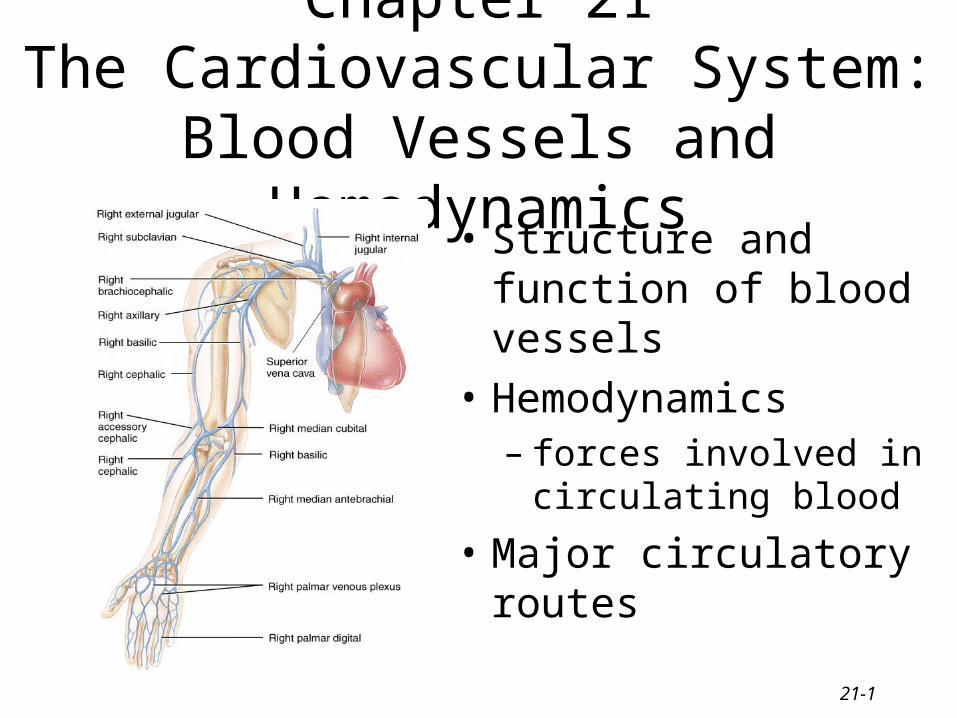
21-1
Chapter 21The Cardiovascular System: Blood
Vessels and Hemodynamics• Structure and function of
blood vessels
• Hemodynamics– forces involved in
circulating blood
• Major circulatory routes
21-2
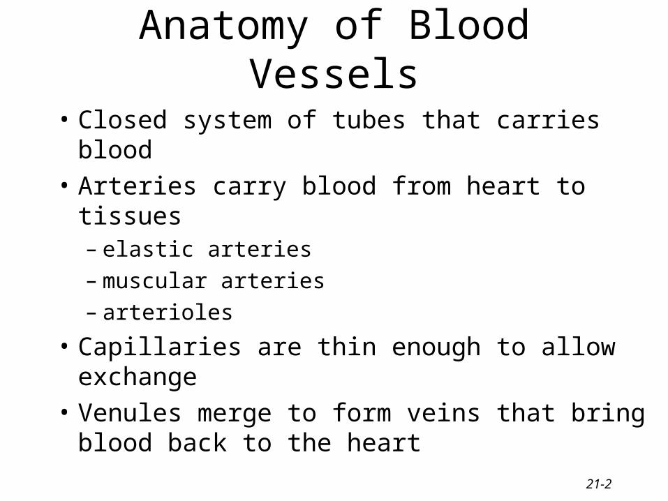
Anatomy of Blood Vessels
• Closed system of tubes that carries blood
• Arteries carry blood from heart to tissues– elastic arteries– muscular arteries– arterioles
• Capillaries are thin enough to allow exchange
• Venules merge to form veins that bring blood back to the heart
21-3
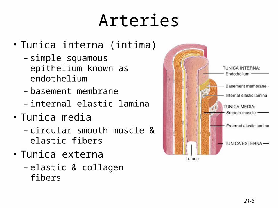
Arteries• Tunica interna (intima)
– simple squamous epithelium known as endothelium
– basement membrane– internal elastic lamina
• Tunica media– circular smooth muscle &
elastic fibers
• Tunica externa– elastic & collagen fibers
21-4
Sympathetic Innervation
• Vascular smooth muscle is innervated by sympathetic nervous system– increase in stimulation causes muscle contraction or
vasoconstriction• decreases diameter of vessel
– injury to artery or arteriole causes muscle contraction reducing blood loss (vasospasm)
– decrease in stimulation or presence of certain chemicals causes vasodilation
• increases diameter of vessel
• nitric oxide, K+, H+ and lactic acid cause vasodilation
21-5
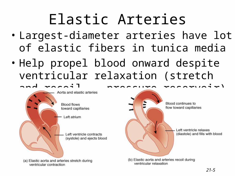
Elastic Arteries• Largest-diameter arteries have lot of elastic fibers
in tunica media
• Help propel blood onward despite ventricular relaxation (stretch and recoil -- pressure reservoir)
21-6
Muscular Arteries
• Medium-sized arteries with more muscle than elastic fibers in tunica media
• Capable of greater vasoconstriction and vasodilation to adjust rate of flow– walls are relatively thick– called distributing arteries because they direct
blood flow
21-7
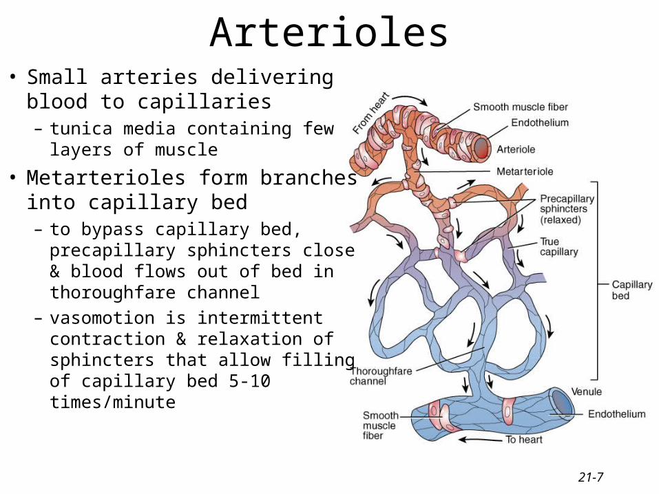
Arterioles• Small arteries delivering blood to
capillaries– tunica media containing few layers
of muscle
• Metarterioles form branches into capillary bed– to bypass capillary bed, precapillary
sphincters close & blood flows out of bed in thoroughfare channel
– vasomotion is intermittent contraction & relaxation of sphincters that allow filling of capillary bed 5-10 times/minute
21-8
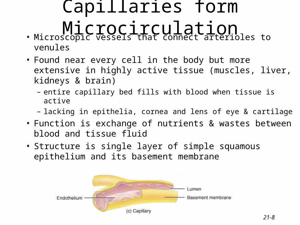
Capillaries form Microcirculation• Microscopic vessels that connect arterioles to venules• Found near every cell in the body but more extensive
in highly active tissue (muscles, liver, kidneys & brain)– entire capillary bed fills with blood when tissue is active
– lacking in epithelia, cornea and lens of eye & cartilage
• Function is exchange of nutrients & wastes between blood and tissue fluid
• Structure is single layer of simple squamous epithelium and its basement membrane
21-9
Venules
• Small veins collecting blood from capillaries
• Tunica media contains only a few smooth muscle cells & scattered fibroblasts– very porous endothelium allows for escape of
many phagocytic white blood cells
• Venules that approach size of veins more closely resemble structure of vein
21-10
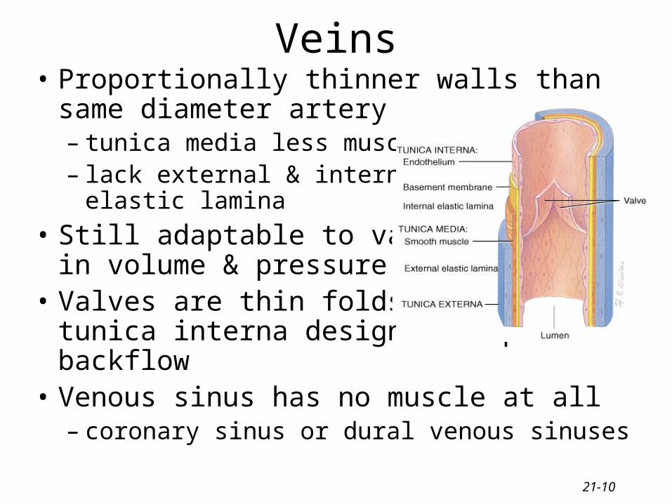
Veins• Proportionally thinner walls than same
diameter artery– tunica media less muscle– lack external & internal
elastic lamina
• Still adaptable to variationsin volume & pressure
• Valves are thin folds of tunica interna designed to prevent backflow
• Venous sinus has no muscle at all– coronary sinus or dural venous sinuses
21-11
Varicose Veins
• Twisted, dilated superficial veins– caused by leaky venous valves
• congenital or mechanically stressed from prolonged standing or pregnancy
– allow backflow and pooling of blood• extra pressure forces fluids into surrounding tissues
• nearby tissue is inflamed and tender
• Deeper veins not susceptible because of support of surrounding muscles
21-12
Anastomoses
• Union of 2 or more arteries supplying the same body region– blockage of only one pathway has no effect
• circle of willis underneath brain
• coronary circulation of heart
• Alternate route of blood flow through an anastomosis is known as collateral circulation– can occur in veins and venules as well
• Alternate routes to a region can also be supplied by nonanastomosing vessels
21-13
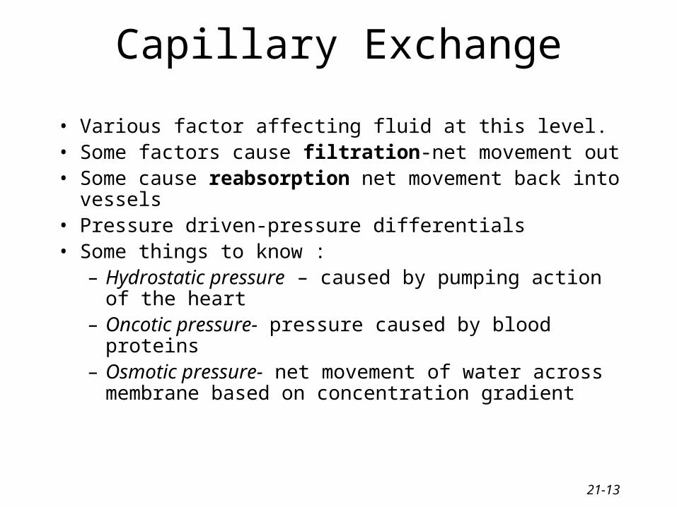
Capillary Exchange
• Various factor affecting fluid at this level. • Some factors cause filtration-net movement out• Some cause reabsorption net movement back into vessels• Pressure driven-pressure differentials• Some things to know :
– Hydrostatic pressure – caused by pumping action of the heart
– Oncotic pressure- pressure caused by blood proteins– Osmotic pressure- net movement of water across
membrane based on concentration gradient
21-14
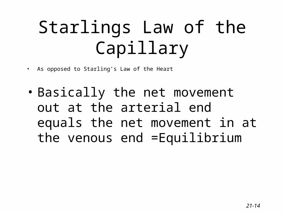
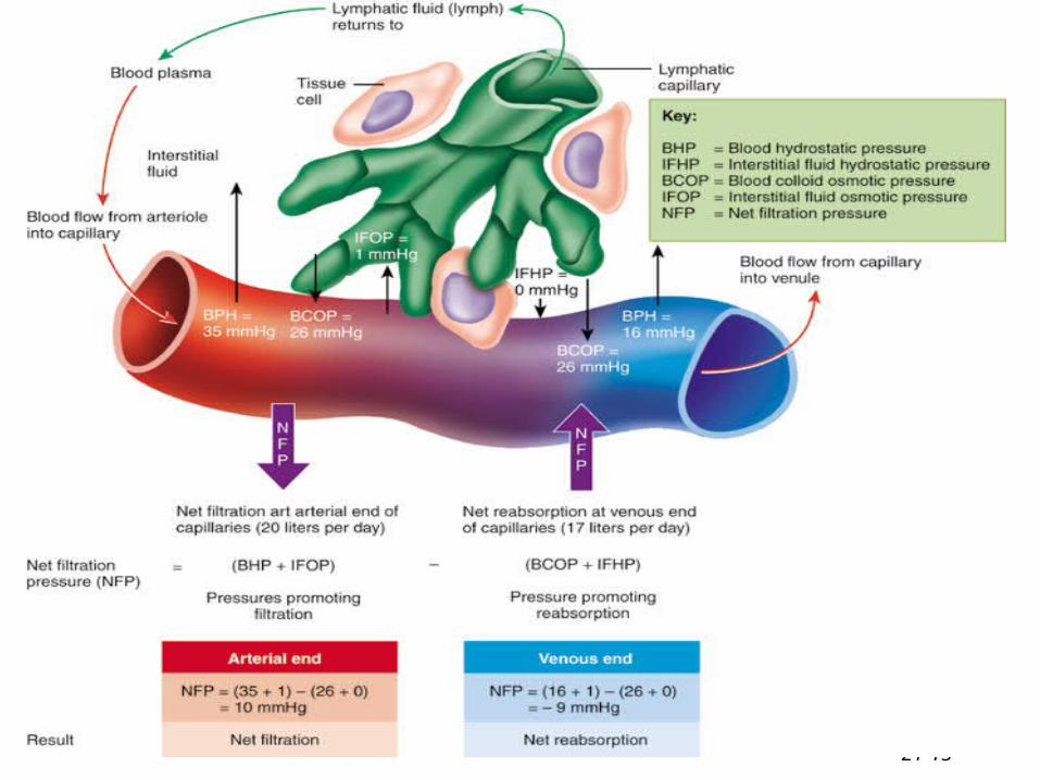
Starlings Law of the Capillary
• As opposed to Starling’s Law of the Heart
• Basically the net movement out at the arterial end equals the net movement in at the venous end =Equilibrium
21-15
21-16
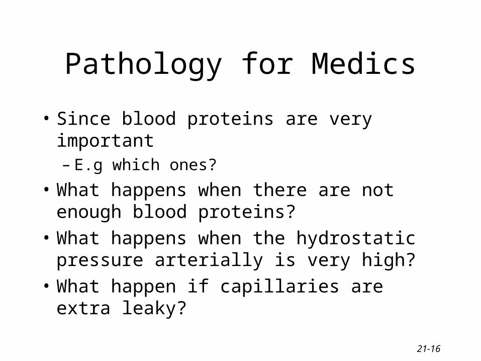
Pathology for Medics
• Since blood proteins are very important – E.g which ones?
• What happens when there are not enough blood proteins?
• What happens when the hydrostatic pressure arterially is very high?
• What happen if capillaries are extra leaky?
21-17
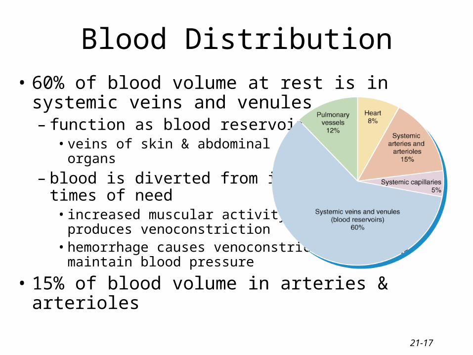
Blood Distribution
• 60% of blood volume at rest is in systemic veins and venules– function as blood reservoir
• veins of skin & abdominalorgans
– blood is diverted from it intimes of need
• increased muscular activityproduces venoconstriction
• hemorrhage causes venoconstriction to help maintain blood pressure
• 15% of blood volume in arteries & arterioles
21-18
Hemodynamics
• Factors affecting circulation– pressure differences that drive the blood flow
• velocity of blood flow
• volume of blood flow
• blood pressure
– resistance to flow– venous return
• An interplay of forces result in blood flow
21-19
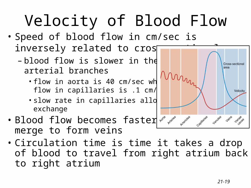
Velocity of Blood Flow• Speed of blood flow in cm/sec is inversely related
to cross-sectional area– blood flow is slower in the
arterial branches• flow in aorta is 40 cm/sec while
flow in capillaries is .1 cm/sec
• slow rate in capillaries allows forexchange
• Blood flow becomes faster when vessels merge to form veins
• Circulation time is time it takes a drop of blood to travel from right atrium back to right atrium
21-20
Volume of Blood Flow
• Cardiac output = stroke volume x heart rate
• Other factors that influence cardiac output– blood pressure– resistance due to friction between blood cells and
blood vessel walls• blood flows from areas of higher pressure to areas of
lower pressure
21-21
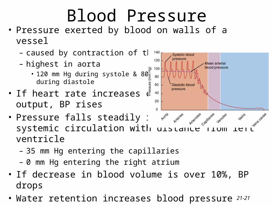
Blood Pressure• Pressure exerted by blood on walls of a vessel
– caused by contraction of the ventricles
– highest in aorta• 120 mm Hg during systole & 80
during diastole
• If heart rate increases cardiacoutput, BP rises
• Pressure falls steadily insystemic circulation with distance from left ventricle– 35 mm Hg entering the capillaries
– 0 mm Hg entering the right atrium
• If decrease in blood volume is over 10%, BP drops• Water retention increases blood pressure
21-22
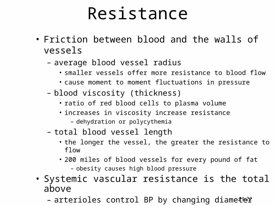
Resistance
• Friction between blood and the walls of vessels– average blood vessel radius
• smaller vessels offer more resistance to blood flow
• cause moment to moment fluctuations in pressure
– blood viscosity (thickness)• ratio of red blood cells to plasma volume
• increases in viscosity increase resistance– dehydration or polycythemia
– total blood vessel length• the longer the vessel, the greater the resistance to flow
• 200 miles of blood vessels for every pound of fat– obesity causes high blood pressure
• Systemic vascular resistance is the total of above– arterioles control BP by changing diameter
21-23
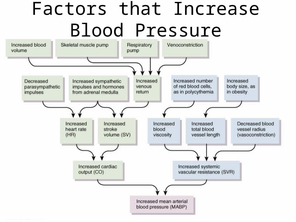
Factors that Increase Blood Pressure
21-24
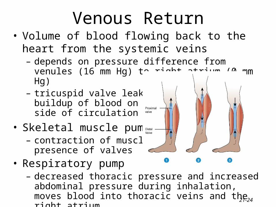
Venous Return• Volume of blood flowing back to the heart from the
systemic veins– depends on pressure difference from venules (16 mm Hg) to
right atrium (0 mm Hg)– tricuspid valve leaky and
buildup of blood on venousside of circulation
• Skeletal muscle pump– contraction of muscles &
presence of valves
• Respiratory pump– decreased thoracic pressure and increased abdominal pressure
during inhalation, moves blood into thoracic veins and the right atrium
21-25
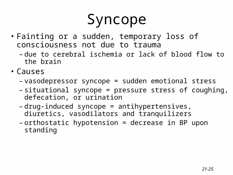
Syncope• Fainting or a sudden, temporary loss of consciousness not
due to trauma– due to cerebral ischemia or lack of blood flow to the brain
• Causes– vasodepressor syncope = sudden emotional stress – situational syncope = pressure stress of coughing, defecation,
or urination– drug-induced syncope = antihypertensives, diuretics,
vasodilators and tranquilizers– orthostatic hypotension = decrease in BP upon standing
21-26
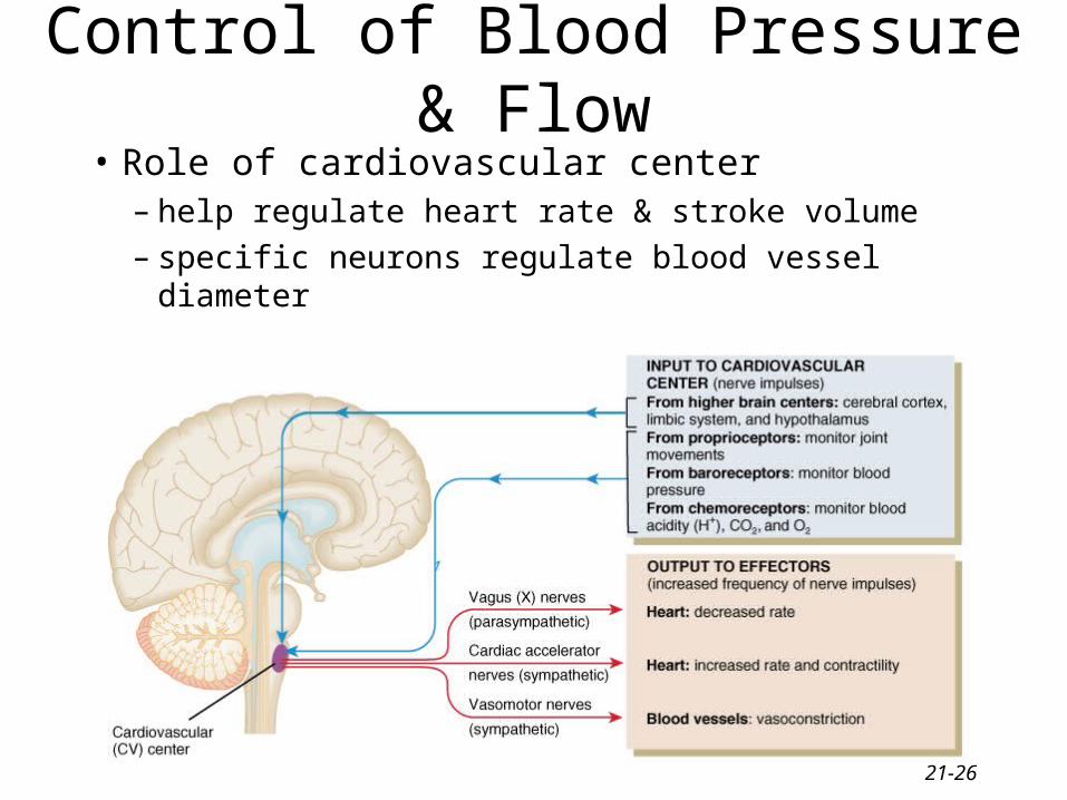
Control of Blood Pressure & Flow
• Role of cardiovascular center– help regulate heart rate & stroke volume– specific neurons regulate blood vessel diameter
21-27
Input to the Cardiovascular Center
• Higher brain centers such as cerebral cortex, limbic system & hypothalamus– anticipation of competition– increase in body temperature
• Proprioceptors– input during physical activity
• Baroreceptors– changes in pressure within blood vessels
• Chemoreceptors– monitor concentration of chemicals in the blood
21-28
Output from the Cardiovascular Center
• Heart– parasympathetic (vagus nerve)
• decrease heart rate
– sympathetic (cardiac accelerator nerves)• cause increase or decrease in contractility & rate
• Blood vessels– sympathetic vasomotor nerves
• continual stimulation to arterioles in skin & abdominal viscera producing vasoconstriction (vasomotor tone)
• increased stimulation produces constriction & increased BP
21-29
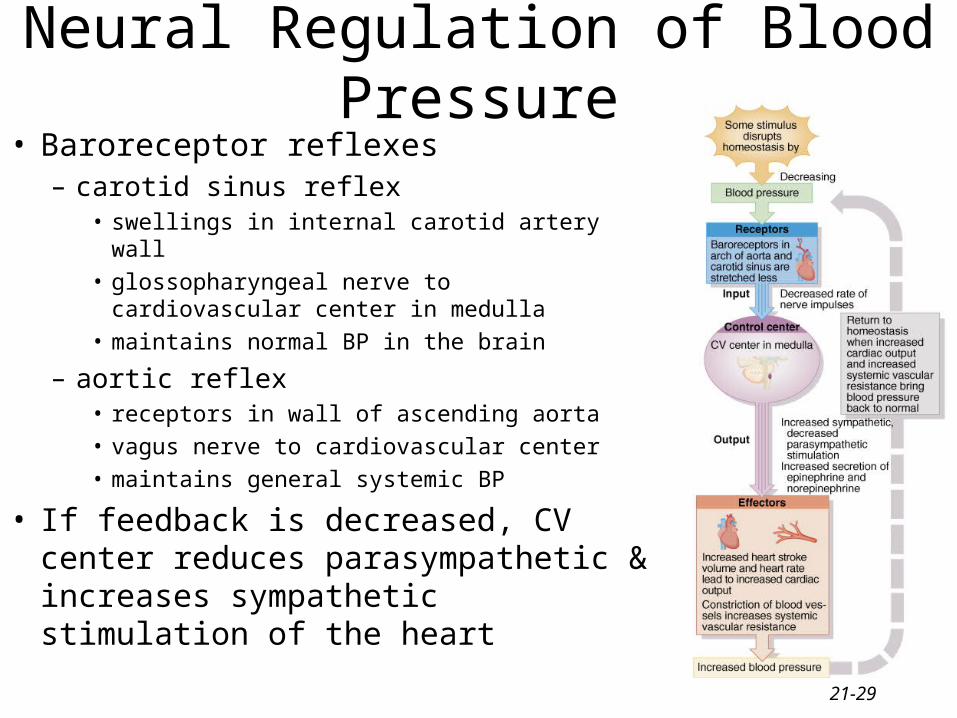
Neural Regulation of Blood Pressure• Baroreceptor reflexes
– carotid sinus reflex• swellings in internal carotid artery wall
• glossopharyngeal nerve to cardiovascular center in medulla
• maintains normal BP in the brain
– aortic reflex• receptors in wall of ascending aorta
• vagus nerve to cardiovascular center
• maintains general systemic BP
• If feedback is decreased, CV center reduces parasympathetic & increases sympathetic stimulation of the heart
21-30
Carotid Sinus Massage & Syncope
• Stimulation (careful neck massage) over the carotid sinus to lower heart rate– paroxysmal superventricular tachycardia
• tachycardia originating from the atria
• Anything that puts pressure on carotid sinus– tight collar or hyperextension of the neck – may slow heart rate & cause carotid sinus
syncope or fainting
21-31
Chemoreceptor Reflexes
• Carotid bodies and aortic bodies– detect changes in blood levels of O2, CO2, and
H+ (hypoxia, hypercapnia or acidosis )– causes stimulation of cardiovascular center– increases sympathetic stimulation to arterioles
& veins– vasoconstriction and increase in blood pressure
• Also changes breathing rates as well
21-32
Hormonal Regulation of Blood Pressure• Renin-angiotensin-aldosterone system
– decrease in BP or decreased blood flow to kidney
– release of renin / results in formation angiotensin II• systemic vasoconstriction
• causes release aldosterone (H2O & Na+ reabsorption)
• Epinephrine & norepinephrine– increases heart rate & force of contraction
– causes vasoconstriction in skin & abdominal organs
– vasodilation in cardiac & skeletal muscle
• ADH causes vasoconstriction• ANP (atrial natriuretic peptide) lowers BP
– causes vasodilation & loss of salt and water in the urine
21-33
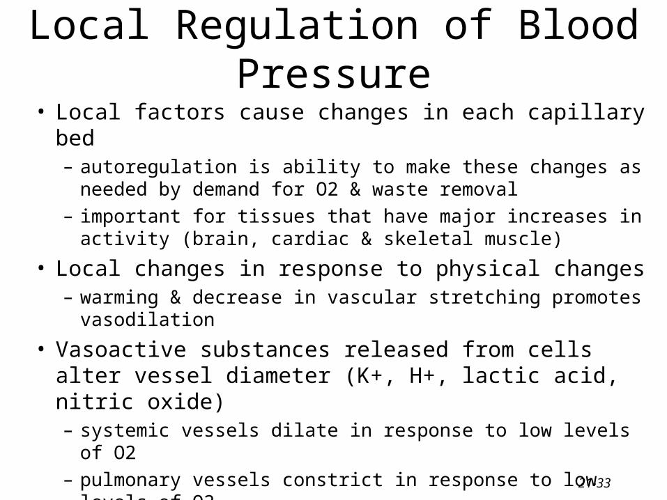
Local Regulation of Blood Pressure
• Local factors cause changes in each capillary bed– autoregulation is ability to make these changes as needed by
demand for O2 & waste removal
– important for tissues that have major increases in activity (brain, cardiac & skeletal muscle)
• Local changes in response to physical changes– warming & decrease in vascular stretching promotes
vasodilation
• Vasoactive substances released from cells alter vessel diameter (K+, H+, lactic acid, nitric oxide)– systemic vessels dilate in response to low levels of O2
– pulmonary vessels constrict in response to low levels of O2
21-34
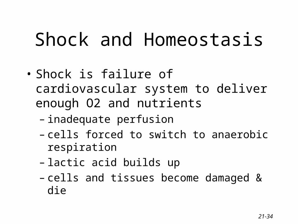
Shock and Homeostasis
• Shock is failure of cardiovascular system to deliver enough O2 and nutrients– inadequate perfusion– cells forced to switch to anaerobic respiration– lactic acid builds up– cells and tissues become damaged & die
21-35
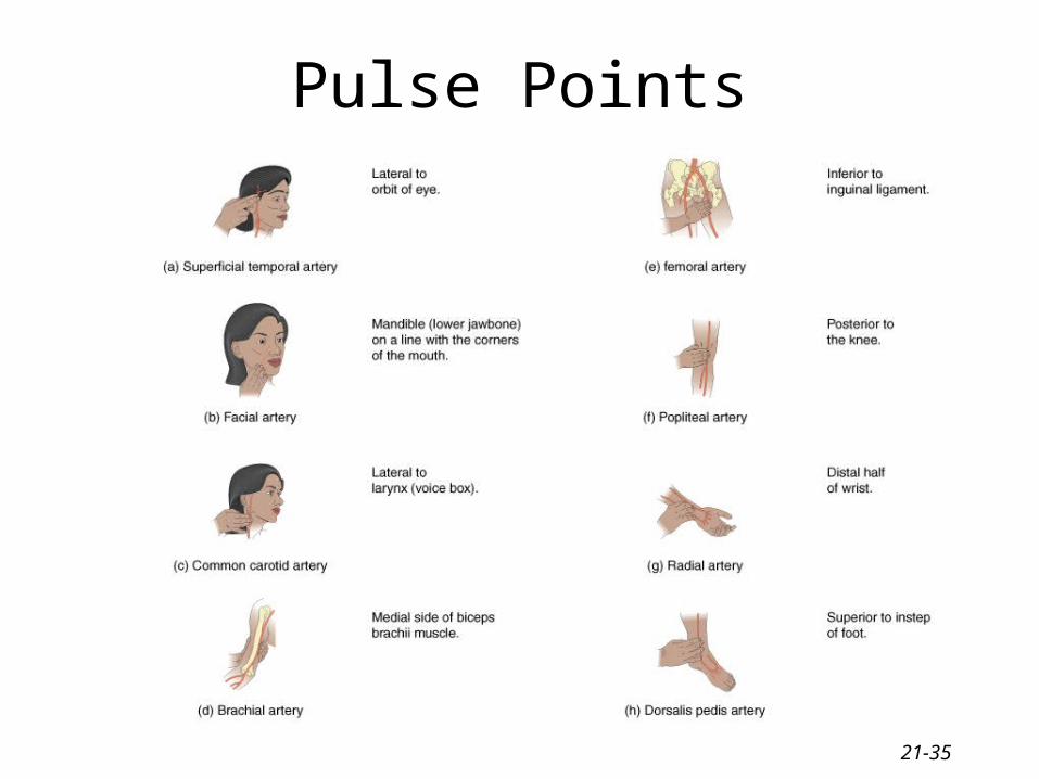
Pulse Points
21-36
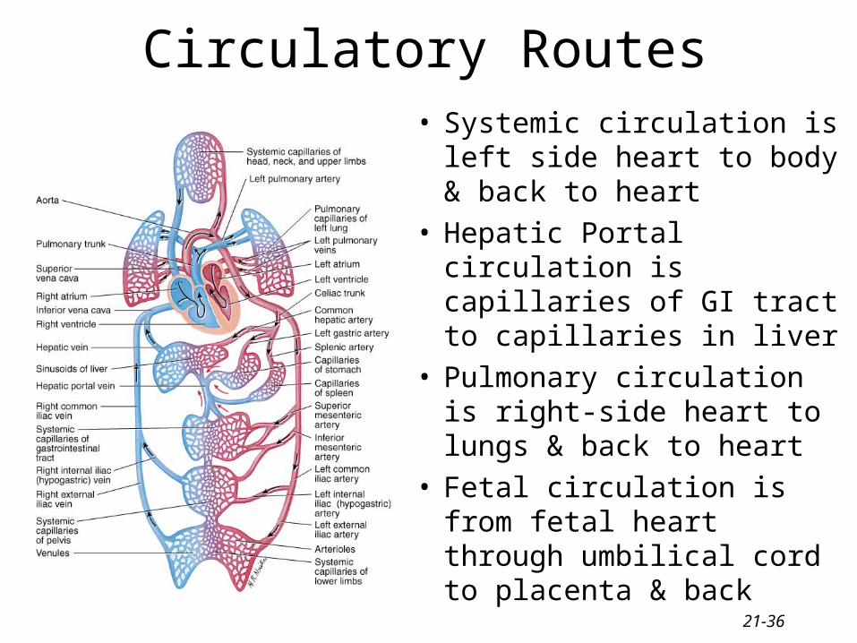
Circulatory Routes• Systemic circulation is left
side heart to body & back to heart
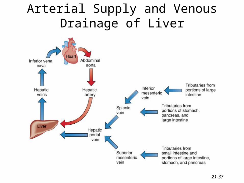
• Hepatic Portal circulation is capillaries of GI tract to capillaries in liver
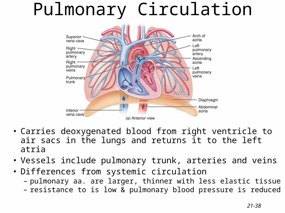
• Pulmonary circulation is right-side heart to lungs & back to heart
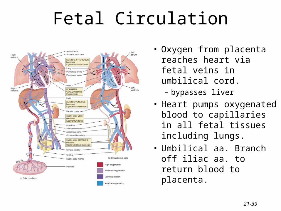
• Fetal circulation is from fetal heart through umbilical cord to placenta & back
21-37
Arterial Supply and Venous Drainage of Liver
21-38
Pulmonary Circulation
• Carries deoxygenated blood from right ventricle to air sacs in the lungs and returns it to the left atria
• Vessels include pulmonary trunk, arteries and veins• Differences from systemic circulation
– pulmonary aa. are larger, thinner with less elastic tissue– resistance to is low & pulmonary blood pressure is reduced
21-39
Fetal Circulation
• Oxygen from placenta reaches heart via fetal veins in umbilical cord.– bypasses liver
• Heart pumps oxygenated blood to capillaries in all fetal tissues including lungs.
• Umbilical aa. Branch off iliac aa. to return blood to placenta.
21-40
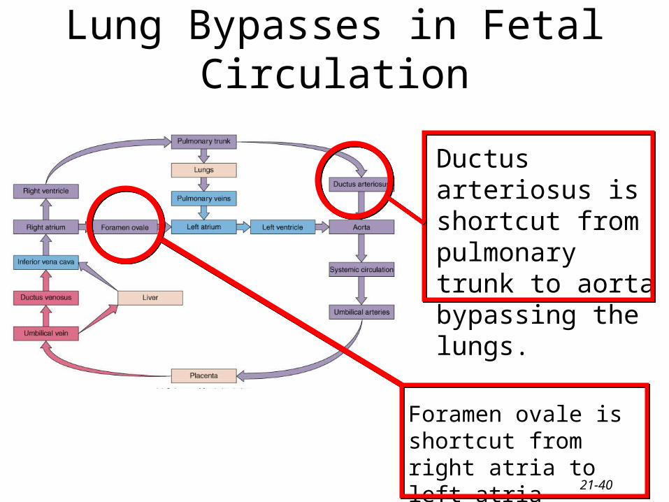
Ductus arteriosus is shortcut from pulmonary trunk to aorta bypassing the lungs.
Lung Bypasses in Fetal Circulation
Foramen ovale is shortcut from right atria to left atria bypassing the lungs.