copyright 2009, john wiley & sons, inc. chapter 21: the cardiovascular system: blood vessels and...
TRANSCRIPT
Copyright 2009, John Wiley & Sons, Inc.
Chapter 21: The Cardiovascular System: Blood Vessels and Hemodynamics
Copyright 2009, John Wiley & Sons, Inc.
Structure and function of blood vessels 5 main types
Arteries – carry blood AWAY from the heart Arterioles Capillaries – site of exchange Venules Veins – carry blood TO the heart
Copyright 2009, John Wiley & Sons, Inc.
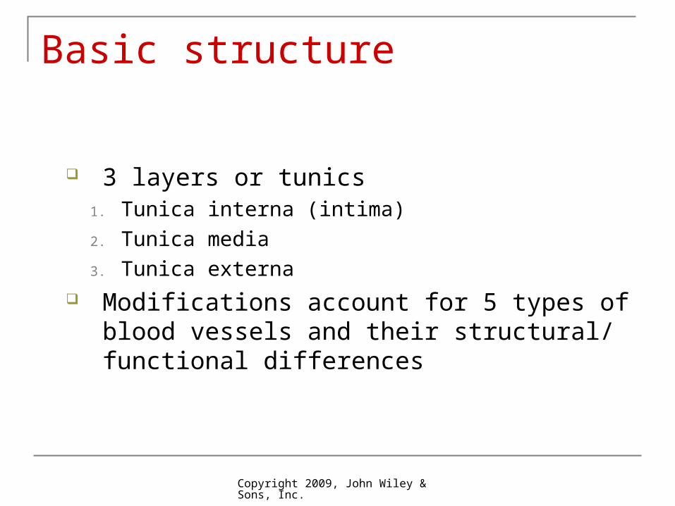
Basic structure
3 layers or tunics1. Tunica interna (intima)
2. Tunica media
3. Tunica externa Modifications account for 5 types of blood
vessels and their structural/ functional differences
Copyright 2009, John Wiley & Sons, Inc.
Copyright 2009, John Wiley & Sons, Inc.
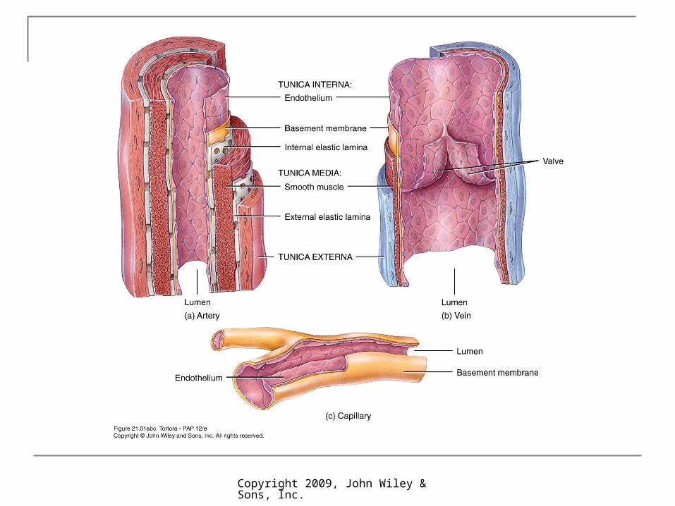
Structure
Tunica interna (intima) Inner lining in direct contact with blood Endothelium continuous with endocardial lining of heart Active role in vessel-related activities
Tunica media Muscular and connective tissue layer Greatest variation among vessel types Smooth muscle regulates diameter of lumen
Tunica externa Elastic and collagen fibers Vasa vasorum Helps anchor vessel to surrounding tissue
Copyright 2009, John Wiley & Sons, Inc.
Arteries
3 layers of typical blood vessel Thick muscular-to-elastic tunica media High compliance – walls stretch and expand in
response to pressure without tearing Vasoconstriction – decrease in lumen diameter
Vasodilation – increase in lumen diameter
Copyright 2009, John Wiley & Sons, Inc.
Elastic Arteries
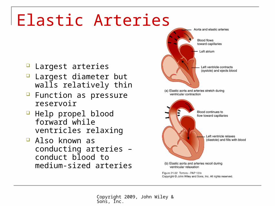
Largest arteries Largest diameter but
walls relatively thin Function as pressure
reservoir Help propel blood
forward while ventricles relaxing
Also known as conducting arteries – conduct blood to medium-sized arteries
Copyright 2009, John Wiley & Sons, Inc.
Arteries
Muscular arteries Tunica media contains more smooth muscle and fewer
elastic fibers than elastic arteries Walls relatively thick Capable of great vasoconstriction/ vasodilatation to adjust
rate of blood flow Also called distributing arteries
Anastomoses Union of the branches of 2 or more arteries supplying the
same body region Provide alternate routes – collateral circulation
Copyright 2009, John Wiley & Sons, Inc.
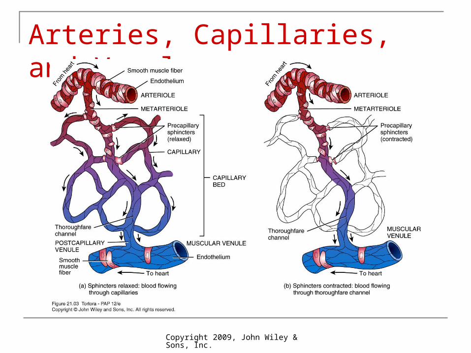
Arterioles
Abundant microscopic vessels Metarteriole has precapillary sphincter which
monitors blood flow into capillary Sympathetic innervation and local chemical
mediators can alter diameter and thus blood flow and resistance
Resistance vessels – resistance is opposition to blood flow
Vasoconstriction can raise blood pressure
Copyright 2009, John Wiley & Sons, Inc.
Capillaries
Capillaries Smallest blood vessels connect arterial outflow and
venous return Microcirculation – flow from metarteriole through
capillaries and into postcapillary venule Exchange vessels – primary function is exchange
between blood and interstitial fluid Lack tunica media and tunica externa
Substances pass through just one layer of endothelial cells and basement membrane
Capillary beds – arise from single metarteriole Vasomotion – intermittent contraction and relaxation Throughfare channel – bypasses capillary bed
Copyright 2009, John Wiley & Sons, Inc.
Arteries, Capillaries, and Venule

Copyright 2009, John Wiley & Sons, Inc.
Types of Capillaries
3 types1. Continuous
Endothelial cell membranes from continuous tube
2. Fenestrated Have fenestrations or
pores3. Sinusoids
Wider and more winding
Unusually large fenestrations
Copyright 2009, John Wiley & Sons, Inc.
Portal vein – blood passes through second capillary bed Hepatic or hypophyseal
Venules Thinner walls than arterial counterparts Postcapillary venule – smallest venule Form part of microcirculatory exchange unit with
capillaries Muscular venules have thicker walls with 1 or 2
layers of smooth muscle
Copyright 2009, John Wiley & Sons, Inc.
Veins
Structural changes not as distinct as in arteries In general, very thin walls in relation to total
diameter Same 3 layers
Tunica interna thinner than arteries Tunica interna thinner with little smooth muscle Tunica externa thickest layer
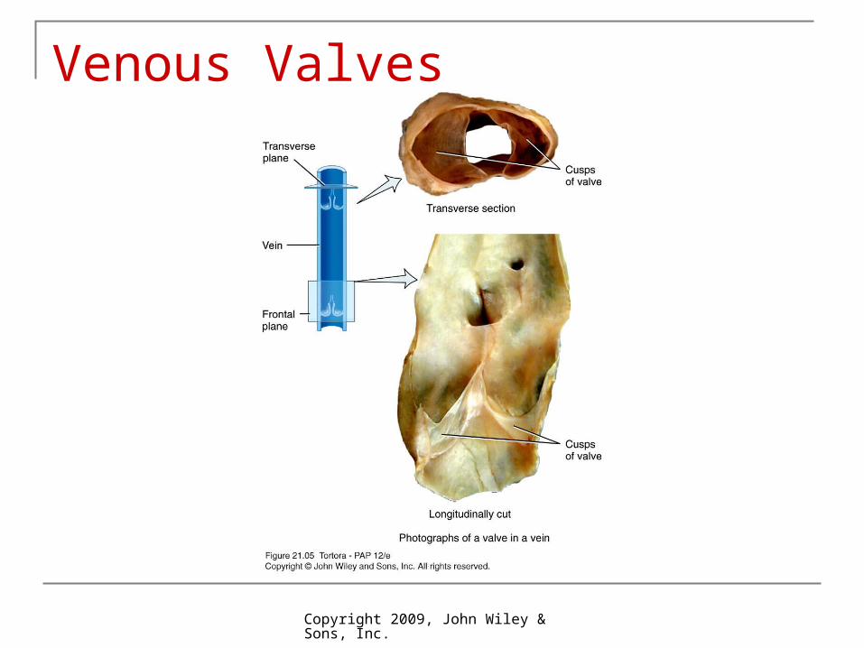
Not designed to withstand high pressure Valves – folds on tunica interna forming cusps
Aid in venous return by preventing backflow
Copyright 2009, John Wiley & Sons, Inc.
Venous Valves

Copyright 2009, John Wiley & Sons, Inc.
Blood Distribution
Largest portion of blood at rest is in systemic veins and venules Blood reservoir
Venoconstriction reduces volume of blood in reservoirs and allows greater blood volume to flow where needed

Copyright 2009, John Wiley & Sons, Inc.
Capillary exchange
Movement of substances between blood and interstitial fluid
3 basic methods1. Diffusion
2. Transcytosis
3. Bulk flow
Copyright 2009, John Wiley & Sons, Inc.
Diffusion
Most important method Substances move down their concentration gradient
O2 and nutrients from blood to interstitial fluid to body cells
CO2 and wastes move from body cells to interstitial fluid to blood
Can cross capillary wall through intracellular clefts, fenestrations or through endothelial cells Most plasma proteins cannot cross Except in sinusoids – proteins and even blood cells leave Blood-brain barrier – tight junctions limit diffusion
Copyright 2009, John Wiley & Sons, Inc.
Transcytosis
Small quantity of material Substances in blood plasma become enclosed
within pinocytotic vessicles that enter endothelial cells by endocytosis and leave by exocytosis
Important mainly for large, lipid-insoluble molecules that cannot cross capillary walls any other way
Copyright 2009, John Wiley & Sons, Inc.
Bulk Flow
Passive process in which large numbers of ions, molecules, or particles in a fluid move together in the same direction
Based on pressure gradient Diffusion is more important for solute exchange Bulk flow more important for regulation of relative
volumes of blood and interstitial fluid Filtration – from capillaries into interstitial fluid Reabsorption – from interstitial fluid into capillaries
Copyright 2009, John Wiley & Sons, Inc.
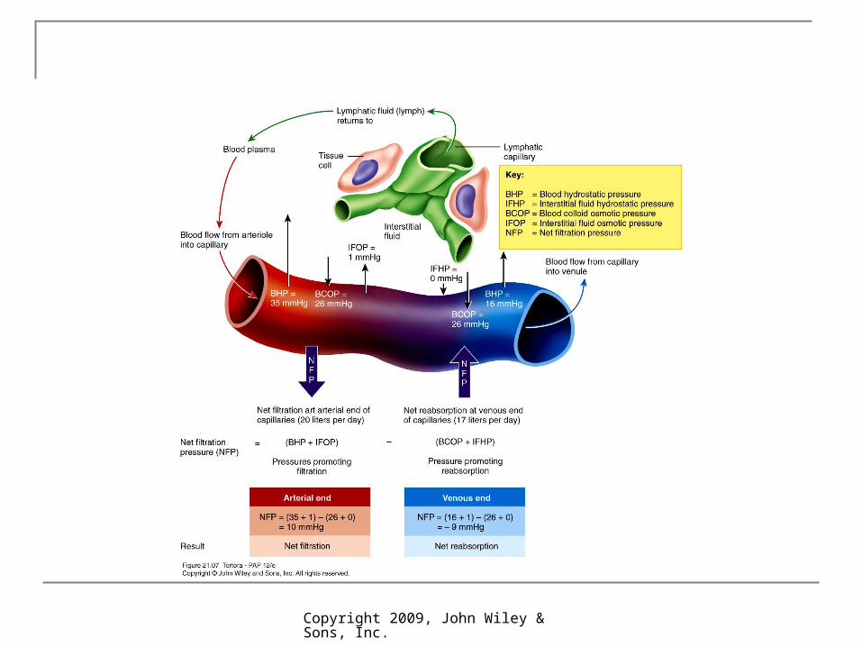
NFP = (BHP + IFOP) – (BCOP + IFHP) Net filtration pressure (NFP) balance of 2
pressures
1. 2 pressures promote filtration Blood hydrostatic pressure (BHP) generated by
pumping action of heart Falls over capillary bed from 35 to 16 mmHg
Interstitial fluid osmotic pressure (IFOP) 1 mmHg
Copyright 2009, John Wiley & Sons, Inc.
NFP = (BHP + IFOP) – (BCOP + IFHP)2. 2 pressures promote reabsorption
Blood colloid osmotic pressure (BCOP) promotes reabsorption
Due to presence of blood plasma proteins to large to cross walls
Averages 36 mmHg Interstitial fluid hydrostatic pressure (IFHP)
Close to zero mmHg
Copyright 2009, John Wiley & Sons, Inc.
Starling’s Law
Nearly as much reabsorbed as filtered At the arterial end, net outward pressure of 10
mmHg and fluid leaves capillary (filtration) At the venous end, fluid moves in (reabsoprtion)
due to -9 mmHg On average, about 85% of fluid filtered in
reabsorpbed Excess enters lymphatic capillaries (about 3L/
day) to be eventually returned to blood
Copyright 2009, John Wiley & Sons, Inc.
Copyright 2009, John Wiley & Sons, Inc.
Dynamics of Capillary Exchange
Copyright 2009, John Wiley & Sons, Inc.
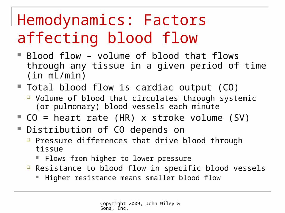
Hemodynamics: Factors affecting blood flow Blood flow – volume of blood that flows through any
tissue in a given period of time (in mL/min) Total blood flow is cardiac output (CO)
Volume of blood that circulates through systemic (or pulmonary) blood vessels each minute
CO = heart rate (HR) x stroke volume (SV) Distribution of CO depends on
Pressure differences that drive blood through tissue Flows from higher to lower pressure
Resistance to blood flow in specific blood vessels Higher resistance means smaller blood flow
Copyright 2009, John Wiley & Sons, Inc.
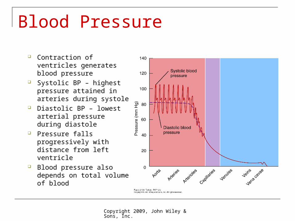
Blood Pressure
Contraction of ventricles generates blood pressure
Systolic BP – highest pressure attained in arteries during systole
Diastolic BP – lowest arterial pressure during diastole
Pressure falls progressively with distance from left ventricle
Blood pressure also depends on total volume of blood
Copyright 2009, John Wiley & Sons, Inc.
Vascular resistance
Opposition to blood flow due to friction between blood and walls of blood vessels
Depends on 1. Size of lumen – vasoconstriction males lumen smaller
meaning greater resistance
2. Blood viscosity – ratio of RBCs to plasma and protein concentration, higher viscosity means higher resistance
3. Total blood vessel length – resistance directly proportional to length of vessel
400 miles of additional blood vessels for each 2.2lb. of fat
Copyright 2009, John Wiley & Sons, Inc.
Venous return
Volume of blood flowing back to heart through systemic veins
Occurs due to pressure generated by constriction of left ventricle
Small pressure difference from venule (16 mmHg) to right ventricle (0 mmHg) sufficient
Copyright 2009, John Wiley & Sons, Inc.
Skeletal Muscle Pump
2 other mechanisms Skeletal muscle pump – milks blood in 1 direction
due to valves Respiratory pump – due to pressure changes in
thoracic and abdominal cavities
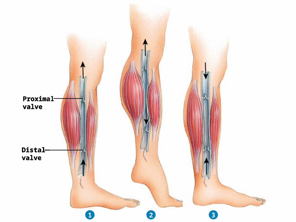
Proximalvalve
Distalvalve
1
Proximalvalve
Distalvalve
1 2
Proximalvalve
Distalvalve
1 2 3
Copyright 2009, John Wiley & Sons, Inc.
Copyright 2009, John Wiley & Sons, Inc.
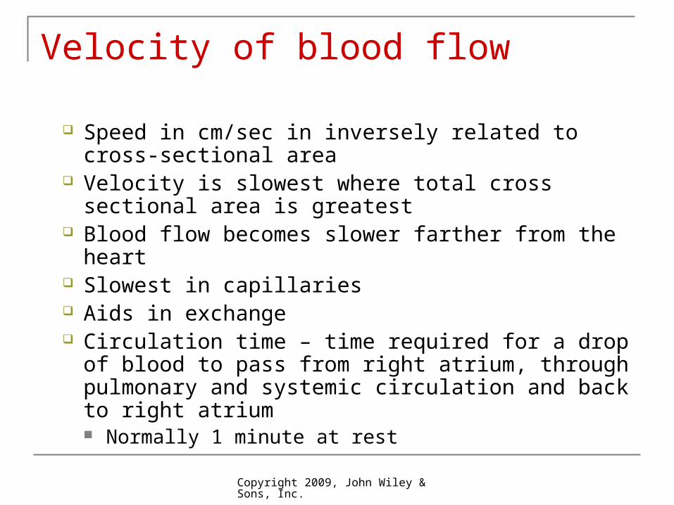
Velocity of blood flow
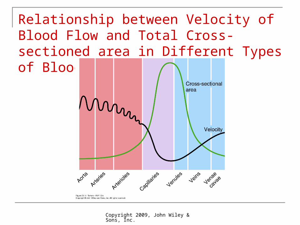
Speed in cm/sec in inversely related to cross-sectional area
Velocity is slowest where total cross sectional area is greatest
Blood flow becomes slower farther from the heart Slowest in capillaries Aids in exchange Circulation time – time required for a drop of blood
to pass from right atrium, through pulmonary and systemic circulation and back to right atrium Normally 1 minute at rest
Copyright 2009, John Wiley & Sons, Inc.
Relationship between Velocity of Blood Flow and Total Cross-sectioned area in Different Types of Blood Vessels
Copyright 2009, John Wiley & Sons, Inc.
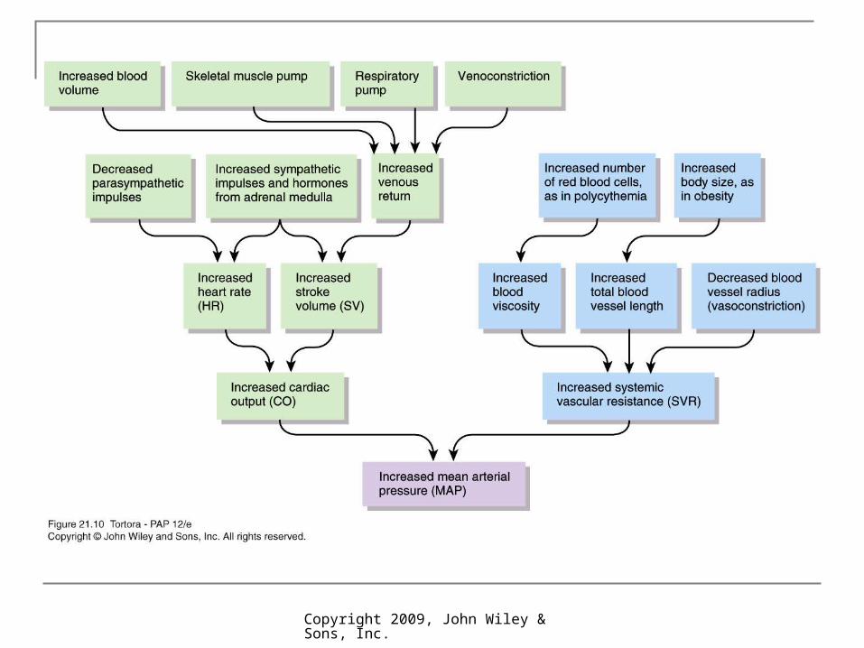
Control of blood pressure and blood flow Interconnected negative feedback systems
control blood pressure by adjusting heart rate, stroke volume, systemic vascular resistance, and blood volume
Some act faster that others Some shorter- or longer-term
Copyright 2009, John Wiley & Sons, Inc.
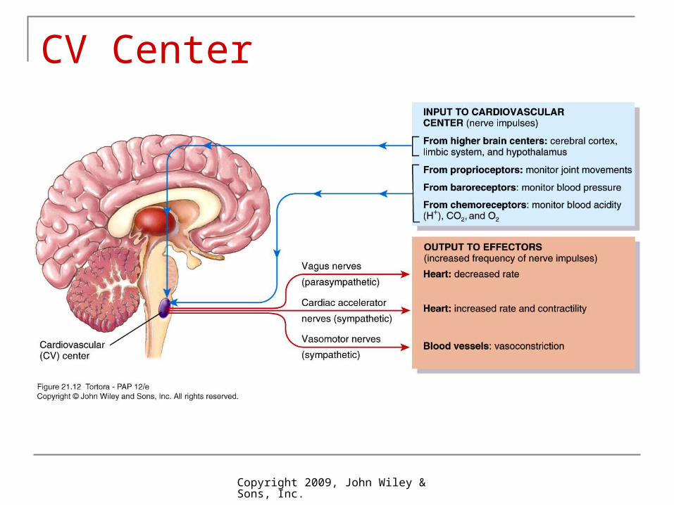
Role of cardiovascular center (CV)
In medulla oblongata Helps regulate heart rate and stroke volume Also controls neural, hormonal, and local negative
feedback systems that regulate blood pressure and blood flow to specific tissues
Groups of neurons regulate heart rate, contractility of ventricles, and blood vessel diameter
Cardiostimulatory and cardioinhibitory centers Vasomotor center control blood vessel diameter Receives input from both higher brain regions and
sensory receptors
Copyright 2009, John Wiley & Sons, Inc.
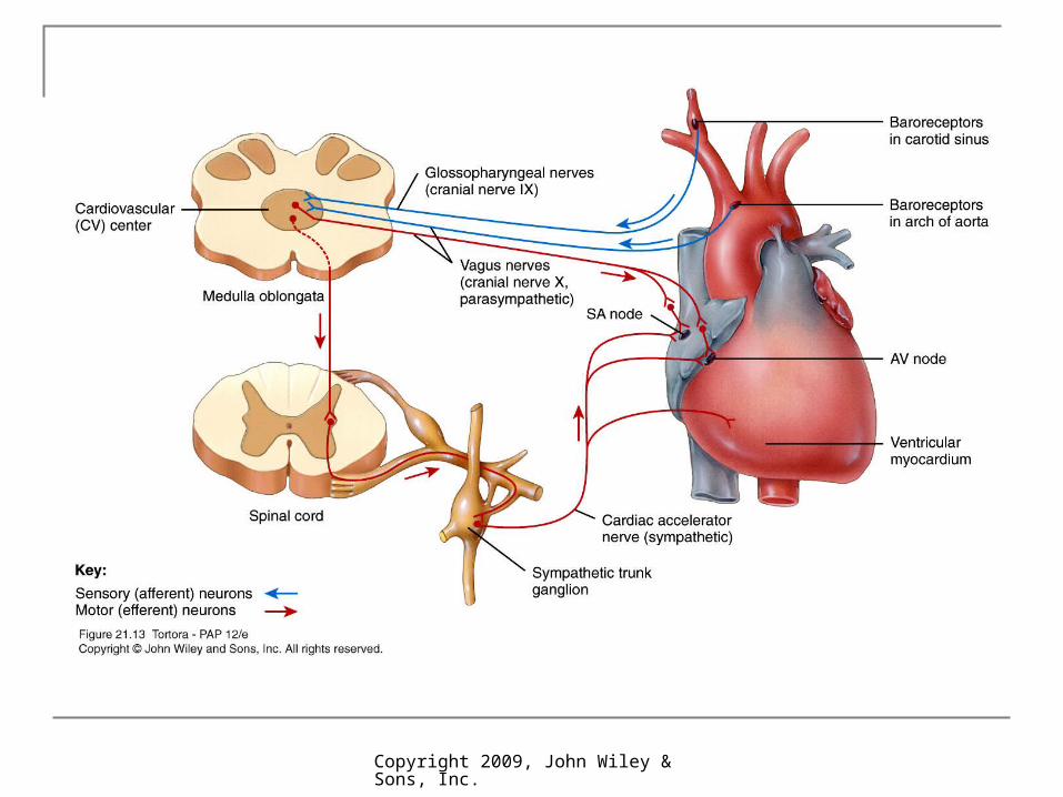
CV Center

Copyright 2009, John Wiley & Sons, Inc.
3 main types of sensory receptors
Proprioceptors – monitor movements of joints and muscles to provide input during physical activity
Baroreceptors – monitor pressure changes and stretch in blood vessel walls
Chemoreceptors – monitor concentration of various chemicals in the blood
Output from CV flows along neurons of ANS Sympathetic (stimulatory) opposes
parasympathetic (inhibitory)
Copyright 2009, John Wiley & Sons, Inc.
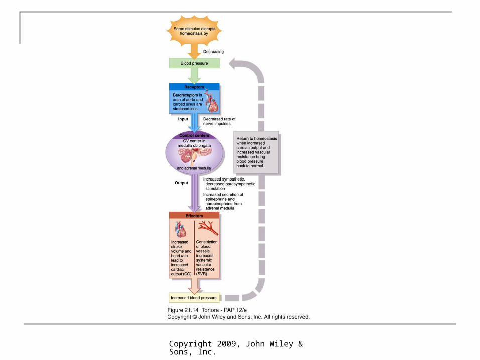
Neural regulation of blood pressure
Negative feedback loops from 2 types of reflexes
1. Baroreceptor reflexes Pressure-sensitive receptors in internal carotid arteries
and other large arteries in neck and chest Carotid sinus reflex helps regulate blood pressure in brain Aortic reflex regulates systemic blood pressure
When blood pressure falls, baroreceptors stretched less, slower rate of impulses to CV
CV decreases parasympathetic stimulation and increases sympathetic stimulation
Copyright 2009, John Wiley & Sons, Inc.
Neural regulation of blood pressure
2. Chemoreceptor reflexes Receptors located close to baroreceptors of carotid
sinus (carotid bodies) and aortic arch (aortic bodies) Detect hypoxia (low O2), hypercapnia (high CO2),
acidosis (high H+) and send signals to CV CV increases sympathetic stimulation to arterioles and
veins, producing vasoconstriction and an increase in blood pressure
Receptors also provide input to respiratory center to adjust breathing rate
Copyright 2009, John Wiley & Sons, Inc.
Copyright 2009, John Wiley & Sons, Inc.
Copyright 2009, John Wiley & Sons, Inc.
Hormonal regulation of blood pressure
Renin-angiotensin-aldosterone (RAA) system Renin (released by kidney when blood volume
falls or blood flow decreases) and angiotensin converting enzyme (ACE) act on substrates to produce active hormone angiotensin II Raises BP by vasoconstriction and secretion of
aldosterone (increases water reabsorption in kidneys to raise blood volume and pressure)
Copyright 2009, John Wiley & Sons, Inc.
Hormonal regulation of blood pressure
Epinephrine and norepinephrine Adrenal medulla releases in response to sympathetic
stimulation Increase cardiac output by increasing rate and force of
heart contractions Antidiuretic hormone (ADH) or vasopressin
Produced by hypothalamus, released by posterior pituitary
Response to dehydration or decreased blood volume Causes vasoconstriction which increases blood
pressure
Copyright 2009, John Wiley & Sons, Inc.
Atrial natriuretic peptide (ANP)
Released by cells of atria Lowers blood pressure by causing vasodilation
and promoting loss of salt and water in urine Reduces blood volume
Copyright 2009, John Wiley & Sons, Inc.
Autoregulation of blood pressure
Ability of tissue to automatically adjust its blood flow to match metabolic demands
Demand of O2 and nutrients can rise tenfold during exercise in heart and skeletal muscles
Also controls regional blood flow in the brain during different mental and physical activities
2 general types of stimuli1. Physical – temperature changes, myogenic response
2. Vasodilating and vasoconstricting chemicals alter blood vessel diameter
Copyright 2009, John Wiley & Sons, Inc.
Circulation
Important difference between pulmonary and systemic circulation in autoregulatory response Systemic blood vessel walls dilate in response to
low O2 to increase O2 delivery Walls of pulmonary blood vessels constrict under
low O2 to ensure most blood flows to better ventilated areas of lung
Copyright 2009, John Wiley & Sons, Inc.
End of Chapter 21
Copyright 2009 John Wiley & Sons, Inc.All rights reserved. Reproduction or translation of this work beyond that permitted in section 117 of the 1976 United States Copyright Act without express permission of the copyright owner is unlawful. Request for further information should be addressed to the Permission Department, John Wiley & Sons, Inc. The purchaser may make back-up copies for his/her own use only and not for distribution or resale. The Publishers assumes no responsibility for errors, omissions, or damages caused by the use of theses programs or from the use of the information herein.