04 infeksi saluran napas
TRANSCRIPT

INFEKSI SALURAN NAPAS
DICKY BAGUS WIDHYATMOKO
25 – 26 MARET 2015DEPARTEMEN MIKROBIOLOGI KLINIK, FAKULTAS KEDOKTERANUNIVERSITAS AIRLANGGA

POKOK BAHASAN
Streptococcus spp
Corynebacterium diphteriae
Mycobacterium tuberculosis

DISEDIAKAN
Streptococcus : S. viridans
S. pneumoniae
S. pyogenes
Corynebacterium spp
M. tuberculosis
Gram Stain
Blood Agar
Chocolate Agar
Optochin Test
Bacitracin test
PAI medium
Neisser Stain (AB, C)
Ziehl-Neelsen Stain
prepared slide

Identifikasi Streptococcus spp.
Blood Agar
Chocolate Agar
Optochin Test
Bacitracin test
Zona hemolisis
Antibiotic Sensitivity Test for Identification

First step
Gram Stain
always do gram stain before continuing to others test
Catalase Test
please learn from previous laboratory practice

Next Step : Hemolysis
hemolysis
hemolysis
hemolysis

hemolysis
partial / incomplete hemolysis
reduction red blood cell hemoglobin methemoglobin in the medium surroundingthe colony.
The greenish color is caused by the presenceof biliverdin, which is a by-product of thebreakdown of hemoglobin
e.g. S. pneumoniae, S. viridans groups

hemolysis
Streptococcus viridans Streptococcus pneumoniae

hemolysis
complete or true lysis of red blood cells
clear zone, approaching the color andtransparency of the base medium, surroundsthe colony
e.g. S. pyogenes

hemolysis
Streptococcus pyogenes
clear zone surround colony

hemolysis
lack of hemolysis / non-hemolysis
e.g. Enterococcus faecalis

hemolysis
non hemolysis, Enterococcus faecalis

Hemolysis Resume
taken from : http://iws2.collin.edu/dcain/

hemolysis
how to differentiate between S. pneumoniaewith S. viridans
Using
Optochin test
Inulin Test
Bile Solubility Test

Optochin Test
Antibiotics Susceptibility Test
Optochin also known as ethyl-hydro-cupreine
To Differentiate between S. pneumoniae withS. viridans
S > 14 mm : S. pneumoniae
No zone inhibition : S. viridans
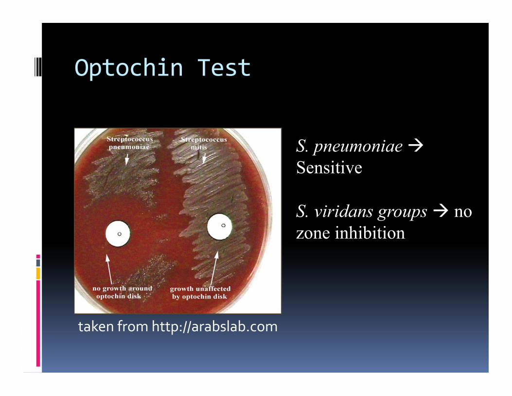
Optochin Test
taken from http://arabslab.com
S. pneumoniae Sensitive
S. viridans groups no zone inhibition

Inulin Test
To differentiate between Streptococcuspneumoniae with all others alpha-hemolyticsstreptococci
pneumoniae hydrolize inulin

Bile Solubility Test
Bile (sodium deoxycholate)
Distinguishes S. pneumoniae from all others alpha-hemolytics streptococci
Sodium deoxycholate (2% in water) will lyse the pneumococcal cell wall

Bile Solubility Test

for feedback mailto : [email protected]