zeolite structure determination from x-ray...
TRANSCRIPT

1 Introduction . . . . . . . . . . . . . . . . . . . . . . . . . . . . . . . . . 1
2 Severe Overlap of Reflections in Powder Data . . . . . . . . . . . . . . 3
3 Incorrect Determination of the Space Group . . . . . . . . . . . . . . . 5
4 Effect of Framework Flexibility . . . . . . . . . . . . . . . . . . . . . . . 8
5 Disorder of Non-Framework Species . . . . . . . . . . . . . . . . . . . 16
6 Faulting within the Framework . . . . . . . . . . . . . . . . . . . . . . . 23
7 Isomorphous Replacement of Framework Atoms . . . . . . . . . . . . . 24
8 Crystal Size Limitations . . . . . . . . . . . . . . . . . . . . . . . . . . . 25
9 Conclusions . . . . . . . . . . . . . . . . . . . . . . . . . . . . . . . . . 25
References . . . . . . . . . . . . . . . . . . . . . . . . . . . . . . . . . . . . 26
1Introduction
Zeolites and related microporous materials are a class of materials with an everwidening range of compositions, structures and uses. Since the earliest days ofzeolite science X-ray diffraction has been one of the basic and most useful toolsfor characterization.
Initially X-ray diffraction was used to answer simple questions such as:“haveI made a new material?”or:“has the crystallization process gone to completion?”Now the questions encompass everything that a researcher might want to knowabout the structure of a material. Early attempts at determining crystal struc-tures using X-ray diffraction were often unsuccessful because many of these ear-ly synthetic materials were available only as powder samples. Fortunately manyof these first synthetic materials had natural counterparts with large singlecrystals, and data from these were used to determine the framework structures
Zeolite Structure Determination from X-Ray Diffraction
H. van Koningsveld 1 and J. M. Bennett 2
1 Laboratory of Organic Chemistry and Catalysis, Delft University of Technology,Julianalaan 136, 2628 BL Delft, The Netherlands; e-mail: [email protected]
2 661 Weadley Road, Radnor, PA 19087, USA; e-mail: [email protected]
Molecular Sieves, Vol. 2© Springer-Verlag Berlin Heidelberg 1999

of their synthetic counterparts. Today, the framework of a new material can beoften determined from powder samples. In addition, single crystal techniqueshave improved considerably leading to increased accuracy in the bond anglesand bond distances and to the ability to study crystals of much smaller size. It isnow possible for a single crystal study to reveal details of the structure that showthe interaction of a sorbed material with the framework or movement of cationswithin the framework and any ensuing distortions of the framework. Structuraldata from powder samples are beginning to reveal similar changes in the crystalstructure with temperature, with sorbed materials and even under catalyticconditions. Even though the technique of X-ray powder diffraction has im-proved greatly since the early days of zeolite science, it is still more accurate todetermine the crystal structure of a new material from single crystal data ratherthan from powder data.
Many of the advances in the structural information derived for zeolitic mate-rials are a direct result of major improvements in powder and single crystal X-ray equipment available, in the development of new structure determinationmethods and in the use of new characterization tools including magic anglespinning NMR, neutron diffraction and electron microscopy, which are de-scribed in subsequent chapters. Two excellent review papers [1, 2] discuss theuse of X-ray diffraction techniques to study zeolites and the problems en-countered, and it is recommended that they be used in combination with thischapter.
The stages in determining the crystal structure of a material have beendescribed as: (i) obtain a suitable sample, (ii) collect the data, (iii) determine atrial structure using ab initio methods, and (iv) refine the data.
However, with zeolites it is not as simple as the above infers since subtle changes in the zeolite framework can influence, to a greater or lesser extent,both the observed intensities and the symmetry. These subtle changes in theobserved intensities and the symmetry can cause serious problems for crystallo-graphers performing a zeolite structure analysis. The crystallographic problemsinclude:
– Severe overlap of reflections in powder data leading to problems with thetechniques used to decompose the peaks into individual reflections
– Incorrect determination of the space group especially when the true symme-try is masked by pseudo-symmetry
– The effect of framework flexibility on the structure analysis– Disorder of the non-framework species and its effect on the structure
solution– Faulting within the framework– Problems caused by isomorphous replacement of framework atoms– The effects due to small crystal size and the limits on the crystal size that can
be used
In order to help those in the zeolite community to better appreciate the beautyof an excellent crystallographic study while learning to evaluate the pitfalls thatare present in an incorrect study, several structures, published in the last decadeand that are examples of the problems listed above, will be reviewed.
2 H. van Koningsveld · J.M. Bennett

2Severe Overlap of Reflections in Powder Data
For a single crystal structure determination one crystal is chosen from the sample and it is assumed that the chosen crystal is both suitable for the studyand typical of the bulk material. Often several crystals have to be evaluated be-fore a “good” crystal for the study is found. In contrast, it is relatively easy toobtain a sample for a powder study and to use a synchrotron source to obtain thebest data. Synchrotron X-ray data are high intensity and high resolution dataand, as such, are far superior to in-house data. The improvements in the qualityof the data obtained from the synchrotron have reduced the magnitude of theproblems that plagued early attempts at structure determination. However,there is still only one dimensional intensity information in the powder patternand it is not a trivial task to determine the correct three-dimensional unit celldimensions especially if a few weak peaks from an unknown impurity phase arepresent.
A successful structure determination starts with a set of accurately deter-mined peak positions. Unfortunately, this task is often left to the computer withdisastrous results. With carefully deconvoluted data the currently used indexingprograms [3, 4] often yield a number of equally probable answers. When com-bined with even partial unit cell information from electron diffraction, it isusually possible to reduce this number to one or two unit cell sets. If no otherdata are available then the wrong choice between two equally probable unit cellsmay prevent the structure from being accurately determined. Even when a unitcell is derived it may later prove to be “incorrect” (too highly symmetric) oncethe structure has been refined. Unfortunately, the only way to know that a chosen unit cell is correct is to solve the crystal structure.
Table 1 lists part of the data obtained from a new material. It was known fromTEM/SEM studies that the synthesis product was impure and that the impuritywas an offretite material based on observed d-spacings and a knowledge of thesynthesis conditions. These offretite peaks were removed from the data beforeusing the indexing programs. However, the best unit cell obtained did not indexall the reflections suggesting that there might be three phases present in thesample which seemed unlikely. The final solution used several common reflec-tions (such as that at 2q = 9.958∞) that came from both the offretite impurity andthe new phase and indexed all 60 observed reflections, out to a d spacing of3.04 Å. The only difference between the first and final unit cell solutions was thevalue for the c dimension. The number of un-indexed reflections now becamezero (see Table 1). Thus it is very important to account for all observed peaks ina pattern even those assigned to other phases and to review even small differ-ences between the observed and calculated 2q values, in order to be sure that the calculated unit cell dimensions are reasonable.
In order to determine the crystal structure, the intensity of the exactly or par-tially overlapping reflections are usually separated by a number of simple tech-niques such as splitting them fifty-fifty. However, these structure determina-tions were often unsuccessful and more sophisticated methods were developedto partition the intensity of the overlapping reflections.
Zeolite Structure Determination from X-Ray Diffraction 3

The use of Direct Methods in determining a weighting scheme for partition-ing the intensities was developed by Jansen, Peschar and Schenk [5, 6]. Themethod was tested on a structure containing 22 atoms in the asymmetric unitcell; of the 527 observed reflections, 317 overlapped within half of the peak fullwidth at half the peak maximum (FWHM) as determined in the fitting process[7]. Estermann et al. [8] described the structure determination of SAPO-40(AFR) 1 using a different method for partitioning the intensities of the overlap-ping reflections. This Fast Iterative Patterson Squaring (FIPS) method indicatedhow to partition the intensity and only after this redistribution did an ab initiostructure determination become possible.
Yet another method was applied to the structure determination of VPI-9(VNI; [9]). This method uses a set of random starting phases for the intensities
4 H. van Koningsveld · J.M. Bennett
Table 1. A partial list of the observed and calculated 2q values for a new phase with an offretite impuritya
2q Values Solution 1b Final solution c
h k l h k lObserved Calculated Difference
2.402 2.396 0.007 1 0 0 1 0 04.142 4.150 0.008 1 1 0 1 1 04.784 4.792 0.009 2 0 0 2 0 06.606 1 0 0 (Off.) 1 0 0 (Off.)6.338 6.341 0.003 2 1 0 2 1 07.189 7.191 0.002 3 0 0 3 0 08.302 8.305 0.003 2 2 0 2 2 09.595 9.593 –0.002 4 0 0 4 0 09.958 9.957 –0.001 0 0 1 (Off.) 0 0 1 (both)
10.244 10.243 –0.001 0 0 1 1 0 110.463 10.456 –0.007 3 2 0 3 2 010.793 10.791 –0.002 U 1 1 111.451 1 1 0 (Off.) 1 1 0 (Off.)11.815 11.813 –0.002 U 2 1 112.002 11.999 –0.003 5 0 0 5 0 012.295 12.293 –0.002 U 3 0 112.478 12.471 –0.007 3 3 0 3 3 012.704 12.701 –0.003 4 2 0 4 2 012.980 12.980 0.000 U 2 2 113.369 13.367 –0.002 5 1 0 5 1 013.843 13.843 0.000 U 4 0 1
Off. indicates an offretite reflection and U an unindexed reflectiona Personal communication, Smith W, Bennett JM.b Solution 1 had a = 36.147(3) and c = 7.329(1) Å.c Final correct solution had a = 36.150(2) and c = 7.541(1) Å.
1 The arrangement of the tetrahedral atoms in most of the zeolite structures is indicated by athree letter code. This code is independent of the composition of the zeolite, the space groupand symmetry. A full list of all currently assigned codes can be found in the ‘Atlas of ZeoliteStructure Types’ by W.M. Meier, D.H. Olson and Ch. Baerlocher, Fourth Revised Edition,published on behalf of the Structure Commission of the International Zeolite Association byElsevier, London, Boston, 1996.

obtained from the powder pattern and is then combined with a topologicalsearch routine in the Fourier recycling procedure. With this method both che-mical and structural information are incorporated into the partitioning proce-dure used for the powder diffraction profile. With seven crystallographicallyunique tetrahedral sites, VPI-9 is the most complex framework arrangementcurrently solved from powder diffraction without manual intervention.
Since one-dimensional intensity data from powders is resolved into three-dimensional intensity data for single crystals, the problem with obtaining indi-vidual intensity data is not present with single crystal data. Therefore, the deter-mination of the unit cell and symmetry is less difficult. Using the correct unitcell dimensions the intensities of all the single crystal reflections can be mea-sured without serious overlap in most cases.
The lack of individually measured reflections with powder data also has adetrimental effect on the structure determination and refinement procedure. Inpowder diffraction the ratio between the number of observations and the num-ber of parameters to be refined is very often less than or equal to one. However,with single crystal data this ratio usually ranges from three to ten. This overabundance of data allows an incomplete, or even partly wrong starting model tobe used to yield a successful solution and final refinement of the structure. Arecent example, illustrating the difference between powder and single crystaldata, is the structure determination of GaPO4(OH)0.25 (–CLO; [10]). Even withhigh-resolution synchrotron powder data, 552 of the first 617 reflections haveexact 2q overlaps. This extreme example of the overlap of the individual inten-sity data could not be overcome until a large single crystal became available forconventional analysis. Then 2776 independent reflections were measured andthe refinement converged smoothly.
3Incorrect Determination of the Space Group
Space groups are determined from a list of hkl reflections that are not observed.This is very difficult with powder data because of the occurrence of overlappingreflections. Without a space group no crystal structure solution can be complet-ed. However, in many cases it is not necessary to determine the space group thatwill result from a successful structure refinement. It is often only necessary todetermine the starting space group that defines the maximum symmetry of thetopology (maximum topological symmetry). For example, it is not necessary todifferentiate between the tetrahedral aluminum and phosphorus atoms in amicroporous aluminophosphate material in order to determine the correct fra-mework topology. Fortunately, there have been found to be only a small numberof maximum topology space groups that are applicable; some of them are C2/m,Cmcm, I41/amd and P63/mmc. Since the choice of unit cell dimensions will affectthe systematic absences and ultimately the space group, this knowledge of appli-cable space groups can be helpful when choosing between two different, butequally possible, unit cells. However, it must be remembered that the spacegroup chosen must account for all of the low hkl systematic absences.
Zeolite Structure Determination from X-Ray Diffraction 5

There are many different techniques used by crystallographers to arrive at thestarting topology of a new material. All techniques, except model building, re-quire that the space group be correctly determined. However, this very importantstep of determining the starting topology is often not adequately reported, pos-sibly because it is the most time consuming step of a powder structure determi-nation. It is possible to spend months to years determining the correct topologywhich, when determined, can lead to spending only days to weeks on the finalrefinement. The powder pattern of the proposed topology can be simulated afterrefinement of the interatomic distances using a Distance Least Squares (DLS)refinement [11] procedure and can then be compared to the experimental pat-tern of the material. Even when there is a passable match between the observedand simulated powder patterns it does not mean that the proposed frameworkarrangement is correct. Probably, any partially incorrect topology can be refinedwith the Rietveld technique [12] to yield an apparently acceptable solution.
ZSM-18 (MEI; [13]) is the only aluminosilicate zeolite that has been reportedto contain a three tetrahedral atom ring (a T3-ring)2. However, similar frame-work structures, such as MAPSO–46 (AFS; [14]), CoAPO–50 (AFY; [14]) andberyllophosphate–H (BPH; [15]), do not support this novel arrangement. Anexamination of the reported framework topology shows that the three ringarrangements can be replaced by a vertical SiOSi unit with practically no changein the positions of the remainder of the framework atoms. Lowering the sym-metry by removing the six-fold axes and changing to orthorhombic symmetryallows the framework to rotate off the original six-fold axis thereby reducing thevertical SiOSi bond angles of 180∞, which are undesirable but observed in theproposed structure. Unfortunately, any DLS refinement of an orthorhombicarrangement always refines back to a pseudo six-fold axis. The final answer tothe question of whether ZSM-18 contains three rings will require a completestructure determination using powder data and consideration of the possibilitythat the original space group used to determine the structure was incorrect.
A postulated framework arrangement based on a DLS refinement shouldalways be treated with suspicion because very few DLS refinements use the fullsymmetry of the chosen space group since the only symmetry operations need-ed are those that generate bonds that lie across the asymmetric unit cell bound-aries. In addition, there is always the possibility that the space group chosen isincorrect and that therefore the final structure is incorrect as well. Several cor-rect structures have been refined in two or more space groups and illustrate thatthere are subtle changes in the framework topology depending on the choice ofspace group [16].
An example showing that the observed distortions of the framework aredependent on the choice of the space group is given by the refinement of SAPO-40 (AFR; [17, 18]). The ordering of aluminum and phosphorus in the structurerequired that the c-axis be doubled and the space group be changed fromorthorhombic Pmmm to monoclinic P112/n. Subsequently, it was realized that
6 H. van Koningsveld · J.M. Bennett
2 The standard method used to describe the number of atoms in a ring of a zeolite structureis to only count the tetrahedral (T) atoms. Thus a three ring opening would have three sili-con atoms and the interconnecting three oxygen atoms for a total of six atoms.

this doubling generated c-glide planes and that the correct space group wasactually orthorhombic Pccn. This change reduced the number of variables from186 (for P112/n) to 95 (for Pccn) without affecting the quality of the profile fit. Inaddition many of the distances and angles, which were different in the P112/nrefinement, become equivalent in Pccn. From a practical point of view it is verydifficult to say which refinement yields a truer picture of the material and whateffect the framework distortions will have on the material properties.
Sometimes the question of how material properties are affected by changes inthe framework can be answered. In the case of VPI-5 (VFI; [19]) the recognitionthat octahedral aluminum is present in the structure of VPI-5 required that thesymmetry be lowered from P63cm to P63. Only after this symmetry change didthe refinement of the structure proceed smoothly. The presence of a triple helixof occluded water molecules became evident because these water moleculeswere required to complete the octahedral coordination of half of the aluminumatoms in the fused 4-rings. The same octahedral coordination of aluminum waspostulated for AlPO4H2 (AHT; [20]), since both structures contain a triplecrankshaft chain with fused 4-rings. Once the similar octahedral configurationwas shown to be present, it was suggested that these octahedral distortions onthe aluminum sites promote the reconstructive phase transition of VPI-5 toAlPO4-8 (AET) above room temperature and of AlPO4H2 to tridymite (Fig. 1) athigher temperatures. The phase transition of AlPO4H2 to tridymite is irrever-
Zeolite Structure Determination from X-Ray Diffraction 7
Fig. 1 a, b. Schematic illustration of the framework transformation of a VPI-5 to AlPO4-8 and b AlPO4-H2 to AlPO4-tridymite. Large dots indicate Al positions. Reproduced by permissionof the Royal Society of Chemistry from [20]
a
b

sible; conflicting reports exist as to whether the transition of VPI-5 to AlPO4-8 isreversible or not [21–24].
4Effect of Framework Flexibility
The difficulty in determining zeolite structures from diffraction data is in-creased when there are changes in cell dimensions and/or symmetry caused by theframework flexion in response to having different cations or other non-frame-work species present. The TO4 tetrahedra are rigid but interconnected throughoxygen atoms which act as flexible hinges [25]. In collapsible frameworks, suchas ABW, GIS, NAT, RHO and SOD, all angles around the TO4 tetrahedra co-rotatein the same sense when cell dimensions and volumes change. The frameworkswrap themselves around occluded non-framework species or collapse until thesmallest angle of the TOT hinges (~126∞) is reached [26]. MFI and MEL are alsocollapsible frameworks but do it through a shearing of the pentasil layers paral-lel to the crystallographic c axis [27]. In non-collapsible frameworks, such asLTA, FAU and KFI, the TOT hinges rotate in opposite directions when the celldimensions and volume change. The frameworks are very flexible at interme-diate values of the cell dimensions [28].
The distortions observed in the collapsible framework structures MAlSiO4(ABW) depend on the exchangeable cation M (where M is Li, Na, K, Rb, Cs, Tl orAg) [29]. Even though it is theoretically possible to double the unit cell volume(Fig. 2), the space group usually remains Pna21 even with structures such asLiGaAlSiO4· H2O [31], LiBePO4· H2O, LiZnPO4· H2O, LiZnAsO4· H2O [32] andLiBeAsO4 · H2O [33]. Because there is no change in the observed space group these framework structures are relatively easily determined from powder dif-fraction data, and, more importantly, they are relatively easily recognized fromtheir powder patterns.
Powder structure determination becomes more complicated when changes inboth cell dimension and symmetry occur and in these cases similar arrange-ments of the framework atoms can go unrecognized. An example is the case ofzeolites with the gismondine-type framework arrangement (GIS). This frame-work is extremely flexible and changes in both the framework and non-frame-work atoms cause structural changes. Several minerals, with different composi-tions, but with the GIS framework arrangement with topological symmetryI41/amd, have been refined using single crystal data in different space groups(such as I112/b, Fddd, P21c, P21, P212121, Pnma, I2, I4̄, Pmn21 and P21/a) [34]. Theframework deformation in dehydrated gismondine from Montalto di Castro,Italy (Ca3.91Al7.77Si8.22O32 · 17.57H2O) was determined by single crystal X-ray dif-fraction (Fig. 3; [36]). On dehydration the symmetry of this material changesfrom P21/c to P212121 with a doubling of the unit cell volume combined with anobserved shrinkage of the (doubled) unit cell volume by 17%. These subtle sym-metry changes, that are easily observed using single crystal data, are much moredifficult to observe from powder data. It is much more difficult to recognizefrom powder data that two framework connectivities are identical when thereare significant changes in the space group and cell dimensions.
8 H. van Koningsveld · J.M. Bennett

Zeolite Structure Determination from X-Ray Diffraction 9
Fig. 2 a – c. The ABW framework. a The maximum volume unit cell projected down [001]:a = 8.96, b =9.50, c=5.49 Å. Approximately the same unit cell is observed in anhydrous Cs-ABW. b The minimum volume unit cell projected down [001]: a = 9.50, b = 5.49, c = 4.48 Å.Approximately the same unit cell is observed in anhydrous Li-ABW. c Six-ring in the maxi-mum (max) and minimum (min) volume unit cell seen along [100] with [001] vertical. Repro-duced by permission of Elsevier Science from [29]
a
b
c max min
Fig. 3 a, b. Framework deformation in gismondine: a non-dehydrated (space group: P21/c),b after dehydration in vacuum at room temperature for 24 h (space group: P212121). Repro-duced by permission of Elsevier Science from [36]
ba

For zeolites with the RHO topology [32, 39] the cubic unit cell dimensionvaries from a = 13.100 Å in the dehydrated beryllophosphate mineral pahasa-paite [44] to a = 15.098 Å in dehydrated deuterium exchanged rho [49]. If a islarger than 14.95 Å the centrosymmetric space group Im3m is observed and if ais smaller than this the acentric space groups I4̄3m or I23 are observed. In thecentrosymmetric form the double 8-rings are essentially circular, while in theacentric forms the 8-rings are elliptical (Fig. 4). The ellipticity parameter (EL) isa measure of the difference between the major and minor axes of the elliptical8-ring [42] and is a function of both the cation type present and the degree ofhydration. A regression analysis of the acentric aluminosilicate frameworkstructures gave EL = 13.265 – 0.798x (unit cell dimension) [48]. A similar linearvariation of the EL parameter against the cubic unit cell dimension is observedfor the beryllophosphates but with an offset from the trend observed for thealuminosilicates due to the differences in radii of beryllium and phosphorus ascompared to those of aluminum and silicon [46]. All the structural details wereobtained from powder diffraction data, except for those for the mineral pahasa-paite. The datum for pahasapaite, determined from a single crystal structuredetermination, lays on the regression line determined from the powder data andso strengthens the significance of these data.
Extensive studies on zeolites with NAT [25, 50] or SOD [54] topology haveshown that, in order to accommodate different sizes of non-framework species,the frameworks collapse by tilting, shearing and/or deformation of the TO4tetrahedra. In the NAT frameworks this flexibility leads to a variety of spacegroups (e.g., I4̄2d, Fdd2, F1d1, Fd11, C112 and F2), and most of these structureshave been determined by single crystal diffraction. In contrast, nearly all SODframeworks exhibit the same (P4̄3n) space group symmetry and nearly all thesestructures have been determined by powder diffraction. Therefore, it seems rea-sonable to state that, when only powder samples are available, a powder patternrefinement is more successful when the choice of the possible space groups islimited and the number of refinable parameters is small.
10 H. van Koningsveld · J.M. Bennett
Fig. 4 a, b. The centric (a) and acentric (b) form of the RHO framework projected down [001].Reproduced by permission of Elsevier Science from [45]
ba

Subtle symmetry changes are frequently deduced from diffraction data.Lowering of symmetry usually increases the number of variable parameters andalso increases the number of reflections which, especially with powder data, cancomplicate the analysis of the symmetry changes enormously because of over-lapping reflections. A successful explanation of such a subtle symmetry change,caused by shearing of TO4 layers, has been given from the single crystal X-raydiffraction data of high-silica zeolite ZSM-5 (MFI). The structure of as-synthe-sized ZSM-5, containing the tetrapropylammonium (TPA) ion, was describedusing the orthorhombic space group Pnma [71]. The empty, calcined frame-work, H-ZSM-5, shows a reversible displacive phase transition at about 340 K.The precise transition temperature is dependent on the number and type ofatoms substituting for the framework silicon atoms [72, 73]. H-ZSM-5 exhibitsmonoclinic symmetry below and orthorhombic symmetry above this transitiontemperature. The high-temperature H-ZSM-5 phase is a single crystal with thesame orthorhombic Pnma symmetry and geometry as the as-synthesized ZSM-5 crystal (containing TPA) (Fig. 5a; [74]) and it is concluded that the templatedoes not deform the framework significantly. Upon cooling, the empty ortho-rhombic Pnma crystal changes into an aggregate of twin domains with mono-clinic P21/n11 symmetry.
Rotation photographs from a H-ZSM-5 crystal at different temperatures(Fig. 6) illustrate this phase transition. At 295 K splitting of the reflection spotsis observed. From these photographs and the framework topology it can be con-cluded that the twin formation can be ascribed to a mutual shift (a shear) of suc-cessive (010) pentasil layers along the + c or – c axis with equal probability
Zeolite Structure Determination from X-Ray Diffraction 11
Fig. 5. a (100) pentasil layer in H-ZSM-5 with orthorhombic Pnma symmetry. b (100) Pentasillayer in monoclinic H-ZSM-5 at room temperature. Random (exaggerated) shift of (010) layers along +c and –c, leading to a twinned crystal with P21/n11 symmetry. The size of thetwin domains in the actual crystal is at least about 50 unit cells (~1000 Å). c Monoclinic H-ZSM-5 after application of mechanical stress. A perfect monoclinic single crystal is shown.d (100) Pentasil layer showing the strictly alternating shift of successive (010) layers along c,leading to orthorhombic P212121 symmetry
a
(010) layer

12 H. van Koningsveld · J.M. Bennett
Fig. 5 (continued)
d
c
b

(Fig. 5b; [27]). H-ZSM-5 appears to be ferroelastic: application of an appropri-ate uniaxial mechanical stress during the orthorhombic/monoclinic transitionchanges the population of the monoclinic twin domains and a monoclinic(nearly) single crystal can be produced (Fig. 5c; [76]). From Fig. 7 it can be seenthat the ratio of the intensities in the 0kl doublets change drastically upon appli-cation of a uniaxial mechanical stress. The volume fraction of one of the twin
Zeolite Structure Determination from X-Ray Diffraction 13
Fig. 6. Details of rotation photographs of a H-ZSM-5 crystal around [100], [010] and [001] (left to right) at 400 K (top) and 295 K (bottom). The rotation axis runs vertical in the plane of the paper

domains changes from 0.5 to 0.06 after application of this mechanical stress tothe crystal used for structure determination [77]. At room temperature, themonoclinic/orthorhombic symmetry change can be reversibly induced by sorp-tion/desorption of various organic molecules (e.g., p-xylene, p-dichloroben-zene, p-nitroaniline and naphthalene [78]). The sorbate loaded and sorbate freeH-ZSM-5 shows orthorhombic and monoclinic symmetry, respectively. At lowsorbate loading, when there are (sufficient) sorbate molecules in the straightchannels only, H-ZSM-5 exhibits the orthorhombic space group Pnma [78, 80,86]. High sorbate loadings, when there are additional sorbate molecules in thesinusoidal channels, bring about yet another symmetry change. The shift ofadjacent (010) pentasil layers along c now strictly alternates and the H-ZSM-5
14 H. van Koningsveld · J.M. Bennett
Fig. 7 a, b. 0kl-Weissenberg photographs before (a) and after (b) application of an appropriateuniaxial mechanical stress on a H-ZSM-5 crystal
a
b

framework transforms to orthorhombic symmetry with space group P212121(Fig. 5 d; [78, 79, 82, 84]). All these symmetry changes were studied using singlecrystal data. It would be nearly impossible to determine these changes usingpowder diffraction data. Moreover, since the ratio between the number of obser-vations and the number of unknowns is dangerously smaller than one, it wouldbe even more difficult to refine the data.
Zeolite A (LTA) is an example of a non-collapsible framework. The flexibilityof this framework has been effectively summarized by Baur (Fig. 8). Singlecrystal and powder structural data from 108 determinations were extractedfrom ZeoBase [87]. In one extreme configuration, the T–O1–T angle is almost180∞ with the corresponding T–O2–T angle of almost 128∞, and in the oppositeconfiguration the angles are reversed. The size and shape of the 8-ring is there-fore almost the same in the two extreme configurations but rotated 45∞ with re-spect to each other (Fig. 9). For a circular ring opening both T–O–T anglesshould be close to 155∞; the framework is very flexible at these intermediate
Zeolite Structure Determination from X-Ray Diffraction 15
Fig. 8. Plots of T–O–T angles against the unit cell constant a0 in 108 zeolites with LTA topolo-gy.As T–O2–T increases, T–O1–T tends to decrease. The T–O3–T angles (not plotted) increasein the same sense as the T–O2–T angles, but their increase with a0 is much less. Reproducedby permission of Academic Press from [28]

angles. Furthermore the effective size of the pore openings in Zeolite A can bemodified by the appropriate choice of exchangeable cations which partiallyblock the pore windows. In such a way pore cross sections of 3 Å (K+ exchangedform), 4 Å (Na+ exchanged form), or 5 Å (Ca2+/Na+ form) can be produced. Thesymmetry changes from Pm3m to approximately Fm3c with a correspondingeight fold increase in unit cell volume. In most cases only a few very weak reflec-tions are available to support refinement in Fm3c. Therefore, many single crystalstructure refinements were carried out in the higher symmetry space groupPm3m [88]. However, because the higher symmetry space group constrains theframework atoms on more special positions, many of the calculated interatomicdistances are in error. A successful refinement in Fm3c has been reported for afluoride containing GaPO4-LTA using powder data [91] illustrating the potentialof current powder diffraction methods.
5Disorder of Non-Framework Species
Another crystallographic problem inherent to zeolite structure analysis is thelocalization of non-framework species. The often high symmetry of the frame-work is rarely obeyed by the guests such as templates, adsorbed molecules orcations leading to partial occupancies, disorder and pseudo-symmetry. In near-ly all zeolite structures presently studied, the occluded material is disordered.When the point group symmetry of the site where the guest molecule resides ismuch higher than the symmetry of the guest molecule itself, the induced dis-order is many fold and an accurate determination of the position and geometry of the extra-framework molecules becomes very difficult. If, in addition, theoccluded material partially occupies two or more positions not related by a sym-metry operation of the space group, the electron density of the atoms is spread
16 H. van Koningsveld · J.M. Bennett
Fig. 9 a, b. The two extremes of possible distortions in LTA: a Dehydrated K-exchanged zeoliteA; a0 = 12.31 Å; T–O1–T = 128.5∞, T–O2–T = 178.4∞, T–O3–T = 153.7∞. b Dehydrated Li-ex-changed zeolite A; a0 = 11.96 Å; T–O1–T = 171.6∞, T–O2–T = 140.4∞, T–O3–T = 133.4∞. Repro-duced by permission of Academic Press from [28]
a b
over many sites within the zeolite and the localization of the material becomesnearly impossible.
Very highly disordered template molecules can sometimes be successfullymodeled as molecules with spherical electron density such as the guest mole-cules quinuclidine in AlPO4-16 (AST; [92]) and 1-aminoadamantane in dodeca-sil-1H (DOH; [93]).A novel chiral zincophosphate, (CZP; [94, 95]), very probablycontains a (disordered) infinite helix built up of sodium cations and water mole-cules. The structural role of the non-framework species is important here be-cause the framework, which is stable under ambient conditions, irreversiblycollapses to a condensed structure on dehydration.
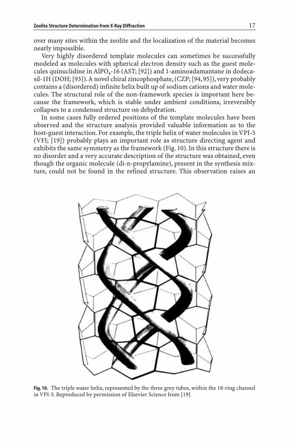
In some cases fully ordered positions of the template molecules have beenobserved and the structure analysis provided valuable information as to thehost-guest interaction. For example, the triple helix of water molecules in VPI-5(VFI; [19]) probably plays an important role as structure directing agent andexhibits the same symmetry as the framework (Fig. 10). In this structure there isno disorder and a very accurate description of the structure was obtained, eventhough the organic molecule (di-n-propylamine), present in the synthesis mix-ture, could not be found in the refined structure. This observation raises an
Zeolite Structure Determination from X-Ray Diffraction 17
Fig. 10. The triple water helix, represented by the three grey tubes, within the 18-ring channelin VPI-5. Reproduced by permission of Elsevier Science from [19]

important question, that cannot be answered here, as to the role of the di-n-pro-pylamine in the synthesis of VPI-5.
Another example of template ordering was observed for cobalticinium nona-sil (NON; [96]). The point group symmetry of the cobalticinium template cation,Co(C5H5)2+, is 2/m and its center of symmetry coincides with the center ofsymmetry of the large cage (point group P1–) and the template ion is perfectlyordered. In the final example of template ordering the framework symmetry ofAlPO4-34 (CHA; [97]) is very low (P1–) and the morpholinium template residesin a fixed general position. The template cations tetraethylammonium in AlPO4-18 (AEI; [98]), tetrapropylammonium in SAPO-40 (AFR; [17, 18]),18-crown-6-Na+ in EMC-2 (EMT; [99]) and DABCO in CoGaPO-5 (CGF; [100]),are only two-fold disordered because of the small difference in symmetry be-tween the templates and the frameworks at the position of the templates. Theresults for NON, CHA and CGF were obtained from single crystal data and those for AEI, AFR and EMT from high-resolution powder data. Thus with either technique, accurate information on the localization of the template can be obtained as long as their positions are completely ordered or only slightly disordered.
The same order/disorder problems arise when (organic) molecules are adsorb-ed into the zeolites and only a few examples of a successful localization of theadsorbate within the framework have been reported. The study on the structureof disordered m-xylene sorbed on barium exchanged X (FAU; [101]) at differentloadings shows that with careful attention to detail excellent results can be ob-tained from powder data. The study revealed that when the loading increases,different molecular orientations are adopted by the m-xylene molecules in orderto maximize methyl–methyl distances and minimize the intermolecular repul-sion. Other examples which illustrate the potential of accurate powder diffrac-tion are the studies of various organic molecules adsorbed into type Y (FAU)zeolites. The sites of aniline and m-dinitrobenzene, simultaneously adsorbed in NaY using selective deuterated organic molecules, were studied by neutronpowder diffraction [102]. UV spectroscopy gives evidence of a charge transferinteraction between aniline and dinitrobenzene. In the rare earth exchangedNa,YbY/1,3,5- trimethylbenzene system [103], the mesitylene molecules occupytwo distinct sites. The molecules on site I are two-fold disordered, while site II isonly singly occupied. In contrast, the other Na,YbY/sorbate systems [104, 105]show highly disordered organic molecules. At the present time these differencesin results cannot be satisfactorily explained.
No serious order/disorder problems are involved in several H-ZSM-5/sorbatesystems. Single crystals of H-ZSM-5 (MFI) have been successfully loaded withseveral organic molecules (see also Sect. 4). The structure of a single crystal oflow-loaded H-ZSM-5, containing about three molecules of p-dichlorobenzene(pdcb) per unit cell, has been determined in the orthorhombic space groupPnma [83]. The sorbed pdcb molecules prefer the position at the intersection ofchannels (Fig. 11a, b). Although the symmetry of the pdcb molecule is compa-tible with the site symmetry of the framework it turns out that the molecularmirror plane perpendicular to the Cl–Cl axis does not coincide with the crystal-lographic mirror plane and a 2-fold positional disorder around the mirror plane
18 H. van Koningsveld · J.M. Bennett

Zeolite Structure Determination from X-Ray Diffraction 19
Fig. 11 a – d. ORTEP drawings [106] of the position and orientation of adsorbed molecules atthe intersection of channels in low-loaded H-ZSM-5. Open bonds connect framework atomsand solid bonds connect atoms in adsorbed molecules. a p-dichlorobenzene molecules in H-ZSM–5/2.6 p-dichlorobenzene, viewed down the straight channel axis. b as in a but vieweddown an axis inclined 20∞ with the straight channel axis. c naphthalene molecules in H-ZSM-5/3.7 naphthalene, viewed as in b. d p-nitroaniline molecules in H-ZSM-5/4.0 p-nitroaniline,viewed as in b
a
c
b
d

occurs. The location and rotational orientation of the sorbate at the intersectioncan, in a first approximation, be described by the fractional coordinates (x,y,z)of its molecular center and the angle a between the positive a axis and the vec-tor normal to the aromatic ring plane (Fig. 11a; Table 2).
In single crystals of H-ZSM-5 loaded with four molecules of naphthalene [81]or four molecules of p-nitroaniline [86] per unit cell, both exhibiting ortho-rhombic Pnma symmetry, the organic molecules at the intersection are in ananalogous orientation as in the low-loaded H-ZSM-5/pdcb system (Fig. 11b, d;Table 2). In H-ZSM-5, fully loaded with eight molecules p-xylene per unit cell,the adsorbate has been found to be ordered in the orthorhombic space groupP212121, allowing its packing determination (Fig. 12a; [79]). One of the p-xylenemolecules lies at the intersection of the straight and sinusoidal channels with itslong molecular axis nearly parallel to (100) and deviating about eight degreesfrom the straight channel axis.
The second p-xylene molecule is in the sinusoidal channel. Its long molecularaxis is practically parallel to (010) and deviates almost six degrees from [100].The structural aspects of H-ZSM-5 loaded with eight p-dichlorobenzene (pdcb)molecules per unit cell [84] are in all details comparable to those in the high-loaded H-ZSM-5/8 p-xylene system (compare Figs. 12a and 12b).
The phase transition from Pnma to P212121 can be connected to a suddenincrease in ordering of the sorbed phase with increasing coverage [79, 107, 108].This commensurate crystallization of molecules within the H-ZSM-5 frame-work is assumed to be stabilized by establishing contacts at the channel inter-section between adjacent molecules (See Fig. 12; [109]). However, the me-thyl(H)–aromatic ring interactions in H-ZSM-5/8 p-xylene are replaced by Cl-ring (C,H) interactions in H-ZSM-5/8 pdcb, which are substantially weaker.The importance of these interactions in stabilizing the guest structure withinthe zeolite host framework might therefore need reconsideration. The ring-(C,H)–framework (O) contacts, which are the same in both structures, might beimportant in stabilizing the actually observed packing arrangement.
20 H. van Koningsveld · J.M. Bennett
Table 2. Orientation of adsorbates at the intersection of channels in H-ZSM-5
Code a PDCB1 NAPH PNAN b PXYL PDCB2
x 0.4860 0.4860 0.4858 0.4894 0.4824y 0.2400 0.2366 0.2332 0.2379 0.2439z –0.0188 –0.0352 –0.0260 –0.0180 –0.0175a c 47.1 40.5 44.3 –31.1 –26.2
a Codes are as follows:PDCB1: H-ZSM-5 containing 2.6 molecules p-dichlorobenzene/u.c.NAPH: H-ZSM-5 containing 3.7 molecules naphthalene/u.c.PNAN: H-ZSM-5 containing 4.0 molecules p-nitroaniline/u.c.PXYL: H-ZSM-5 containing 8.0 molecules p-xylene/u.c.PDCB2: H-ZSM-5 containing 8.0 molecules p-dichlorobenzene/u.c.
b Molecular center calculated disregarding the oxygen atoms.c The angle a is defined in the text and illustrated in Fig. 11a.

The longest dimension of the adsorbed molecule, being similar to the lengthof the straight channel axis, very probably determines the commensuratecrystallization by a flexible response of the channel pores in the zeolite host framework. The rotational orientation of pdcb in the low-loaded system differs73.3∞ (= 47.1 + 26.2) and 78.2∞ (= 47.1+31.1) from the rotational orientation ofthe pdcb and p-xylene molecules trapped at the intersection in the high-loadedH-ZSM-5/sorbate systems (see Table 2). These two rotational orientations corre-
Zeolite Structure Determination from X-Ray Diffraction 21
Fig. 12 a, b. Position and orientation of adsorbed molecules in high-loaded H-ZSM-5. a p-xy-lene molecules at the intersection of channels and in the sinusoidal channel in the high-loadedH-ZSM-5/8 p-xylene system. The angle between the view direction and the straight channelaxis is 15∞. b As in a with p-xylene replaced by p-dichlorobenzene and viewed down thestraight channel axis
a
b
spond to the directions of the two maximal pore dimensions observed in theclover-like window in the empty H-ZSM-5 framework at 350 K [74].
From Table 2 it can be seen that the molecular center (disregarding the oxy-gen atoms) is approximately the same “off-intersection center” position in allstructures. The same type of disorder is observed in all low-loaded systems.Refinements of the low-loaded systems show that the starting orientation of thesorbate molecule may be rather far away from its minimized orientation as longas its geometry is reasonable and its molecular center is not too far away fromthe minimized value.The examples illustrate that sorbed molecules can be locat-ed within the zeolite framework by single crystal X-ray diffraction methodswhen the symmetry of the adsorption site is compatible with the symmetry ofthe sorbed molecule and also in some cases when these symmetries do not coin-cide and disorder occurs.
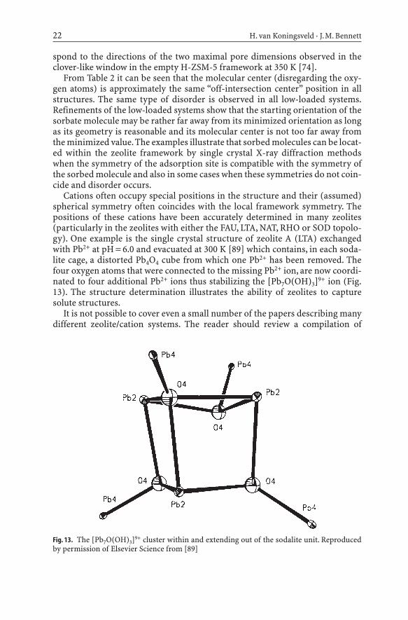
Cations often occupy special positions in the structure and their (assumed)spherical symmetry often coincides with the local framework symmetry. Thepositions of these cations have been accurately determined in many zeolites(particularly in the zeolites with either the FAU, LTA, NAT, RHO or SOD topolo-gy). One example is the single crystal structure of zeolite A (LTA) exchangedwith Pb2+ at pH = 6.0 and evacuated at 300 K [89] which contains, in each soda-lite cage, a distorted Pb4O4 cube from which one Pb2+ has been removed. Thefour oxygen atoms that were connected to the missing Pb2+ ion, are now coordi-nated to four additional Pb2+ ions thus stabilizing the [Pb7O(OH)3]9+ ion (Fig.13). The structure determination illustrates the ability of zeolites to capturesolute structures.
It is not possible to cover even a small number of the papers describing manydifferent zeolite/cation systems. The reader should review a compilation of
22 H. van Koningsveld · J.M. Bennett
Fig. 13. The [Pb7O(OH)3]9+ cluster within and extending out of the sodalite unit. Reproducedby permission of Elsevier Science from [89]

extra-framework sites in zeolites [110] published by the Structure Commissionof the International Zeolite Association. It is planned to publish an updated issuein 1998, which will include the most recent results.An inexperienced user shouldbe very careful in assigning extra-framework sites to maxima in a differenceelectron density map because simulations in pseudo-symmetric sodalites [60]and refinement of cation positions in Linde Q (BPH; [111]) have shown thatsome of the extra-framework electron density observed in a difference map isan artifact caused by the Fourier technique used in the refinement of the frame-work structure.
6Faulting Within the Framework
Faulting within the zeolite framework is one of the difficulties encountered inzeolite structure analysis. When faulting occurs repeatedly but in an irregularmanner the resulting structure is disordered. In cases where the repeat distancesbetween the faults become small enough diffuse stripes perpendicular to thefault planes are observed in the diffraction pattern.When the fault planes repeatin every unit cell a new framework topology emerges and the resulting diffrac-tion pattern again contains peaks only at Bragg positions. Faulting on a verysmall and/or irregular frequency will seriously hamper a precise structuredetermination. Such faulting is usually evident when a powder pattern has broad and sharp peaks. Powder patterns can also have broader and sharper lines when one dimension of the crystal is very thin but this is usually evidentfrom an electron microscopy photograph.
In the classical sense the explanation of faulting and the calculation of theresulting powder pattern should not be called a structure determination, yet itrepresents one of the more difficult tasks that can be undertaken. Examples arethe studies on zeolite beta, FAU-EMT, MFI-MEL, OFF-ERI, RUB-n and the SSZ-n/CIT-n series. It must be emphasized that any time the powder pattern consistsof sharp peaks only then faulting is an unsatisfactory explanation for difficultieswith a structure determination. An examination of the three proposed endmembers for zeolite beta [112] shows why the material faults. There are severaldifferent sequences of five and six rings around a 12-ring. The only connectionbetween one arrangement and another would be the chosen crystallographicsymmetry. But this is an artificial choice, because crystallographic symmetry isa result of the arrangement of the atoms in a structure and not the driving force. The structure of beta has been shown to be an almost random arrange-ment of sequences and has an almost uniform powder pattern. The accessibilityof the pore system is not affected by the degree of faulting nor does it change thediameter of the pore openings.Single crystals of the pure end-members have notbeen synthesized yet and therefore no single crystal structure analysis of thepolymorphs of zeolite beta has been performed [115]. Even the beta mineralanalogue, Tschernichite [116], has a powder pattern almost identical to those ofthe synthesized beta materials.
In contrast, both a wide range of different intermediate phases and unfaultedexamples of the end-members can be obtained for other intergrown materials.
Zeolite Structure Determination from X-Ray Diffraction 23

Thus, end members of FAU and EMT [117], MFI and MEL, OFF and ERI, someend-members in the RUB-n series (the zincosilicates RUB-17 (RSN; [120]) andVPI-7 (VSV; [121, 122]), the beryllosilicate lovdarite (LOV; [123]), the decasilRUB-3 (RTE; [124]), the borosilicate RUB-13 (RTH; [125])), and one end-mem-ber in the SSZ/CIT series (the borosilicate CIT-1 (CON; [126, 127])) can all beproduced by a proper choice of synthesis conditions and templates. Blocking of the pores can occur in some of the faulted intergrowths thereby drasticallychanging the properties of the materials. Only the syntheses of zeolite X (FAU)and ZSM-5 (MFI) currently give single crystals suitable for an accurate X-raystructure analysis [71, 128].
7Isomorphous Replacement of Framework Atoms
Isomorphous replacement of framework atoms has been studied in several zeo-lites. The problem with this type of diffraction experiments is that the differencein scattering power of a tetrahedral site with and without the substitution is often small. For example, replacement of 10% of the aluminum atoms at onesite by manganese ((Al9Mn)P10O40; AEL; [129]) is equivalent of looking for oneextra electron on a site where there is already an atom. The detection of partialreplacement of phosphorous by silicon, such as in SAPO-43 (GIS; [35]), is evenmore difficult.
The incorporation into the framework of tetrahedral atoms with a radius dif-ferent from the radius of the substituted tetrahedral atom can lead to a changein unit cell volume and X-ray diffraction can be used to follow these changes.Cell expansion or contraction does not conclusively establish the actual incor-poration of tetrahedral atoms into the framework, since extra framework spe-cies may also change the cell volume. The actual distribution of the incorporat-ed tetrahedral atoms on the framework sites has been studied using diffractiondata by examining both the refined T–O distances (and sometimes the O–T–Oangles) and the refined population parameters. From statistical studies on theT–O distances it was inferred that in substituted Nu-1-type frameworks (RUT;[130]) the boron atoms were uniformly distributed over all the tetrahedral sites.This conclusion may be in error because both the accepted value for a Si–Odistance was used and it was assumed that the T–O–T angle would be un-changed. A recent paper on a material with the same topology (RUB-10; [131])now indicates that the boron atoms may be ordered but the space group has been changed to P21/a.
In several of the cited papers the population parameters (or site occupancies)of the framework atoms have been refined. The population parameter is fre-quently used as a parameter which mimics the success/failure of isomorphoussubstitution. However, it is dangerous to draw too many conclusions from changes in the refined population parameters because, like the (anisotropic) displacement parameters, they act as a waste-basket for all systematic and non-systematic errors occurring in a refinement. For example, missing tetrahedralatoms influence the population parameter in a non-systematic way and even theactual scattering factors used in the refinement affect the site occupancies. It has
24 H. van Koningsveld · J.M. Bennett

also been shown that in dodecasil-1H (DOH; [93]) when the oxygen atoms aredisordered, the population factors of the tetrahedral atoms appear smaller than1.0 and this was shown to be an artifact of the least squares algorithm used.Other examples include the single crystal determination of the structure ofZAPO-M1 (ZON; [132]), where both the distribution of the T–O bond lengthsand the population parameters were consistent with a non-uniform distributionof the zinc atoms. It has also been claimed that from very precise single crystaldiffraction studies it was observed that the substitution of silicon for phos-phorus in SAPO-31 (ATO; [133]) and of cobalt for (exclusively) aluminum inboth CoAPO-5 (AFI; [134]) and CoSAPO-34 (CHA; [135]) had taken place. Inmany other structure reports the opposite conclusion was reached, namely thatreplacement of framework atoms could not be established from the analysis ofX-ray diffraction data alone.
8Crystal Size Limitations
The possibility of using single crystal data rather than powder X-ray diffractionbecame much more feasible with the development of area detectors for use with high-intensity synchrotron X-ray sources. A 30 ¥ 30 ¥ 30 µm crystal of(Mg,Al)PO4-STA-1 (SAO; [136]) was successfully used for structure determina-tion at the ESRF in Grenoble using a diffractometer equipped with a CCD detec-tor. Unfortunately, no low angle data were collected which probably accountedfor why the template could not be localized. Data from crystals of this size havebeen previously collected using rotating anode generators [34] and even sealedtube data from crystals of about the same volume [137] have been measured.Unfortunately, the use of these data was restricted to only determining the framework structure. In the structure analysis of a 35 ¥ 20 ¥ 15 µm crystal ofAlPO4-34 (CHA; [97]) synchrotron diffraction data were collected at the SRS atDaresbury and used to determine the location of the morpholinium cation.Theoretically the coherence length of about 0.1 µm determines the lower sizelimit for single crystal diffraction [138] and the physical ability to handle suchsmall crystals may finally prove to be the limiting factor in their use.
As the size of the crystal that can be used decreases it can be expected that theuse of single crystal diffraction facilities at synchrotron sources by the zeolitecommunity will increase enormously in the coming years. However, many newmicroporous materials are synthesized with crystals that are only 1 µm in size –still too small to be utilized for single crystal work – and only can be studied bypowder techniques.
9Conclusions
As hopefully explained here, the problems with attempting structure deter-minations of unknown materials requires that an accurate starting model beobtained and all of the possible problems that could cause subtle framework
Zeolite Structure Determination from X-Ray Diffraction 25

changes be evaluated. This requires that the unit cell dimensions and spacegroup be correctly determined. Molecular modeling techniques have started tobe applied to determining starting models. They are currently restricted bybeing difficult to use to compose a framework arrangement within a definedunit cell and space group. Other techniques are focusing on using the power ofcurrent computers to try to determine a framework arrangement by startingwith random arrangements of atoms and hoping that the program can refine thestarting models to the correct topology.
While all these techniques will continue to improve over the next few years,the quality and accuracy of the final crystal structure will depend on how goodthe data are and how well the unit cell and space group have been determined.While not covered in this paper, it should be also realized that input from manyother characterization techniques should be used as an aid in the determinationof an unknown structure, that the final solution should be in agreement withdata from all the different techniques employed and that the solution shouldmake chemical sense. When the structure has been correctly determined, it ispossible to use the information to explain framework distortions, cation andsorbate locations, isomorphous substitutions and other subtle variations in thebehavior of the structure. But, the use of the structure of a material requires acorrect structure determination and obtaining that solution is both a scienceand an art and not a trivial exercise.
References
1. McCusker LB (1991) Acta Cryst A47:2972. Baerlocher Ch, McCusker LB (1994) Practical aspects of powder diffraction data analy-
sis. In: Jansen JC, Stöcker M, Karge HG, Weitkamp J (eds) Advanced zeolite science andapplications. Stud Surf Sci Catal Vol 85. Elsevier Science, Amsterdam, p 391
3. Visser JW (1993) A fully automatic program for finding the unit cell from poweder data.Version 15. Method described in (1969) J Appl Cryst 2 :89
4. Werner PE (1990) Treor 90. A trial and error program for indexing of unknown powderpatterns. Department of Structural Chemistry, Arrhenius Laboratory, University ofStockholm
5. Jansen J, Peschar R, Schenk H (1992) J Appl Cryst 25: 2376. Jansen J, Peschar R, Schenk H (1993) Z Kristallogr 206:337. Spengler R, Zimmermann H, Burzlaff H, Jansen J, Peschar R, Schenk H (1994) Acta Cryst
B50: 5788. Estermann MA, McCusker LB, Baerlocher Ch (1992) J Appl Cryst 25:5399. McCusker LB, GrosseKunstleve RW, Baerlocher Ch, Yoshikawa M, Davis ME (1996)
Microporous Mater 6 :29510. Estermann MA, McCusker LB, Baerlocher Ch, Merrouche A, Kessler H (1991) Nature 352:
32011. Baerlocher Ch, Hepp A, Meier WM (1977) DLS. A Fortran program for the simulation of
crystal structures by geometric refinement. Institut für Kristallographie, ETH, Zürich,Switzerland
12. Young RA (1993) The Rietveld Method, Oxford University Press, Oxford13. Lawton SL, Rohrbaugh WJ (1990) Science 247:131914. Bennett JM, Marcus BK (1988) The crystal structures of several metal aluminophosphate
molecular sieves. In: Grobet PJ et al. (eds) Innovation in zeolite material science. ElsevierScience, Amsterdam, p 269
26 H. van Koningsveld · J.M. Bennett

15. Harvey G, Baerlocher Ch (1992) Z Kristallogr 201:11316. Kirchner RM, Bennett JM (1994) Zeolites 14:52317. Dumont N, Gabelica Z, Derouane EG, McCusker LB (1993) Microporous Mater 1:14918. McCusker LB, Baerlocher Ch (1996) Microporous Mater 6 :5119. McCusker LB, Baerlocher Ch, Jahn E, Bülow M (1991) Zeolites 11:30820. Li HX, Davis ME, Higgins JB, Dessau RM (1993) J Chem Soc Chem Commun 1993:40321. Vogt ETC, Richardson JW Jr (1990) J Solid State Chem 87:46922. Annen MJ, Young D, Davis ME (1991) J Phys Chem 95: 138023. Prasad S, Balakrishnan I (1990) Inorg Chem 29: 483024. Richardson JW Jr,Vogt ETC (1992) Zeolites 12: 1325. Baur WH (1995) Framework mechanics: limits to the collapse of tetrahedral frameworks.
In: Rozwadowski M (ed) Proceedings of the 2nd Polish-German zeolite colloquium.Nicholas Copernicus University Press, Torun, p 171
26. Baur WH (1992) Why the open framework of zeolite A does not collapse, while the dense framework of natrolite is collapsible. In: Rozwadowski M (ed) Proceedings of thePolish-German zeolite colloquium. Nicholas Copernicus University Press, Torun, p 11
27. Koningsveld H van, Jansen JC, Bekkum H van (1987) Zeolites 7 : 56428. Baur WH (1992) J Solid State Chem 97: 24329. Norby P, Fjellvåg H (1992) Zeolites 12: 89830. Krogh Anderson IG, Krogh Anderson E, Norby P, Colella C, de’Gennaro M (1991) Zeolites
11: 14931. Newsam JM (1988) J Phys Chem 92: 44532. Gier TE, Stucky GD (1991) Nature 349: 50833. Harrison WT, Gier TE, Stucky GD (1995) Acta Cryst C51: 18134. Pluth JJ, Smith JV, Bennett JM (1989) J Am Chem Soc 111: 169235. Helliwell M, Kauèiè V, Cheetham GMT, Harding MM, Kariuki BM, Rizkallah PJ (1993)
Acta Cryst B49: 41336. Vezallini G, Quartieri S, Alberti A (1993) Zeolites 13: 3437. Hansen S, Håkansson U, Fälth L (1990) Acta Cryst C46: 136138. Hansen S, Håkansson U, Landa-Canovas AR, Fälth L (1993) Zeolites 13: 27639. Baur WH, Fischer RX, Shannon RD (1988) Relations and correlations in zeolite RHO and
computer simulations of its crystal structure. In: Grobet PJ et al. (eds) Innovation in zeo-lite materials science. Elsevier Science, Amsterdam, p 281
40. Fischer RX, Baur WH, Shannon RD, Staley RH, Abrams L, Vega AJ, Jorgensen JD (1988)Acta Cryst B44: 321
41. Baur WH, Bieniok A, Shannon RD, Prince E (1989) Z Kristallogr 187: 25342. Fischer RX, Baur WH, Shannon RD, Parise JB, Faber J, Prince E (1989) Acta Cryst C45: 98343. Bieniok A, Baur WH (1993) Acta Cryst B49: 81744. Corbin DR, Abrams L, Jones GA, Harlow RL, Dunn PJ (1991) Zeolites 11: 36445. Parise JB, Corbin DR, Gier TE, Harlow RL, Abrams L, Dreele RB von (1992) Zeolites 12:
36046. Parise JB, Corbin DR, Abrams L, Northrup P, Rakovan J, Nenoff TM, Stucky GD (1994)
Zeolites 14: 2547. Parise JB, Corbin DR, Abrams L (1995) Microporous Mater 4 : 9948. Newsam JM, Vaughan DEW, Strohmaier KG (1995) J Phys Chem 99: 992449. Baur WH, Fischer RX, Shannon RD, Staley RH, Vega AJ, Abrams L, Corbin DR (1987) Z
Kristallogr 179: 28150. Ståhl K, Hanson J (1994) J Appl Cryst 27: 54351. Ståhl K, Thomassen R (1994) Zeolites 14: 1252. Joswig W, Baur WH (1995) N Jb Miner Mh 1995: 2653. Baur WH, Joswig W (1996) N Jb Miner Mh 1996: 17154. Richardson JW Jr, Pluth JJ, Smith JV, Dytrych WJ, Bibby DM (1988) J Phys Chem 92: 24355. Fleet ME (1989) Acta Cryst C45: 84356. Kempa PB, Engelhardt G, Buhl JCh, Felsche J, Harvey G, Baerlocher Ch (1991) Zeolites 11:
558
Zeolite Structure Determination from X-Ray Diffraction 27

57. Nenoff TM, Harrison WTA, Gier TE, Stucky GD (1991) J Am Chem Soc 113: 37858. Sieger P, Wiebcke M, Felsche J (1991) Acta Cryst C47: 49859. Depmeier W (1992) Z Kristallogr 199: 7560. Hu X, Depmeier W (1992) Z Kristallogr 201: 9961. Depmeier W, Melzer R, Hu X (1993) Acta Cryst B49: 48362. Harrison WT, Gier TE, Stucky GD (1994) Acta Cryst C50: 47163. Féron B, Guth JL, Mimouni-Erddalane N (1994) Zeolites 14: 17764. Brenchley ME, Weller MT (1994) Zeolites 14: 68265. Mead PJ, Weller MT (1994) Microporous Mater 3 : 28166. Fütterer K, Depmeier W, Altorfer F, Behrens P, Felsche J (1994) Z Kristallogr 209: 51767. Duke CVA, Hill SJ, Williams CD (1995) Zeolites 15: 41368. Mead PJ, Weller MT (1995) Zeolites 15: 56169. Hassan I (1996) Z Kristallogr 211: 22870. Dann SE, Weller MT (1996) Inorg Chem 35: 555 71. Koningsveld H van, Bekkum H van, Jansen JC (1987) Acta Cryst B43: 12772. Gabelica Z, Guth JL (1989) Angew Chem 101: 6073. Lopez A, Soulard M, Guth JL (1990) Zeolites 10: 13474. Koningsveld H van (1990) Acta Cryst B46: 73175. Aizu K (1970) Phys Rev B 2: 754 76. Koningsveld H van, Tuinstra F, Jansen JC, Bekkum H van (1989) Zeolites 9: 25377. Koningsveld H van, Jansen JC, Bekkum H van (1990) Zeolites 10: 23578. Mentzen BF (1988) J Appl Cryst 21: 266 79. Koningsveld H van, Tuinstra F, Bekkum H van, Jansen JC (1989) Acta Cryst B45: 42380. Mentzen BF, Sacerdote-Peronnet M, Berar JF, Lefebvre F (1993) Zeolites 13: 48581. Koningsveld H van, Jansen JC (1996) Microporous Mater 6 : 15982. Mentzen BF, Sacerdote-Peronnet M (1993) Mat Res Bull 28: 116183. Koningsveld H van, Jansen JC, Man AJM de (1996) Acta Cryst B52: 13184. Koningsveld H van, Jansen JC, Bekkum H van (1996) Acta Cryst B52: 14085. Reck G, Marlow F, Kornatowski J, Hill W, Caro J (1996) J Phys Chem 100: 169886. Koningsveld H van, Koegler JH (1997) Microporous Mater 9: 7187. Baur WH, Fischer RX (1995) ZeoBase, Frankfurt and Mainz88. Heo NH, Seff K (1992) Zeolites 12: 81989. Ronay Ch, Seff K (1993) Zeolites 13: 9790. Jang SB, Kim Y, Seff K (1994) Zeolites 14: 26291. Simmen A, Patarin J, Baerlocher Ch (1993) Rietveld refinement of F-containing GaPO4–
L TA. In: Ballmoos R von, Higgins JB, Treacy MMJ (eds) Proceedings from the NinthInternational Zeolite Conference, Montreal 1992. Butterworth-Heinemann, StonehamMA, p 433
92. Bennett JM, Kirchner RM (1991) Zeolites 11: 50293. Miehe G, Vogt T, Fuess H, Müller U (1993) Acta Cryst B49: 74594. Rajiè N, Logar NZ, Kauèiè V (1995) Zeolites 15: 672 95. Harrison WTA, Gier TE, Stucky GD, Broach RW, Bedard RA (1996) Chem Mater 8: 14596. Goor G van de, Freyhardt CC, Behrens P (1995) Z Anorg Allg Chem 621: 31197. Harding MM, Kariuki BM (1994) Acta Cryst C50: 85298. Simmen A, McCusker LB, Baerlocher Ch, Meier WM (1991) Zeolites 11: 65499. Baerlocher Ch, McCusker LB, Chiapetta R (1994) Microporous Mater 2 : 269
100. Chippindale AM, Cowley AR. Paper to be submitted101. Mellot C, Espinat D, Rebours B, Baerlocher Ch, Fischer P (1994) Catal Lett 27: 159102. Kirschhock C, Fuess H (1997) Microporous Mater 8 : 19103. Czjzek M, Vogt T, Fuess H (1992) Zeolites 12: 237104. Czjzek M, Vogt T, Fuess H (1991) Zeolites 11: 832105. Czjzek M, Fuess H, Vogt T (1991) J Phys Chem 95: 5255106. Johnson CK (1965) ORTEP, Report ORNL. Oak Ridge Nat Lab, Oak Ridge TN, revised
June 1970107. Reischman PT, Schmitt KD, Olson DH (1988) J Phys Chem 92: 5165
28 H. van Koningsveld · J.M. Bennett

108. Richards RE, Rees LVC (1988) Zeolites 8 : 35109. Thamm H (1987) J Phys Chem 91: 8110. Mortier WJ (1982) Compilation of extra-framework sites in zolites. Butterworth Scientific,
Guildford 111. Andries KJ, Bosmans HJ, Grobet PJ (1991) Zeolites 11: 124112. Newsam JM, Treacy MMJ, Koetsier WT, Gruyter CB de (1988) Proc Roy Soc Lond A420:
375113. Treacy MMJ, Newsam JM (1988) Nature 332: 249114. Higgins JB, LaPierre RB, Schlenker JL, Rohrman AC, Wood JD, Kerr GT, Rohrbaugh WJ
(1988) Zeolites 8: 446115. Marler B, Böhme R, Gies H (1993) Single crystal structure analysis of zeolite beta: the
superposition structure. In: Ballmoos R von, Higgins JB, Treacy MMJ (eds) Proceedingsfrom the Ninth International Zeolite Conference, Montreal 1992. Butterworth-Heine-mann, Stoneham MA, p 425
116. Boggs RC, Howard DG, Smith JV, Klein GL (1993) Am Mineral 78: 822117. Delprato F, Delmotte L, Guth JL, Huve L (1990) Zeolites 10: 546118. Burkett SL, Davis ME (1993) Microporous Mater 1: 265119. Chatelain T, Patarin J, Soulard M, Guth JL (1995) Zeolites 15: 90120. Röhrig C, Gies H (1995) Angew Chem Int Ed 34: 63121. Annen MJ, Davis ME, Higgins JB, Schlenker JL (1991) J Chem Soc Chem Commun 1991:
1175122. Röhrig C, Gies H, Marler B (1994) Zeolites 14: 498123. Merlino S (1990) Eur J Mineral 2 : 809124. Marler B, Grünewald-Lüke A, Gies H (1995) Zeolites 15: 388125. Vortmann S, Marler B, Gies H, Daniels P (1995) Microporous Mater 4 : 111126. Lobo RF, Pan M, Chan I, Li HX, Medrud RC, Zones SI, Crozier PA, Davis ME (1993) Science
262: 1543127. Lobo RF, Davis ME (1995) J Am Chem Soc 117: 3766128. Olson DH (1995) Zeolites 15: 439129. Pluth JJ, Smith JV, Richardson JW Jr (1988) J Phys Chem 92: 2734130. Bellusi G, Millini R, Carati A, Maddinelli G, Gervasini A (1990) Zeolites 10: 642131. Gies H, Rius J (1995) Z Kristallogr 210: 475132. Marler B, Patarin J, Sierra L (1995) Microporous Mater 5 : 151133. Baur WH, Joswig W, Kassner D, Kornatowski J, Finger G (1994) Acta Cryst B50: 290134. Chao KJ, Sheu SP, Sheu HS (1992) J Chem Soc Faraday Trans 88: 2949135. Nardin G, Randaccio L, Kauèiè V, Rajiè N (1991) Zeolites 11: 192136. Noble GW, Wright PA, Lightfoot P, Morris RE, Hudson KJ, Kvick A, Graafsma H (1997) To
be published137. Flanigen EM, Bennett JM, Grose RW, Cohen JP, Patton RL, Kirchner RM (1978) Nature
271: 512138. Schlenker JL, Peterson BK (1996) J Appl Cryst 29: 178
Zeolite Structure Determination from X-Ray Diffraction 29