zeiss axio lab - ctk instrumentstypical applications, typical specimens task zeiss axio lab.a1...
TRANSCRIPT

ZEISS Axio Lab.A1TÜV-Certified Ergonomics for Your Efficient Day in the Laboratory
Product Information
Version 1.0

2
Axio Lab.A1 was developed for your daily work in the laboratory. The compact
microscope operates continuously, reliably, and economically while providing the
highest performance. You can use all conventional contrast methods: brightfield,
darkfield, phase contrast, and fluorescence. Experience intuitive imaging with the
free ZEN lite software. Or use Labscope, the imaging app from ZEISS. In combination
with the Axiocam ERc 5s microscope camera, you can transform your Axio Lab.A1
into a Wi-Fi imaging system. The particular advantage of Axio Lab.A1 for those who
spend long hours at the microscope is its TÜV-certified ergonomics. You can view
your specimens from the most comfortable height. Your neck and shoulders stay
relaxed even during long days in the laboratory.
TÜV-Certified Ergonomics for Your Efficient Day in the Laboratory
› In Brief
› The Advantages
› The Applications
› The System
› Technology and Details
› Service

3
Simpler. More Intelligent. More Integrated.
TÜV-Certified Ergonomics for Relaxed Work
in the Laboratory
It is possible to work long hours at the microscope
without strain in your neck and shoulders: The
viewing height of your Axio Lab.A1 is individually
adjustable – you can alter the height and angle of
view by adjusting the ergotube to suit your own
body height. This allows you to always view your
specimens from the most comfortable position,
eliminating strain on your neck and shoulders.
You can access all the main controls of your
Axio Lab.A1 with one hand, including the stage
drive, fine focus, and brightness control.
Versatile in Use, Brilliant Results
Axio Lab.A1 shines in laboratories for microbiology,
cytology, hematology, and pathology. The micro-
scope is ideally suited for work in parasitology and
immunofluorescence and is a valuable working tool
in schools and universities. With the multidiscussion
equipment, up to three observers can view the
same image. As the main observer, you highlight
interesting regions in the sample using a light
marker in white, red, or green. The excellent optics
from ZEISS ensure brilliant, color-corrected, high-
contrast results. The quality of the image leaves
nothing to be desired.
LED Illumination with Push & Click
Your Axio Lab.A1 is equipped with a fivefold Abbe
turret condenser with darkfield and Ph1, Ph2, and
Ph3. With the polarization contrast, you can capture
birefringent structures such as crystals and fibers.
For fluorescence applications, Axio Lab.A1 utilizes
two LED positions and the proven Push & Click
modules from ZEISS. LED fluorescence is more
energy efficient and easier to handle than conven-
tional HBO illumination. You avoid warm-up and
cool-down times, lamp replacement, and lamp
adjustment. In brightfield, you use the warm, white
light effect of energy-efficient LEDs even while
benefiting from their longer service life.
› In Brief
› The Advantages
› The Applications
› The System
› Technology and Details
› Service

4
Your Insight into the Technology Behind It
TÜV-Certified Ergonomics – Perfectly Adapted
Laboratory microscopes such as Axio Lab.A1 are
often used several hours a day for routine
applications in hematology, pathology and cytology.
The unique character of your Axio Lab.A1 stems
from its excellent ergonomic qualities, stability, and
sophisticated details. Axio Lab.A1 was developed
and designed in collaboration with occupational
health physicians and TÜV Rheinland to meet your
most stringent ergonomic requirements for micro-
scope workplaces. It is the first light microscope
in the world to be available in a special ergonomic
configuration that carries the “Ergonomics
Approved” certificate (ID:0000025994) from TÜV
Rheinland. Continuous adjustment of the viewing
height of the ergotube through 50 mm lets you
avoid neck and shoulder strain. The tube can also
be pivoted continuously between 8 and 33 degrees.
› In Brief
› The Advantages
› The Applications
› The System
› Technology and Details
› Service

5
Tailored Precisely to Your Applications
Typical Applications, Typical Specimens Task ZEISS Axio Lab.A1 Offers
Genetic laboratory examinations Examination of heparinized blood for cytogenetic (chromosome analysis) and molecular cytogenetic investigations (fluorescence in situ hybridization, FISH)
LED fluorescence with two LED positions and Push & Click modules: LED fluorescence is reliable, energy efficient, quick, and easy to use; additional advantages: no warm-up or cool-down times, no need for adjustment
Immunology In vivo examination of the physiology of immune cells under normal and inflamed conditions in their natural environment
Fluorescent FITC markers for antigen/antibody reactions; FITC can be linked with antibodies (immunoglobulins); under LED excitation of 470 nm, FITC emits a green fluorescent signal, which allows detection of specific surface characteristics (antigens) in fluids, cells, or tissues, as well as pathogens
Hematology Laboratory examination of blood samples (EDTA blood) for quantity, shape, and condition of blood cells
Axio Lab.A1 makes it easier to count erythrocytes and leu-kocytes; you can access all the main controls with one hand, including the stage drive, fine focus, and brightness control, leaving your other hand free for operating the leukocyte- counting device
Pathology Examination of specimens such as cell smears, bone marrow, and blood samples as to structure, composition, and growth of the cells during laboratory work; efficiency and sample throughput are of major importance
TÜV-certified ergonomics specially aimed at long-term users – you observe your specimens from the most comfortable height your neck and shoulder muscles stay relaxed
› In Brief
› The Advantages
› The Applications
› The System
› Technology and Details
› Service

6
Hematology: Plasmodium malariae, daisy stage, brightfield; courtesy of A. Michelsen, Ortenau Klinikum in Lahr-Ettenheim, Germany
ZEISS Axio Lab.A1 at Work
Pig gut, staining MG; objective: A-Plan 10×/0.25
Hematology: blood smear, darkfield Gout examination: uric acid crystal, polarization
Mouse kidney, stained with FITC Urine analysis: calcium carbonate, brightfield; courtesy of G. Spengler-Schulz, Alexander Fleming Schule in Stuttgart, Germany
› In Brief
› The Advantages
› The Applications
› The System
› Technology and Details
› Service

7
ZEN lite for Your Imaging Tasks
Download ZEN lite, your free version of the
ZEN Imaging Software. View your .czi files and take
advantage of the many possibilities. With ZEN lite,
you control your Axiocam microscope cameras,
capture images and simple video sequences,
measure lengths and areas interactively, and
generate simple reports. Expand ZEN lite with
selected modules and capture time-lapse images
of your specimens or create multichannel
fluorescent images.
Expand Your Possibilities
› In Brief
› The Advantages
› The Applications
› The System
› Technology and Details
› Service

8
Expand Your Possibilities
ZEISS Labscope – Your Doorway
to the Digital World
With the imaging app Labscope from ZEISS, you
can display all the live images from your connected
microscopes. With just one click, select the image
from one of your students. You can record images
and videos in high five-megapixel resolution.
Annotate your images and measure distances,
for example. Then share your images, reports, and
videos with others via e-mail, social media, or cloud
services. With Labscope, you save your images in the
ZEN-compatible .czi file format including all meta-
data and a separate annotation layer. Or select the
space-saving .jpg format. Download Labscope from
the Apple App Store – easily, quickly, and free of
charge.
› In Brief
› The Advantages
› The Applications
› The System
› Technology and Details
› Service

9
Multidiscussion equipment
Using the multidiscussion equipment from ZEISS,
you create identical image-viewing positions for
all co-observers. Up to two co-observers see the
same image in the same orientation as the main
observer – this prevents confusion through image
rotation or mirroring. The main observer and co-
observer benefit from homogenous lighting in the
field of view.
The movable light marker allows you to mark
interesting structures or notable histological altera-
tions in the prepared specimen. You provide an
optimum orientation guide for differently stained
specimens by continuously regulating the intensity
of the light marker and selecting between the
various color settings (white, green, red).
You can use the multidiscussion system for training
and consulting situations and in the medical field,
for example when teaching students and doctoral
candidates, when performing consultations, or
when jointly evaluating difficult specimens. By
receiving a direct impression of the microscopic
image, you are often better able to detect fine
structures and nuances of color, particularly in the
case of histologically stained transmitted-light
specimens, such as tissue sections or blood smears,
in the microscopic image.
Expand Your Possibilities
› In Brief
› The Advantages
› The Applications
› The System
› Technology and Details
› Service

1
2
36
3
4
10
1 Microscope
Axio Lab.A1:
• Stand with transmitted light
• Stand with transmitted light polarization
• Stand with transmitted light and reflected light
fluorescence
2 Objectives
Recommended classes of objectives:
• A-Plan
• N-Achroplan
• EC Plan-NEOFLUAR
5 Software
• ZEN lite
• Labscope imaging app
• Labscope
6 Tubes
• Standard tubes
• Tubes with adjustable angle
• Tubes with adjustable height
• Tubes with adjustable angle and height
Your Flexible Choice of Components
3 Illumination
Transmitted light:
• HAL 35 (halogen, integrated)
• LED illumination (warm white)
Reflected light:
• LED illumination (fluorescence)
4 Cameras
Recommended cameras:
• Axiocam MRc
• Axiocam ICc 5
• Axiocam ICc 1
• Axiocam ERc 5s
› In Brief
› The Advantages
› The Applications
› The System
› Technology and Details
› Service

Intermediate plate Axio Lab.A1 for tubes 423732-9040-000
Ultra condenser 1.2/1.4 (0.75-1.0)465500-0000-000
Dry darkfield condenser 0.8/0.95 (0.6/0.75)465505-0000-000
Condenser 0.9/1.25 H, D, Ph1, Ph2, Ph3 for Axio Lab.A1424227-9010-000
Mechanical stage 75x30 Lwith hardcoat anodized surface432035-9161-000
Mechanical stage 75x30 R ergon. drive, fixed positionwith hardcoat anodized surface432035-9180-000
Specimen holder for counting chambers 75x25, spring lever left 432323-9030-000
Mechanical stage 75x30 R with hardcoat anodized surface432035-9151-000
Specimen holder for one-hand operation, spring lever left432323-9000-000
Condenser 0.9/1.25 H for Axio Lab.A1424227-9000-000
Center comp. co-observation for Axio Scope.A1/Axio Lab.A1425145-9010-000
Tube carrier for 1 co-observer, light-intensive, end panel, left425145-9060-000
Binocular ergo tube 5-30°/23 (co-obs.), upright image425520-9080-000
Eyepiece eyecup 444801-0000-000
Eyepiece PL 10x/22 Br. foc.444036-9010-000
Eyepiece micrometer see price list
Condenser holder Z for darkfield condenser445323-0000-000
Crossline micrometer see price list
Eyepiece PL 16x/16 Br. foc.444054-9000-000Eyepiece E-PL 10x/20 Br. foc.444232-9904-000
Auxiliary microscope, d=30444830-9902-000
Pinhole diaphragm, d=30 444020-0000-000
Modulator disk H, D 0.65, Ph 1, 2, 3 424225-9010-000
Condenser, achrom.-aplan. 0,9 H424225-9060-000Condenser, achrom.-aplan. 0,9 H D Ph DIC424225-9070-000
Condenser 0.9 H Pol424225-9080-000
Eyepiece PL 10x/22 Br. foc. Pol with crossline graticule444036-9020-000
Binocular photo tube 30°/23 (50:50), reversed image425520-9010-000Binocular photo tube 30°/23 (100:0/0:100), reversed image425520-9020-000
Binocular tube 30°/23, reversed image425520-9000-000
Binocular photo tube 20°/23 (100:0/0:100), upright imagewith sliding prism425520-9030-000
Comfortable binocular ergo photo tube 15°/23 (50:50)vertically adjustable and extendable, upright image425520-9050-000
Binocular tube 20°/23, upright image425520-9090-000
Tube carrier multidiscussion for 2 tubes, end panel linear, l/r425145-9050-000
Adaptation set for tubes Axio Scope.A1 425145-9070-000
Specimen holder plate for slides suitable for transmitted and reflected light432323-9020-000
including:Base plate for microscope stand Axio Lab.A1
Microscope stand Axio Lab.A1HAL 35, 4x H, rotary stage Pol 360°430037-9031-000
Binocular tube 30°/20, reversed image425522-9000-000
Binocular phototube 30°/20 (50:50), reversed image425522-9010-000
Binocular ergophototube 8-38°/20 (50:50), reversed image425522-9030-000
Intermediate plate Axio Lab.A1 for tubes 423732-9040-000
Rotary stage Pol, 360° with clamping device for Axio Lab432035-9191-000
Attachable object guide Pol, 45x25 mm432323-9011-000
Microscope stand Axio Lab.A1 HAL 35, 5x H430037-9000-000Microscope stand Axio Lab.A1HAL 35, 5x H, mechanical stage R430037-9010-000
Binocular ergotube 8-38°/20, reversed image425522-9020-000including:Base plate for microscope stand Axio Lab.A1
Base plate for microscope stand Axio Lab.A1430037-9100-000
Binocular phototube 30°/23 (50:50), reversed image425520-9010-000
Binocular phototube 30°/23 (100:0/0:100), reversed image425520-9020-000
Binocular tube 30°/23, reversed image425520-9000-000
Binocular phototube 20°/23 (100:0/0:100), upright image425520-9030-000
*
*
* Recommended to use:Base plate for microscope stand Axio Lab.A1430037-9100-000
*
1 1
Fine drive knob with scale, changeable430051-9000-000
Analyzer to be screwed into tubes Axio Lab.A1 or in intermediate plate for tubes Axio Scope.A1428107-9000-000
Quartz depolarizer to be screwed into tubes Axio Lab.A1 or into intermediate plate for tubes Axio Scope.A1428106-9020-000
Analyzer to be screwed into tubes Axio Lab.A1 or in intermediate plate for tubes Axio Scope.A1428107-9000-000
Quartz depolarizer to be screwed into tubes Axio Lab.A1 or into intermediate plate for tubes Axio Scope.A1428106-9020-000
Fine drive knob with scale, changeable430051-9000-000
Binocular tube 20°/23, upright image425520-9090-000
Quartz depolarizer with tube lens for tubes Axio Scope.A1428106-9010-000
11
System Overview
› In Brief
› The Advantages
› The Applications
› The System
› Technology and Details
› Service

mandatory:Base plate for microscope stand Axio Lab.A1430037-9100-000
Adapter video 60 C 1/3" 0.4x456108-0000-000Camera adapter 60N-C 2/3" 0.5x426112-0000-000Camera adapter 60N-C 2/3" 0.63x426113-0000-000Camera adapter 60N-C 1" 1.0x426114-0000-000
Camera adapter T2-C 1" 1.0x426104-0000-000Camera adapter T2-C 1" 1.0x,adjustable 426105-0000-000
T2 adapter for SLR cameraon request
Camera adapter T2-T2 DSLR 1.6x426115-0000-000Camera adapter T2-T2 SLR 2.5x426116-0000-000
Adapter 60N-T2 1.0x426103-0000-000
Camera with SLR bayonet
AxioCam ERcAxioCam ICAxioCam MRother AxioCam on request
Double adapter 60N - 2x 60N with slider mount426141-9902-000
Slider with beam splitter mount for double adapter426141-9021-000
Slider with 100 % mirror for double adapter426141-9011-000
Circular polarizer Dincluding slider 6x20 with compensator Lambda/4453623-0000-000
Polarizer D, fixed, removable427701-0000-000
Polarizer fixed with lambda plate rotatable445226-0000-000
Polarizer D, 90° rotatable, removable427706-0000-000
Polarizer rotatable with color filter carrier427707-0000-000
Color filter carrier 3x for filter d=32 mm428305-0000-000
Low-power system for objectives 2.5x/4x for condenser 0.9/1.25 H424225-9050-000
Phototube / ergophototubeaccording to system configuration
1
12
System Overview
› In Brief
› The Advantages
› The Applications
› The System
› Technology and Details
› Service

mandatory:Base plate for microscope stand Axio Lab.A1430037-9100-000
Adapter video 60 C 1/3" 0.4x456108-0000-000Camera adapter 60N-C 2/3" 0.5x426112-0000-000Camera adapter 60N-C 2/3" 0.63x426113-0000-000Camera adapter 60N-C 1" 1.0x426114-0000-000
Camera adapter T2-C 1" 1.0x426104-0000-000Camera adapter T2-C 1" 1.0x,adjustable 426105-0000-000
T2 adapter for SLR cameraon request
Camera adapter T2-T2 DSLR 1.6x426115-0000-000Camera adapter T2-T2 SLR 2.5x426116-0000-000
Adapter 60N-T2 1.0x426103-0000-000
Camera with SLR bayonet
AxioCam ERcAxioCam ICAxioCam MRother AxioCam on request
Double adapter 60N - 2x 60N with slider mount426141-9902-000
Slider with beam splitter mount for double adapter426141-9021-000
Slider with 100 % mirror for double adapter426141-9011-000
Circular polarizer Dincluding slider 6x20 with compensator Lambda/4453623-0000-000
Polarizer D, fixed, removable427701-0000-000
Polarizer fixed with lambda plate rotatable445226-0000-000
Polarizer D, 90° rotatable, removable427706-0000-000
Polarizer rotatable with color filter carrier427707-0000-000
Color filter carrier 3x for filter d=32 mm428305-0000-000
Low-power system for objectives 2.5x/4x for condenser 0.9/1.25 H424225-9050-000
Phototube / ergophototubeaccording to system configuration
1
Objectives A-Plan M27see price list
Objectives W 0.8see price list
TIC slider 6x20000000-1105-190(usable with reflector module C-DIC/TIC ACR P&C424941-9000-000)
Reflector module FL EC P&C424931-0000-000
Filter set for reflector modules FLsee price list
Optovar modulessee price list
Analyzer slide fixed for transmitted light, 6x20433605-0000-000
Compensator Lambda/4, 6x20473714-0000-000Analyzer slide D, fixed with lambda plate453681-0000-000
Adapter M 27x0.75 auf W 0.8 H "0"000000-1095-168
Object marker000000-1105-072Refill set for object marker000000-0428-327
Reflector module DIC/Pol ACR P&C for reflected light 424939-9000-000Reflector module DIC/Pol red I Lambda ACR P&C for reflected light 424938-0000-000Reflector module polarizer ACR P&Cfor reflected light424923-9901-000
for fluorescence:
LED module 365 nm423052-9511-000LED module 625 nm423052-9521-000LED module 590 nm423052-9542-000LED module 530 nm423052-9552-000LED module 505 nm423052-9561-000LED module 470 nm423052-9572-000LED module 455 nm423052-9582-000LED module 380 nm423052-9592-000LED module neutral white 540 - 580 nm423052-9601-000
Whitelight LED lamp 3x2 W, daylight000000-0552-377Whitelight LED lamp 3x2 W, warmlight000000-0552-376
Objectives N-Achroplan Polsee price listObjective N-Achroplan M27see price listObjectives EC-Epiplan M27see price list
Bulb 12 V 50 W halogen reflector GU5.3000000-0488-372
Filter slider included in stand
Bulb 12 V 35 W halogen reflector GU5.3000000-0425-360
DIC slider C 6x20 for EC EPN 5x-20x 000000-1105-192DIC slider C 6x20 for EC EPN 50x-100x000000-1105-193
Reflector module brightfield ACR P&C for reflected light424928-9901-000Reflector module darkfield ACR P&C for reflected light424922-9901-000Reflector module C-DIC/TIC ACR P&C for reflected light 424941-9000-000
Analyzer module 424937-9901-000
Antiglare screen452163-0000-000
not usable with microscope stand Axio Lab.A1 MAT430037-9051-000:Compensator Lambda, 6x20473704-0000-000
13
System Overview
› In Brief
› The Advantages
› The Applications
› The System
› Technology and Details
› Service
339
(13.
4)
14
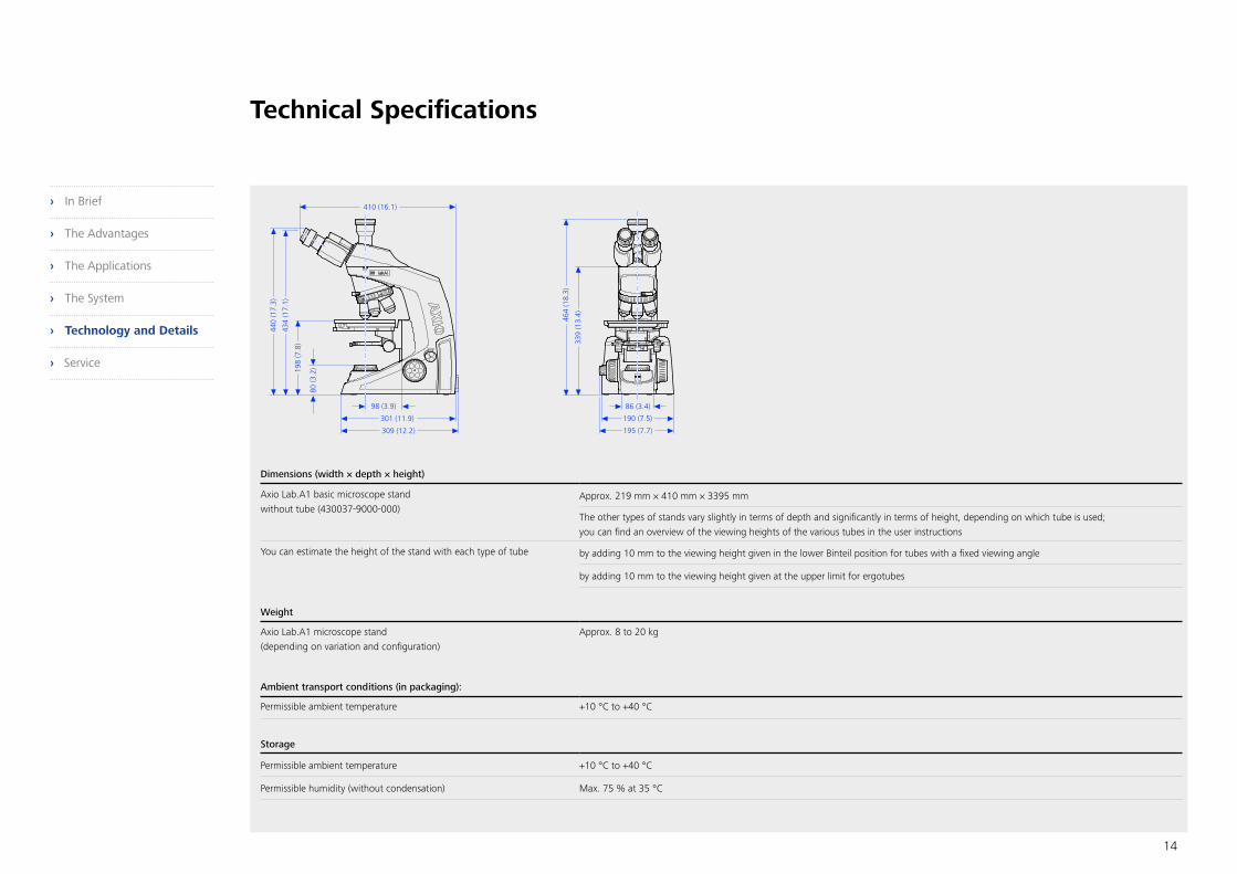
Technical Specifications
Dimensions (width × depth × height)
Axio Lab.A1 basic microscope standwithout tube (430037-9000-000)
Approx. 219 mm × 410 mm × 3395 mm
The other types of stands vary slightly in terms of depth and significantly in terms of height, depending on which tube is used; you can find an overview of the viewing heights of the various tubes in the user instructions
You can estimate the height of the stand with each type of tube by adding 10 mm to the viewing height given in the lower Binteil position for tubes with a fixed viewing angle
by adding 10 mm to the viewing height given at the upper limit for ergotubes
Weight
Axio Lab.A1 microscope stand (depending on variation and configuration)
Approx. 8 to 20 kg
Ambient transport conditions (in packaging):
Permissible ambient temperature +10 °C to +40 °C
Storage
Permissible ambient temperature +10 °C to +40 °C
Permissible humidity (without condensation) Max. 75 % at 35 °C
› In Brief
› The Advantages
› The Applications
› The System
› Technology and Details
› Service

15
Technical Specifications
Operation
Permissible ambient temperature +10 °C to +40 °C
Permissible relative humidity (without condensation) Max. 75 % at 35 °C
Max. altitude 2,000 m
Atmospheric pressure 800 hPa to 1,060 hPa
Degree of pollution 2
Technical specifications
Area of use Closed spaces
Protection class I
Protection type IP 20
Electrical safety in accordance with CSA and UL standards In accordance with DIN EN 61010-1 (IEC 61010-1)
Overvoltage category II
Radio interference suppression In accordance with EN 55011, class B
Noise immunity In accordance with DIN EN 61326
Mains voltage for Axio Lab.A1 100 to 240 V ±10%
Mains voltage conversion is not necessary
Power frequency 50/60 Hz
Power consumption for Axio Lab.A1 110 VA
Fuses in accordance with IEC 127
Axio Lab.A1 microscope stand 2× T 3.15 A/H, 5 mm × 20 mm
› In Brief
› The Advantages
› The Applications
› The System
› Technology and Details
› Service

16
Technical Specifications
Light sources
LED transmitted light illumination Power consumption Max. 3 W
Light source adjustment range Continuously variable, approx. 0.5 to 12 V
Transmitted halogen light Power consumption Max. 35 W
Reflected halogen light Power consumption 50 W
Light source adjustment range Continuously variable, approx. 0.5 to 12 V
LED reflected fluorescent illumination with exchangeable LED modules Choose between wavelengths 365 nm, 380 nm, 455 nm, 470 nm, 505 nm, 530 nm, 590 nm, 625 nmor neutral white (540 nm–580 nm)
LED classification LED risk group 2 in accordance with IEC 62471
Axio Lab.A1
Stand with manual stage focus Coarse focus Approx. 4 mm/rotation
Fine focus Approx. 0.4 mm/rotation; approx. 4 μm scale spacing
Stroke range 30 mm
Height stop Preset at factory
Choice between condenser 0.9/1.25 H with or without modulator disc for brightfield, darkfield, and phase contrast Ph1, Ph2, Ph3
Manual change of objective Using nosepiece turret, 4x H Pol or 5x H D, M27
Manual change of reflector module Using reflector turret, fourfold
› In Brief
› The Advantages
› The Applications
› The System
› Technology and Details
› Service

Profit from the optimized performance of your microscope system with a Carl Zeiss service contract – now and for years to come.
Because the ZEISS microscope system is one of your most important tools, we make sure it is always ready
to perform. What’s more, we’ll see to it that you are employing all the options that get the best from
your microscope. You can choose from a range of service products, each delivered by highly qualified
ZEISS specialists who will support you long beyond the purchase of your system. Our aim is to enable you
to experience those special moments that inspire your work.
Repair. Maintain. Optimize.
Attain maximum uptime with your microscope. A ZEISS Protect Service Agreement lets you budget for
operating costs, all the while reducing costly downtime and achieving the best results through the improved
performance of your system. Choose from service agreements designed to give you a range of options and
control levels. We’ll work with you to select the service program that addresses your system needs and
usage requirements, in line with your organization’s standard practices.
Our service on-demand also brings you distinct advantages. ZEISS service staff will analyze issues at hand and
resolve them – whether using remote maintenance software or working on site.
Enhance Your Microscope System.
Your ZEISS microscope system is designed for a variety of updates: open interfaces allow you to maintain
a high technological level at all times. As a result you’ll work more efficiently now, while extending the
productive lifetime of your microscope as new update possibilities come on stream.
Count on Service in the True Sense of the Word
>> www.zeiss.com/microservice
17
› In Brief
› The Advantages
› The Applications
› The System
› Technology and Details
› Service

Carl Zeiss Microscopy GmbH 07745 Jena, Germany [email protected] www.zeiss.de/axiolab
Not
all
prod
ucts
are
ava
ilabl
e in
all
coun
trie
s. T
he u
se o
f pro
duct
s fo
r med
ical
dia
gnos
is, t
hera
py, o
r tre
atm
ent m
ay b
e su
bjec
t to
loca
l res
tric
tions
. For
mor
e in
form
atio
n, c
onta
ct y
our Z
EISS
sal
es re
pres
enta
tive.
EN_4
1_01
1_04
0 | C
Z 06
-201
6 | D
esig
n, s
cope
of d
eliv
ery
and
tech
nica
l pro
gres
s su
bjec
t to
chan
ge w
ithou
t not
ice.
| ©
Car
l Zei
ss M
icro
scop
y G
mbH
866-464-1005