you say mitosis and i say meiosis.. or how cells reproduce
TRANSCRIPT

You Say Mitosis and I Say Meiosis..
or How Cells Reproduce

The ability of organisms to reproduce their kind is one characteristic that distinguishes living things from nonliving matter.
The continuity of life from one cell to another is based on the reproduction of cells through cell division.
This process occurs as part of the life of a cell from its origin in the division of a parent cell until its own division into two.
A. Introduction

The division of a unicellular organism reproduces an entire organism, increasing the population.
Cell division on a larger scale can produce offspring for some multicellular organisms.
B. Cell division functions in reproduction, growth, and repair
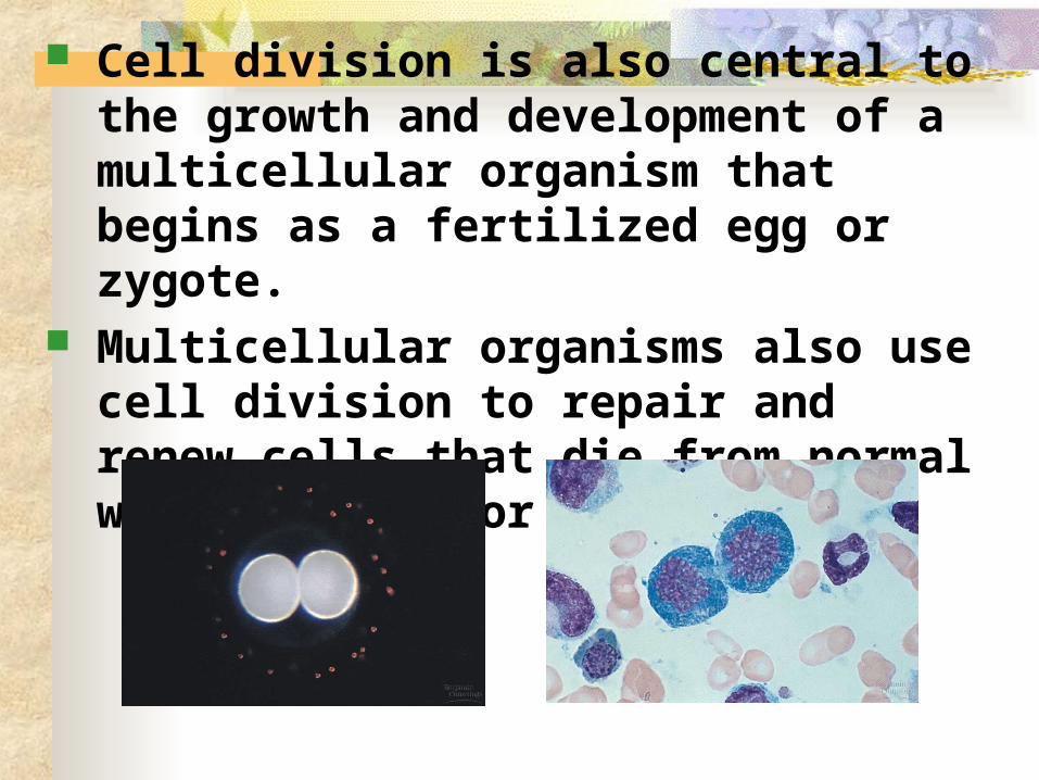
Cell division is also central to the growth and development of a multicellular organism that begins as a fertilized egg or zygote.
Multicellular organisms also use cell division to repair and renew cells that die from normal wear and tear or accidents.

Cell division requires the distribution of identical genetic material - DNA - to two daughter cells. What is remarkable is the continuity with
which DNA is passed along, without much change from one generation to the next.
A dividing cell duplicates its DNA, distributes the two copies to opposite ends of the cell, and then splits into two daughter cells.

A cell’s genetic information, packaged as DNA, is called its genome. In prokaryotes, the genome is often a single long
DNA molecule. In eukaryotes, the genome consists of several
DNA molecules. A human cell must duplicate about 3 meters
of DNA and separate the two copies such that each daughter cell ends up with a complete genome.
C. Cell division distributes identical sets of chromosomes to daughter cells
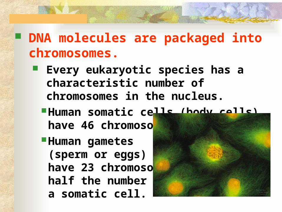
DNA molecules are packaged into chromosomes. Every eukaryotic species has a characteristic
number of chromosomes in the nucleus.Human somatic cells (body cells) have 46
chromosomes.Human gametes
(sperm or eggs) have 23 chromosomes, half the number in a somatic cell.
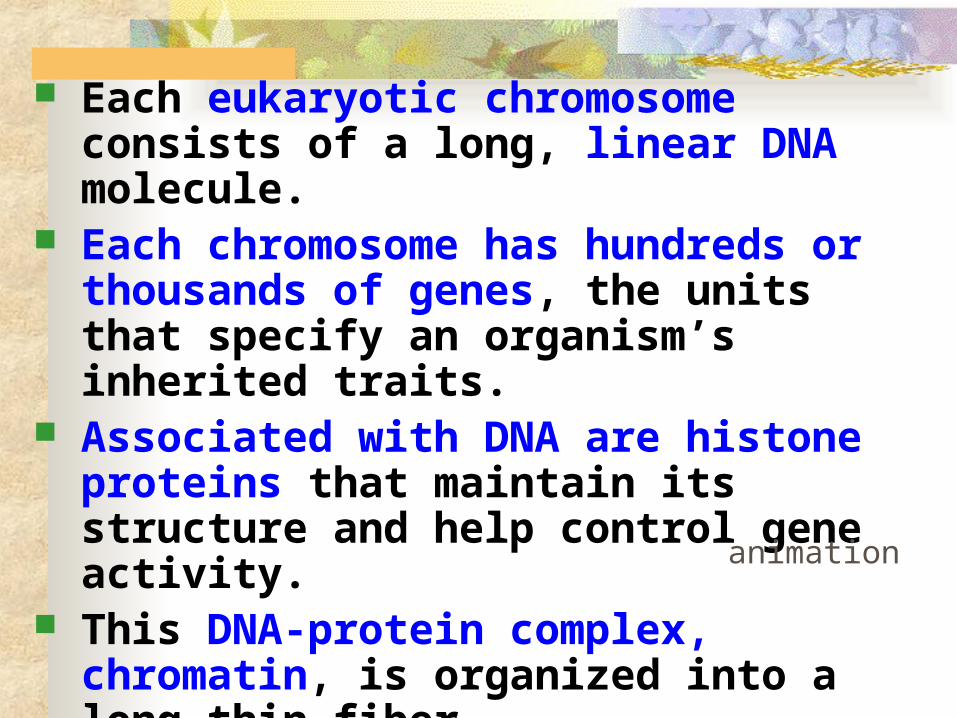
Each eukaryotic chromosome consists of a long, linear DNA molecule.
Each chromosome has hundreds or thousands of genes, the units that specify an organism’s inherited traits.
Associated with DNA are histone proteins that maintain its structure and help control gene activity.
This DNA-protein complex, chromatin, is organized into a long thin fiber.
After the DNA duplication, chromatin condenses, coiling and folding to make a smaller package.
animation

Each duplicated chromosome consists of two sister chromatids which contain identical copies of the chromosome’s DNA.
The area where the chromatids connect (like a
thumbtack) is the centromere.
Later, the sister chromatids are pulled apart and repackaged into two new nuclei at opposite ends of the parent cell.
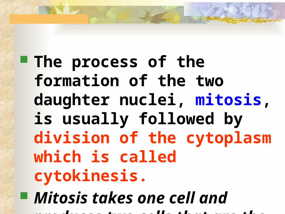
The process of the formation of the two daughter nuclei, mitosis, is usually followed by division of the cytoplasm which is called cytokinesis.
Mitosis takes one cell and produces two cells that are the genetic equivalent of the parent.

Each of us inherited 23 chromosomes from each parent: one set in an egg and one set in sperm.
The fertilized egg or zygote underwent trillions of cycles of mitosis and cytokinesis to produce a fully developed multicellular human.
These processes continue every day to replace dead and damaged cell.
Essentially, these processes produce clones - cells with the same genetic information.

The mitotic (M) phase of the cell cycle alternates with the much longer interphase. The M phase includes mitosis and
cytokinesis (about one hour in length for eukaryotes)
Interphase accounts for 90% of the cell cycle or 23 hours out of the day.
During Interphase the cell is conducting all life activities and getting ready for the next round of mitosis to occur (in most cells)
D: A Day in the Life of a Cell

animation
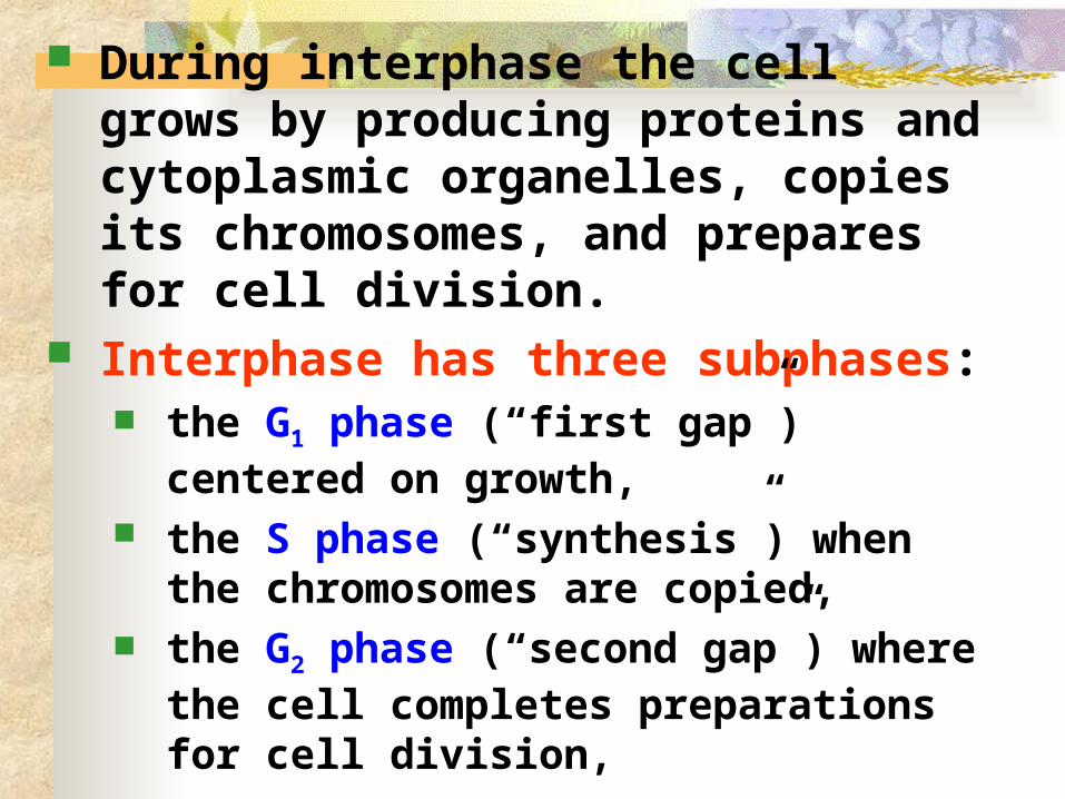
During interphase the cell grows by producing proteins and cytoplasmic organelles, copies its chromosomes, and prepares for cell division.
Interphase has three subphases: the G1 phase (“first gap”) centered on growth,
the S phase (“synthesis”) when the chromosomes are copied,
the G2 phase (“second gap”) where the cell completes preparations for cell division,
The cell divides (M). The daughter cells may then repeat the
cycle.
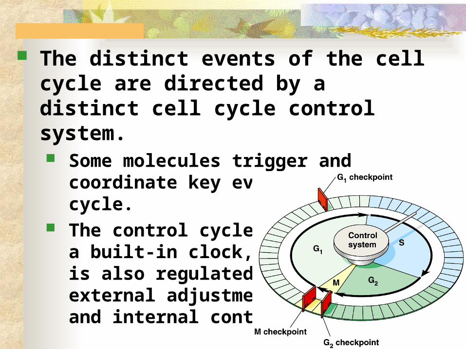
The distinct events of the cell cycle are directed by a distinct cell cycle control system. Some molecules trigger and coordinate key
events in the cell cycle. The control cycle has
a built-in clock, but it is also regulated by external adjustments and internal controls.

A checkpoint in the cell cycle is a critical control point where stop and go signals regulate the cycle.
Three major checkpoints are found in the G1, G2, and M phases.

For many cells, the G1 checkpoint, the restriction point in mammalian cells, is the most important. If the cells receives a go-ahead signal, it usually
completes the cell cycle and divides. If it does not receive a go-ahead signal, the cell
exits the cycle and switches to a nondividing state, the G0 phase.
Most human cells are in this phase.Liver cells can be “called back” to the cell
cycle by external cues (growth factors), but highly specialized nerve and muscle cells never divide.
Click here
Great animation

Regulation of the cell cycle Protein signals inhibit or activate the cell cycle. Molecules called cyclins together with CDKs
(cyclin dependent kinases) form MPFs (mitotic promoting factors)
These proteins are important in regulating the cell cycle and cause cell proliferation.
Inhibitors of CDKs are used to treat cancer

E. Mitosis is a continuum of changes. For description, mitosis is usually
broken into five subphases: prophase, prometaphase, metaphase, anaphase, and telophase.

By late interphase (after the “S” phase), the chromosomes have been duplicated but are loosely packed.
The centrosomes with centrioles in animal cells have been duplicated
and begin to organize
microtubules into an
aster (“star”).

In prophase, the chromosomes are tightly coiled and become visible, with sister chromatids joined together.
The nucleoli and nuclear membrane begin to disappear. The mitotic spindle begins
to form and appears to push the centrosomes away from each other toward opposite ends (poles) of the cell.

During prometaphase, the nuclear envelope fragments and microtubules from the spindle interact with the chromosomes.
Microtubules from one pole attach to one of two kinetochores, special regions of the centromere,
kinetochores

In metaphase the sister chromatids line up at the metaphase plate, an imaginary plane equidistant between the poles, defining metaphase.

At anaphase, the centromeres divide, separating the sister chromatids.
Each is now pulled toward the pole to which it is attached by spindle fibers.
By the end, the two poles have equivalent collections of chromosomes.

At telophase, the cell continues to elongate as free spindle fibers from each centrosome push off each other.
Two nuclei begin for form, as the nuclear envelope begins to
reappear. Cytokinesis, division
of the cytoplasm, ends.


Animation
Animation #2
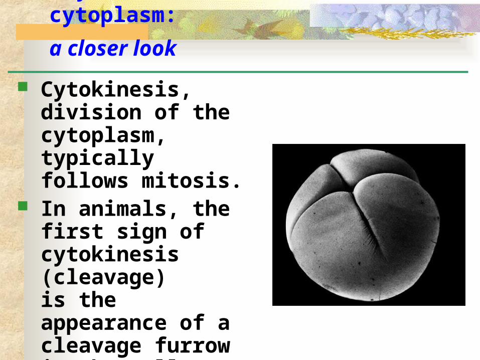
Cytokinesis, division of the cytoplasm, typically follows mitosis.
In animals, the first sign of cytokinesis (cleavage) is the appearance of a cleavage furrow in the cell surface near the old metaphase plate.
F.Cytokinesis divides the cytoplasm:
a closer look

On the cytoplasmic side of the cleavage furrow a contractile ring of actin microfilaments and the motor protein myosin form.
Contraction of the ring pinches the cell in two.

Cytokinesis in plants, which have cell walls, involves a completely different mechanism.
During telophase, vesicles from the Golgi fuse at the metaphase plate, forming a cell plate. The plate enlarges until its
membranes fuse with the plasma membrane at the perimeter, with the contents of the vesicles forming new wall material in between.

Animation of cell plate formation


Prokaryotes reproduce by binary fission, not mitosis.
Most bacterial genes are located on a single bacterial chromosome which consists of a circular DNA molecule and associated proteins.
While bacteria do not have as many genes or DNA molecules as long as those in eukaryotes, their circular chromosome is still highly folded and coiled in the cell.
G. Mitosis in eukaryotes may have evolved from
binary fission in bacteria

In binary fission, chromosome replication begins at one point in the circular chromosome, the origin of replication site.
These copied regions begin to move to opposite ends of the cell.

The mechanism behind the movement of the bacterial chromosome is still an open question.
As the bacterial chromosome is replicating and the copied regions are moving to opposite ends of the cell, the bacterium continues to grow until it reaches twice its original size.

Cell division involves inward growth of the plasma membrane, dividing the parent cell into two daughter cells, each with a complete genome.

It is quite a jump from binary fission to mitosis.
Possible intermediate evolutionary steps are seen in the division of two types of unicellular algae. In dinoflagellates, replicated
chromosomes are attached to the nuclear envelope.
In diatoms, the spindle develops within the nucleus.


Cancer is uncontrolled mitosis! Cancer cells divide excessively and invade
other tissues because they are free of the body’s control mechanisms. Cancer cells do not stop dividing when growth
factors are depleted either because they manufacture their own, have an abnormality in the signaling pathway, or have a problem in the cell cycle control system.
If and when cancer cells stop dividing, they do so at random points, not at the normal checkpoints in the cell cycle.
H. How is Cancer Related to Mitosis?

Cancer cell may divide indefinitely if they have a continual supply of nutrients. In contrast, nearly all mammalian cells
divide 20 to 50 times under culture conditions before they stop, age, and die.
Cancer cells may be “immortal”.Cells from a tumor removed from a
woman (Henrietta Lacks) in 1951 are still reproducing in culture.

In 1951, a scientist at Johns Hopkins Hospital in Baltimore, Maryland, created the first immortal human cell line with a tissue sample taken from a young black woman with cervical cancer. Those cells, called HeLa cells, quickly became invaluable to medical research.

They were essential to developing the polio vaccine. They went up in the first space missions to see what would happen to cells in zero gravity. Many scientific landmarks since then have used her cells, including cloning, gene mapping and in vitro fertilization.
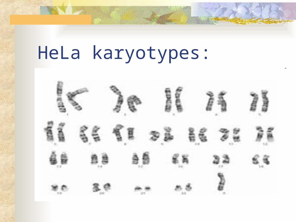
HeLa karyotypes:


The abnormal behavior of cancer cells begins when a single cell in a tissue undergoes a transformation that converts it from a normal cell to a cancer cell. Normally, the immune system recognizes and
destroys transformed cells. However, cells that evade destruction
proliferate to form a tumor, a mass of abnormal cells.
If the abnormal cells remain at the originating site, the lump is called a benign tumor. Most do not cause serious problems and can be
removed by surgery.

An overview with several animations showing how cancer develops. Click here…
In a malignant tumor, the cells leave the original site and impairs the functions of one or more organs.
animation

animation
Treatments for metastasizing cancers include high-energy radiation and chemotherapy with toxic drugs. These treatments target actively dividing cells.
Researchers are beginning to understand how a normal cell is transformed into a cancer cell. The causes are diverse. However, cellular transformation always
involves the alteration of genes that influence the cell cycle control system.

How can Natural Killer Cells in Our Immune System help to fight cancer? See this youtube video…

Cancer and gene regulation http://learn.genetics.utah.edu/content/
epigenetics/control/

Where does cancer (uncontrolled mitosis) come from? Faulty signaling pathways Mutations in human genome (p53 site)

Click here to learn about the p53 site of the genome and cancer

In asexual reproduction, a single individual passes along copies of all its genes to its offspring. Single-celled eukaryotes reproduce
asexually by mitotic cell division to produce two identical daughter cells.
Even some multicellular eukaryotes, like hydra, can reproduce by budding cells produced by mitosis.
I. A close look at mitosis as a form of reproduction.

In animals, there are many examples of asexual reproduction
Invertebrates: Fission: asexual
reproduction in which a parent separates into two or more approximately equal sized individuals.
Budding: asexual reproduction in which new individuals split off from existing ones.
Video of hydra budding

Fragmentation: the breaking of the body into several pieces, some or all of which develop into complete adults. Requires regeneration of lost body parts. Example – a lizard trying to escape a
predator biting its tail can lose this part of its body and regrow a new tail.
Stories have been told of oyster fisherman who try to rid their oyster beds of predatory starfish by slicing them up and throwing them back to sea. Why would this technique backfire?
Lizard loses tail video

Parthenogenesis (virgin birth) is the process by which an unfertilized egg develops into (often) haploid adult or the individual produces a diploid fertilized egg. Parthenogenesis plays a role in the social
organization of species of bees, wasps, and ants. Male honeybees are haploid and female honeybees are
diploid.
Several genera of fishes, amphibians, and lizards produce by a form of parthenogenesis that produces diploid zygotes.

What are the benefits of asexual reproduction

Many plants clone themselves by asexual reproduction, also called vegetative propagation. This occurs when a part separates from the
overall plant and eventually develops into a whole plant.
This clone would be identical to the parent.
J. Many plants clone themselves by asexual reproduction

Asexual reproduction is an extension of the capacity of plants for growth.Meristematic tissues with dividing
undifferentiated cells can sustain or renew growth indefinitely- usually found at the tips of a stem or the bottom of a root…
Parenchyma cells throughout the plant can divide and differentiate into various types of specialized cells.

In fragmentation, a parent plant separates into parts that reform whole plants.
A variation of this occurs in some dicots, in which the root system of a single parent gives rise to many adventitious shoots that become separate root systems, forming a clone. (aka-runners) A ring of creosote bushes
in the Mojave Desert of California is believed to be at least 12,000 years old.
One advantage of asexual reproduction is that a plant well suited to a particular environment can clone many copies of itself rapidly.
Moreover, the offspring of vegetative reproduction are not as fragile as seedlings produced by sexual reproduction.

Various methods have been developed for the asexual propagation of crop plants, orchards, and ornamental plants.These can be reproduced asexually
from plant fragments called cuttings.
These are typically pieces of shoots or stems.
K. Vegetative propagation of plants is common in agriculture

Some plants, including African violets, can be propagated from single leaves (known as “cuttings”)
In others, specialized storage stems can be cut into several pieces and develop into clones.For example, a piece of a potato
including an “eye” can regenerate a whole plant.
A twig or bud from one plant can be grafted onto a plant of a closely related species or a different variety of the same species. This makes it possible to combine the best
properties of different species or varieties into a single plant.
The plant that provides the root system is called the stock and the twig grafted onto the stock is the scion.
For example, a “cocktail fruit tree” can be made from grafting pears, apples, apricots and oranges on the same tree!

Advantages of asexual reproduction: Can reproduce without needing to find a
mate Can have numerous offspring in a short
period of time In stable environments, allows for the
perpetuation of successful genotypes.

L. Meiosis is another type of cell reproduction used in sexually reproducing organisms Meiosis is a type of cell division whereby
gametes –sex cells are made. Meiosis occurs in the gonads- sex organs
of sexually reproducing organismsIn animals, the gonads are the
ovaries and testes.In flowering plants (angiosperms),
the female gonad is the ovary and the male gonad is the anther.

M. Why is Meiosis Necessary for Organisms that Reproduce Sexually? Meiosis produces cells with half of the
chromosome number. When gametes unite during fertilization, the
chromosome number must remain constant through generations.
For example, if fruit flies have a chromosome number of 8, what would happen if their gametes had 8 chromosomes as well and fertilization occurred?

Sexual reproduction results in greater variation among offspring than does asexual reproduction which is key to evolution.
Two parents give rise to offspring that have unique combinations of genes inherited from the parents.
Offspring of sexualreproduction vary genetically from their siblings and from both parents.
N. Meiosis results in variation in the species.

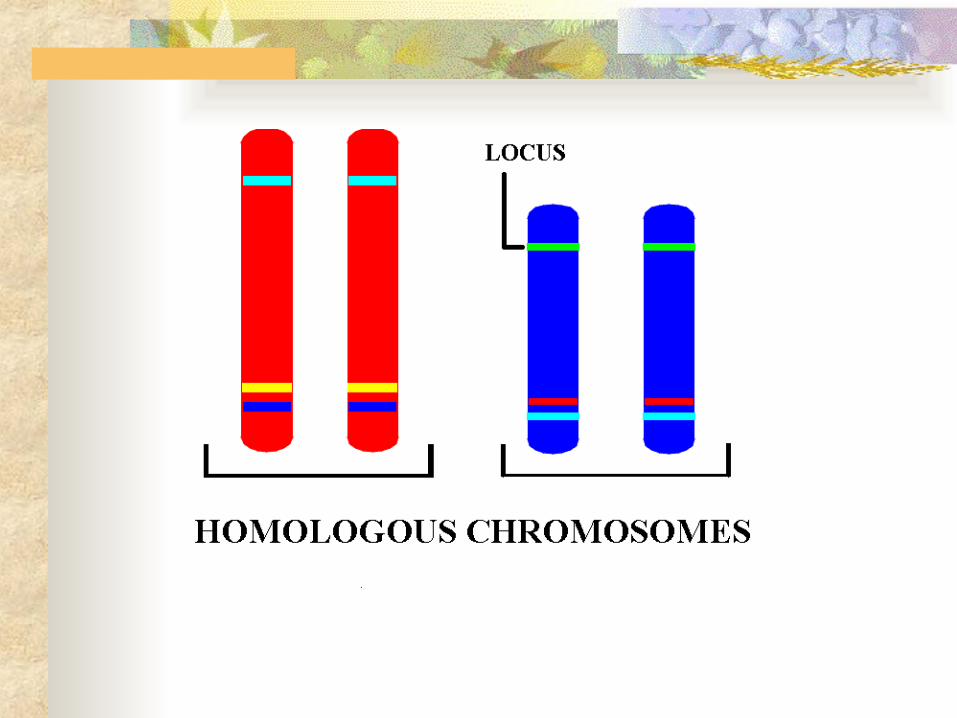
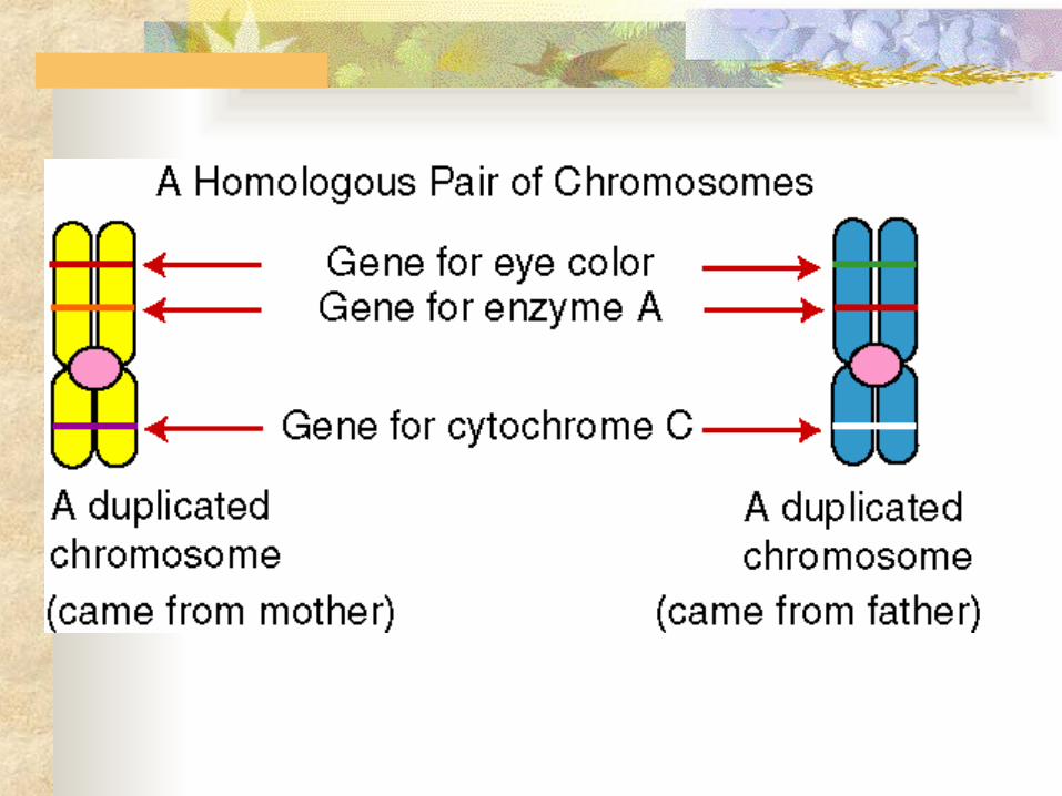
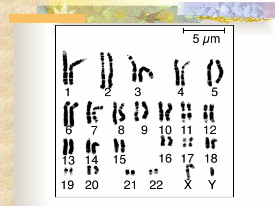
In humans, each somatic cell (all cells other than sperm or ovum) has 46 chromosomes.
A karyotype display of the 46 chromosomes shows 23 pairs of chromosomes, each pair with the same length, centromere position, and staining pattern.
These homologous chromosome pairs carry genes that control the same inherited characters.
O. Background information and vocabulary needed for our understanding of meiosis
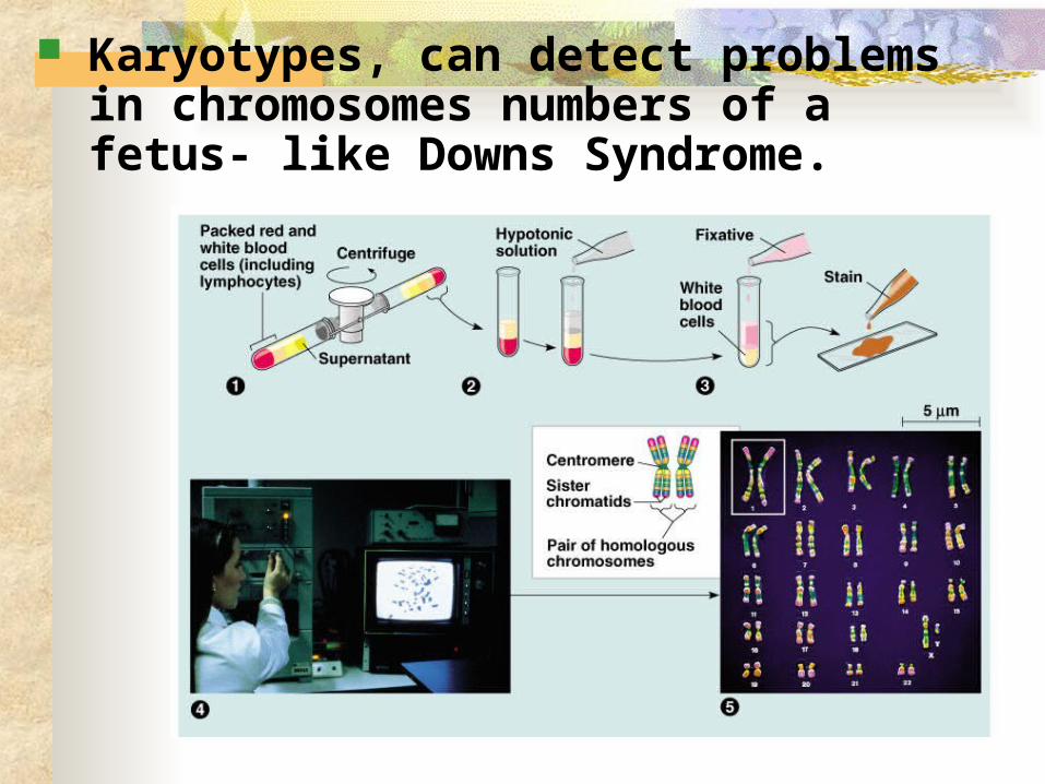
Karyotypes, can detect problems in chromosomes numbers of a fetus- like Downs Syndrome.

An exception to the rule of homologous chromosomes is found in the sex chromosomes, the X and the Y.
The pattern of inheritance of these chromosomes determine an individual’s sex. Human females have a homologous pair of X
chromosomes (XX). Human males have an X and a Y chromosome
(XY).
Because only small parts of these have the same genes, most of their genes have no counterpart on the other chromosome.
The other 22 pairs are called autosomes.
The occurrence of homologous pairs of chromosomes is a consequence of sexual reproduction.
We inherit one chromosome of each homologous pair from each parent. The 46 chromosomes in a somatic cell can be
viewed as two sets of 23, a maternal set and a paternal set.
Sperm cells or ova (gametes) have only one set of chromosomes - 22 autosomes and an X or a Y.
A cell with a single chromosome set is haploid -symbol is “n”where n is the number of homologous pairs For humans, the haploid number of
chromosomes is 23 (n = 23).

By means of sexual intercourse, a haploid sperm reaches and fuses with a haploid ovum.
These cells fuse resulting in fertilization. The fertilized egg (zygote) now has two
haploid sets of chromosomes bearing genes from the maternal and paternal family lines.
The zygote and all cells with two sets of chromosomes are diploid cells symbolized as “2n” (twice the # of homologous pairs) For humans, the diploid number of
chromosomes is 46 (2n = 46).
As an organism develops from a zygote to a sexually mature adult, the zygote’s genes are passes on to all somatic cells by mitosis.
Gametes, which develop in the gonads, are not produced by mitosis. If gametes were produced by mitosis, the
fusion of gametes would produce offspring with four sets of chromosomes after one generation, eight after a second and so on.
Instead, gametes undergo the process of meiosis in which the chromosome number is halved. Human sperm or ova have a haploid set of 23
different chromosomes, one from each homologous pair.

Fertilization restores the diploid condition by combining two haploid sets of chromosomes.
Fertilization and
meiosis alternate in sexual
life cycles.

The timing of meiosis and fertilization does vary among species.
The life cycle of humans and other animals is typical of one major type. Gametes, produced by meiosis,
are the only haploid cells. Gametes undergo no divisions
themselves, but fuse to form a diploid zygote that divides by mitosis to produce a multicellular organism.

Most fungi and some protists have a second type of life cycle. The zygote is the only diploid phase. After fusion of two gametes to form a zygote,
the zygote undergoes meiosis to produce haploid cells.
These haploid cells undergo mitosis to develop into a haploid multicellular adult organism.
Some haploid cells develop into gametes by mitosis.

Look at the following life cycle of a plant and of some algae. How is it different from the life cycle of a human? How is it different from the life cycle of a fungus?
Difference from human:
Difference from fungus:
Multicellular haploid organism
Multicellular diploid organism

Plants and some algae have a third type of life cycle, alternation of generation.
•This life cycle includes both haploid (gametophyte) and diploid (sporophyte) multicellular stages.
•Meiosis by the sporophyte produces haploid spores that develop by mitosis into the gametophyte.
•Gametes produced via mitosis by the gametophyte fuse to form the zygote which produces the sporophyte by mitosis.


Label the diagram with the following words: gametophyte, sporophyte, spores, zygote, mitosis, meiosis, fertilization

Sporophyte
(2n)
Gametophyte
(n)
Spores (n)
Zygote (2n)

Many steps of meiosis resemble steps in mitosis.
Both are preceded by the replication of chromosomes.
However, in meiosis, there are two consecutive cell divisions, meiosis I and meiosis II, which results in four daughter cells.
Each final daughter cell has only half as many chromosomes as the parent cell.
P. Meiosis reduces chromosome number from diploid to haploid: a closer look
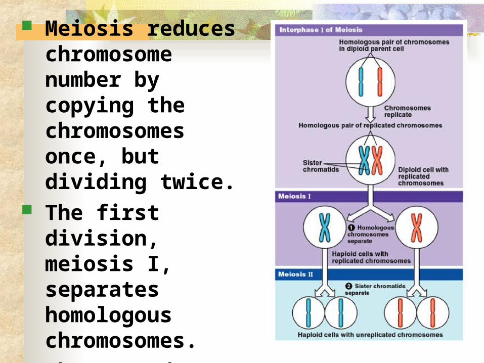
Meiosis reduces chromosome number by copying the chromosomes once, but dividing twice.
The first division, meiosis I, separates homologous chromosomes.
The second, meiosis II, separates sister chromatids.

Division in meiosis I occurs in four phases: prophase, metaphase, anaphase, and telophase.
During the preceding interphase the chromosomes are replicated to
form sister chromatids. These are genetically identical
and joined at the centromere. Also, the single centrosome
is replicated.
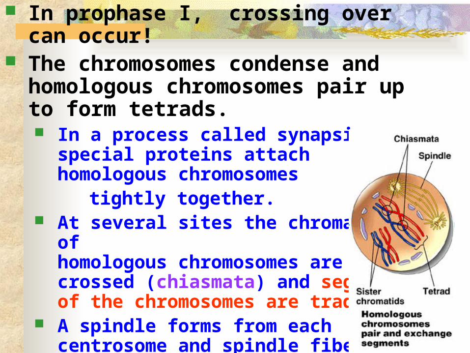
In prophase I, crossing over can occur! The chromosomes condense and
homologous chromosomes pair up to form tetrads. In a process called synapsis, special proteins
attach homologous chromosomes tightly together. At several sites the chromatids of
homologous chromosomes are crossed (chiasmata) and segments of the chromosomes are traded.
A spindle forms from each centrosome and spindle fibers attached to kinetochores on the chromosomes begin to move the tetrads around.
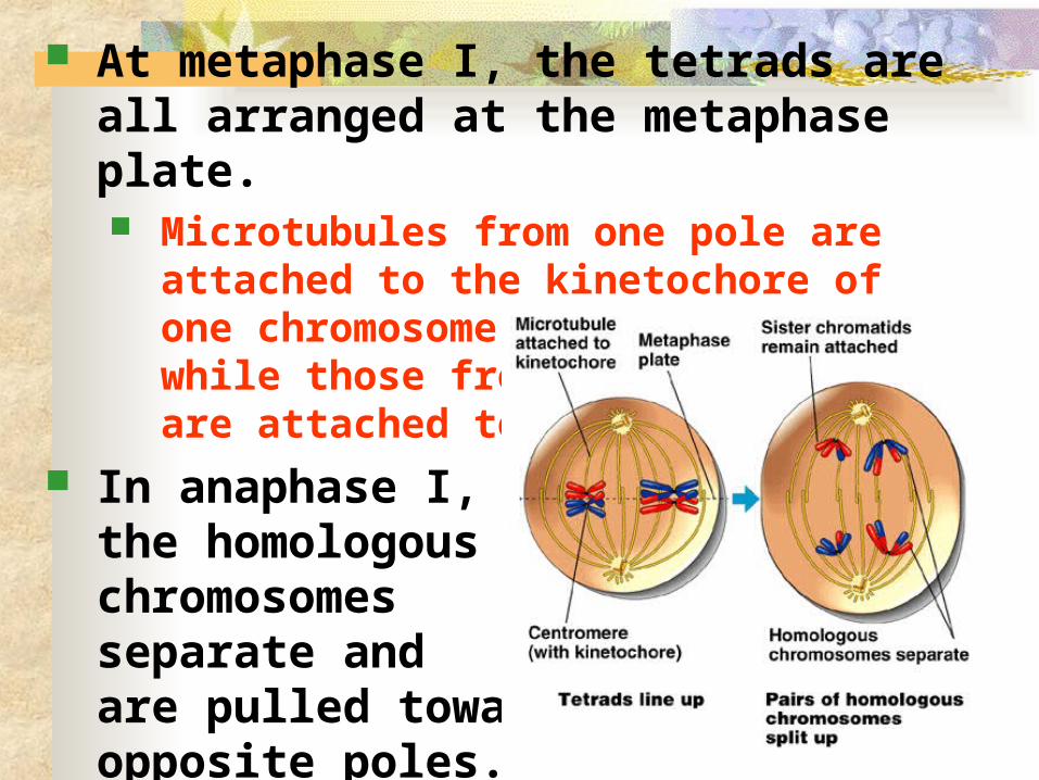
At metaphase I, the tetrads are all arranged at the metaphase plate. Microtubules from one pole are attached to the
kinetochore of one chromosome of each tetrad, while those from the other pole are attached to the other.
In anaphase I, the homologous chromosomes separate and are pulled toward opposite poles.

In telophase I, movement of homologous chromosomes continues until there is a haploid set at each pole. Each chromosome consists of linked sister
chromatids. Cytokinesis by the same
mechanisms as mitosis usually occurs simultaneously.
Interkinesis occurs-a brief
intermission - but there is no further replication of chromosomes.
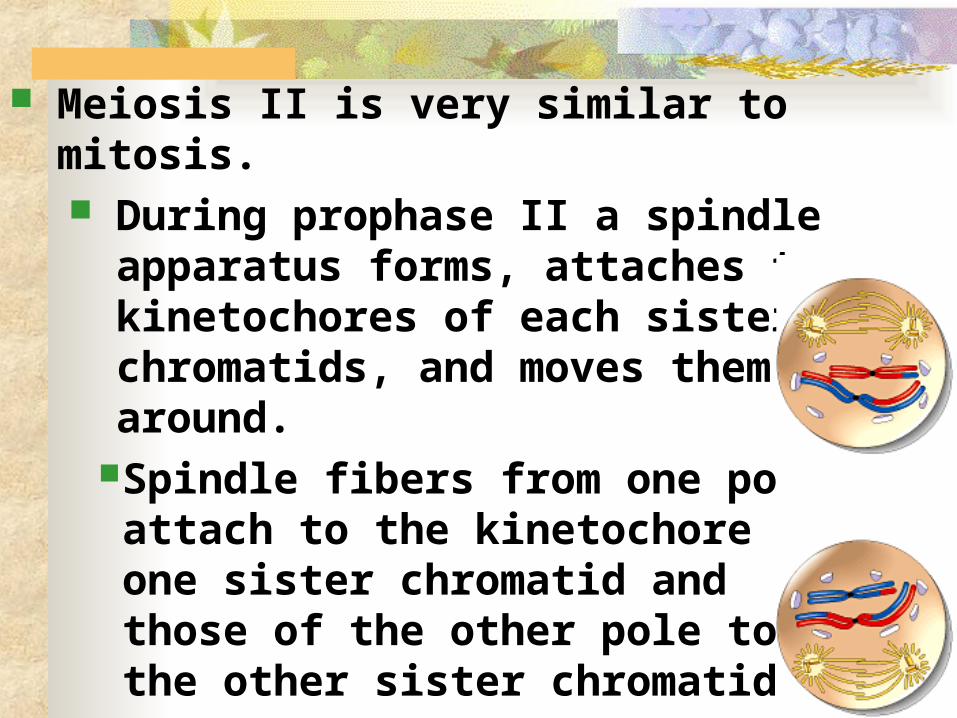
Meiosis II is very similar to mitosis. During prophase II a spindle apparatus
forms, attaches to kinetochores of each sister chromatids, and moves them around.
Spindle fibers from one pole attach to the kinetochore of one sister chromatid and those of the other pole to the other sister chromatid.

At metaphase II, the sister chromatids are arranged at the metaphase plate.
At anaphase II, the centomeres of sister chromatids separate and the now separate sisters travel toward opposite poles.

In telophase II, separated sister chromosomes arrive at opposite poles. Nuclei form around
the chromosomes Cytokinesis separates
the cytoplasm. At the end of meiosis,
there are four haploid daughter cells.
Click on animation
Click on animation
Q. Mitosis and meiosis have several key differences. The chromosome number is reduced
by half in meiosis, but not in mitosis. Mitosis produces daughter cells that
are genetically identical to the parent and to each other.
Meiosis produces cells that differ from the parent and each other.

Three events, unique to meiosis, occur during the first division cycle.1. During prophase I, homologous chromosomes pair up in a process called synapsis when crossing over occurs. A protein zipper, the synaptonemal complex,
holds homologous chromosomes together tightly.
Later in prophase I, the joined homologous chromosomes are visible as a tetrad.
At X-shaped regions called chiasmata, sections of nonsister chromatids are exchanged.
CROSSING OVER LEADS TO GENETIC VARIATION!!!

2. At metaphase I homologous pairs of chromosomes, not individual chromosomes are aligned along the metaphase plate.
In humans, you would see 23 tetrads.
3. At anaphase I, it is homologous chromosomes, not sister chromatids, that separate and are carried to opposite poles of the cell. Sister chromatids remain attached at the
centromere until anaphase II. The processes during the second meiotic
division are virtually identical to those of mitosis.

Mitosis produces two identical daughter cells, but meiosis produces 4 very different cells.


Found on a Biology t-shirt

Animation showing mitosis vs. meiosis

The behavior of chromosomes during meiosis and fertilization is responsible for most of the variation that arises each generation during sexual reproduction.
Three mechanisms contribute to genetic variation: independent assortment crossing over random fertilization
R. Sexual life cycles produce genetic variation among offspring
Independent assortment of chromosomes contributes to genetic variability due to the random orientation of tetrads at the metaphase plate. There is a fifty-fifty chance that a
particular daughter cell of meiosis I will get the maternal chromosome of a certain homologous pair and a fifty-fifty chance that it will receive the paternal chromosome.


Each homologous pair of chromosomes is positioned independently of the other pairs at metaphase I.
Therefore, the first meiotic division results in independent assortment of maternal and paternal chromosomes into daughter cells.
The number of combinations possible when chromosomes assort independently into gametes is 2n, where n is the haploid number of the organism. If n = 3, there are eight possible combinations. For humans with n = 23, there are 223 or about
8 million possible combinations of chromosomes.
Click on animation

Independent assortment alone would find each individual chromosome in a gamete that would be exclusively maternal or paternal in origin.
However, crossing over produces recombinant chromosomes which combine genes inherited from each parent.

Crossing over begins very early in prophase I as homologous chromosomes pair up gene by gene.
In crossing over, homologous portions of two nonsister chromatids trade places. For humans, this occurs two to three times per
chromosome pair. One sister chromatid may undergo different
patterns of crossing over than its match. Independent assortment of these
nonidentical sister chromatids during meiosis II increases still more the number of genetic types of gametes that can result from meiosis.

The random nature of fertilization adds to the genetic variation arising from meiosis.
Any sperm can fuse with any egg. A zygote produced by mating of a woman and
man has a unique genetic identity. An ovum is one of approximately 8 million
possible chromosome combinations (actually 223).
The successful sperm represents one of 8 million different possibilities (actually 223).
The resulting zygote is composed of 1 in 70 trillion (223 x 223) possible combinations of chromosomes.
Crossing over adds even more variation to this.
The three sources of genetic variability in a sexually reproducing organism are: Independent assortment of homologous
chromosomes during meiosis I and of nonidentical sister chromatids during meiosis II.
Crossing over between homologous chromosomes during prophase I.
Random fertilization of an ovum by a sperm. All three mechanisms reshuffle the various genes
carried by individual members of a population. Mutations, still to be discussed, are what
ultimately create a population’s diversity of genes.

Darwin recognized the importance of genetic variation in evolution via natural selection.
A population evolves through the differential reproductive success of its variant members.
Those individuals best suited to the local environment leave the most offspring, transmitting their genes in the process.
This natural selection results in adaptation, the accumulation of favorable genetic variations.
S. Evolutionary adaptation depends on a population’s genetic variation
As the environment changes or a population moves to a new environment, new genetic combinations that work best in the new conditions will produce more offspring and these genes will increase. The formerly favored genes will decrease.
Sex and mutations are two sources of the continual generation of new genetic variability.
Gregor Mendel, a contemporary of Darwin, published a theory of inheritance that helps explain genetic variation. However, this work was largely unknown for
over 40 years until 1900.

T. Meiosis in males vs. females Meiosis in males is called
“spermatogenesis” : the birth of sperm Meiosis in females is called
“oogenesis” : the birth of an egg or ovum
The timing of meiosis, and the size and number of gametes formed differ in males and females.

Spermatogenesis is the production of mature sperm cells from spermatogonia.
A continuous and prolific process in the adult male. Each ejaculation contains 100 – 650 million sperm. Occurs in seminiferous tubules. As spermatogenesis progresses the developing sperm
cells move from the seminiferous tubule to the epididymis where they can be temporarily stored.
Spermatogenesis and oogenesis both involve meiosis but differ in significant ways


Sperm structure: Haploid nucleus. Tipped with an acrosome.
Contains enzymes that help the sperm penetrate to the egg.
A large numberof mitochondriaprovide ATP topower theflagellum.

Oogenesis is the production of ova from oogonia.
Differs from spermatogenesis in three major ways: At birth an ovary contains all of the
primary oocytes it will ever have. Unequal cytokinesis during meiosis
results in the formation of a single large secondary oocyte and three small polar bodies.
The polar bodies degenerate. Oogenesis has long “resting” periods.

animation
Animation on nondisjunction in meiosis I
Animation on nondisjunction in meiosis II

Click here to see a youtube video that summarizes our unit!