yet another numbering scheme for immunoglobulin variable ... · yet another numbering scheme for...
TRANSCRIPT
doi:10.1006/jmbi.2001.4662 available online at http://www.idealibrary.com on J. Mol. Biol. (2001) 309, 657±670
Yet Another Numbering Scheme for ImmunoglobulinVariable Domains: An Automatic Modeling andAnalysis Tool
Annemarie Honegger* and Andreas PluÈ ckthun
Biochemisches Institut derUniversitaÈt ZuÈ richWinterthurerstrasse 190CH-8057 ZuÈ richSwitzerland
E-mail address of the [email protected]
Abbreviations used: CDR, compldetermining region; FR, frameworkimmunoglobulin variable domains;Bank; Fv, heterodimeric fragment ca VH domain; Fab, heterodimeric fran antibody light chain and the ®rsheavy chain; scFv, single-chain Fv fantibody, VL and VH connected bylinker; TCR, T-cell receptor; VL anddomains of the antibody light and hCH, constant domains antibody lighVl and Vk, lambda and kappa subtvariable domains; Va, Vb, Vg, and Vof T-cell receptor alpha, beta, gamm
0022-2836/01/030657±14 $35.00/0
A common residue numbering scheme for all immunoglobulin variabledomains (immunoglobulin light chain lambda (Vl) and kappa (Vk)variable domains, heavy chain variable domains (VH) and T-cell recep-tor alpha (Va), beta (Vb), gamma (Vg) and delta (Vd) variable domains)has been devised. Based on the spatial alignment of known three-dimensional structures of immunoglobulin domains, it places the align-ment gaps in a way that minimizes the average deviation from theaveraged structure of the aligned domains. This residue numberingscheme was applied to the immunoglobulin variable domain structuresin the PDB database to automate the extraction of information onstructural variations in homologous positions of the different mol-ecules. A number of methods are presented that allow the automatedprojection of information derived from individual structures or fromthe comparison of multi-structure alignments onto a graphical rep-resentation of the sequence alignment.
# 2001 Academic Press
Keywords: immunoglobulin variable domains; numbering scheme; proteinengineering; antibody engineering
*Corresponding authorIntroduction
When analyzing an individual protein struc-ture, sequential numbering of the amino acidresidues within each chain, combined with aunique chain label for each chain in multi-chainstructures, is the most convenient method to
ing author:
ementarity-region ofPDB, Protein Data
onsisting of a VL andagment consisting oft two domains of theragment of ana ¯exible peptideVH, variableeavy chains; CL andt and heavy chains;ypes of light chain
d, variable domainsa and delta chains.
identify individual residues within the proteinstructure and sequence. When comparing a largenumber of related structures showing multiplesequence insertions and deletions, such a simplescheme becomes very cumbersome. To facilitatediscussion of common sequence and structuralfeatures within a protein family, a uni®ed resi-due nomenclature, in which structurally equival-ent residues in different family members areidenti®ed by the same residue labels, is veryuseful. In an attempt to automate the compara-tive analysis of a large number of relatedsequences and structures, the unambiguousidenti®cation of structurally equivalent residuesis of crucial importance. The quality of the ®nalresults of such an analysis is limited by theaccuracy with which the input sequences andstructures have been aligned. As Protein DataBank (PDB) ®les (coordinates derived from X-rayand NMR structures) represent the major inputfor this type of analysis, such a numberingscheme should be compatible with the speci®ca-tions of the PDB ®le format. This format allowsa one-character chain identi®er, a four-digitresidue number and a one-character insertion
# 2001 Academic Press

658 Immunoglobulin Numbering Scheme
code. However, the ®eld allocated to the inser-tion code according to the most recent de®nitionof the PDB ATOM and HETATM records{ isfrequently used to indicate alternate atomlocations or, occasionally, used for the chainidenti®er, depending on the computer programthat generated the ®le.
In this analysis, we are concentrating on theimmunoglobulin superfamily, and especially onthe family of immunoglobulin variable domains,which consists of the immunoglobulin light chainlambda (Vl) and kappa (Vk) variable domains, theheavy chain variable domains (VH) and the T-cellreceptor alpha (Va), beta (Vb), gamma (Vg) anddelta (Vd) variable domains, which share suf®cientstructural and functional similarity that a compara-tive analysis can yield valuable insights into therules governing the correlation between sequence,structure and domain folding behavior. The immu-noglobulin variable domains represent a particu-larly well-suited target for such a study. Thecreation, selection and evolution of functional anti-bodies and T-cell receptors from the natural combi-natorial libraries encoded in the immunoglobulinloci by genetic recombination and somatic hyper-mutation in the time-course of an immuneresponse create a degree of variability within asingle organism unseen in any other protein famil-y.1,2
The usefulness of antibodies and of antibody-derived arti®cial constructs in various medical andbiochemical applications has made them a primetarget for protein engineering.3 Fv fragments andsingle-chain Fv fragments, in which the antibodyVL and VH domain are connected by a ¯exible lin-ker, represent the minimal building blocks neededto preserve the antigen-recognition function.4 ± 6
ScFv or Fab fragments can be obtained from givenmonoclonal antibodies, from libraries derived fromimmunized animals, from the naõÈve B-cell reper-toire or from germline genes.7,8 Although some ofthe fragments generated in this way perform verywell, many others show insuf®cient stability andproduction yields for the intended application andhave to be improved by rational engineering9 ± 12 orin vitro evolution schemes.13,14 Feedback from theseexperiments provides valuable input to be com-bined with the data derived from public domaindatabases.
Currently (June 2000) the PDB{ contains the 3Dstructures of 281 VH domains representing 181non-identical sequences, 269 Vk domains represent-ing 183 non-identical sequences and 56 Vl-domainsrepresenting 28 non-identical sequences. Inaddition, 13 (eight non-identical) TCR Va, 16 (six
{ http://www.rcsb.org/pdb/docs/format/pdbguide2.2/guide2.2_frame.html
{ http://www.rcsb.org/} http://imgt.cnusc.fr:8104/
VBase, http://www.mrc-cpe.cam.ac.uk/imt-doc/} http://www.ibt.unam.mx/vir/V_mice.html|| http://immuno.bme.nwu.edu/
non-identical) TCR Vb and 1 TCR Vd domain struc-tures are available. Combined with the knowledgeof the sequence variability allowed by the immu-noglobulin germline repertoire (IMGT,15} VBaseand ABG}), and the thousands of rearrangedsequences collected in the Kabat database||, awealth of information is available, which just hasto be combined and visualized in a form suitableto facilitate interpretation.
The combined information can be used toimprove the structural modeling and functionalpredictions involved in the optimization of engin-eered antibody fragments.16 The insights gainedfrom the analysis can then be applied in the con-struction of synthetic antibody libraries,17 and usedto help in the interpretation of in vitro evolutionexperiments.18
To facilitate this analysis, the data derived fromthe different databases has been rearranged in sucha way that it can be accessed by molecule and byresidue. With the new scheme proposed here,using PDB coordinate sets that are consistentlynumbered and superimposed, the features can beextracted and displayed for the entire family.Queries and analyses are easily possible, such as todisplay the superimposed structures of all VL
kappa domains from residue 20 to 47 (all comple-mentarity-determining region (CDR) L1 loops withadjacent beta sheets), or show the main-chain tor-sion angles of the VH residue H7 color-coded bydomain subtype, show the position-dependentamino acid compositions of all human and murineVl sequences contained in the Kabat database, justto illustrate some examples.
Current Residue Numbering Schemes
In their compilation of Sequences of Proteins ofImmunological Interest, of which several printededitions had appeared, Kabat et al.19 collected andaligned the sequences of different members of theimmunoglobulin superfamily. They proposednumbering schemes for many of the different pro-tein families, which make up the immunoglobulinsuperfamily. The placement of sequence gaps wasbased on sequence variability rather than on thespatial structure, as far fewer structures wereknown at the time. Chothia & Lesk20 corrected thepositioning of CDR L1 and CDR H1 sequencelength variability in the antibody variable domainsto better ®t their actual position in the three-dimen-sional structure. In 1989, Chothia and colleagueschanged the insertion point in CDR L1,21 butreturned to the old de®nition in 1997.22 The differ-ent families of immunoglobulin domains are trea-ted separately by both Kabat and Chothia, despitethe high degree of sequence and structural hom-ology. Gelfand and colleagues.23 ± 25 studied indetail the structurally invariant core of antibody VL
and VH domains, but they basically restricted theiranalysis to a set of residues that is almost identicalwith the core residues we use for least-squares

Immunoglobulin Numbering Scheme 659
superpositions and ignored the less conserved pos-itions. They identi®ed the residues by their second-ary structure position, resulting in a complexnomenclature incompatible with the PDB format,indicating the b-strand in which a residue islocated and its position in this strand. Lefranc andcolleagues (IMGT15,26{) proposed a uni®ed num-bering scheme for immunoglobulin variabledomain germline sequences,27 including antibodylambda and kappa light and heavy chain variabledomains as well as T-cell receptor alpha, beta,gamma and delta chain variable domains. How-ever, since they deal only with germ-linesequences, their numbering scheme reaches onlyinto CDR 3 and does not address the residues inCDR 3 or framework 4. An important differencefrom the numbering scheme presented here (AHo)is that in the IMGT scheme insertions and deletions``grow'' unidirectionally, as in the original Chothiade®nition,20 while in the AHo scheme, insertionsand deletions are placed symmetrically around thekey position marked in yellow (Figure 1(a)). Fur-thermore, length variations in CDR 1 and CDR 2are represented by a single gap in IMGT and byone or two gaps in AHo (Figures 1 and 3). Thedifferent numbering schemes are compared inFigure 1(a), together with the sequences(Figure 1(b) and (c)) of the different variabledomain PDB structures representing the structuraldiversity of immunoglobulin and T-cell receptorvariable domains.
In the course of our efforts to study the in¯uenceof individual sequence features on functionality,stability and folding ef®ciency of antibody single-chain constructs, as well as of constructs derivedfrom T-cell receptor variable domains, we had toautomate the comparison of a large number ofimmunoglobulin variable domain X-ray structures,including residues from the loop regions. Thisnecessitated the introduction of a uni®ed residuelabeling scheme carefully designed to preserve thepositional information derived from the compari-son of the experimental structures and models.Based on structural criteria, it seemed clear thatsequence insertions and deletions, indicated byalignment gaps, should be placed symmetrically,centered on turn and loop positions in the struc-ture, as this is the only location where they can beaccommodated without distorting the surroundingstructure. In contrast, the existing numberingschemes all imply a unidirectional insertion.
In order to facilitate automated processing, wetried to avoid alphabetic modi®ers to the residuenumbers (insertion numbering, such as 30A, 30B,etc.), but chose to allow large enough gaps in thenumbering scheme to accommodate the length
variation of all known germline sequences aswell as to give ample space for long CDR H3loops. Sequence insertions due to somaticmutations28 and extremly long CDR H3 loops29
exceeding the length provided for in ournumbering scheme still may nessecitate theuse of insertion numbering. Its use would,however, mark rare exceptions that defy thestatistical norm. Antibody light and heavy chainsare identi®ed by the chain identi®er L and H,T-cell receptor alpha, beta, gamma and deltachains by A, B, C and D, respectively, placedbefore the residue number (e.g. H6 for position6 in the antibody heavy chain, A6 for the struc-turally equivalent position in the TCR alphachain).
Immunoglobulin variable domain sequencesextracted from the Kabat{, IMGT{, VBase},ABG} and PDB|| databases were aligned usingthe GCG (Wisconsin Package Version 9.0, Gen-etics Computer Group, Madison, WI) modulePILEUP. Sequence alignments were tabulatedusing the GCG module PRETTY and importedinto EXCEL98 (Microsoft). To determine the opti-mal placement of the alignment gaps, the 3Dstructures of several representative members ofthe different families covering the observedlength variability were aligned by a least-squares®t of Ca positions in the structurally most con-served core region of the domains, consisting ofresidues 3-7, 20-24, 41-47, 51-57, 78-82, 89-93,102-108 and 138-144, and indicated in dark grayin Figure 1(a) and in white in Figure 2(a). TheCa coordinates of the aligned structures wereimported into EXCEL. The sequence alignmentof the residues not used for the structural align-ment was then optimized in such a way thatthe sum of the average deviation from the meanCa position for each position was minimized,resulting in the sequence alignment shown inFigure 1(b) and (c). The positions of sequencegaps in the other alignments were corrected toconform to the AHo numbering scheme usingEXCEL Visual Basic macros. These alignmentswere color-coded according to different criteria:type of amino acid (Figure 1(b)), hydropathy,predicted secondary structure, and similarity to areference sequence (not shown). Other macroswere written to calculate consensus sequences,distance matrices and to determine the sequencevariability and position-dependent amino acidcomposition of sequences grouped according todifferent criteria (see Figure 5(b) and (c)).
Description of the AHoNumbering Scheme
The ®rst gap to be placed concerns the one-amino acid insertions in the VL kappa framework 1compared to VL lambda and VH (Figure 3(a)).Kabat placed this insertion/deletion at the tenthresidue from the amino terminus. However, as a

Figure 1 (legend shown on page 672)
660 Immunoglobulin Numbering Scheme
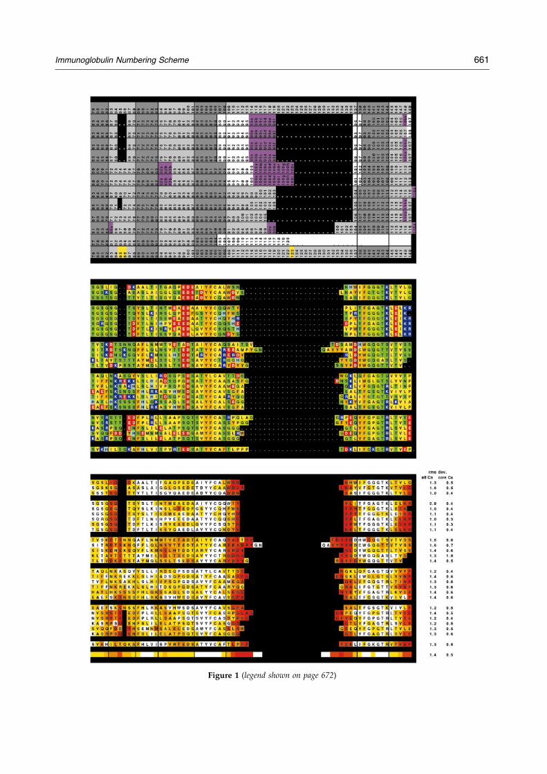
Figure 1 (legend shown on page 672)
Immunoglobulin Numbering Scheme 661

Figure 1. (a) The different numbering schemes for immunoglobulin variable domains (Kabat,19 Chothia,20,22
IMGT,27 AHo (this study)) have been aligned. Boxes with dark gray background indicate the residues whose Ca coor-dinates were used for the least-squares alignment of the representative structures of each type of variable domain(Figure 2). Pink background indicates the placement of insertions/deletions implied by the numbering scheme. Whitedots on black background indicate additional gaps introduced to bring the different numbering schemes into struc-tural alignment. White background indicates the position of the complementarity-determining regions. In the AHonumbering scheme, gaps are centered on the positions indicated in yellow. (b) Sequences representing the differenttypes of immunoglobulin variable domains (immunoglobulin VH, Vk and Vl, T-cell receptor Va, Vb and Vd) shown inFigure 2 were aligned so as to minimize the average deviation from the average structure. The amino acids are colorcoded according to residue type: aromatic residues (Tyr, Phe, Trp), orange; hydrophobic residues (Leu, Ile, Val, Met,Cys, Pro, Ala), yellow; uncharged hydrophilic residues (Ser, Thr, Gln, Asn, Gly), green; acidic residues (Asp, Glu),red; basic residues (Arg, Lys, His), blue. The corresponding 3D structures are shown in Figures 2 and 3. (c) Averagedeviation from the mean Ca position. Individual domains were excised from the corresponding PDB ®les and alignedby a least-squares ®t of the Ca positions of the core residues (3-7, 20-24, 41-47, 51-57, 78-82, 89-93, 102-108 and 138-144) to the corresponding Ca positions of a reference structure (Vk domain of PDB entry 1F5836). The mean Ca coordi-nates for each position in the alignment were calculated and the deviation of each residue in each structure from theaverage structure calculated and color coded in the corresponding sequence alignment (white, rms deviation <0.5 AÊ ;yellow, 0.5-1 AÊ ; yellow-orange, 1-1.5 AÊ ; orange, 1.5-2 AÊ ; orange-red, 2-4 AÊ ; red, >4 AÊ ). The Ca rms deviations of eachstructure from the average and the rms deviation of the Ca positions used for the alignment (dark gray boxes in (a))are indicated in the last two columns.
Figure 2. (a) Representative structures of Vl (PDB entries 1MFA,37 2FB4,38 and 8FAB39 pink), Vk (PDB entries1A2Y,40 1F58,36 1FLR,41 1HIL,42 25C8,43 and 2FBJ44 magenta), VH (PDB entries 1A2Y,40 1A6V,45 1F58,36 1FLR,41
1MFA,37 1MRC,46 and 2HMI47 cyan), TCR Va (PDB entries 1A07,48 1B88,49 1BD2,50 1KB5,51 1NFD,52 and 1TCR53
orange), TCR Vb (PDB entries 1A07,48 1BD2,50 1KB5,51 1NFD,52 1TCR53 green) and TCR Vd (PDB entry 1TVD31 red)were aligned by a least-squares ®t of the Ca atoms of residues 3-7, 20-24, 41-47, 51-57, 78-82, 89-93, 102-108 and 138-144 (indicated in white). Numbers indicate residue positions of the aligned b-strands. (b) A representation of the con-sensus structure and main-chain hydrogen bonding pattern of immunoglobulin variable domains. Arrows indicatehydrogen bonds that are present in the majority of the structures in all types of immunoglobulin variable domains.The loop and turn regions that accommodate gaps are indicated in yellow. Gray areas underlie the residues whoseCa positions were used for least-squares superposition of the structures.
662 Immunoglobulin Numbering Scheme

Figure 3. Placement of sequence length variability in the 3D structure of immunoglobulin variable domains. Thedomains were structurally aligned and color coded as described in the legend to Figure 2. The chain segments usedfor least-squares alignment are white, the loop regions pink (Vl ), magenta (Vk) , cyan (VH), green (TCR Va ) ororange (TCR Vb), depending on the domain type. The symbol � indicates the positions where gaps are placed tokeep the numbering scheme consistent between different types of variable domains. Larger gaps are placed symmetri-cally, centered on this position. (a) Framework 1 region. Vl and VH have a one-residue gap in position 8 compared toVk. The TCR Va and Vb domains whose structure have been solved so far are all Vk-like in length, although an align-ment of TCR germlines shows that Va sequences with a Vl-like length also exist. Most of TCR Vb and 50 % of the Vk-like TCR Va germlines have Pro in position 8. The available structures suggest that this Pro is a cis-Pro, as it is in Vkdomains. (b) Complementarity-determining region 1 (CDR 1). The segment containing the complementary-determin-ing region 1 is divided into two loops by residue 31, which intercalates between the outer and inner (dimer interface)beta sheet of the immunoglobulin variable domain beta sandwich. The length variability between CDR 1 loops of thesame class (e.g. Vk, VH) is mainly accommodated in the inner, C-terminal loop, centered around position 36, whilethe different classes of molecules differ in the length of the outer, N-terminal loop, accommodated by a gap centeredaround position 28. (c) CDR 2. While the length difference between VL and VH can be accommodated at the turn cen-tered on positions 62 and 63, the descending branch of CDR 2 in TCR Va is retracted from the dimer interface insuch a way that the structural alignment is best described by placing an additional two-residue gap in positions 74and 75. (d) Framework 3 region. The length variability of the framework 3 sequence is accommodated in the hairpinloop sometimes called CDR 4 in T-cell receptors. VL domains have a two-residue gap in positions 85 and 86 com-pared to VH and Va domains, Vb domains have a one residue gap in position 86.
Immunoglobulin Numbering Scheme 663
superposition of representative lambda and kappachain structures shows, the amide nitrogen atom ofthe seventh residue assumes a structurally equival-ent position in Vl and Vk chains, while the C1Ooxygen of the seventh residue of Vl is structurallyequivalent to the C1O oxygen of the eighthresidue in Vk. Structurally, the gap in Vl relative toVk should therefore be placed either in position L7or L8.30 The corresponding segment of TCR Vbchains is VL kappa-like in length and structure,including the very frequent occurrence of a cis-pro-line residue in position B8, while some Va-chainsare Vk-like and others resemble Vl with its one-residue deletion. For Vg and Vd there is not enough
structural information available yet, althoughthe single delta chain for which a structure hasbeen solved31 (PDB entry 1TVD) is Vk-like inlength. VH domains, while corresponding in lengthto Vl, show a larger structural variation in thisregion,32 ± 34 and the gap could equally well beplaced in position H7, H8 or H9. We thereforechose to place the gap in position H8. The con-served cysteine residue forming the intradomaindisul®de bridge therefore always carries the label23, as in the IMGT numbering scheme, whileaccording to Kabat, it was labeled L23 in Vk andVl, H22 in VH, 23 in TCR Va and Vb and 21 in TCRVg and Vd.
664 Immunoglobulin Numbering Scheme
The ®rst complementarity-determining region,CDR L1 and CDR H1 in antibodies, is treated as asingle loop in all existing numbering schemes.With CDR 1, the amino acid chain switches fromthe ``outer'' beta-sheet to the ``inner'' one, whichforms the heterodimer interface between the anti-body light and heavy chain, between the alphaand beta chains in alpha-beta TCRs and betweenthe gamma and delta chains in gamma-delta TCRs.Structurally, in the CDR 1 segment, one usuallyhydrophobic residue assumes a distinguished pos-ition. It intercalates between the two beta sheetsand divides the CDR 1 into two loops (Figure 3(b)).In our numbering scheme, the residue number ofthis amino acid is 31. The outer, N-terminal loop islongest in some TCR Va domains and shortest insome Vl domains, which both show some lengthvariability in this part of CDR 1. VH domains, aswell as the majority of the Va domains, have a one-residue gap in position 28, Vk and Vb domains atwo-residue gap in positions 27 and 28. CDR L1 ofthe kappa chain shows the largest degree of lengthvariability, affecting only the loop C-terminal tothe intercalating residue 31. Here, the gap is cen-tered on position 36. The highly conserved coretryptophan residue (Kabat H36, L35, A34, B34, C35or D35, IMGT 41) is always numbered 43 in ourscheme.
Since the two gap positions, centered on residues28 and 36, are treated separately and the interca-lated residue is always kept ®xed at position 31, ashorter N-terminal loop cannot compensate a long-er C-terminal loop. Compared to the IMGTscheme, two positions more are needed to rep-resent the length variability, since in the IMGTalignment, an insertion in the N-terminal loop ofCDR 1 can be compensated by a short C-terminalloop (Figure 1(a)).
The gap correcting for the length variability inthe CDR 2 region has been placed to be centeredon position 63, resulting in an eight residue gap inthe VL domains (L59-L66, placed between L50 andL51 according to Kabat nomenclature) and a one tofour residue gap in VH domains (between H52 andH53 according to Kabat) (Figure 3(c)). In Vadomains, a second gap has been placed in pos-itions A74 and A75 to re¯ect the way in which theC-terminal branch of the CDR A2 loop is retractedfrom the dimer interface compared to the CDR 2 ofother immunoglobulin variable domains. While inall other variable domains the descending branchof CDR 2 is part of the inner beta-sheet (whichforms the dimer interface), the descending branchof the Va CDR 2 (residues 67-73) is more closelyassociated with the outer beta-sheet, although thehydrogen bonds to the strand formed by residues78-82 are not very well conserved in the few Vastructures known. Again, this splitting of the CDR2 length variability into two insertion pointsnecessitates more positions than allowed for in theIMGT nomenclature.
The outer loop, sometimes called CDR 4 in T-cellreceptors, contains a two residue gap in VL (L74,
L75, between Kabat L68 and L69). VH and Va haveno gap, while Vb has a one residue gap in positionB86 (Figure 3(d)). This places the second cysteineresidue in position 106 (Kabat L88, H92, A90, B92,C94 and D88, IMGT 104). The CDR 3 residues aredivided symmetrically between this cysteine resi-due and the ®rst glycine residue of the framework4 beta bulge in position 140 (Kabat L99, H104,A107, B109, C109 and D107, unde®ned in IMGT),leaving ample space for long CDR 3 sequences andcentering the CDR 3 gap around position 123. Thenumber of residues allowed for the CDR 3 lengthvariability may seem excessive, but CDR3 loopsclose to that length can be found in the Kabat data-base.
Verification and Practical Applicationsof the New Numbering Scheme
To prove that the new numbering scheme isindeed a better representation of the structuralequivalence of residues in the immunoglobulindomains than the classical schemes, the X-raystructures of the individual domains wereextracted from the corresponding PDB ®les andstructurally aligned by a least-squares ®t betweenthe least variable Ca positions (3-7, 20-24, 41-47, 51-57, 78-82, 89-93, 102-108 and 138-144) using theprogram Insight II (MSI/Biosym). The Ca coordi-nates of the aligned domains were exported toEXCEL98 (Microsoft), preserving the new relativeorientations. This allowed the calculation of themean Ca coordinates for each position and thedeviation of the corresponding residue in theactual structures from this consensus position,taking into account the alignments implied by thedifferent numbering schemes. To minimize theeffects of the choice of reference structure on thequality of the ®nal alignment, the structural align-ment was performed in two steps: ®rst, an arbi-trary structure was used as a reference structure.From the alignment to this structure, the mean Ca
coordinates for each position were calculated andall structures compared to this mean structure. Thestructure with the lowest deviation from the aver-age was selected as new reference structure andthe analysis repeated. The numerical values for thestructural deviations were translated into a colorcode projected onto the sequence alignment asshown in Figure 1(c) and plotted against the resi-due number in a graphical representation (notshown). This representation also provides an indi-cation of which positions can truly be consideredstructurally equivalent (deviation from average Ca
position 5 distance between two neighbouring Ca
positions).Such a coloring of an alignment of individual
structures within a family serves well to quicklyidentify outliers. Larger deviations from the con-sensus of individual sequences or groups ofsequences help to identify residues that cause aconformational change. For instance, a strong

Immunoglobulin Numbering Scheme 665
structural deviation in the outer loop of Vk (L83-L87) from the average Vk conformation correlateswith the presence of a non-Gly residue in positionL82. This residue usually assumes a positive f tor-sion angle disallowed for residues with bulkierside-chains, and a non-Gly residue in this positionresults in an outward kink of the loop (Figure 5(a)).The mean Ca deviation for each position, indicatedin the header of an alignment of related sequences,serves as a quick reminder of which parts of thesequence represent structurally conserved residues,and which positions are more variable (Figure 4(a)-(c)).
The core Ca positions of the different variabledomain structures shown in Figure 2 ®t with a rmsdeviation of 0.52 AÊ . Using all Ca positions, withgaps positioned as de®ned by the AHo numberingscheme, the rms deviation obtained was 1.38 AÊ ,with gaps needed to align the different types ofvariable domains positioned as implied by theIMGT numbering scheme, 1.5 AÊ . Since only thesmall fraction of the residues that align differentlyin the two schemes contribute to this difference,this represents quite a large difference in the resi-dues affected. The core residues of 185 Vk domains®t with an average rms deviation of 0.3 AÊ , the coreresidues of 23 Vl with an average rms deviation of0.3 AÊ , and the core residues of 206 VH domainswith an average rms deviation of 0.4 AÊ . The aver-age structural variability for all Ca positions was0.6 AÊ for the Vk domains, 0.7 AÊ for the Vldomains, and 1.2 AÊ for the VH domains.
The residue and side-chain solvent-accessiblesurfaces of the residues were calculated from theindividual VL and VH domain structures as well asfor liganded and unliganded Fv fragments and Fabfragments using the program NACCESS{. To cor-rect for the different absolute surface areas of thedifferent residue types, accessibilities wereexpressed as a percentage of the exposed surfacearea of the same amino acid in the context of apoly(Ala)peptide in extended conformation. Thus,highly exposed residues, e.g. in turns or at the ter-mini, can have relative accessibilities of more than100%. The relative side-chain accessibilites werecolor-coded onto the sequence alignment to indi-cate buried and solvent-exposed residues(Figure 4(e)). From the relative reduction of theabsolute solvent-accessible surface area of eachresidue in the Fv fragment upon antigen binding,the average contribution of the different residuepositions to the antigen binding site could be eval-uated (Figure 4(f)). This automation enabled us toexpand the study by Padlan et al.35 to enumeratethe antigen/antibody contacts for a total of 45 anti-body/protein complex structures, 30 antibody/oli-gomer complexes and 52 antibody/haptencomplexes and investigate which residues contrib-ute to the interface, and to link this positional
information to the sequence variability observed ineach position in murine and human germlinesequences, and in the rearranged sequences,subdivided according to germline families(Figure 5(b) and (c)). The same method of evaluat-ing differences in residue solvent-accessibilityallowed to identify the residues contributing tothe VL/VH CL/CH dimer interface, the VL/CL andVH/CH interface11 (Figure 4(g) and (h)).
Lists of main-chain and side-chain hydrogenbonds, torsion angles, hydrogen bonds and otherproperties were extracted from the individualdomain structures and correlated with sequencepattern and structural properties (Figure 5(b)). InFigure 5(c), preferred residue types for differentantigen types were extracted. Having the differentdata tables in an interactive spreadsheet appli-cation and the structurally equivalent sequencepositions linked by a common residue numberingscheme allowed us to sort the data according tovarious criteria intrinsic or extrinsic to the datatables analyzed, and thus to test for the in¯uenceof different factors on the data.
One example is given by Langedijk et al.,32
where the in¯uence of particular sequence pos-itions on framework conformation and hydrogenbonding pattern was analyzed. In the work byHonegger & PluÈ ckthun,34 these observations weregeneralized to allow the prediction of the in¯uenceof frequently observed primer-induced mutationson the conformation of the framework 1 regionof VH. As another example, the analysis of theinteraction modes and antibody sequence prefer-ences of different types of antigens allowed us toextract rules for the optimization of randomizationstrategies to be employed in the re®nement ofsynthetic antibody libraries such as the humancombinatorial antibody library (HuCAL).17
While the numbering of the sequence by itselfmay not seem like an important step in the anal-ysis, these examples show that the correct structur-al alignment, which forms the basis for the newnumbering scheme, simpli®es all analyses enor-mously. By having a strict structural correspon-dence in residue numbers across all variabledomains, very fast access to all comparative andstructural parameters is possible, using standardmolecular modeling packages and spreadsheetapplications.
The aligned and renumbered PDB coordinates,sequence alignments and derived information willbe collected in an appropriate database system tobe made available on the Internet{. A set ofEXCEL macros will be provided, which allow thesemi-automatic renumbering of multiple PDB-®les:reading the ®les into an EXCEL workbook, extract-ing the sequence (from the data part, not from theheader) and displaying a sequence alignmentungapped or gapped according to the residuelabels present in the PDB ®le. The alignment canbe changed manually or exported to be alteredby external programs (e.g. GCG module PILEUP,or for a better automated ®t to the proposed

Figure 4. Standard header for VL and VH sequence alignments, summarizing the consensus structural properties and interaction residues of the domains, easily extractablefrom the aligned domains with the new numbering scheme. (a)-(c) Structural variability: average rms deviation from mean Ca position (average of 185 Vk and 206 VH struc-tures representing >100 non-identical sequences, all the experimental Fv and Fab structures with a resolution better than 3.0 AÊ available in the PDB database at the time ofthe analysis) for structures aligned according to the (a) AHo, (b) Chotia and (c) Kabat alignment. Individual domains were excised from the corresponding PDB ®les andaligned by a least-squares ®t of the Ca positions of the core residues (3-7, 20-24, 41-47, 51-57, 78-82, 89-93, 102-108 and 138-144) to the corresponding Ca-positions of a refer-ence structure 1YEH54 for Vk and 1MFD55 for VH. The mean Ca positions for each residue were calculated and the average deviation for each residue position in the align-ment is indicated by a color code (white, rms deviation <0.5 AÊ , yellow; 0.5-1 AÊ , yellow-orange; 1-1.5 AÊ , orange; 1.5-2 AÊ , orange-red, 2-4 AÊ ; red, >4 AÊ ). (d) Sequencevariability. From the position-dependent amino acid composition, the sequence variability was calculated according to Kabat et al.:19 Variability � 100 � (number of differentamino acids at position n)/(frequency of the most common amino acid at that position). Color code: white, <10; yellow, 10-25; yellow-orange, 25-50; orange, 50-75; red-orange, 75-100; red, >100. (e) Average relative side-chain accessibility. The side-chain solvent-accessible surface of each residue was calculated as a percentage of the sol-vent-accessible surface that the same residue would have in the context of a poly(Ala) peptide in extended conformation (program NACCESS (http://wolf.bms.umist.ac.uk/naccess)) (yellow, 0-10 %; yellow-green, 10-25 %; buried; green, 25-50 %; green-blue, 50-75 %, semi-buried; blue, 75-100 %; dark blue, >100 %, exposed). (f) Reduction ofthe side-chain accessible surface upon formation of the complex of the Fv fragment with a protein antigen. Relative reduction of the side-chain accessible surface of eachresidue in the complex of antigen-Fv fragment compared to the same residue in the Fv fragment without antigen (white, 0 % reduction; yellow, 0-20 %; yellow-orange, 20-40 %; orange, 40-60 %; red-orange, 60-80 %; red, 80-100 %). (g) Reduction of the side-chain accessible surface upon formation of the dimer interface between VL and VH.Average relative reduction of the side-chain accessible surface of each residue in the Fv fragment compared to its accessible surface in the isolated VL or VH domain (white,0 % reduction; yellow, 0-20 %; yellow-orange, 20-40 %; orange, 40-60 %; red-orange, 60-80 %; red, 80-100 %). (h) Reduction of the side-chain accessible surface upon formationof the interface between VL and CL or between VH and CH. Average relative reduction of the side-chain accessible surface of each residue in the Fab fragment compared toits accessible surface in the Fv fragment (white, 0 % reduction; yellow, 0-20 %; yellow-orange, 20-40 %; orange, 40-60 %; red-orange, 60-80 %; red, 80-100 %).

Figure 5. Examples of analyses greatly facilitated by the common residue numbering scheme. (a) Ramachandran plot of torsion angles VL lambda and kappa residue L82.Magenta triangles, Vl; blue diamonds, Vk with a Gly residue in position L82; dark blue diamonds, Vk with a non-Gly residue in position L82 (usually Arg). Such residue-by-residue correlation between sequence and structural features can be very valuable in avoiding errors in homology modeling and assessing the effects of point mutationsacquired as somatic point mutations or during in vitro evolution on structure and function. (b) Position-dependent amino acid composition of human and murine antibodyvariable domain sequences collected in the Kabat database (complete sequences only). The rearranged human sequences were sorted according to the major germline sub-types to analyze the proportional representation, consensus sequence and sequence variability of the different classes as a base for the construction of synthetic consensusframeworks.17 (Sequences in the Kabat database were considered to be rearranged if the sequence information extended beyond CDR 3 into FR 4; shorter sequences wereomitted from the analysis). Fields containing values of exactly zero were colored white and the number was supressed if the amino acid was not observed in any sequence.Those containing higher values were color-coded from light blue (values 0 %-1 %) to very dark blue (90 %-100 %), and the amino acids were sorted according to their proper-ties (charged, uncharged hydrophilic, aliphatic, aromatic side chains) to facilitate the perception of pattern of position-dependent residue properties and sequence conserva-tion for each position in the domain. (c) Position-dependent amino acid composition of structures represented in the PDB database. The sequences were sorted according tothe type and size of the antigen (hapten, oligomer (peptide or oligosaccharide) and protein), in order to analyze the correlation between sequence, antibody structure andtype of antigen.

668 Immunoglobulin Numbering Scheme
numbering scheme, module PROFILEGAP) and re-imported. All PDB ®les in the workbook can thenbe renumbered taking into account the gap pos-itions in the sequence alignment and the residuelabels indicated in the header line of the alignment.The renumbered coordinates can be re-exported asPDB ®les. Other macros allow color sequencealignment according to properties intrinsic (e.g.residue type, hydropathy, similarity to a referencesequence) or extrinsic (according to data providedin a separate table) to the information contained inthe sequence alignment and to calculate consensussequences and residue statistics.
Acknowledgements
This work was supported by a grant from the Schwei-zerische Nationalfonds 3100-046624.
References
1. Frippiat, J. P., Williams, S. C., Tomlinson, I. M.,Cook, G. P., Cherif, D., Le Paslier, D., Collins, J. E.,Dunham, I., Winter, G. & Lefranc, M. P. (1995).Organization of the human immunoglobulin lambdalight-chain locus on chromosome 22q11. 2. Hum.Mol. Genet. 4, 983-991.
2. Barbie, V. & Lefranc, M. P. (1998). The humanimmunoglobulin kappa variable (IGKV) genes andjoining (IGKJ) segments. Expt. Clin. Immunogenet. 15,171-183.
3. Huston, J. S., Tai, M. S., McCartney, J., Keck, P. &Oppermann, H. (1993). Antigen recognition andtargeted delivery by the single-chain Fv. Cell.Biophys. 22, 189-224.
4. Bird, R. E., Hardman, K. D., Jacobson, J. W.,Johnson, S., Kaufman, B. M., Lee, S. M., Lee, T.,Pope, S. H., Riordan, G. S. & Whitlow, M. (1988).Single-chain antigen-binding proteins. Science, 242,423-426.
5. Huston, J. S., Levinson, D., Mudgett-Hunter,M., Tai, M. S., Novotny, J., Margolies, M. N.,Ridge, R. J., Bruccoleri, R. E., Haber, E. & Crea, R.et al. (1988). Protein engineering of antibodybinding sites: recovery of speci®c activity in an anti-digoxin single-chain Fv analogue produced inEscherichia coli. Proc. Natl Acad. Sci. USA, 85, 5879-5883.
6. Glockshuber, R., Malia, M., P®tzinger, I. &PluÈ ckthun, A. (1990). A comparison of strategies tostabilize immunoglobulin Fv-fragments. Biochemistry,29, 1362-1367.
7. Winter, G., Grif®ths, A. D., Hawkins, R. E. &Hoogenboom, H. R. (1994). Making antibodies byphage display technology. Annu. Rev. Immunol. 12,433-455.
8. Vaughan, T. J., Williams, A. J., Pritchard, K.,Osbourn, J. K., Pope, A. R., Earnshaw, J. C.,McCafferty, J., Hodits, R. A., Wilton, J. & Johnson,K. S. (1996). Human antibodies with sub-nanomolaraf®nities isolated from a large non-immunizedphage display library. Nature Biotechnol. 14, 309-314.
9. Pantoliano, M. W., Bird, R. E., Johnson, S., Asel,E. D., Dodd, S. W., Wood, J. F. & Hardman, K. D.(1991). Conformational stability, folding, and ligand-
binding af®nity of single-chain Fv immunoglobulinfragments expressed in Escherichia coli. Biochemistry,30, 10117-10125.
10. Young, N. M., MacKenzie, C. R., Narang, S. A.,Oomen, R. P. & Baenziger, J. E. (1995). Thermalstabilization of a single-chain Fv antibody fragmentby introduction of a disulphide bond. FEBS Letters,377, 135-139.
11. Nieba, L., Honegger, A., Krebber, C. & PluÈ ckthun,A. (1997). Disrupting the hydrophobic patches at theantibody variable/constant domain interface. ProteinEng. 10, 435-444.
12. Jung, S. & PluÈ ckthun, A. (1997). Improving in vivofolding and stability of a single-chain Fv antibodyfragment by loop grafting. Protein Eng. 10, 959-966.
13. Hanes, J., Jermutus, L., Weber-Bornhauser, S.,Bosshard, H. R. & PluÈ ckthun, A. (1998). Ribosomedisplay ef®ciently selects and evolves high-af®nityantibodies in vitro from immune libraries. Proc. NatlAcad. Sci. USA, 95, 14130-14135.
14. Jung, S., Honegger, A. & PluÈ ckthun, A. (1999). Selec-tion for improved protein stability by phage display.J. Mol. Biol. 294, 163-180.
15. Lefranc, M. P., Giudicelli, V., Ginestoux, C., Bodmer,J., MuÈ ller, W., Bontrop, R., Lemaitre, M., Malik, A.,Barbie, V. & Chaume, D. (1999). IMGT, the inter-national ImMunoGeneTics database. Nucl. Acids Res.27, 209-212.
16. WoÈrn, A. & PluÈ ckthun, A. (2001). Stability engi-neeering of antibody single-chain Fv fragments.J. Mol. Biol. 305, 989-1010.
17. Knappik, A., Ge, L., Honegger, A., Pack, P., Fischer,M., Wellnhofer, G., Hoess, A., WoÈ lle, J., PluÈ ckthun,A. & VirnekaÈs, B. (2000). Fully synthetic humancombinatorial antibody libraries (HuCAL) based onmodular consensus frameworks and CDRs random-ized with trinucleotides. J. Mol. Biol. 296, 57-86.
18. Proba, K., WoÈrn, A., Honegger, A. & PluÈ ckthun, A.(1998). Antibody scFv fragments without disul®debonds made by molecular evolution. J. Mol. Biol.275, 245-253.
19. Kabat, E. A., Wu, T. T., Perry, H. M., Gottesmann,K. S. & Foeller, C. (1991). Sequences of Proteins ofImmunological Interest, 5th edit., NIH Publication no.91-3242 U.S. Department of Health and HumanServices.
20. Chothia, C. & Lesk, A. M. (1987). Canonical struc-tures for the hypervariable regions of immunoglobu-lins. J. Mol. Biol. 196, 901-917.
21. Chothia, C., Lesk, A. M., Tramontano, A., Levitt, M.,Smith-Gill, S. J., Air, G., Sheriff, S., Padlan, E. A.,Davies, D. & Tulip, W. R., et al. (1989). Confor-mations of immunoglobulin hypervariable regions.Nature, 342, 877-883.
22. Al-Lazikani, B., Lesk, A. M. & Chothia, C. (1997).Standard conformations for the canonical structuresof immunoglobulins. J. Mol. Biol. 273, 927-948.
23. Gelfand, I., Kister, A., Kulikowski, C. & Stoyanov,O. (1998). Algorithmic determination of core pos-itions in the VL and VH domains of immunoglobulinmolecules. J. Comput. Biol. 5, 467-477.
24. Gelfand, I., Kister, A., Kulikowski, C. & Stoyanov,O. (1998). Geometric invariant core for the VL andVH domains of immunoglobulin molecules. ProteinEng. 11, 1015-1025.
25. Gelfand, I. M., Kister, A. E. & Leshchiner, D. (1996).The invariant system of coordinates of antibodymolecules: prediction of the ``standard'' C alpha
Immunoglobulin Numbering Scheme 669
framework of VL and VH domains. Proc. Natl Acad.Sci. USA, 93, 3675-3678.
26. Ruiz, M., Giudicelli, V., Ginestoux, C., Stoehr, P.,Robinson, J., Bodmer, J., Marsh, S. G., Bontrop, R.,Lemaitre, M., Lefranc, G., Chaume, D. & Lefranc,M. P. (2000). IMGT, the international ImMuno-GeneTics database. Nucl. Acids Res. 28, 219-221.
27. Lefranc, M. P. (1997). Unique database numberingsystem for immunogenetic analysis. Immunol. Today,18, 509.
28. De Wildt, R. M., van Venrooij, W. J., Winter, G.,Hoet, R. M. & Tomlinson, I. M. (1999). Somaticinsertions and deletions shape the human antibodyrepertoire. J. Mol. Biol. 285, 701-710.
29. Saini, S. S., Allore, B., Jacobs, R. M. & Kaushik, A.(1999). Exceptionally long CDR3H region withmultiple cysteine residues in functional bovine IgMantibodies. Eur. J. Immunol. 29, 2420-2426.
30. Spada, S., Honegger, A. & PluÈ ckthun, A. (1998).Reproducing the natural evolution of proteinstructural features with the selectively infective phage(SIP) technology. The kink in the ®rst strand of anti-body kappa domains. J. Mol. Biol. 283, 395-407.
31. Li, H., Lebedeva, M. I., Llera, A. S., Fields, B. A.,Brenner, M. B. & Mariuzza, R. A. (1998). Structureof the Vdelta domain of a human gamma-deltaT-cell antigen receptor. Nature, 391, 502-506.
32. Langedijk, A. C., Honegger, A., Maat, J., Planta, R. J.,van Schaik, R. C. & PluÈ ckthun, A. (1998). The natureof antibody heavy chain residue H6 strongly in¯u-ences the stability of a VH domain lacking the disul-®de bridge. J. Mol. Biol. 283, 95-110.
33. Jung, S., Spinelli, S., Schimmele, B., Honegger, A.,Pugliese, L., Cambillau, C. & PluÈ ckthun, A. (2001).The importance of framework residues H6, H7 andH10 in antibody heavy chains: experimental evi-dence for a new structural subclassi®cation of anti-body VH domain. J. Mol. Biol. 309, 701-716.
34. Honegger, A. & PluÈ ckthun, A. (2001). The in¯uenceof the buried glutamine or glutamate residue in pos-ition 6 on the structure of immunoglobin variabledomains. J. Mol. Biol. 309, 687-699.
35. Padlan, E. A., Abergel, C. & Tipper, J. P. (1995).Identi®cation of speci®city-determining residues inantibodies. FASEB J. 9, 133-139.
36. Stan®eld, R. L., Cabezas, E., Satterthwait, A. C.,Stura, E. A., Profy, A. T. & Wilson, I. A. (1999).Dual conformations for the HIV-1 Gp120 V3 loop incomplexes with different neutralizing Fabs. Struct.Fold. Des. 7, 131-142.
37. Zdanov, A., Li, Y., Bundle, D. R., Deng, S. J.,MacKenzie, C. R., Narang, S. A., Young, N. M. &Cygler, M. (1994). Structure of a single-chain anti-body variable domain (Fv) fragment complexedwith a carbohydrate antigen at 1.7-AÊ resolution.Proc. Natl Acad. Sci. USA, 91, 6423-6427.
38. Kratzin, H. D., Palm, W., Stangel, M., Schmidt,W. E., Friedrich, J. & Hilschmann, N. (1989). Die Pri-maerstruktur des kristallisierbaren monoklonalenImmunoglobulins IgG1 Kol. II. Aminosaeuresequenzder L-Kette, Lambda-Typ, Subgruppe I. Biol. Chem.Hoppe-Seyler, 370, 263-272.
39. Strong, R. K., Campbell, R., Rose, D. R., Petsko,G. A., Sharon, J. & Margolies, M. N. (1991).Three-dimensional structure of murine anti-p-azo-phenylarsonate Fab 36-71.1. X-ray crystallography,site-directed mutagenesis, and modeling of thecomplex with hapten. Biochemistry, 30, 3739-3748.
40. Dall'Acqua, W., Goldman, E. R., Lin, W., Teng, C.,Tsuchiya, D., Li, H., Ysern, X., Braden, B. C., Li, Y.,Smith-Gill, S. J. & Mariuzza, R. A. (1998). A muta-tional analysis of binding interactions in an antigen-antibody protein-protein complex. Biochemistry, 37,7981-7991.
41. Whitlow, M., Howard, A. J., Wood, J. F., Voss, E. W.,Jr & Hardman, K. D. (1995). 1.85 AÊ structure ofanti-¯uorescein 4-4-20 Fab. Protein Eng. 8, 749-761.
42. Rini, J. M., Schulze-Gahmen, U. & Wilson, I. A.(1992). Structural evidence for induced ®t as a mech-anism for antigen-antibody recognition. Science, 255,959-965.
43. Gruber, K., Zhou, B., Houk, K. N., Lerner, R. A.,Shevlin, C. G. & Wilson, I. A. (1999). Structuralbasis for antibody catalysis of a disfavored ringclosure reaction. Biochemistry, 38, 7062-7074.
44. Navia, M. A., Segal, D. M., Padlan, E. A., Davies,D. R., Rao, N., Rudikoff, S. & Potter, M. (1979).Crystal structure of galactan-binding mouse immu-noglobulin J539 Fab at 4.5 AÊ resolution. Proc. NatlAcad. Sci. USA, 76, 4071-4074.
45. Simon, T. & Rajewsky, K. (1992). A functional anti-body mutant with an insertion in the frameworkregion 3 loop of the VH domain: implications forantibody engineering. Protein Eng. 5, 229-234.
46. Pokkuluri, P. R., Bouthillier, F., Li, Y., Kuderova, A.,Lee, J. & Cygler, M. (1994). Preparation, characteriz-ation and crystallization of an antibody Fab frag-ment that recognizes RNA. Crystal structures ofnative Fab and three Fab- mononucleotide com-plexes. J. Mol. Biol. 243, 283-297.
47. Ding, J., Das, K., Yu, H., Sara®anos, S. G., Clark,A. D., Jr, Jacobo-Molina, A., Tantillo, C., Hughes,S. H. & Arnold, E. (1998). Structure and functionalimplications of the polymerase active site region in acomplex of HIV-1 RT with a double-stranded DNAand an Antibody Fab fragment at 2.8 AÊ ngstromresolution. J. Mol. Biol. 284, 1095-1111.
48. Garboczi, D. N., Ghosh, P., Utz, U., Fan, Q. R.,Biddison, W. E. & Wiley, D. C. (1996). Structure ofthe complex between human T-cell receptor, viralpeptide and HLA-A2. Nature, 384, 134-141.
49. Plaksin, D., Chacko, S., McPhie, P., Bax, A., Padlan,E. & Margulies, D. (1996). A T cell receptor V alphadomain expressed in bacteria: does it dimerize insolution? J. Exp. Med. 184, 1251-1258.
50. Ding, Y. H., Smith, K. J., Garboczi, D. N., Utz, U.,Biddison, W. E. & Wiley, D. C. (1998). Two humanT Cell receptors bind in a similar diagonal mode tothe HLA-A2/Tax peptide complex using differentTCR amino acids. Immunity, 8, 403-411.
51. Housset, D., Mazza, G., Gregoire, C., Piras, C.,Malissen, B. & Fontecilla-Caps, J. C. (1997). Thethree-dimensional structure of a T-cell antigen recep-tor V alpha/V beta heterodimer reveals a novelarrangement of the V beta domain. EMBO J. 16,4205-4216.
52. Wang, J. H., Lim, K., Smolyar, A., Teng, M. K., Liu,J. H., Tse, A. G. D., Liu, J., Hussey, R. E., Chishti,Y., Thomson, C. T., Sweet, R. M., Nathenson, S. G.,Chang, H.-C., Sacchettini, J. C. & Reinherz, E. L.(1998). Atomic structure of an alpha/beta T-cellreceptor (TCR) heterodimer in complex with ananti-TCR Fab fragment derived from a mitogenicantibody. EMBO J. 17, 10-26.
53. Garcia, K. C., Degano, M., Stan®eld, R. L.,Brunmark, A., Jackson, M. R., Peterson, P. A.,Teyton, L. & Wilson, I. A. (1996). An alpha/beta T
670 Immunoglobulin Numbering Scheme
cell receptor structure at 2.5 AÊ and its orientation inthe TCR-MHC complex. Science, 274, 209-219.
54. Gigant, B., Charbonnier, J. B., Eshhar, Z., Green,B. S. & Knossow, M. (1997). X-ray structures of ahydrolytic antibody and of complexes elucidatecatalytic pathway from substrate binding and tran-sition state stabilization through water attack and
product release. Proc. Natl Acad. Sci. USA, 94,7857-7861.
55. Bundle, D. R., Baumann, H., Brisson, J. R., Gagne,S. M., Zdanov, A. & Cygler, M. (1994). Solutionstructure of a trisaccharide-antibody complex:comparison of NMR measurements with a crystalstructure. Biochemistry, 33, 5183-5192.
Edited by I. Wilson
(Received 4 December 2000; received in revised form 28 March 2001; accepted 29 March 2001)