monoclonal immunoglobulin disorders
TRANSCRIPT

Approach to
Patients with Monoclonal Immunoglobulin
Disorders

Ig abnormalities can be classified as one of three
main categories
hypogammaglobulinemia,
polyclonal Ig elevations and
monoclonal Ig disorders

For any of these abnormalities, a careful medical
history and thorough laboratory evaluation are
needed to confirm a suspected diagnosis,
particularly because of the major implications of
the diagnosis and potential prognosis for a
person with a monoclonal Ig disorder


General Principles


After these questions have been answered, the
laboratory information is combined with information
from the history and physical examination to narrow
the diagnosis

IMMUNOGLOBULIN
ABNORMALITIES: METHODS OF
DETECTION
Clinical Scenarios
Monoclonal Ig disorders are diagnosed in the
asymptomatic stage, with many of the patients
having MGUS or SMM
These patients require no immediate therapy and
must be distinguished from those with active MM.
Monoclonal protein may be detected when the
clinical suspicion of MM is high
One has to be alert to the possibility that in a small
fraction of patients with MM (<2%), no monoclonal
protein can be detected in the serum or urine

If the patient has monoclonal light chains (no heavy
chains) in the blood only (not in the urine) and no
symptoms, the condition has been called idiopathic
Bence Jones proteinemia.

Protein Electrophoresis
The best method for detecting a monoclonal protein
is high-resolution agarose gel electrophoresis.
This test detects abnormalities in the migration of the
proteins on electrophoresis
The test can be performed with samples from the
serum (SPEP) or urine (UPEP).
The resulting densitometric tracing shows a spike
that is commonly referred to as the monoclonal
spike, or M spike

After a localized band or spike has been recognized
on electrophoresis, immunofixation or
immunosubtraction with capillary electrophoresis
should be performed to determine the type of
monoclonal protein

A monoclonal spike is seen as a discrete band that usually migrates to the ɣ or ß region of the electrophoretic strip, and rarely the α2 region.
A polyclonal increase in Igs produces a broad-band or broad-based peak and is limited to the ɣ region.
Two monoclonal proteins (biclonal gammopathy) occur in 8 to 9% of sera containing monoclonal protein abnormalities.
Rarely, a triclonal gammopathy (three monoclonal proteins) is found


Immunofixation,
Immunoelectrophoresis, and
Immunodiffusion
Structural type of a monoclonal protein is determined best by immunofixation
Thus immunofixation should be performed whenever a monoclonal protein of unknown identity is detected with SPEP
Immunofixation may also be performed when a B-cell or plasma cell neoplasm is suspected but no monoclonal spike is apparent, as is frequently the case with AL or nonsecretoryMM
Immunofixation may also be used to monitor for disease relapse in patients who have achieved a complete response to therapy

In the past, immunoelectrophoresis was used to
characterize monoclonal proteins. This test is
more difficult to perform, and it is not as sensitive
as immunofixation.
Other methods for characterizing monoclonal
proteins include capillary zone electrophoresis
and immunosubtraction.
Monoclonal proteins of the IgD and IgE isotypes
usually are measured by immunodiffusion.

Quantitative Immunoglobulins
(Nephelometry)
At the time of diagnosis and to monitor disease, Igs
may be quantified directly with rate nephelometry.
This test is frequently ordered as “quantitative Igs.”
In addition to determining the serum concentration of
the same isotype as the monoclonal protein, the test
can detect a decrease in uninvolved, normal Igs.

Nephelometry is rapid and reliable.
However, it is not useful when the concentration
of the monoclonal protein is low, because
nephelometry cannot differentiate between
monoclonal and polyclonal Igs.

Free Light Chain Assays
Tests for serum free light chains are an important
part of the workup of patients with suspected
plasma cell neoplasms or AL.
These tests measure both free K and λ light
chains and provide a ratio between them.
Excessive skewing indicates the possibility of a
monoclonal protein. It is noteworthy that for
serum free light chains, the ratio between K and λis skewed towards λ

Urine Studies The excretion of light chains in the urine has been
referred to as Bence Jones proteinuria.
If patients have measurable monoclonal Igs (usually =10 g/L) in the serum or if MM or AL are suspected, urine should be collected for determining protein excretion
A timed 24-hour collection is best.
For patients in whom this is done for the first time, immunofixation is also recommended to characterize fully the monoclonal protein present.
Immunofixation is also recommended if no monoclonal protein is detected with UPEP.

Generally, the amount of monoclonal light chain in
the urine reflects the tumor mass of the patient.
However, the presence of Bence Jones
proteinuria alone does not necessarily indicate a
diagnosis of MM, as it also occurs in up to 30% of
patients with MGUS.
Patients with a pattern of nonselective proteinuria
(i.e., albumin predominance) are more likely to
have AL with kidney involvement

DIAGNOSIS

Bone Marrow Examination
For most patients with evidence of a monoclonal
protein, an examination of the bone marrow is
recommended.
This examination should include both an aspirate
and a biopsy sample and an MM-specific test

Bone Marrow Plasma Cell
Labeling Index (PCLI)
The PCLI is a surrogate of cells undergoing mitosis and is the most useful prognostic marker in multiple myeloma.
This is a slide-based test (one slide is labeled with anti-k and one with anti-λ fluorescent antibodies) that scores the number of plasma cells (usually out of 500) that incorporate bromodeoxyuridine.
Bromodeoxyuridine is taken up by cells undergoing DNA synthesis and can be detected with fluorescent-specific antibodies.

The test can also be performed on blood samples mainly to detect circulating plasma cells. Because the test is also done with clone-specific fluorescent antibodies, it provides an estimate of the plasmacytosis and the ratio between k and λ (to detect clonality).
The test can provide information about clonality, plasmacytosis, and labeling index (mitotic activity).
The major limitation to the test is that it can be done only on samples incubated with bromodeoxyuridine at the time of bone marrow aspiration

Radiologic Tests
Patients with an IgM monoclonal protein should have abdominal and chest computed tomography to exclude organomegaly (hepatomegaly and splenomegaly) and bulky lymphadenopathy .
Patients with evidence of MM should have a radiologic examination of the axial and appendicularskeleton (“bone survey”).
In some special circumstances, more sophisticated tests such as magnetic resonance imaging (MRI) have been recommended .
Other tests such as positron emission tomography are being explored

NEOPLASTIC IMMUNOGLOBULIN
DISORDERS

Non–Immunoglobulin M Monoclonal Gammopathy of Undetermined
Significance
MGUS represents the earliest stage of a monoclonal
plasma/lymphoid cell proliferation.
It is critical to distinguish between IgM MGUS and
non-IgM MGUS.

By definition, non-IgM MGUS is a discrete
proliferation of monoclonal plasma cells.
Multiple diagnostic criteria have been applied but
the essence is the same—a discrete proliferation
of plasma cells without evidence of the clone
resulting in disease for the host
One criterion for diagnosis is fewer than 10%
plasma cells in the bone marrow

Because of the heterogeneous nature of disease
involving the bone marrow and different ways for
determining the percentage of plasma cells, the
size of the monoclonal proteins is usually
considered a diagnostic criterion
MGUS to have evolved into a more advanced
plasma cell neoplasm if the serum monoclonal
spike is greater than 30 g/L

An important focus of the physical examination in non-IgM MGUS is the search for bone disease.
Percussion tenderness (spine and sternum) should raise the diagnostic possibility of MM. This is particularly important in patients with IgA MGUS, which may be more difficult to differentiate from MM.
Also, clinicians must be aware of the classic signs and symptoms of AL. AL may be present even with the smallest concentration of monoclonal proteins (occasionally not even detectable), and this possibility should always be kept in mind

CLINICAL MANAGEMENT AND
MONITORING
Because of the nonmalignant nature of MGUS,
patients do not experience end-organ damage
If a patient has evidence suggestive of possible
bone disease (pain in the shaft of extremities that
worsens with movement or weight bearing), a
bone radiographic survey should be done
It has long been the practice not to examine the
bone marrow of patients with suspected MGUS

Monoclonal gammopathy of the IgD or IgE type
presents unique problems because the size of the
monoclonal spike is usually small and cannot be
used to estimate tumor burden. In these cases, a
bone marrow examination is mandatory.
Patients with IgE monoclonal proteins often
present with the most aggressive phenotype of
plasma cell neoplasms, namely, plasma cell
leukemia

Although MGUS includes the term “undetermined
significance,” non-IgM MGUS clearly is the most
important risk factor for the subsequent
development of MM
MGUS is not only a marker of persons at high risk
for the development of MM, but, more important,
is the latent, premalignant condition

When MGUS evolves to MM, the same Ig isotype is expressed in MGUS and MM.
Patients should be followed indefinitely because the risk of progression to MM is never eliminated and has been estimated to be almost 1% per year.
Globally, the risk is greater for patients with higher concentrations of monoclonal proteins.
No good biologic markers are available that can discern or predict which patients will develop into MM
For these patients the recommendation is to have a repeat SPEP in 6 months and, if that is normal, at least yearly thereafter.

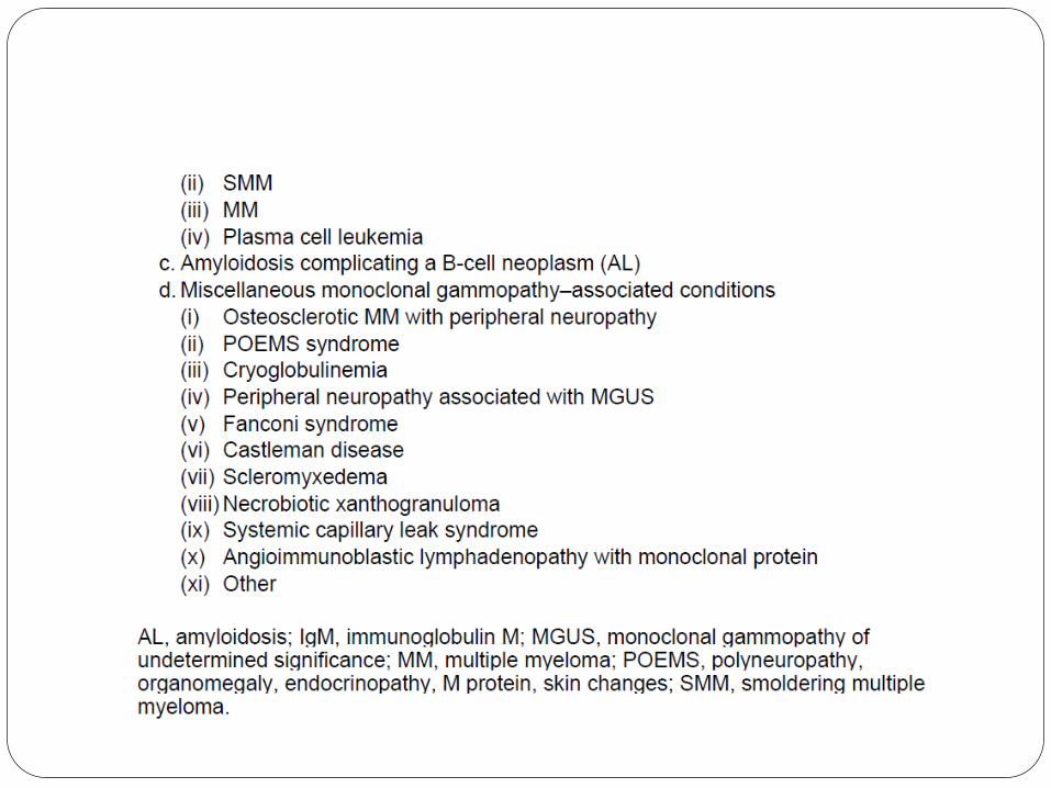
Smoldering Multiple Myeloma
(Asymptomatic Multiple Myeloma)
SMM is an intermediate stage between MGUS
and MM
Conceptually, SMM is almost identical to
MGUS—a plasma cell neoplasm without disease
in the host—but represents a more advanced
clone than that found in MGUS, usually with little
evidence of proliferation
Diagnostic criteria have been proposed, including
a clonal plasmacytosis greater than 10% or a
serum monoclonal protein of 30 g/L or greater.
For the diagnosis of SMM to be made, no
anemia, hypercalcemia, bone disease, or renal
insufficiency related only to the plasma cell
neoplasm must be present.
A related diagnosis is indolent MM, which is
nearly identical to SMM but with a small number
of discrete bone lesions

EVOLUTION TO MULTIPLE
MYELOMA
It is important to note that plasma cell neoplasms
do not need to go through all the stages in an
orderly fashion, and patients may quickly
advance to the malignant phase of the disease
from MGUS without a recognizable SMM stage.
Similarly, patients may present to the
hematologist with MM, without MGUS having
been recognized previously


MONITORING
Monitoring should occur every 3 months initially,
and if the condition is stable, at least every 6
months thereafter.
Follow-up tests should include a complete blood
count, serum levels of calcium and creatinine,
SPEP with measurement of the monoclonal
spike, and quantitative measurement of the
involved Ig (e.g., IgG levels in patients with IgG
SMM)

Multiple Myeloma

Magnitude
MM is the most common malignancy of plasma
cells
The diagnosis of MM is straightforward when the
patient has advanced bone lesions and anemia.
However, a high level of suspicion is needed to
make the diagnosis promptly in a patient who
presents with hypercalcemia or renal insufficiency

HYPERCALCEMIA
Hypercalcemia must be diagnosed and treated promptly in MM.
A classic complex may include polydipsia, polyuria, nausea, constipation, somnolence, confusion, and anorexia. Patients may have this constellation of signs and symptoms and believe they have a viral infection of the upper respiratory tract.
Although the workup of other causes of hypercalcemia may be needed, the possibility of MM should take precedence, and empirical treatment may be considered while the diagnosis is being confirmed.
Confirmatory tests can be performed rapidly, and hypercalcemiashould be treated as soon as possible, preferably within hours after its diagnosis if MM is suspected

BONE DISEASE
Bone destruction is an integral part of MM and is seen in at least 70% of patients.
When more sensitive tests are used, nearly all patients have evidence of bone destruction, and its complications
Bone disease associated with MM may involve the extremities, but most frequently it involves the spine.
A characteristic feature is that movement and weight bearing exacerbate the pain


RENAL FAILURE Patients may present because of symptoms of uremia
and only later report other symptoms of MM such as extensive bone pain.
It is critical to assess renal function quickly by measuring the serum level of creatinine.
Renal function (e.g., creatinine clearance) may be investigated in detail after therapy has been initiated.
Patients with MM and renal failure (assuming they are not oliguric) commonly have Bence Jones proteinuriain excess of 1 g/24 hours

BACK PAIN Approximately 5 to 10% of patients with MM have
back pain as a presenting feature.
This pain may be severe and require hospitalization.
Characteristically, it is movement related and
aggravated by cough, sneeze, and strain. Patients
may walk stiffly and have great difficulty getting onto
and off the examination or x-ray table.
MRI is the only reliable way to examine for a possible
epidural tumor with potential spinal cord compression

SOLITARY PLASMACYTOMA
Plasma cell neoplasms may present as a
localized growth of plasma cells referred to as
plasmacytomas
Plasmacytomas can occur in association with
bony structures ( medullary) or in other areas,
most commonly the nasopharynx (
extramedullary)
The term solitary plasmacytoma is applied to
tumor growth in patients with no other evidence of
MM.
Patients with plasmacytoma frequently present
with back pain, and, subsequently, a vertebral
mass is discovered

A site-directed biopsy is usually recommended except in patients with overt MM
Plasmacytomas that arise from bone marrow have a high propensity (approximately 75%) for progressing to MM, but those originating from extramedullary sites are less likely to do so
With the disappearance of a detectable monoclonal protein after radiation treatment for plasmacytoma, the likelihood of definitive control is higher

MULTIPLE MYELOMA MIMICRY
Without overt MM, it is erroneous to assume that
a person with a monoclonal protein in the serum
and a bone lesion identified on radiologic study
has a plasmacytoma.
Thus, needle aspiration or biopsy of the mass is
recommended.
A patient with metastatic cancer may present with
lytic bone lesions

Light Chain–Associated
Amyloidosis
AL is a serious complication of any B-cell
neoplasm capable of producing a monoclonal
protein
AL is associated most commonly with non-IgM
MGUS and, in many cases, the amount of
monoclonal protein may be minimal.
Although AL is slightly more common with λ-type
light chains, it is also seen with k-type light chains


Other features include post proctoscopic
periorbital purpura (4P sign), carpal tunnel
syndrome, macroglossia, jaw claudication,
orthostatic hypotension, diarrhea, and muscle
pseudohypertrophy.
Tissue diagnosis of AL should be sought,
including Congo red staining of suspicious
amyloid deposits
In some patients, AL is diagnosed on the basis of
symptoms, and then tests for monoclonal protein
are performed

Macroglobulinemia and Other
Immunoglobulin M–Producing
Neoplasms
IgM monoclonal gammopathy is rarely a
presenting sign of MM and should immediately
point to the possibility of a lymphoproliferative
disorder, particularly Waldenström
macroglobulinemia

IMMUNOGLOBULIN M MONOCLONAL GAMMOPATHY OF
UNDETERMINED SIGNIFICANCE
A small proportion of patients with MGUS have an IgMmonoclonal protein.
In this case, lymphadenopathy, hepatomegaly, and splenomegaly are evidence of malignant disease, usually Waldenström macroglobulinemia, CLL, or a malignant lymphoma.
All patients should have computed tomography of the abdomen and a bone marrow examination performed unless they are completely asymptomatic and the monoclonal protein level is less than 10 g/L.
The probability of IgM MGUS evolving to one of these disorders is greater than 1% per year

WALDENSTRöM
MACROGLOBULINEMIA
Hepatomegaly, lymphadenopathy, splenomegaly, epistaxis, retinal
hemorrhages, tortuosity of the retinal veins and “sausaging” are clues to
the diagnosis of Waldenström macroglobulinemia.
These signs are characteristic of Waldenström macroglobulinemia and
are distinctly uncommon in MM.
In most patients with Waldenström macroglobulinemia, the bone marrow
is replaced extensively by monoclonal lymphocytes or cells with
lymphoplasmacytic differentiation.
In typical cases of Waldenström macroglobulinemia, the IgM
monoclonal spike is greater than 30 g/L, although there is no reliable
cut-off value for excluding or making the diagnosis.
Hyperviscosity is the hallmark of the disease, but it is clinically
significant in only 15% of patients

NONNEOPLASTIC
IMMUNOGLOBULIN DISORDERS

Polyclonal Gammopathy By definition, polyclonal gammopathy represents an
accumulation of nonneoplastic plasma cells
It is important to confirm with SPEP that the increased Igsare polyclonal; if the SPEP results are equivocal, immunofixation should be used.
Polyclonal increases in the Igs can involve all or just one class of Igs .
For most cases, no major diagnostic workup is needed because the cause is apparent.
For other patients, a complete history and physical examination may provide clues to the origin of the polyclonal gammopathy.

An increase in the erythrocyte sedimentation rate may be
due to the increase in Igs; it is not a good surrogate
marker of inflammation.
Other markers of inflammation, such as C-reactive
protein, may be used.
A special condition to keep in mind as a cause of
polyclonal gammopathy is temporal arteritis, for which
effective treatment is available.
Even if liver enzyme abnormalities are mild, liver disease
should be suspected.

Hypogammaglobulinemia
In patients with recurrent encapsulated bacterial
infections, humoral immune deficiency should be
suspected. In these situations, it is advisable to
perform SPEP and quantitative Igs. Knowing the
age at onset of the problem facilitates diagnosis.
In adults with recurrent sinus or pulmonary
infections and low levels of IgG, IgA, and IgM, the
most likely diagnosis is common variable
immunodeficiency. Replacement therapy with
intravenous Igs is corrective

It is especially important to rule out MM in patients older than 50 years because Ig depression of the uninvolved Igs is frequently seen. This scenario is particularly common in patients with non-secretoryMM.
UPEP and immunofixation are also recommended because patients who have MM that produces only light chains may not have an apparent monoclonal protein in the serum.
Children with hypogammaglobulinemia most likely have a hereditary condition such as Bruton type hypogammaglobulinemia, which is usually an X-linked recessive disorder
