yellow lupine gene encoding stearoyl-acp desaturase
TRANSCRIPT
Yellow lupine gene encoding stearoyl-ACP desaturase —
organization, expression and potential application�
¯aneta Zaborowska1, Micha³ Starzycki2, Iwona Femiak1, Micha³ Œwiderski1 and
Andrzej B. Legocki1 �
1Institute of Bioorganic Chemistry, Polish Academy of Sciences, Z. Noskowskiego 12/14,
61-704 Poznañ, Poland;2Plant Breeding and Acclimatization Institute, Branch Division Poznañ,
Poznañ, Poland
Received: 19 September, 2001; revised: 3 December, 2001; accepted: 4 February, 2002
Key words: stearoyl-ACP desaturase, fatty acids, yellow lupine, nitrogen fixation, root nodules
A gene for the �9
desaturase specific to stearoyl-ACP (acyl carrier protein) was iden-
tified from yellow lupine (Lupinus luteus) cDNA and genomic libraries through the dif-
ferential display method. The desaturase transcript appears in plants infected with
Bradyrhizobium sp. (Lupinus) as revealed by Northern hybridization, RT-PCR and ex-
pression of �-glucuronidase under the desaturase promoter. A small amount of
desaturase transcript was also detected in uninfected plants, which suggests that the
gene does not belong to the strict nodule-specific sequences. The desaturase provides
unsaturated fatty acids for additional cell membrane synthesis. During nodule and
symbiosome development a peribacteroid membrane is formed and the requirement
for membrane surface increases, thus the level of desaturase expression is also
higher. Transgenic plants of Nicotiana tabacum with overexpression of the full-length
lupine stearoyl-ACP desaturase sequence were obtained. They revealed higher con-
tent of unsaturated fatty acids (especially oleic acid) in comparison with control
plants.
Desaturases catalyze the conversion of satu-rated fatty acids to unsaturated acids intro-ducing the first double bond into saturatedfatty acids (Harwood, 1980; Stumpf, 1980).This group of enzymes has been identified inall eukaryotes, cyanobacteria and in some Ba-
cillus bacteria (Bloomfield & Bloch, 1960;
Fulco, 1974). Three types of desaturases aredistinguishable depending on the kind of com-pounds esterified to fatty acid. The fatty acidcan be attached to acyl carrier protein (ACP),to coenzyme A (CoA) or to lipid molecules.The only known soluble desaturase is theplant stearoyl-ACP desaturase specific to
Vol. 49 No. 1/2002
29–42
QUARTERLY
�This research was supported by the Polish-French Plant Biotechnology Center and by the UNESCO-PASNetwork for Molecular and Cellular Biology.
�Corresponding author: [email protected]: ACP, acyl carrier protein; GUS, �-glucuronidase.
stearic acid, localized in plastids. Desaturasesidentified in plants, animals and yeast aremembrane-bound proteins with acyl chain at-tached to CoA or lipids.
Desaturation proceeds under oxidizing con-ditions (Bloomfield & Bloch, 1960; Mudd &Stumpf, 1961) and requires a short electrontransport chain. Two electrons are necessaryfor the formation of one double bond. Thereare two systems of electron transport. One ispresent in plastids, while the other is localizedin the endoplasmic reticulum. In plants, therole of an electron donor is played by NADPH,while ferredoxin-NADP+ oxidoreductase func-tions as a flavoprotein and ferredoxin with itsiron-sulfur center is the electron carrier(Nagai & Bloch, 1966; Schmidt & Heinz, 1990;Wada et al., 1993). The source of electrons inphotosynthetic tissues under light conditionis photosystem I (Jacobson et al., 1974). In theendoplasmic reticulum, the electron donor isNADH and cytochrome b5 reductase plays therole of flavoprotein, while cytochrome b5 (aheme protein) functions as an electron carrier(Dailey & Strittmatter, 1979; Hackett &Strittmatter, 1984; Spatz & Strittmatter,1971).The chemical bond between carbon and hy-
drogen in a fatty acid chain is one of the moststable bonds. A metal cofactor is essential forthe breakage and further modification of thisbond. Crystallization of plastid �9-18:0-ACPdesaturase from Ricinus communis has re-vealed that this enzyme requires two iron at-oms for its catalytic activity (Shanklin &Sommerville, 1991; Thompson et al., 1991;Fox et al., 1993). The conservative region(D/EX2H)2 was identified in soluble plantdesaturases and other di-iron proteins, suchas methane monooxygenase and ribonucleo-tide reductase (Fox et al., 1993). The ste-aroyl-ACP desaturase from Ricinus communis
is a homodimer consisting of two 41.6 kDaunits. The secondary structure of the desa-turase consists of 11 � helices and two �strands. The active site is localized within atetrahelix core bundle. A hydrophobic channel
is necessary for substrate orientation in thevicinity of di-iron center. Glutamic acid andhistidine residues coordinate two iron atomsessential for catalytic activity.Plant desaturases have gained a wide inter-
est due to their potential biotechnological sig-nificance. These enzymes expressed in trans-genic crops can modify fatty acid spectrum,which might be useful for the production ofhealthy edible oils with an increased level ofunsaturated fatty acids.
MATERIALS AND METHODS
Identification of stearoyl-ACP desaturase
cDNA, screening of a genomic library and
Southern hybridization
Identification of stearoyl-ACP desaturasecDNA clone (Gen Bank acc. no. AF 139377)and sequence alignment with other plantdesaturases were published earlier (Swiderskiet al., 2000). RNA differential display reac-tions were carried out according to (Liang et
al., 1993; Liang & Pardee, 1992) as describedelsewhere (Swiderski et al., 2000). A genomiclibrary of yellow lupine DNA constructed in �EMBL-3 (Stratagene) was screened using an[�-32P]dATP labeled desaturase cDNA probe.Nitrocellulose membranes were hybridized at60�C for 16–20 h and washed two times in 4 �SSC/0.1% SDS at 60�C, two times in 2 �SSC/0.1% SDS at 60�C and two times in 1 �SSC/0.1% SDS at room temperature.
Northern hybridization
Total RNA isolated from lupine tissues wasseparated on 1.2% agarose gel with 2% formal-dehyde. After nucleic acid transfer to a nylonmembrane it was hybridized with an [�-32P]dATP labeled desaturase cDNA at 42�C for 48h. The membrane was washed two times in 2� SSC/0.1% SDS at room temperature, oncein 1 � SSC/0.1% SDS at 68�C and once in 0.1� SSC/0.1% SDS at 68�C.
30 ¯. Zaborowska and others 2002

RT-PCR reaction
Two micrograms of total RNA isolated fromyellow lupine tissues was mixed with reversetranscriptase buffer and 50 pmol of oligo-nucleotide primer specific to the 3� end ofdesaturase cDNA. The mixture was incubatedat 65�C for 10 min and finally at 42�C for 1 h.Reverse transcription reaction was startedwith the addition of 20 units of MMLV tran-scriptase and 1 mM of each dNTP. The reac-tion was performed at 37�C for 1 h. The en-zyme was inactivated at 65�C for 10 min.
Plant transformation
Alfalfa transformation. Alfalfa transfor-mation was obtained by wound inoculation.Leaf explants of alfalfa were cocultivated witha bacterial suspension of Agrobacterium
tumefaciens EHA105 strain grown overnight(YEB medium, kanamycin and rifampicin re-sistance). Leaf fragments were incubated for72 h in the dark on a nonselective medium andfor 4–5 weeks in the dark on the selective me-dium SHMab with kanamycin and car-benicylin. Then the growth conditions werechanged to light/dark (16 h/8 h) and after3 weeks an embriogenic callus was obtainedon SHM2 medium with kanamycin and car-benicylin. Growing plants were transferred tosoil. After 3 weeks the plants were inoculatedwith Bradyrhizobium meliloti bacteria to in-duce symbiotic root nodule formation. Alfalfaroots, stems, leaves, flowers, seeds and nod-ules between 3–4 weeks after rhizobial inocu-lation were used for further analyses.Tobacco transformation. Transformation
of tobacco was performed by cocultivation ofleaf explants with a bacterial suspension ofA. tumefaciens EHA105 and LBA4404 straingrown overnight (YEB medium, BASTA her-bicide and rifampicin resistance). Leaf frag-ments of tobacco were grown on MS mediumfor two days. Then the explants were trans-ferred onto T1 medium with BASTA herbicideand rifampicin for transgenic plant selection.
Leaves were used for lipid content analysis us-ing gas chromatography.
RESULTS
cDNA clone encoding stearoyl-ACP
desaturase from yellow lupine
The yellow lupine desaturase cDNA wasidentified by the differential display as a se-quence expressed in root nodules after inocu-lation with symbiotic bacteria (Swiderski et
al., 2000). Alignment, based on putativeamino-acid sequence allowed us to classify thelupine enzyme as a desaturase specific tostearic acid attached to acyl carrier protein.Two di-iron motifs separated by 100 amino ac-ids were found within the active center of theenzyme. These motifs are essential for cata-lytic activity and their amino-acid sequence ischaracteristic of a soluble group of acyl-ACPdesaturases (Fox et al., 1993).
Screening of a genomic library
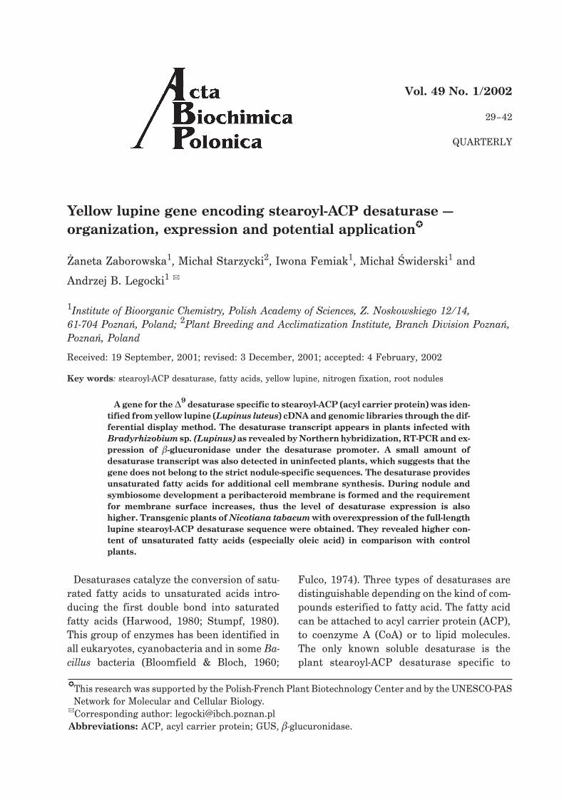
The yellow lupine desaturase cDNA clonewas labeled with [�-32P]dATP and used as aprobe to screen genomic library constructedin � EMBL-3 phage. Strong single hybridiza-tion signals were used as a material to phageDNA isolation and restriction analysis(Fig. 1A). Southern hybridization with labeleddesaturase cDNA revealed several hybridiza-tion signals providing additional indicationthat the analyzed sequence encodes thedesaturase (Fig. 1B).
Organization and nucleotide sequence of
the genomic clone encoding stearoyl-ACP
desaturase
To establish the orientation of the genomicclone, PCR (Expand Long Template PCR Sys-tem) was performed using phage template andtwo oligonucleotide primers specific to theright and left arms of � EMBL-3 phage and a
Vol. 48 Yellow lupine gene encoding stearoyl-ACP desaturase 31
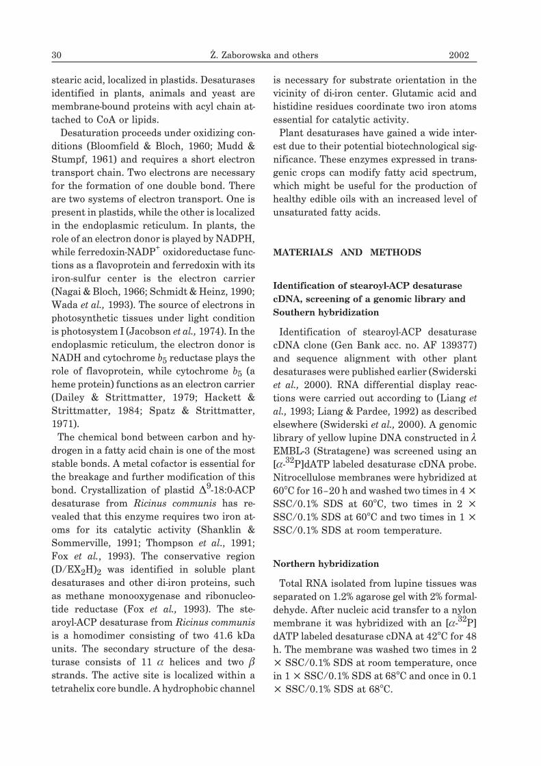
primer specific to the 5� end of desaturasecDNA clone. The reaction revealed that the 5�end of the genomic clone of desaturase is local-ized close to the left arm of the phage. Se-quencing results indicated that the clone didnot contain the complete sequence of thestearoyl-ACP desaturase gene and includedonly the coding region with promoter. To ob-tain the complete genomic sequence, PCR wasperformed using yellow lupine genomic DNAtemplate (Fig. 2). Figure 3 presents the organi-zation of the cDNA and the genomic clone ofthe stearoyl-ACP desaturase gene from lupine.
Northern blot and RT-PCR analyses of lu-
pine desaturase gene expression
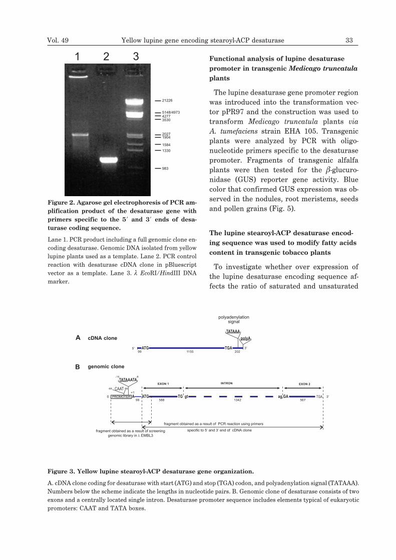
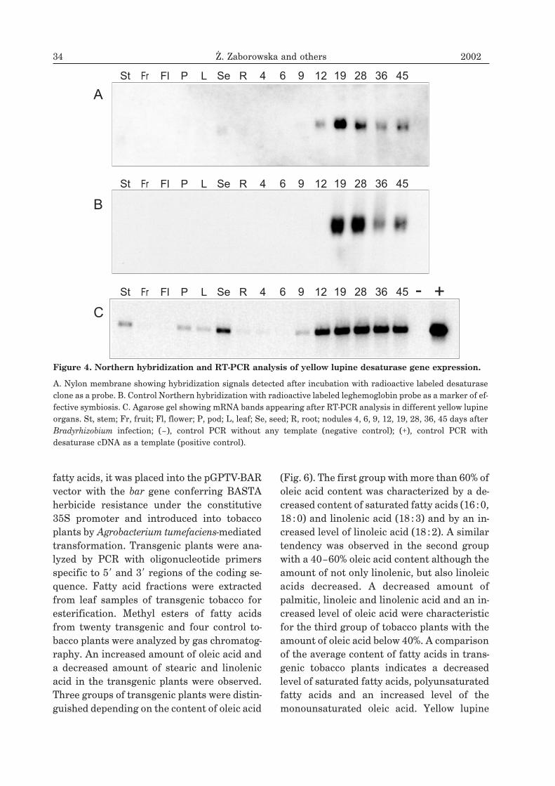
Total RNA preparations isolated from differ-ent organs of yellow lupine (stem, petiole,leaf, flower, pod, green seeds, root, and nod-ule-like structures or nodules 4, 6, 9, 12, 19,
28, 36, 45 days after inoculation with Brady-
rhizobium sp. (Lupinus) were transferred on anylon membrane and hybridized with radioac-tive desaturase probe (Fig. 4A). Control hy-bridization was performed with radioactive la-beled leghemoglobin probe that served as amarker of effective symbiosis (Fig. 4B). North-ern hybridization confirmed nodule specific-ity of the desaturase. Its transcript appears onday 12 after symbiotic bacteria inoculationand the highest expression is observed 19days after the infection. The expression pat-tern of the desaturase gene as revealed byNorthern blot hybridization is similar to thatof leghemoglobin gene. RT-PCR analysis re-vealed that desaturase mRNA appears 9 daysafter inoculation. Moreover, from this analy-sis it follows that this enzyme cannot be quali-fied as a strict nodulin since a low amount ofthe transcript was detected in stem, pod, leafas well as in uninfected root (Fig. 4C).
32 ¯. Zaborowska and others 2002
1 2 3 4 5 6 7 8 1 2 3 4 5 6
A. B.
23130
9416
6557
4361
800
500
Figure 1. Southern hybridization of a yellow lupine desaturase genomic clone in phage vector with
digoxygenin labeled desaturase cDNA.
A. Agarose gel electrophoresis with separated restriction fragments of a desaturase genomic clone digested with thefollowing enzymes: Lane 1, SalI; Lane 2, SalI/EcoRI; Lane 3, EcoRI; Lane 4, SalI/HindIII; Lane 5, SalI/PstI; Lane6, PstI; Lane 7, � HindIII DNA marker; Lane 8, DNA marker consisting of two bands: 800 and 500 nucleotides. B. Ny-lon membrane with digested DNA fragments of phage genomic clone isolated from � EMBL 3 library. Hybridizationsignals appeared after incubation with a digoxygenin labeled desaturase cDNA clone.
Functional analysis of lupine desaturase
promoter in transgenic Medicago truncatula
plants
The lupine desaturase gene promoter regionwas introduced into the transformation vec-tor pPR97 and the construction was used totransform Medicago truncatula plants via
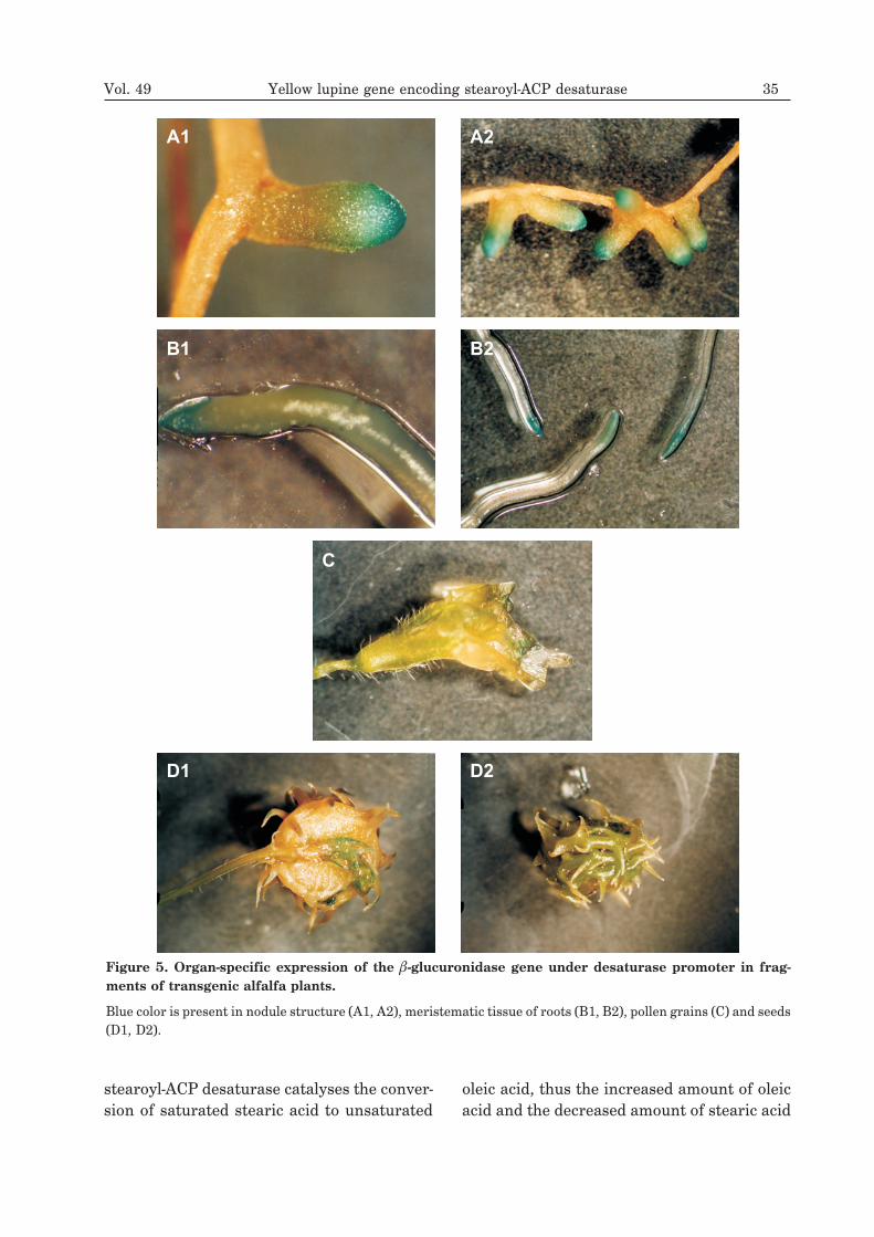
A. tumefaciens strain EHA 105. Transgenicplants were analyzed by PCR with oligo-nucleotide primers specific to the desaturasepromoter. Fragments of transgenic alfalfaplants were then tested for the �-glucuro-nidase (GUS) reporter gene activity. Bluecolor that confirmed GUS expression was ob-served in the nodules, root meristems, seedsand pollen grains (Fig. 5).
The lupine stearoyl-ACP desaturase encod-
ing sequence was used to modify fatty acids
content in transgenic tobacco plants
To investigate whether over expression ofthe lupine desaturase encoding sequence af-fects the ratio of saturated and unsaturated
Vol. 49 Yellow lupine gene encoding stearoyl-ACP desaturase 33
21226
5148/497342773530
20271904
1584
1330
983
1 2 3
Figure 2. Agarose gel electrophoresis of PCR am-
plification product of the desaturase gene with
primers specific to the 5� and 3� ends of desa-
turase coding sequence.
Lane 1. PCR product including a full genomic clone en-coding desaturase. Genomic DNA isolated from yellowlupine plants used as a template. Lane 2. PCR controlreaction with desaturase cDNA clone in pBluescriptvector as a template. Lane 3. � EcoRI/HindIII DNAmarker.
A
TATAAATA
CAAT
TATAAA
polyA
ATG
ATG TG gt ag
TGA
GA TGA
EXON 1
-15 -8
-66-69
INTRON EXON 2
genomic clone
cDNA clone
5'
5'
+1
3'
3'
99 1155 202
polyadenylationsignal
99 588 1342 567
fragment obtained as a result of PCR reaction using primers
fragment obtained as a result of screening
genomic library in EMBL3�
specific to 5’ and 3’ end of cDNA clone
PROMOTER
A
B
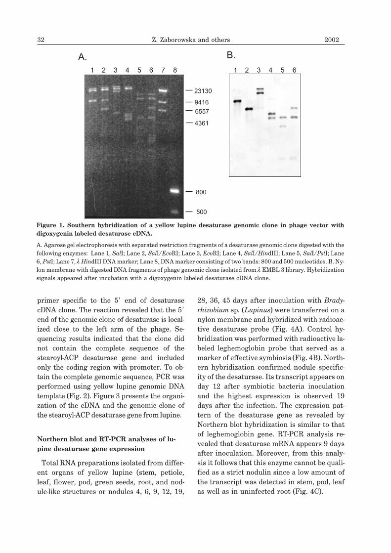
Figure 3. Yellow lupine stearoyl-ACP desaturase gene organization.
A. cDNA clone coding for desaturase with start (ATG) and stop (TGA) codon, and polyadenylation signal (TATAAA).Numbers below the scheme indicate the lengths in nucleotide pairs. B. Genomic clone of desaturase consists of twoexons and a centrally located single intron. Desaturase promoter sequence includes elements typical of eukaryoticpromoters: CAAT and TATA boxes.
fatty acids, it was placed into the pGPTV-BARvector with the bar gene conferring BASTAherbicide resistance under the constitutive35S promoter and introduced into tobaccoplants by Agrobacterium tumefaciens-mediatedtransformation. Transgenic plants were ana-lyzed by PCR with oligonucleotide primersspecific to 5� and 3� regions of the coding se-quence. Fatty acid fractions were extractedfrom leaf samples of transgenic tobacco foresterification. Methyl esters of fatty acidsfrom twenty transgenic and four control to-bacco plants were analyzed by gas chromatog-raphy. An increased amount of oleic acid anda decreased amount of stearic and linolenicacid in the transgenic plants were observed.Three groups of transgenic plants were distin-guished depending on the content of oleic acid
(Fig. 6). The first group with more than 60% ofoleic acid content was characterized by a de-creased content of saturated fatty acids (16 :0,18 :0) and linolenic acid (18 :3) and by an in-creased level of linoleic acid (18 :2). A similartendency was observed in the second groupwith a 40–60% oleic acid content although theamount of not only linolenic, but also linoleicacids decreased. A decreased amount ofpalmitic, linoleic and linolenic acid and an in-creased level of oleic acid were characteristicfor the third group of tobacco plants with theamount of oleic acid below 40%. A comparisonof the average content of fatty acids in trans-genic tobacco plants indicates a decreasedlevel of saturated fatty acids, polyunsaturatedfatty acids and an increased level of themonounsaturated oleic acid. Yellow lupine
34 ¯. Zaborowska and others 2002
4
4
4
6
6
6
9
9
9
12
12
12
19
19
19
28
28
28
36
36
36
45
45
45
St
St
St
Fr
Fr
Fr
Fl
Fl
Fl
P
P
P
L
L
L
Se
Se
Se
R
R
R - +
A
B
C
Figure 4. Northern hybridization and RT-PCR analysis of yellow lupine desaturase gene expression.
A. Nylon membrane showing hybridization signals detected after incubation with radioactive labeled desaturaseclone as a probe. B. Control Northern hybridization with radioactive labeled leghemoglobin probe as a marker of ef-fective symbiosis. C. Agarose gel showing mRNA bands appearing after RT-PCR analysis in different yellow lupineorgans. St, stem; Fr, fruit; Fl, flower; P, pod; L, leaf; Se, seed; R, root; nodules 4, 6, 9, 12, 19, 28, 36, 45 days afterBradyrhizobium infection; (–), control PCR without any template (negative control); (+), control PCR withdesaturase cDNA as a template (positive control).
stearoyl-ACP desaturase catalyses the conver-sion of saturated stearic acid to unsaturated
oleic acid, thus the increased amount of oleicacid and the decreased amount of stearic acid
Vol. 49 Yellow lupine gene encoding stearoyl-ACP desaturase 35
A1
B1
D1
A2
B2
D2
C
Figure 5. Organ-specific expression of the �-glucuronidase gene under desaturase promoter in frag-
ments of transgenic alfalfa plants.
Blue color is present in nodule structure (A1, A2), meristematic tissue of roots (B1, B2), pollen grains (C) and seeds(D1, D2).
in transgenic tobacco plants are consistentwith expectations. The decreased level of poly-
unsaturated fatty acids and the increasedamount of monounsaturated fatty acids were
36 ¯. Zaborowska and others 2002
0
10
20
30
40
50
60
70
palm
itic
stea
ric
olei
c
linol
ic
linol
enic
eico
seni
c
control
group 1
group 2
group 3
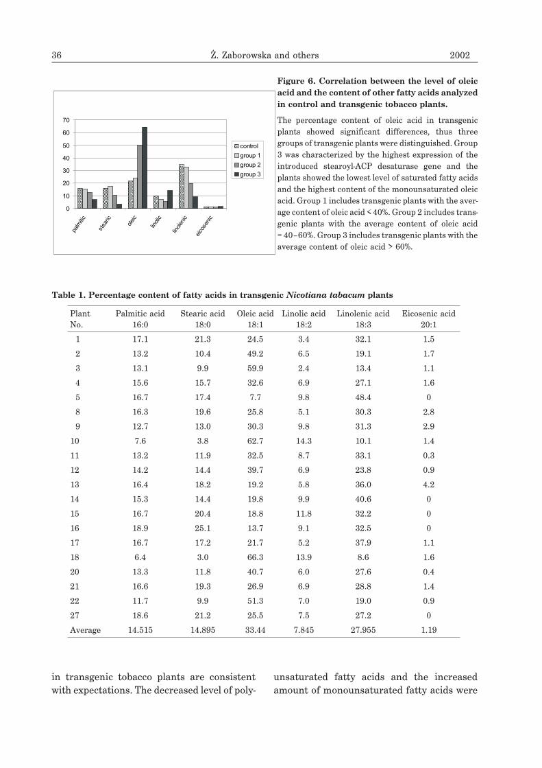
Figure 6. Correlation between the level of oleic
acid and the content of other fatty acids analyzed
in control and transgenic tobacco plants.
The percentage content of oleic acid in transgenicplants showed significant differences, thus threegroups of transgenic plants were distinguished. Group3 was characterized by the highest expression of theintroduced stearoyl-ACP desaturase gene and theplants showed the lowest level of saturated fatty acidsand the highest content of the monounsaturated oleicacid. Group 1 includes transgenic plants with the aver-age content of oleic acid < 40%. Group 2 includes trans-genic plants with the average content of oleic acid= 40–60%. Group 3 includes transgenic plants with theaverage content of oleic acid > 60%.
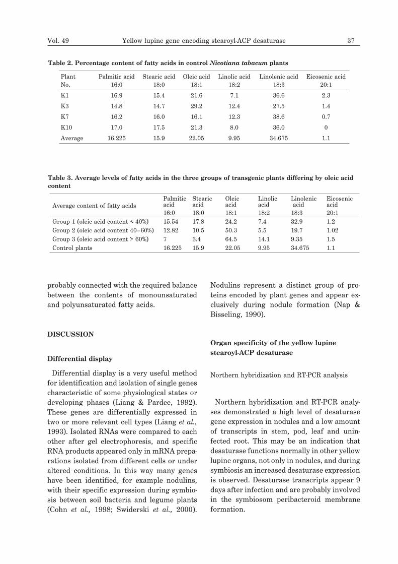
Table 1. Percentage content of fatty acids in transgenic Nicotiana tabacum plants
PlantNo.
Palmitic acid16:0
Stearic acid18:0
Oleic acid18:1
Linolic acid18:2
Linolenic acid18:3
Eicosenic acid20:1
1 17.1 21.3 24.5 3.4 32.1 1.5
2 13.2 10.4 49.2 6.5 19.1 1.7
3 13.1 9.9 59.9 2.4 13.4 1.1
4 15.6 15.7 32.6 6.9 27.1 1.6
5 16.7 17.4 7.7 9.8 48.4 0
8 16.3 19.6 25.8 5.1 30.3 2.8
9 12.7 13.0 30.3 9.8 31.3 2.9
10 7.6 3.8 62.7 14.3 10.1 1.4
11 13.2 11.9 32.5 8.7 33.1 0.3
12 14.2 14.4 39.7 6.9 23.8 0.9
13 16.4 18.2 19.2 5.8 36.0 4.2
14 15.3 14.4 19.8 9.9 40.6 0
15 16.7 20.4 18.8 11.8 32.2 0
16 18.9 25.1 13.7 9.1 32.5 0
17 16.7 17.2 21.7 5.2 37.9 1.1
18 6.4 3.0 66.3 13.9 8.6 1.6
20 13.3 11.8 40.7 6.0 27.6 0.4
21 16.6 19.3 26.9 6.9 28.8 1.4
22 11.7 9.9 51.3 7.0 19.0 0.9
27 18.6 21.2 25.5 7.5 27.2 0
Average 14.515 14.895 33.44 7.845 27.955 1.19
probably connected with the required balancebetween the contents of monounsaturatedand polyunsaturated fatty acids.
DISCUSSION
Differential display
Differential display is a very useful methodfor identification and isolation of single genescharacteristic of some physiological states ordeveloping phases (Liang & Pardee, 1992).These genes are differentially expressed intwo or more relevant cell types (Liang et al.,
1993). Isolated RNAs were compared to eachother after gel electrophoresis, and specificRNA products appeared only in mRNA prepa-rations isolated from different cells or underaltered conditions. In this way many geneshave been identified, for example nodulins,with their specific expression during symbio-sis between soil bacteria and legume plants(Cohn et al., 1998; Swiderski et al., 2000).
Nodulins represent a distinct group of pro-teins encoded by plant genes and appear ex-clusively during nodule formation (Nap &Bisseling, 1990).
Organ specificity of the yellow lupine
stearoyl-ACP desaturase
Northern hybridization and RT-PCR analysis
Northern hybridization and RT-PCR analy-ses demonstrated a high level of desaturasegene expression in nodules and a low amountof transcripts in stem, pod, leaf and unin-fected root. This may be an indication thatdesaturase functions normally in other yellowlupine organs, not only in nodules, and duringsymbiosis an increased desaturase expressionis observed. Desaturase transcripts appear 9days after infection and are probably involvedin the symbiosom peribacteroid membraneformation.
Vol. 49 Yellow lupine gene encoding stearoyl-ACP desaturase 37
Table 2. Percentage content of fatty acids in control Nicotiana tabacum plants
PlantNo.
Palmitic acid16:0
Stearic acid18:0
Oleic acid18:1
Linolic acid18:2
Linolenic acid18:3
Eicosenic acid20:1
K1 16.9 15.4 21.6 7.1 36.6 2.3
K3 14.8 14.7 29.2 12.4 27.5 1.4
K7 16.2 16.0 16.1 12.3 38.6 0.7
K10 17.0 17.5 21.3 8.0 36.0 0
Average 16.225 15.9 22.05 9.95 34.675 1.1
Table 3. Average levels of fatty acids in the three groups of transgenic plants differing by oleic acid
content
Average content of fatty acidsPalmiticacid16:0
Stearicacid18:0
Oleicacid18:1
Linolicacid18:2
Linolenicacid18:3
Eicosenicacid20:1
Group 1 (oleic acid content < 40%) 15.54 17.8 24.2 7.4 32.9 1.2Group 2 (oleic acid content 40–60%) 12.82 10.5 50.3 5.5 19.7 1.02Group 3 (oleic acid content > 60%) 7 3.4 64.5 14.1 9.35 1.5Control plants 16.225 15.9 22.05 9.95 34.675 1.1
The symbiosom consists of the bacteroid celland the surrounding peribacteroid membrane(Roth et al., 1988). The bacteroid membrane isformed from plasmalemma during endo-cytosis of symbiotic bacteria from the infec-tious strand inside the plant cell. The surfaceof the peribacteroid membrane increases20–40 times during bacteroids cell divisionand becomes larger by including, for example,Golgi structure. Stearoyl-ACP desaturase in-
troduces a double bond into stearic acid andprovides an unsaturated fatty acid that maybe essential for the membrane structure, flu-ency and functions.In our earlier studies on characterization of
the lupine plant-Bradyrhizobium sp. (Lupinus)
symbiotic system we have detected and de-scribed plant genes that were induced duringendosymbiotic interaction (Strozycki et al.,
2000; Strozycki & Legocki, 1995) and those
38 ¯. Zaborowska and others 2002
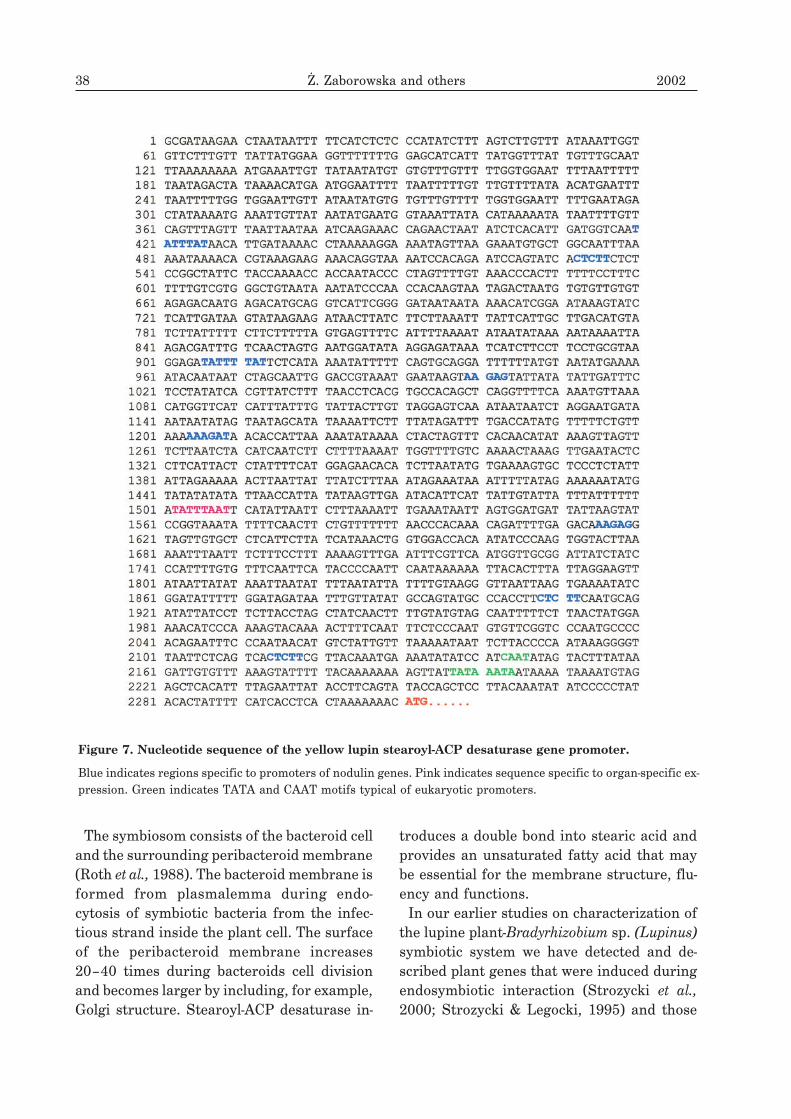
Figure 7. Nucleotide sequence of the yellow lupin stearoyl-ACP desaturase gene promoter.
Blue indicates regions specific to promoters of nodulin genes. Pink indicates sequence specific to organ-specific ex-pression. Green indicates TATA and CAAT motifs typical of eukaryotic promoters.
Vol. 49 Yellow lupine gene encoding stearoyl-ACP desaturase 39
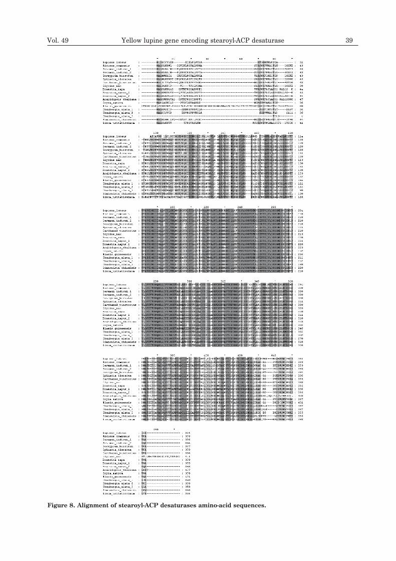
Figure 8. Alignment of stearoyl-ACP desaturases amino-acid sequences.

which were down regulated (Biesiadka et al.,1999; Sikorski et al., 1999). Although symbi-otic regulation of plant genes seems to behighly specific, some of nodulin genes are ex-pressed at a low level in other organs than theroot nodule.
Stearoyl-ACP desaturase promoter sequence
Stearoyl-ACP desaturase promoters are themost active in developing tissues (Slocombe et
al., 1994). Expression of desaturase genes istemporally regulated and organospecific. Ananalysis of canola stearoyl-ACP desaturasepromoter in transgenic tobacco plants has re-vealed high expression of �-glucuronidase re-porter gene in developing seeds, flowers andpollen grains (Slocombe et al., 1994). Expres-sion of the �-glucuronidase gene under yellowlupine stearoyl-ACP desaturase promoter wasobserved in nodules, root meristems, seedsand pollen grains. This suggests an importantrole of desaturase in division processes re-quiring unsaturated fatty acids essential forthe construction of the cell membrane. Thepromoter sequence of yellow lupine ste-aroyl-ACP desaturase contains regions spe-cific to other nodule gene promoters (Fig. 7).
Alignment of yellow lupine stearoyl-ACP
desaturase with other known plant
desaturases
Stearoyl-ACP desaturase in stroma plastidsconverts stearic acid connected with acyl car-rier protein to oleic acid (Slocombe et al.,
1994; Stumpf, 1980). Oleic acid is transportedto tylakoid membranes or to the cytoplasmand then is attached to lipids and desaturated(Roughan, 1987; Roughan & Slack, 1982). Thefirst double bond can be generated at the �4,�6 and �9 position. Each stearoyl-ACP desa-turase requires two iron atoms essential forreactive complex formation with oxygen(Fe–O–Fe) necessary for the catalytic activity(Fox et al., 1994; Shanklin et al., 1994). A crys-tallographic analysis of stearoyl-ACP
desaturase from Ricinus communis has re-vealed that desaturase forms an active di-ironcluster (Lindqvist et al., 1996). One of theseiron atoms interacts with side chains of E196and H232 residues, whereas the other withside chains of E105 and H146 (Lindqvist et al.,
1996). A di-iron cluster was identified alsowithin the active center of yellow lupine ste-aroyl-ACP desaturase (Swiderski et al., 2000).This motif characteristic of acyl-ACP desa-turases contains two EXXH sequences sepa-rated by about 100 amino acids. In yellow lu-pine stearoyl-ACP desaturase the di-iron clus-ter includes two EKRH motifs separated by 82amino-acid residues.Plant stearoyl-ACP desaturases are the only
known soluble desaturases. These enzymesare transported to the stroma plastid by thepresence of a signal peptide localized at theN-end of the protein, consisting of about 30amino acids — most of them hydrophobic orpositively charged. After a hydropathy analy-sis, the signal peptide was also identified atthe amino end of yellow lupine stearoyl-ACPdesaturase. An alignment of stearoyl-ACPdesaturase amino-acid sequences indicates ahigh percentage of identity (Fig. 8).
R E F E R E N C E S
Biesiadka, J., Sikorski, M.M., Bujacz, G. &Jaskólski, M. (1999) Crystallization and pre-liminary X-ray structure determination ofLupinus luteus PR10 protein. Acta Crystallo-
graphica D55, 1925–1927.
Bloomfield, D.K. & Bloch, K. (1960) Formation of�9
-unsaturated fatty acids. J. Biol. Chem.235, 337–345.
Cohn, J., Day, R.B. & Stacey, G. (1998) Legumenodule organogenesis. Trends Plant Sci. 3,
105–110.
Dailey, H.A. & Strittmatter, P. (1979) Modificationand identification of cytochrome b5 carboxylgroups involved in protein–protein interac-tion with cytochrome b5 reductase. J. Biol.
Chem. 254, 5388–5396.
40 ¯. Zaborowska and others 2002
Fox, B.G., Shanklin, J., Sommerville, Ch. &Munck, E. (1993) Stearoyl-acyl carrier protein�9 desaturase from Ricinus communis is adi-iron-oxo protein. Proc. Natl. Acad. Sci.U.S.A. 90, 2486–2490.
Fox, B.G., Shanklin, J., Jingyuan, A., Loehr, T.M.& Sanders-Loehr, J. (1994) Resonance Ramanevidence for an Fe-O-Fe center in stearoyl-ACPdesaturase. Primary sequence identity withother di-iron-oxo proteins. Biochemistry 33,12776–12786.
Fulco, A.J. (1974) Metabolic alterations of fatty ac-ids. Annu. Rev. Biochem. 43, 215–240.
Hackett, C.S. & Strittmatter, P. (1984) Covalentcross-linking of the active sites of vesicle-bound cytochrome b5 and NADH-cytochromeb5 reductase. J. Biol. Chem. 259, 3275–3282.
Harwood, J.L. (1980) Plant acyl lipids: Structure,distribution, and analysis; in The Biochemistry
of Plants (Stumpf, P.K. & Conn, E.E., eds.) vol.4, pp. 1–55, Academic Press, New York.
Jacobson, B.S., Jaworski, J.G. & Stumpf, P.K.(1974) Fat metabolism in plants. LXII.Stearoyl-acyl carrier protein desaturase fromspinach chloroplast. Plant Physiol. 54,484–486.
Liang, P. & Pardee, A.B. (1992) Differential dis-play of eukaryotic messenger RNA by meansof the polymerase chain reaction. Science 257,967–971.
Liang, P., Averboukh, L. & Pardee, A.B. (1993) Dis-tribution and cloning of eukaryotic mRNAs bymeans of differential display: Refinementsand optimization. Nucleic Acids Res. 21,3269–3275.
Lindqvist, Y., Huang, W., Schneider, G. &Shanklin, J. (1996) Crystal structure of �9
stearoyl-acyl carrier protein desaturase fromcastor seed and its relationship to otherdi-iron proteins. EMBO J. 15, 4081–4092.
Mudd, J.B. & Stumpf, P.K. (1961) Fat metabolismin plants. XIV. Factors affecting the synthesisof oleic acid by particulate preparations fromavocado mesocarp. J. Biol. Chem. 236,2602–2609.
Nagai, J. & Bloch, K. (1966) Enzymatic desa-turation of stearoyl-acyl carrier protein. J.
Biol. Chem. 241, 1925–1927.
Nap, J.P. & Bisseling, T. (1990) Developmental bi-ology of plant-prokaryote symbiosis: The le-gume root nodule. Science 250, 948–954.
Roth, E., Jeon, K. & Stacey, G. (1988) Homology inendosymbiotic systems: The term “symbio-some”; in Molecular Genetics of Plant-Microbe
Interactions (Palacios, R. & Verma, D.P.S.,eds.) pp. 220–225, American Phytopath. Soc.Press, St. Paul.
Roughan, P.G. (1987) On the control of fatty acidcompositions of plant glycerolipids; in Metabo-
lism, Structure, and Function of Plant Lipids
(Stumpf, P.K.J., Mudd, B. & Nes, W.D., eds.)pp. 247–254, Plenum Press, New York.
Roughan, P.G. & Slack, C.R. (1982) Cellular orga-nization of glycerolipid metabolism. Annu.
Rev. Plant Physiol. 33, 97–132.
Schmidt, H. & Heinz, E. (1990) Involvement offerredoxin in desaturation of lipid boundoleate in chloroplast. Plant Physiol. 94,214–220.
Shanklin, J. & Sommerville, Ch. (1991) Stearoyl-acyl-carrier-protein desaturase from higherplants is structurally unrelated to the animaland fungal homologs. Proc. Natl. Acad. Sci.U.S.A. 88, 2510–2514.
Shanklin, J., Whittle, E. & Fox, B.G. (1994) Eighthistidine residues are catalytically essential ina membrane-associated iron enzyme, stearoyl-CoA desaturase, and are conserved in alkanehydroxylase and xylene monooxygenase. Bio-
chemistry 33, 12787–12794.
Sikorski, M.M., Biesiadka, J., Kasperska, A.E.,Kopcinska, J., Lotocka, B., Golinowski, W. &Legocki, A.B. (1999) Expression of genes en-coding PR10 class pathogenesis-related pro-teins is inhibited in yellow lupine root nodules.Plant Sci. 149, 125–137.
Slocombe, S.P., Piffanelli, P., Fairbairn, D.,Bowra, S., Hatzopoulos, P., Tsiantis, M. &Murphy, D.J. (1994) Temporal and tis-sue-specific regulation of a Brassica napus
stearoyl-acyl carrier protein desaturase gene.Plant Physiol. 104, 1167–1176.
Vol. 49 Yellow lupine gene encoding stearoyl-ACP desaturase 41
Spatz, L. & Strittmatter, P. (1971) A form ofcytochrome b5 that contains an additional hy-drophobic sequence of 40 amino acid residues.Proc. Natl. Acad. Sci. U.S.A. 68, 1042–1046.
Strozycki, P.M. & Legocki, A.B. (1995)Leghemoglobins from an evolutionarily old le-gume Lupinus luteus. Plant Sci. 110, 83–93.
Strozycki, P.M., Karlowski, W.M., Dessaux, Y., Pe-tit, A. & Legocki, A.B. (2000) Lupine leghe-moglobin I: Expression in transgenic lotus andtobacco tissues. Mol. Gen. Genet. 263,
173–182.
Stumpf, P.K. (1980) Biosynthesis of saturated andunsaturated fatty acids; in The Biochemistry of
Plants (Stumpf, P.K. & Conn, E.E., eds.) vol. 4,pp. 177–204, Academic Press, New York.
Swiderski, M., Zaborowska, Z. & Legocki, A.B.(2000) Identification of new nodulin cDNAsfrom yellow lupine by differential display.Plant Sci. 151, 75–78.
Thompson, G.A., Scherer, D.E., Foxall-Van Aken,S., Kenny, J.W., Young, H.L., Shintani, D.K.,Kridl, J.C. & Knauf, V.C. (1991) Primary struc-tures of the precursor and mature forms ofstearoyl-acyl carrier protein desaturase fromsaff lower embryos and requirement offerredoxin for enzyme activity. Proc. Natl.
Acad. Sci. U.S.A. 88, 2578–2582.
Wada, H., Schmidt, H., Heinz, E. & Murata, N.(1993) In vitro ferredoxin-dependent desatu-ration of fatty acids in cyanobacterial thyl-akoid membranes. J. Bacteriol. 175, 544–547.
42 ¯. Zaborowska and others 2002