yeast rab gtpase-activating protein gyp1p localizes to the ... filerab gtpases, strongly inhibits...
TRANSCRIPT
Molecular Biology of the CellVol. 12, 1215–1226, May 2001
Yeast Rab GTPase-activating Protein Gyp1p Localizesto the Golgi Apparatus and Is a Negative Regulatorof Ypt1pLi-Lin Du* and Peter Novick†‡
*Departments of Molecular Biophysics and Biochemistry and †Cell Biology, Yale University School ofMedicine, New Haven, Connecticut 06520
Submitted December 20, 2000; Revised February 12, 2001; Accepted February 22, 2001Monitoring Editor: Suzanne R. Pfeffer
A family of related proteins in yeast Saccharomyces cerevisiae is known to have in vitro GTPase-activating protein activity on the Rab GTPases. However, their in vivo function remains obscure.One of them, Gyp1p, acts on Sec4p, Ypt1p, Ypt7p, and Ypt51p in vitro. Here, we present data toreveal its in vivo substrate and the role that it plays in the function of the Rab GTPase. Redfluorescent protein-tagged Gyp1p is concentrated on cytoplasmic punctate structures that largelycolocalize with a cis-Golgi marker. Subcellular fractionation of a yeast lysate confirmed that Gyp1pis peripherally associated with membranes and that it cofractionates with Golgi markers. Thislocalization suggests that Gyp1p may only act on Rab GTPases on the Golgi. A gyp1D straindisplays a growth defect on synthetic medium at 37°C. Overexpression of Ypt1p, but not otherRab GTPases, strongly inhibits the growth of gyp1D cells. Conversely, a partial loss-of-functionallele of YPT1, ypt1-2, can suppress the growth defect of gyp1D cells. Furthermore, deletion ofGYP1 can partially suppress growth defects associated with mutants in subunits of transportprotein particle complex, a complex that catalyzes nucleotide exchange on Ypt1p. These resultsestablish that Gyp1p functions on the Golgi as a negative regulator of Ypt1p.
INTRODUCTION
Rab GTPases form the largest subfamily of small GTPases inthe Ras superfamily (Novick and Zerial, 1997). They performessential functions in different membrane transport path-ways in the cell. In the budding yeast Saccharomyces cerevi-siae, Ypt1p, Ypt31p/32p, and Sec4p function on the exocyticpathway, whereas Ypt6p, Ypt7p, and Ypt51p/52p/53p func-tion on the endocytic/vacuolar pathway (Lazar et al., 1997).Similar to other small GTPases, Rab GTPases act as molec-ular switches, cycling between a GTP-bound state and aGDP-bound state. The intrinsic rate of conversion betweenthese two states is low. Guanine nucleotide exchange factors(GEFs) catalyze the exchange of GDP for GTP, whereasGTPase-activating proteins (GAPs) stimulate the hydrolysisof GTP to GDP. The activity of these regulatory proteins candetermine where and when the GTPases are active.
Most of the known Rab GAPs share a region of homology,which is likely to represent the catalytic domain (Neuwald,1997). In S. cerevisiae, there are at least 10 genes encodingproteins containing this RabGAP domain. They are GYP6,
GYP7, GYP1, MDR1/MIC1/GYP2, MSB3/GYP3, MSB4/GYP4,YPL249C, YOL112W, YMR192W, and BUB2. The proteinproducts of the first six genes have been shown to have RabGAP activities in vitro (Strom et al., 1993; Du et al., 1998;Albert and Gallwitz, 1999, 2000; Vollmer et al., 1999). Bub2p,on the other hand, is likely to be one subunit of a two-component GAP for Tem1p, a small GTPase involved in exitfrom mitosis (reviewed by Hoyt, 2000). One mammalian RabGAP, GAPCenA, also shares this domain (Cuif et al., 1999).However, the other known mammalian Rab GAPs are notrelated in their primary sequences to the yeast Rab GAPs(Fukui et al., 1997; Xiao et al., 1997; Liu and Li, 1998).
In vitro mutagenesis studies of Gyp1p and Gyp7p (Albertet al., 1999) and the determination of the structure of theGyp1p catalytic domain (Rak et al., 2000) have revealeddetailed biochemical and structural properties of this RabGAP family. However, very little is known about their invivo function, mainly due to the lack of an observable phe-notype of their mutants. Deletion of both MSB3 and MSB4results in slow growth and a partial disorganization of theactin cytoskeleton in a fraction of cells (Bi et al., 2000). Therelationship between these phenotypes and the GAP activityof Msb3p and Msb4p is not clear.
The known yeast Rab GAPs have broad and overlappingin vitro substrate specificity. For example, Gyp1p acts almostequally well on Sec4p, Ypt1p, Ypt7p, and Ypt51p (Du et al.,
‡ Corresponding author: E-mail: [email protected] used: CEN, centromere; GAP, GTPase-activatingprotein; GEF, guanine nucleotide exchange protein; GFP, greenfluorescent protein; RFP, red fluorescent protein.
© 2001 by The American Society for Cell Biology 1215
1998; Albert et al., 1999), and Ypt1p is a substrate for bothGyp1p and Msb3p (Albert and Gallwitz, 1999). This over-lapping specificity makes it difficult to distinguish the activ-ity of different GAPs in a yeast lysate. Therefore, the previ-ously observed GAP activity for individual Rab proteins inyeast lysates may reflect the combined activity of severalGAPs (Walworth et al., 1992; Jones et al., 1998). One questionthat has not been addressed is whether these GAPs have thesame specificity in vivo.
The GTP hydrolysis reactions catalyzed by Rab GAPscould occur at multiple steps of the membrane association/disassociation cycle of Rab GTPases. For example, GTP hy-drolysis after vesicle fusion may facilitate the recycling ofRab GTPases by guanine nucleotide disassociation inhibi-tors; on the other hand, GTP hydrolysis on the vesicle-attached Rab GTPases may prevent fusion. Because hydro-lysis at different steps may require distinct Rab GAPs, it isconceivable that Rab GAPs could be either positive or neg-ative regulators of the function of Rab GTPases. We havestudied the subcellular localization of Gyp1p and examinedits in vivo function by using genetic approaches. In thisarticle, we show that Gyp1p localizes to the yeast Golgiapparatus and functions in vivo as a negative regulator ofYpt1p.
MATERIALS AND METHODS
MediaYPD and synthetic complete (SC) media were as described in Sher-man (1991).
Strains and PlasmidsTable 1 lists the genotype of the yeast strains used in this study. Theconstruction of gyp1D strains was previously reported (Du et al.,1998). pep4::HIS3 strains were made by polymerase chain reaction(PCR) amplification of the DNA containing the pep4::HIS3 locusfrom BJ5622 (Jones, 1991) and then introducing the PCR productinto our strains by transformation. The vps1D::kanMX strain was
generated using the same method, transferring the locus from avps1D strain created by the genome deletion project (Winzeler et al.,1999). The vps21D::kanMX strain was made by transforming withXhoI/XbaI-digested pSRG97 (Gerrard et al., 2000). The coding regionof red fluorescent protein was amplified by PCR from the pDsRedvector (CLONTECH, Palo Alto, CA) and inserted into the BamHIsite of a pRS416-based vector containing the TEF promoter and theCYC1 terminator (Mumberg et al., 1995). The resulting plasmid isnamed pNB1091. The GYP1 open reading frame was cloned inframe between the BamHI and SalI sites in the pNB1091 plasmid togenerate pNB1092. To express proteins under the control of theGYP1 promoter and terminator, we cloned 1560 bp of the GYP1promoter region and 250 bp of the terminator region into pRS315,and created BamHI and SalI sites between the promoter and termi-nator. The resulting plasmid is designated pNB1093. Fragmentsencoding Gyp1p(1–637), Gyp1p(212–637), Gyp1p(273–637),Gyp1p(212–630), and Gyp1p(212–620) were cloned into pNB1093 tomake constructs expressing Gyp1p of different lengths. R286A andR343K mutations in Gyp1p(212–637) were created using a mega-primer PCR method (Boles and Miosga, 1995). The mutagenic prim-ers were 59-CAAACAACAGGCGCGTGTATTTTTGGGATA-39 forthe R286A mutation and 59-GGGGATTTGTCTTCGGTATATC-TATCTCTA-39 for the R343K mutation. All of the above-mentionedconstructs were confirmed by sequencing. Plasmids overexpressingRab GTPases were made by cloning the coding regions from thebacteria expression plasmids (Du et al., 1998) into a pRS413-basedvector containing the GPD promoter and the CYC1 terminator(Mumberg et al., 1995). The CEN plasmid containing a genomicclone of YPT1 was made by cloning a 1080-bp BglII/BamHI frag-ment from pSFNB43 to pRS313.
MicroscopyYeast cells were grown in selective medium at 25°C to log phase.Culture (1 ml) was briefly centrifuged in a microfuge tube to pelletthe cells. Medium (0.95 ml) was removed and cells were resus-pended in the remaining medium. Cell suspension (2 ml) wasdropped on a slide and covered with a coverslip. Samples wereviewed on a Zeiss Axiophot 2 microscope using a 633 oil-immer-sion objective (NA 1.4). Images were acquired with a PhotometricsQuantix charge coupled device camera by using IPLab for Macin-tosh software (Scanalytics, Fairfax, VA).
Table 1. List of yeast strains used in this study
Strains Genotype
NY1210 MATa ura3-52 his3-D200 leu2-3,112NY1211 MATa ura3-52 his3-D200 leu2-3,112NY2291 MATa ura3-52 his3-D200 leu2-3,112 gyp1D<LEU2NY2292 MATa ura3-52 his3-D200 leu2-3,112 gyp1D<URA3NY2393 MATa ura3-52 his3-D200 leu2-3,112 gyp1D<URA3NY2294 MATa ura3-52 his3-D200 leu2-3,112 bet3<HIS3 1 [LEU2 CEN BET3-GFP; pSFNB516] 1 [URA3 CEN TEFp-RFP-GYP1;
pNB1092]NY2295 MATa ura3-52 his3-D200 leu2-3,112 pep4<HIS3NY2296 MATa ura3-52 his3-D200 leu2-3,112 pep4<HIS3 gyp1D<URA3NY2297 MATa/MATa ura3-52/ura3-52 his3-D200/his3-D200 leu2-3,112/leu2-3,112 gyp1D<URA3/gyp1D<URA3 YPT1/ypt1-2NY2298 MATa ura3-52 his3-D200 leu2-3,112<[LEU2 bet5-1] bet5D<HIS3NY2399 MATa ura3-52 his3-D200 leu2-3,112<[LEU2 bet5-1] bet5D<HIS3 gyp1D<URA3NY2300 MATa ura3-52 his3-D200 leu2-3,112 vps1D<kanMXNY2301 MATa ura3-52 his3-D200 leu2-3,112 vps21D<kanMXNY2302 MATa ura3-52<[URA3 GALp-YPT1; pSFNB544] his3-D200 leu2-3,112NY2303 MATa ura3-52<[URA3 GALp-YPT1; pSFNB544] his3-D200 leu2-3,112 gyp1D<LEU2NY2304 MATa ura3-52 his3-D200 leu2-3,112 ypt1-2NY2305 MATa ura3-52 his3-D200 leu2-3,112 ypt1-2 gyp1D<URA3
L.-L. Du et al.
Molecular Biology of the Cell1216

Fractionation of Yeast LysatePep4::HIS3 cells (NY2295 and NY2296) grown in YPD at 25°C (100A600 units) were harvested at log phase. The cells were washed oncewith 20 ml of 20 mM HEPES-NaOH, pH 7.5, 20 mM NaN3, 20 mMNaF. The cells were resuspended in 0.95 ml of spheroplasting solu-tion (1.4 M sorbitol, 50 mM KPi, pH 7.5, 10 mM NaN3, 0.4%2-mercaptoethanol, 0.1 mg/ml zymolyase 100-T [ICN Biomedicals,Irvine, CA]) and incubated at 37°C for 45 min. After cooling it on ice,the suspension was loaded on top of 4 ml of ice-cold 1.7 M sorbitol,50 mM KPi, pH 7.5, 13 protease inhibitors cocktail (10 mM antipain,1 mg/ml aprotinin, 30 mM leupeptin, 30 mM chymostatin, 20 mMpepstatin A, 2 mM benzamidine, 1 mM phenylmethylsulfonyl flu-oride), and centrifuged at 3000 rpm for 10 min in a GH-3.8 swing-bucket rotor in a Beckman GS-6 centrifuge. Both layers of liquidwere removed, and pellet was resuspended in 1 ml of lysis buffer (20mM tetraethylammonium [TEA]-acetate, pH 7.2, 0.4 M sorbitol, 1mM EDTA, 13 protease inhibitors cocktail). The suspension wastransferred to a 1-ml Dounce grinder cooled on ice. Cells weredisrupted with 50 strokes by using the tight pestle. The suspensionwas centrifuged in a GH-3.8 rotor at 2000 rpm for 3 min to removeunlysed cells. The supernatant was transferred to a fresh tube andused as the total lysate. The protein concentration in the lysate was;10 mg/ml.
For the iodixanol floatation experiment, 80 ml of NY2295 lysatewas mixed with 320 ml of 50% iodixanol, 20 mM TEA-acetate, pH7.2, 0.4 M sorbitol, 1 mM EDTA, so that the final concentration ofiodixanol was 40%. The mixture (0.1 ml) was loaded to the bottomof an 11 3 34-mm polycarbonate tube (Beckman Instruments, PaloAlto, CA) underneath 0.9 ml of 35% iodixanol, 20 mM TEA-acetate,pH 7.2, 0.4 M sorbitol, 1 mM EDTA. The tubes were centrifuged ina TLA 120.2 rotor at 120,000 rpm for 3 h. Fractions of 130 ml weretaken from the top by using a P200 pipette. The protein concentra-tion was determined using the Bio-Rad (Richmond, CA) proteinassay. The concentration of iodixanol was determined by absor-bance at 244 nm (Schroder et al., 1997).
For the extraction experiment, 100 ml of NY2295 lysate was mixedwith 500 ml of lysis buffer, or lysis buffer containing 2.4% TritonX-100, or 4.8 M urea, or 0.12 M Na2CO3, so that the final concen-tration of the extracting reagents was 2% Triton X-100, 4 M urea, or0.1 M Na2CO3, respectively. After a 40-min incubation on ice, themixtures were centrifuged in a TLA 120.2 rotor at 55,000 rpm(100,000 gav) for 30 min.
For the differential centrifugation experiment, 600 ml of NY2295lysate was centrifuged in an Eppendorf 5402 centrifuge at 11,000rpm (10,000 3 g) for 10 min. The supernatant was transferred to an11 3 34-mm polycarbonate tube and centrifuged at 55,000 rpm for20 min.
The sucrose gradient was prepared by layering 1 ml each of 60, 50,40, 30, 20% sucrose (wt/wt) in 20 mM TEA-acetate, pH 7.2, 1 mMEDTA on top of each other in a 13 3 51-mm ultra-clear tube(Beckman Instruments) and then allowing a gradient to form bydiffusion overnight at 4°C. NY2295 lysate (0.3 ml) was loaded on thetop of the gradient and centrifuged in a SW 50.1 rotor at 35,000 rpm(120,000 gav) for 20 h. Fractions of 15 drops (;300 ml) were collectedfrom the bottom of the tube by tube puncturing. The sucrose con-centration of each fraction was determined by measuring its refrac-tive index. Densitometric measurement of the Western blot wasperformed using a GS-700 densitometer (Bio-Rad) and QuantityOne software (Bio-Rad).
AntibodiesPurified recombinant Gyp1p (Du et al., 1998) was used to immunizerabbits by Yale Biotechnology Services (New Haven, CT). The anti-Gyp1p serum was affinity purified with Gyp1p coupled to Affi-Gel10 (Bio-Rad). Antibodies against Ssop, Sncp, and Pep12p have beendescribed (Grote and Novick, 1999). Antibodies against Sed5p,Bet3p, and Trs33p were from Dr. S. Ferro-Novick (Yale University,New Haven, CT). Antibody against Pma1p was from Dr. C.W.
Slayman (Yale University). Antibody against yeast alcohol dehydro-genase was purchased from Chemicon International.
CPY Transport AssaysThe pulse-chase experiment was performed as described (Govindanet al., 1995). For the overlay assay on YPD plates, freshly saturatedcultures were diluted in YPD to A600 5 2. 3 ml of the dilutedsuspension was spotted on the surface of a YPD plate. After a 3-hincubation at 30°C, a piece of wet nitrocellulose membrane wasoverlaid on the plate. After 18-h incubation at 30°C, the membranewas lifted and washed with water to remove all the cells. Proteinsabsorbed on the membrane were detected by immunoblot. For theoverlay experiment with YP-raffinose-galactose plates, cells werepregrown and diluted in YP-raffinose medium.
RESULTS
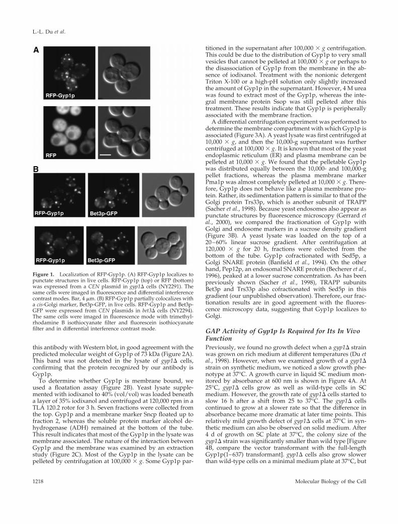
Gyp1p Localizes to Golgi ApparatusTo examine the localization of Gyp1p in yeast, we added agreen fluorescent protein (GFP) tag to the N terminus ofGyp1p. The GFP-tagged Gyp1p (GFP-Gyp1p) expressedfrom the GYP1 promoter showed punctate cytoplasmic lo-calization in live cells (our unpublished observation). Thisdistribution pattern of GFP-Gyp1p was very sensitive tofixation. Once cells were fixed, only evenly diffuse cytoplas-mic fluorescence was observed. This sensitivity to fixationwas not compatible with colocalization studies that rely onimmunofluorescence staining. To perform colocalization ex-periments, we tagged Gyp1p with red fluorescent protein(RFP, i.e., DsRed; CLONTECH). Because of the low intensityof RFP fluorescence, RFP-tagged Gyp1p (RFP-Gyp1p) had tobe expressed at 20 times higher than the endogenous Gyp1plevel (our unpublished observation). Nevertheless, RFP-Gyp1p showed the same punctate localization as GFP-Gyp1p, whereas RFP alone gave only a diffuse signal (Figure1A). Both GFP-Gyp1p and RFP-Gyp1p are fully functionalas determined by a plate assay that will be described below.
Because the punctate localization of Gyp1p is similar tothe localization pattern of yeast Golgi proteins, we per-formed double labeling experiments by using RFP-taggedGyp1p and a known resident of the Golgi apparatus, GFP-tagged Bet3p (Bet3p-GFP) (Sacher et al., 1998). Bet3p is asubunit of yeast transport protein particle complex (TRAPP)localized to the cis-Golgi (Sacher et al., 1998; Barrowman etal., 2000). In cells expressing both RFP-Gyp1p and Bet3p-GFP, the distribution of the RFP signal significantly over-lapped with that of the GFP signal (Figure 1B). We countedfluorescent spots in 45 cells. There were 228 Gyp1p-positivespots and 202 Bet3p-positive spots. Among them, 140 spotswere labeled by both GFP and RFP. In other words, 61% ofthe RFP-Gyp1 spots were labeled by Bet3-GFP, and 69% ofthe Bet3-GFP spots were labeled by RFP-Gyp1. The substan-tial colocalization of Gyp1p and Bet3p indicates that Gyp1pat least partially localizes to Golgi. It is noteworthy that thepunctate localization of Bet3-GFP also disappears upon fix-ation, suggesting that this sensitivity to fixation may be aproperty of many peripherally bound Golgi proteins.
To use an independent approach to assess the localizationof Gyp1p, we carried out subcellular fractionation studies.First, we prepared affinity-purified anti-Gyp1p antibody andexamined its specificity. When wild-type yeast lysate wasseparated by SDS-PAGE, a 70-kDa band was detected by
Gyp1p Is a Negative Regulator of Ypt1p
Vol. 12, May 2001 1217
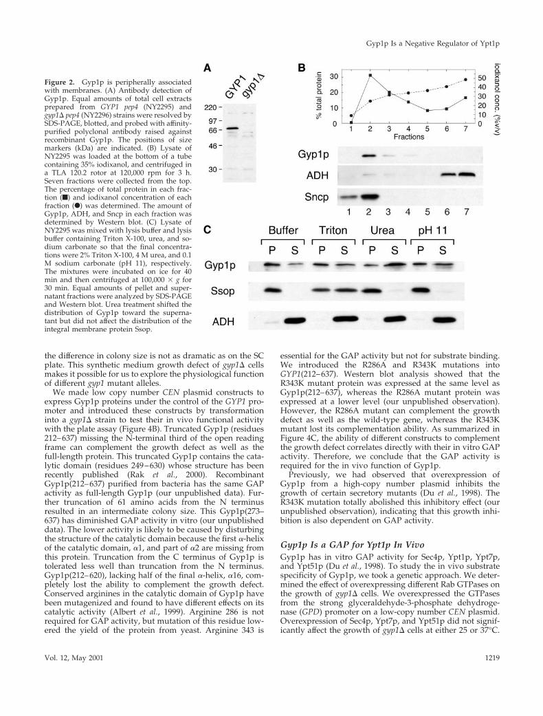
this antibody with Western blot, in good agreement with thepredicted molecular weight of Gyp1p of 73 kDa (Figure 2A).This band was not detected in the lysate of gyp1D cells,confirming that the protein recognized by our antibody isGyp1p.
To determine whether Gyp1p is membrane bound, weused a floatation assay (Figure 2B). Yeast lysate supple-mented with iodixanol to 40% (vol/vol) was loaded beneatha layer of 35% iodixanol and centrifuged at 120,000 rpm in aTLA 120.2 rotor for 3 h. Seven fractions were collected fromthe top. Gyp1p and a membrane marker Sncp floated up tofraction 2, whereas the soluble protein marker alcohol de-hydrogenase (ADH) remained at the bottom of the tube.This result indicates that most of the Gyp1p in the lysate wasmembrane associated. The nature of the interaction betweenGyp1p and the membrane was examined by an extractionstudy (Figure 2C). Most of the Gyp1p in the lysate can bepelleted by centrifugation at 100,000 3 g. Some Gyp1p par-
titioned in the supernatant after 100,000 3 g centrifugation.This could be due to the distribution of Gyp1p to very smallvesicles that cannot be pelleted at 100,000 3 g or perhaps tothe disassociation of Gyp1p from the membrane in the ab-sence of iodixanol. Treatment with the nonionic detergentTriton X-100 or a high-pH solution only slightly increasedthe amount of Gyp1p in the supernatant. However, 4 M ureawas found to extract most of the Gyp1p, whereas the inte-gral membrane protein Ssop was still pelleted after thistreatment. These results indicate that Gyp1p is peripherallyassociated with the membrane fraction.
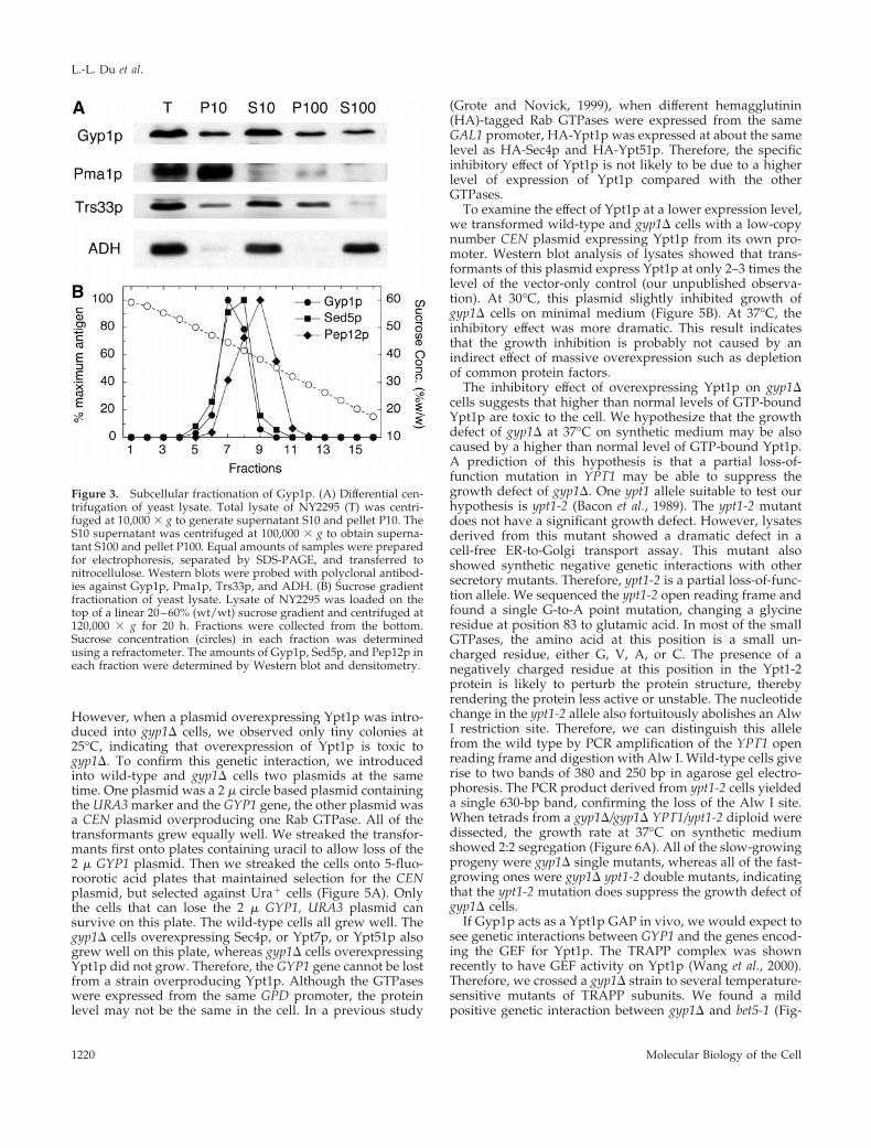
A differential centrifugation experiment was performed todetermine the membrane compartment with which Gyp1p isassociated (Figure 3A). A yeast lysate was first centrifuged at10,000 3 g, and then the 10,000-g supernatant was furthercentrifuged at 100,000 3 g. It is known that most of the yeastendoplasmic reticulum (ER) and plasma membrane can bepelleted at 10,000 3 g. We found that the pelletable Gyp1pwas distributed equally between the 10,000- and 100,000-gpellet fractions, whereas the plasma membrane markerPma1p was almost completely pelleted at 10,000 3 g. There-fore, Gyp1p does not behave like a plasma membrane pro-tein. Rather, its sedimentation pattern is similar to that of theGolgi protein Trs33p, which is another subunit of TRAPP(Sacher et al., 1998). Because yeast endosomes also appear aspunctate structures by fluorescence microscopy (Gerrard etal., 2000), we compared the fractionation of Gyp1p withGolgi and endosome markers in a sucrose density gradient(Figure 3B). A yeast lysate was loaded on the top of a20–60% linear sucrose gradient. After centrifugation at120,000 3 g for 20 h, fractions were collected from thebottom of the tube. Gyp1p cofractionated with Sed5p, aGolgi SNARE protein (Banfield et al., 1994). On the otherhand, Pep12p, an endosomal SNARE protein (Becherer et al.,1996), peaked at a lower sucrose concentration. As has beenpreviously shown (Sacher et al., 1998), TRAPP subunitsBet3p and Trs33p also cofractionated with Sed5p in thisgradient (our unpublished observation). Therefore, our frac-tionation results are in good agreement with the fluores-cence microscopy data, suggesting that Gyp1p localizes toGolgi.
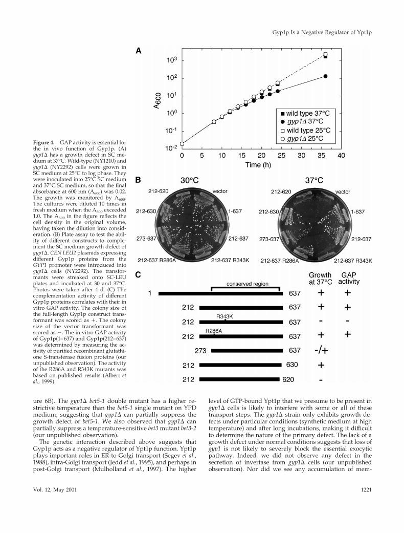
GAP Activity of Gyp1p Is Required for Its In VivoFunctionPreviously, we found no growth defect when a gyp1D strainwas grown on rich medium at different temperatures (Du etal., 1998). However, when we examined growth of a gyp1Dstrain on synthetic medium, we noticed a slow growth phe-notype at 37°C. A growth curve in liquid SC medium mon-itored by absorbance at 600 nm is shown in Figure 4A. At25°C, gyp1D cells grow as well as wild-type cells in SCmedium. However, the growth rate of gyp1D cells started toslow 16 h after a shift from 25 to 37°C. The gyp1D cellscontinued to grow at a slower rate so that the difference inabsorbance became more dramatic at later time points. Thisrelatively mild growth defect of gyp1D cells at 37°C in syn-thetic medium can also be observed on solid medium. After4 d of growth on SC plate at 37°C, the colony size of thegyp1D strain was significantly smaller than wild type [Figure4B, compare the vector transformant with the full-lengthGyp1p(1–637) transformant]. gyp1D cells also grow slowerthan wild-type cells on a minimal medium plate at 37°C, but
Figure 1. Localization of RFP-Gyp1p. (A) RFP-Gyp1p localizes topunctate structures in live cells. RFP-Gyp1p (top) or RFP (bottom)was expressed from a CEN plasmid in gyp1D cells (NY2291). Thesame cells were imaged in fluorescence and differential interferencecontrast modes. Bar, 4 mm. (B) RFP-Gyp1p partially colocalizes witha cis-Golgi marker, Bet3p-GFP, in live cells. RFP-Gyp1p and Bet3p-GFP were expressed from CEN plasmids in bet3D cells (NY2294).The same cells were imaged in fluorescence mode with trimethyl-rhodamine B isothiocyanate filter and fluorescein isothiocyanatefilter and in differential interference contrast mode.
L.-L. Du et al.
Molecular Biology of the Cell1218
the difference in colony size is not as dramatic as on the SCplate. This synthetic medium growth defect of gyp1D cellsmakes it possible for us to explore the physiological functionof different gyp1 mutant alleles.
We made low copy number CEN plasmid constructs toexpress Gyp1p proteins under the control of the GYP1 pro-moter and introduced these constructs by transformationinto a gyp1D strain to test their in vivo functional activitywith the plate assay (Figure 4B). Truncated Gyp1p (residues212–637) missing the N-terminal third of the open readingframe can complement the growth defect as well as thefull-length protein. This truncated Gyp1p contains the cata-lytic domain (residues 249–630) whose structure has beenrecently published (Rak et al., 2000). RecombinantGyp1p(212–637) purified from bacteria has the same GAPactivity as full-length Gyp1p (our unpublished data). Fur-ther truncation of 61 amino acids from the N terminusresulted in an intermediate colony size. This Gyp1p(273–637) has diminished GAP activity in vitro (our unpublisheddata). The lower activity is likely to be caused by disturbingthe structure of the catalytic domain because the first a-helixof the catalytic domain, a1, and part of a2 are missing fromthis protein. Truncation from the C terminus of Gyp1p istolerated less well than truncation from the N terminus.Gyp1p(212–620), lacking half of the final a-helix, a16, com-pletely lost the ability to complement the growth defect.Conserved arginines in the catalytic domain of Gyp1p havebeen mutagenized and found to have different effects on itscatalytic activity (Albert et al., 1999). Arginine 286 is notrequired for GAP activity, but mutation of this residue low-ered the yield of the protein from yeast. Arginine 343 is
essential for the GAP activity but not for substrate binding.We introduced the R286A and R343K mutations intoGYP1(212–637). Western blot analysis showed that theR343K mutant protein was expressed at the same level asGyp1p(212–637), whereas the R286A mutant protein wasexpressed at a lower level (our unpublished observation).However, the R286A mutant can complement the growthdefect as well as the wild-type gene, whereas the R343Kmutant lost its complementation ability. As summarized inFigure 4C, the ability of different constructs to complementthe growth defect correlates directly with their in vitro GAPactivity. Therefore, we conclude that the GAP activity isrequired for the in vivo function of Gyp1p.
Previously, we had observed that overexpression ofGyp1p from a high-copy number plasmid inhibits thegrowth of certain secretory mutants (Du et al., 1998). TheR343K mutation totally abolished this inhibitory effect (ourunpublished observation), indicating that this growth inhi-bition is also dependent on GAP activity.
Gyp1p Is a GAP for Ypt1p In VivoGyp1p has in vitro GAP activity for Sec4p, Ypt1p, Ypt7p,and Ypt51p (Du et al., 1998). To study the in vivo substratespecificity of Gyp1p, we took a genetic approach. We deter-mined the effect of overexpressing different Rab GTPases onthe growth of gyp1D cells. We overexpressed the GTPasesfrom the strong glyceraldehyde-3-phosphate dehydroge-nase (GPD) promoter on a low-copy number CEN plasmid.Overexpression of Sec4p, Ypt7p, and Ypt51p did not signif-icantly affect the growth of gyp1D cells at either 25 or 37°C.
Figure 2. Gyp1p is peripherally associatedwith membranes. (A) Antibody detection ofGyp1p. Equal amounts of total cell extractsprepared from GYP1 pep4 (NY2295) andgyp1D pep4 (NY2296) strains were resolved bySDS-PAGE, blotted, and probed with affinity-purified polyclonal antibody raised againstrecombinant Gyp1p. The positions of sizemarkers (kDa) are indicated. (B) Lysate ofNY2295 was loaded at the bottom of a tubecontaining 35% iodixanol, and centrifuged ina TLA 120.2 rotor at 120,000 rpm for 3 h.Seven fractions were collected from the top.The percentage of total protein in each frac-tion (f) and iodixanol concentration of eachfraction (F) was determined. The amount ofGyp1p, ADH, and Sncp in each fraction wasdetermined by Western blot. (C) Lysate ofNY2295 was mixed with lysis buffer and lysisbuffer containing Triton X-100, urea, and so-dium carbonate so that the final concentra-tions were 2% Triton X-100, 4 M urea, and 0.1M sodium carbonate (pH 11), respectively.The mixtures were incubated on ice for 40min and then centrifuged at 100,000 3 g for30 min. Equal amounts of pellet and super-natant fractions were analyzed by SDS-PAGEand Western blot. Urea treatment shifted thedistribution of Gyp1p toward the superna-tant but did not affect the distribution of theintegral membrane protein Ssop.
Gyp1p Is a Negative Regulator of Ypt1p
Vol. 12, May 2001 1219
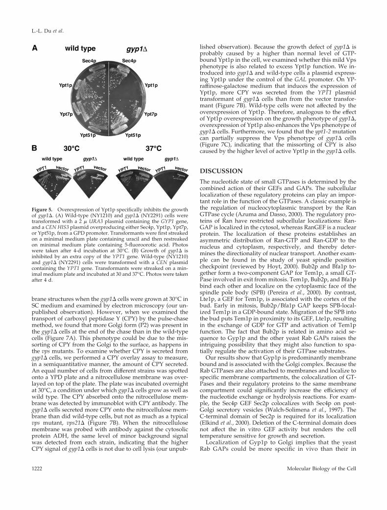
However, when a plasmid overexpressing Ypt1p was intro-duced into gyp1D cells, we observed only tiny colonies at25°C, indicating that overexpression of Ypt1p is toxic togyp1D. To confirm this genetic interaction, we introducedinto wild-type and gyp1D cells two plasmids at the sametime. One plasmid was a 2 m circle based plasmid containingthe URA3 marker and the GYP1 gene, the other plasmid wasa CEN plasmid overproducing one Rab GTPase. All of thetransformants grew equally well. We streaked the transfor-mants first onto plates containing uracil to allow loss of the2 m GYP1 plasmid. Then we streaked the cells onto 5-fluo-roorotic acid plates that maintained selection for the CENplasmid, but selected against Ura1 cells (Figure 5A). Onlythe cells that can lose the 2 m GYP1, URA3 plasmid cansurvive on this plate. The wild-type cells all grew well. Thegyp1D cells overexpressing Sec4p, or Ypt7p, or Ypt51p alsogrew well on this plate, whereas gyp1D cells overexpressingYpt1p did not grow. Therefore, the GYP1 gene cannot be lostfrom a strain overproducing Ypt1p. Although the GTPaseswere expressed from the same GPD promoter, the proteinlevel may not be the same in the cell. In a previous study
(Grote and Novick, 1999), when different hemagglutinin(HA)-tagged Rab GTPases were expressed from the sameGAL1 promoter, HA-Ypt1p was expressed at about the samelevel as HA-Sec4p and HA-Ypt51p. Therefore, the specificinhibitory effect of Ypt1p is not likely to be due to a higherlevel of expression of Ypt1p compared with the otherGTPases.
To examine the effect of Ypt1p at a lower expression level,we transformed wild-type and gyp1D cells with a low-copynumber CEN plasmid expressing Ypt1p from its own pro-moter. Western blot analysis of lysates showed that trans-formants of this plasmid express Ypt1p at only 2–3 times thelevel of the vector-only control (our unpublished observa-tion). At 30°C, this plasmid slightly inhibited growth ofgyp1D cells on minimal medium (Figure 5B). At 37°C, theinhibitory effect was more dramatic. This result indicatesthat the growth inhibition is probably not caused by anindirect effect of massive overexpression such as depletionof common protein factors.
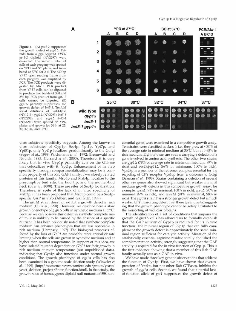
The inhibitory effect of overexpressing Ypt1p on gyp1Dcells suggests that higher than normal levels of GTP-boundYpt1p are toxic to the cell. We hypothesize that the growthdefect of gyp1D at 37°C on synthetic medium may be alsocaused by a higher than normal level of GTP-bound Ypt1p.A prediction of this hypothesis is that a partial loss-of-function mutation in YPT1 may be able to suppress thegrowth defect of gyp1D. One ypt1 allele suitable to test ourhypothesis is ypt1-2 (Bacon et al., 1989). The ypt1-2 mutantdoes not have a significant growth defect. However, lysatesderived from this mutant showed a dramatic defect in acell-free ER-to-Golgi transport assay. This mutant alsoshowed synthetic negative genetic interactions with othersecretory mutants. Therefore, ypt1-2 is a partial loss-of-func-tion allele. We sequenced the ypt1-2 open reading frame andfound a single G-to-A point mutation, changing a glycineresidue at position 83 to glutamic acid. In most of the smallGTPases, the amino acid at this position is a small un-charged residue, either G, V, A, or C. The presence of anegatively charged residue at this position in the Ypt1-2protein is likely to perturb the protein structure, therebyrendering the protein less active or unstable. The nucleotidechange in the ypt1-2 allele also fortuitously abolishes an AlwI restriction site. Therefore, we can distinguish this allelefrom the wild type by PCR amplification of the YPT1 openreading frame and digestion with Alw I. Wild-type cells giverise to two bands of 380 and 250 bp in agarose gel electro-phoresis. The PCR product derived from ypt1-2 cells yieldeda single 630-bp band, confirming the loss of the Alw I site.When tetrads from a gyp1D/gyp1D YPT1/ypt1-2 diploid weredissected, the growth rate at 37°C on synthetic mediumshowed 2:2 segregation (Figure 6A). All of the slow-growingprogeny were gyp1D single mutants, whereas all of the fast-growing ones were gyp1D ypt1-2 double mutants, indicatingthat the ypt1-2 mutation does suppress the growth defect ofgyp1D cells.
If Gyp1p acts as a Ypt1p GAP in vivo, we would expect tosee genetic interactions between GYP1 and the genes encod-ing the GEF for Ypt1p. The TRAPP complex was shownrecently to have GEF activity on Ypt1p (Wang et al., 2000).Therefore, we crossed a gyp1D strain to several temperature-sensitive mutants of TRAPP subunits. We found a mildpositive genetic interaction between gyp1D and bet5-1 (Fig-
Figure 3. Subcellular fractionation of Gyp1p. (A) Differential cen-trifugation of yeast lysate. Total lysate of NY2295 (T) was centri-fuged at 10,000 3 g to generate supernatant S10 and pellet P10. TheS10 supernatant was centrifuged at 100,000 3 g to obtain superna-tant S100 and pellet P100. Equal amounts of samples were preparedfor electrophoresis, separated by SDS-PAGE, and transferred tonitrocellulose. Western blots were probed with polyclonal antibod-ies against Gyp1p, Pma1p, Trs33p, and ADH. (B) Sucrose gradientfractionation of yeast lysate. Lysate of NY2295 was loaded on thetop of a linear 20–60% (wt/wt) sucrose gradient and centrifuged at120,000 3 g for 20 h. Fractions were collected from the bottom.Sucrose concentration (circles) in each fraction was determinedusing a refractometer. The amounts of Gyp1p, Sed5p, and Pep12p ineach fraction were determined by Western blot and densitometry.
L.-L. Du et al.
Molecular Biology of the Cell1220
ure 6B). The gyp1D bet5-1 double mutant has a higher re-strictive temperature than the bet5-1 single mutant on YPDmedium, suggesting that gyp1D can partially suppress thegrowth defect of bet5-1. We also observed that gyp1D canpartially suppress a temperature-sensitive bet3 mutant bet3-2(our unpublished observation).
The genetic interaction described above suggests thatGyp1p acts as a negative regulator of Ypt1p function. Ypt1pplays important roles in ER-to-Golgi transport (Segev et al.,1988), intra-Golgi transport (Jedd et al., 1995), and perhaps inpost-Golgi transport (Mulholland et al., 1997). The higher
level of GTP-bound Ypt1p that we presume to be present ingyp1D cells is likely to interfere with some or all of thesetransport steps. The gyp1D strain only exhibits growth de-fects under particular conditions (synthetic medium at hightemperature) and after long incubations, making it difficultto determine the nature of the primary defect. The lack of agrowth defect under normal conditions suggests that loss ofgyp1 is not likely to severely block the essential exocyticpathway. Indeed, we did not observe any defect in thesecretion of invertase from gyp1D cells (our unpublishedobservation). Nor did we see any accumulation of mem-
Figure 4. GAP activity is essential forthe in vivo function of Gyp1p. (A)gyp1D has a growth defect in SC me-dium at 37°C. Wild-type (NY1210) andgyp1D (NY2292) cells were grown inSC medium at 25°C to log phase. Theywere inoculated into 25°C SC mediumand 37°C SC medium, so that the finalabsorbance at 600 nm (A600) was 0.02.The growth was monitored by A600.The cultures were diluted 10 times infresh medium when the A600 exceeded1.0. The A600 in the figure reflects thecell density in the original volume,having taken the dilution into consid-eration. (B) Plate assay to test the abil-ity of different constructs to comple-ment the SC medium growth defect ofgyp1D. CEN LEU2 plasmids expressingdifferent Gyp1p proteins from theGYP1 promoter were introduced intogyp1D cells (NY2292). The transfor-mants were streaked onto SC-LEUplates and incubated at 30 and 37°C.Photos were taken after 4 d. (C) Thecomplementation activity of differentGyp1p proteins correlates with their invitro GAP activity. The colony size ofthe full-length Gyp1p construct trans-formant was scored as 1. The colonysize of the vector transformant wasscored as 2. The in vitro GAP activityof Gyp1p(1–637) and Gyp1p(212–637)was determined by measuring the ac-tivity of purified recombinant glutathi-one S-transferase fusion proteins (ourunpublished observation). The activityof the R286A and R343K mutants wasbased on published results (Albert etal., 1999).
Gyp1p Is a Negative Regulator of Ypt1p
Vol. 12, May 2001 1221
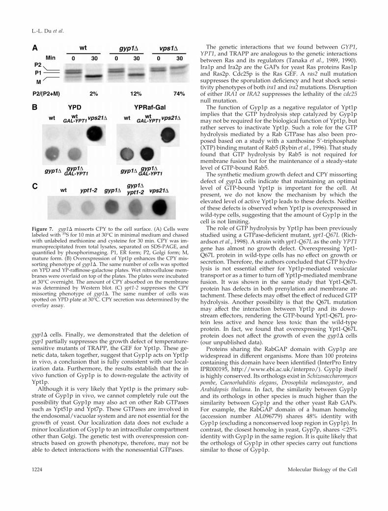
brane structures when the gyp1D cells were grown at 30°C inSC medium and examined by electron microscopy (our un-published observation). However, when we examined thetransport of carboxyl peptidase Y (CPY) by the pulse-chasemethod, we found that more Golgi form (P2) was present inthe gyp1D cells at the end of the chase than in the wild-typecells (Figure 7A). This phenotype could be due to the mis-sorting of CPY from the Golgi to the surface, as happens inthe vps mutants. To examine whether CPY is secreted fromgyp1D cells, we performed a CPY overlay assay to measure,in a semiquantitative manner, the amount of CPY secreted.An equal number of cells from different strains was spottedonto a YPD plate and a nitrocellulose membrane was over-layed on top of the plate. The plate was incubated overnightat 30°C, a condition under which gyp1D cells grow as well aswild type. The CPY absorbed onto the nitrocellulose mem-brane was detected by immunoblot with CPY antibody. Thegyp1D cells secreted more CPY onto the nitrocellulose mem-brane than did wild-type cells, but not as much as a typicalvps mutant, vps21D (Figure 7B). When the nitrocellulosemembrane was probed with antibody against the cytosolicprotein ADH, the same level of minor background signalwas detected from each strain, indicating that the higherCPY signal of gyp1D cells is not due to cell lysis (our unpub-
lished observation). Because the growth defect of gyp1D isprobably caused by a higher than normal level of GTP-bound Ypt1p in the cell, we examined whether this mild Vpsphenotype is also related to excess Ypt1p function. We in-troduced into gyp1D and wild-type cells a plasmid express-ing Ypt1p under the control of the GAL promoter. On YP-raffinose-galactose medium that induces the expression ofYpt1p, more CPY was secreted from the YPT1 plasmidtransformant of gyp1D cells than from the vector transfor-mant (Figure 7B). Wild-type cells were not affected by theoverexpression of Ypt1p. Therefore, analogous to the effectof Ypt1p overexpression on the growth phenotype of gyp1D,overexpression of Ypt1p also enhances the Vps phenotype ofgyp1D cells. Furthermore, we found that the ypt1-2 mutationcan partially suppress the Vps phenotype of gyp1D cells(Figure 7C), indicating that the missorting of CPY is alsocaused by the higher level of active Ypt1p in the gyp1D cells.
DISCUSSION
The nucleotide state of small GTPases is determined by thecombined action of their GEFs and GAPs. The subcellularlocalization of these regulatory proteins can play an impor-tant role in the function of the GTPases. A classic example isthe regulation of nucleocytoplasmic transport by the RanGTPase cycle (Azuma and Dasso, 2000). The regulatory pro-teins of Ran have restricted subcellular localizations: Ran-GAP is localized in the cytosol, whereas RanGEF is a nuclearprotein. The localization of these proteins establishes anasymmetric distribution of Ran-GTP and Ran-GDP to thenucleus and cytoplasm, respectively, and thereby deter-mines the directionality of nuclear transport. Another exam-ple can be found in the study of yeast spindle positioncheckpoint (reviewed by Hoyt, 2000). Bub2p and Bfa1p to-gether form a two-component GAP for Tem1p, a small GT-Pase involved in exit from mitosis. Tem1p, Bub2p, and Bfa1pbind each other and localize on the cytoplasmic face of thespindle pole body (SPB) (Pereira et al., 2000). By contrast,Lte1p, a GEF for Tem1p, is associated with the cortex of thebud. Early in mitosis, Bub2p/Bfa1p GAP keeps SPB-local-ized Tem1p in a GDP-bound state. Migration of the SPB intothe bud puts Tem1p in proximity to its GEF, Lte1p, resultingin the exchange of GDP for GTP and activation of Tem1pfunction. The fact that Bub2p is related in amino acid se-quence to Gyp1p and the other yeast Rab GAPs raises theintriguing possibility that they might also function to spa-tially regulate the activation of their GTPase substrates.
Our results show that Gyp1p is predominantly membranebound and is associated with the Golgi complex. Because theRab GTPases are also attached to membranes and localize tospecific membrane compartments, the colocalization of GT-Pases and their regulatory proteins to the same membranecompartment could significantly increase the efficiency ofthe nucleotide exchange or hydrolysis reactions. For exam-ple, the Sec4p GEF Sec2p colocalizes with Sec4p on post-Golgi secretory vesicles (Walch-Solimena et al., 1997). TheC-terminal domain of Sec2p is required for its localization(Elkind et al., 2000). Deletion of the C-terminal domain doesnot affect the in vitro GEF activity but renders the celltemperature sensitive for growth and secretion.
Localization of Gyp1p to Golgi implies that the yeastRab GAPs could be more specific in vivo than their in
Figure 5. Overexpression of Ypt1p specifically inhibits the growthof gyp1D. (A) Wild-type (NY1210) and gyp1D (NY2291) cells weretransformed with a 2 m URA3 plasmid containing the GYP1 gene,and a CEN HIS3 plasmid overproducing either Sec4p, Ypt1p, Ypt7p,or Ypt51p, from a GPD promoter. Transformants were first streakedon a minimal medium plate containing uracil and then restreakedon minimal medium plate containing 5-fluoroorotic acid. Photoswere taken after 4-d incubation at 30°C. (B) Growth of gyp1D isinhibited by an extra copy of the YPT1 gene. Wild-type (NY1210)and gyp1D (NY2291) cells were transformed with a CEN plasmidcontaining the YPT1 gene. Transformants were streaked on a min-imal medium plate and incubated at 30 and 37°C. Photos were takenafter 4 d.
L.-L. Du et al.
Molecular Biology of the Cell1222
vitro substrate specificity suggests. Among the known invitro substrates of Gyp1p, Sec4p, Ypt1p, Ypt7p, andYpt51p, only Ypt1p localizes predominantly to the Golgi(Segev et al., 1988; Wichmann et al., 1992; Brennwald andNovick, 1993; Gerrard et al., 2000). Therefore, it is verylikely that in vivo Gyp1p primarily acts on the GTPasethat colocalizes with it, Ypt1p. Enhancement of in vivospecificity through compartmentalization may be a com-mon property of this Rab GAP family. Two closely relatedproteins of this family, Msb3p and Msb4p, localize to thepresumptive bud site, the bud tip, and the mother-budneck (Bi et al., 2000). These are sites of Sec4p localization.Therefore, in spite of the lack of in vitro specificity ofMsb3p, it has been proposed that Msb3p could be a Sec4p-specific GAP in vivo (Albert and Gallwitz, 1999).
The gyp1D strain does not exhibit a growth defect in richmedium (Du et al., 1998). However, we describe here a slowgrowth phenotype of gyp1D cells in synthetic medium at 37°C.Because we can observe this defect in synthetic complete me-dium, it is unlikely to be caused by the absence of a specificnutrient. It has been previously noted that synthetic completemedium can enhance phenotypes that are less noticeable inrich medium (Hampsey, 1997). The biological processes af-fected by the loss of GYP1 are probably more critical or ratelimiting when the cells are grown in synthetic medium and athigher than normal temperature. In support of this idea, wehave isolated mutants dependent on GYP1 for their growth inrich medium at room temperature (our unpublished data),indicating that Gyp1p also functions under normal growthconditions. The growth phenotype of gyp1D cells has alsobeen examined in a genome-scale deletion study (Winzeler etal., 1999) (http://sequence-www.stanford.edu/group/yeast/yeast_deletion_project/Enter_function.html). In that study, thegrowth rates of homozygous diploid null mutants of 558 non-
essential genes were examined in a competitive growth assay.Ten strains were classified as class 0, i.e., they grew at ,80% ofthe average rate in minimal medium at 30°C, but at .95% inrich medium. Eight of them are strains carrying a deletion of agene involved in amino acid synthesis. The other two strainsare gyp1D (78% of average rate in minimum medium, 99% inrich) and vps29/pep11D (69% in minimum, 100% in rich).Vps29p is a member of the retromer complex essential for therecycling of CPY receptor Vps10p from endosomes to Golgi(Seaman et al., 1998). Strains containing a deletion of severalother vps genes also showed significant but weaker minimalmedium growth defects in this competitive growth assay; forexample, vps5D (91% in minimal, 100% in rich), vps8D (90% inminimal, 99% in rich), and vps21D (91% in minimal, 98% inrich). The gyp1D strain has a stronger growth defect but a muchweaker CPY missorting defect than these vps mutants, suggest-ing that the growth phenotype cannot be solely attributed tothe missorting of vacuolar proteins.
The identification of a set of conditions that impairs thegrowth of gyp1D cells has allowed us to formally establishthat the GAP activity of Gyp1p is required for its in vivofunction. The minimal region of Gyp1p that can fully com-plement the growth defect is approximately the same min-imal region sufficient for catalytic activity. Mutation of thecatalytically essential arginine residue totally abolished thecomplementation activity, strongly suggesting that the GAPactivity is required for the in vivo function of Gyp1p. This isthe first evidence showing that a member of this Rab GAPfamily actually acts as a GAP in vivo.
We have made three key genetic observations that addressthe function of Gyp1p. First, we have shown that overex-pression of Ypt1p, but not other Rab GTPases, inhibits thegrowth of gyp1D cells. Second, we found that a partial loss-of-function allele of ypt1 suppresses the growth defect of
Figure 6. (A) ypt1-2 suppressesthe growth defect of gyp1D. Tet-rads from a gyp1D/gyp1D YPT1/ypt1-2 diploid (NY2297) weredissected. The same number ofcells of each progeny was spottedon YPD and SC plates and incu-bated at 37°C for 2 d. The 630-bpYPT1 open reading frame fromeach progeny was amplified byPCR. The PCR products were di-gested by Alw I. PCR productfrom YPT1 cells can be digestedto produce two bands of 380 and250 bp. PCR product from ypt1-2cells cannot be digested. (B)gyp1D partially suppresses thegrowth defect of bet5-1. Tenfoldserial dilutions of wild-type(NY1211), gyp1D (NY2293), bet5-1(NY2298), and gyp1D bet5-1(NY2299) were spotted on YPDplates and grown for 36 h at 25,30, 32, 34, and 37°C.
Gyp1p Is a Negative Regulator of Ypt1p
Vol. 12, May 2001 1223
gyp1D cells. Finally, we demonstrated that the deletion ofgyp1 partially suppresses the growth defect of temperature-sensitive mutants of TRAPP, the GEF for Ypt1p. These ge-netic data, taken together, suggest that Gyp1p acts on Ypt1pin vivo, a conclusion that is fully consistent with our local-ization data. Furthermore, the results establish that the invivo function of Gyp1p is to down-regulate the activity ofYpt1p.
Although it is very likely that Ypt1p is the primary sub-strate of Gyp1p in vivo, we cannot completely rule out thepossibility that Gyp1p may also act on other Rab GTPasessuch as Ypt51p and Ypt7p. These GTPases are involved inthe endosomal/vacuolar system and are not essential for thegrowth of yeast. Our localization data does not exclude aminor localization of Gyp1p to an intracellular compartmentother than Golgi. The genetic test with overexpression con-structs based on growth phenotype, therefore, may not beable to detect interactions with the nonessential GTPases.
The genetic interactions that we found between GYP1,YPT1, and TRAPP are analogous to the genetic interactionsbetween Ras and its regulators (Tanaka et al., 1989, 1990).Ira1p and Ira2p are the GAPs for yeast Ras proteins Ras1pand Ras2p. Cdc25p is the Ras GEF. A ras2 null mutationsuppresses the sporulation deficiency and heat shock sensi-tivity phenotypes of both ira1 and ira2 mutations. Disruptionof either IRA1 or IRA2 suppresses the lethality of the cdc25null mutation.
The function of Gyp1p as a negative regulator of Ypt1pimplies that the GTP hydrolysis step catalyzed by Gyp1pmay not be required for the biological function of Ypt1p, butrather serves to inactivate Ypt1p. Such a role for the GTPhydrolysis mediated by a Rab GTPase has also been pro-posed based on a study with a xanthosine 59-triphosphate(XTP) binding mutant of Rab5 (Rybin et al., 1996). That studyfound that GTP hydrolysis by Rab5 is not required formembrane fusion but for the maintenance of a steady-statelevel of GTP-bound Rab5.
The synthetic medium growth defect and CPY missortingdefect of gyp1D cells indicate that maintaining an optimallevel of GTP-bound Ypt1p is important for the cell. Atpresent, we do not know the mechanism by which theelevated level of active Ypt1p leads to these defects. Neitherof these defects is observed when Ypt1p is overexpressed inwild-type cells, suggesting that the amount of Gyp1p in thecell is not limiting.
The role of GTP hydrolysis by Ypt1p has been previouslystudied using a GTPase-deficient mutant, ypt1-Q67L (Rich-ardson et al., 1998). A strain with ypt1-Q67L as the only YPT1gene has almost no growth defect. Overexpressing Ypt1-Q67L protein in wild-type cells has no effect on growth orsecretion. Therefore, the authors concluded that GTP hydro-lysis is not essential either for Ypt1p-mediated vesiculartransport or as a timer to turn off Ypt1p-mediated membranefusion. It was shown in the same study that Ypt1-Q67Lprotein has defects in both prenylation and membrane at-tachment. These defects may offset the effect of reduced GTPhydrolysis. Another possibility is that the Q67L mutationmay affect the interaction between Ypt1p and its down-stream effectors, rendering the GTP-bound Ypt1-Q67L pro-tein less active and hence less toxic than the wild-typeprotein. In fact, we found that overexpressing Ypt1-Q67Lprotein does not affect the growth of even the gyp1D cells(our unpublished data).
Proteins sharing the RabGAP domain with Gyp1p arewidespread in different organisms. More than 100 proteinscontaining this domain have been identified (InterPro EntryIPR000195, http://www.ebi.ac.uk/interpro/). Gyp1p itselfis highly conserved. Its orthologs exist in Schizosaccharomycespombe, Caenorhabditis elegans, Drosophila melanogaster, andArabidopsis thaliana. In fact, the similarity between Gyp1pand its orthologs in other species is much higher than thesimilarity between Gyp1p and the other yeast Rab GAPs.For example, the RabGAP domain of a human homolog(accession number AL096779) shares 48% identity withGyp1p (excluding a nonconserved loop region in Gyp1p). Incontrast, the closest homolog in yeast, Gyp7p, shares ,25%identity with Gyp1p in the same region. It is quite likely thatthe orthologs of Gyp1p in other species carry out functionssimilar to those of Gyp1p.
Figure 7. gyp1D missorts CPY to the cell surface. (A) Cells werelabeled with 35S for 10 min at 30°C in minimal medium and chasedwith unlabeled methionine and cysteine for 30 min. CPY was im-munoprecipitated from total lysates, separated on SDS-PAGE, andquantified by phosphorimaging. P1, ER form; P2, Golgi form; M,mature form. (B) Overexpression of Ypt1p enhances the CPY mis-sorting phenotype of gyp1D. The same number of cells was spottedon YPD and YP-raffinose-galactose plates. Wet nitrocellulose mem-branes were overlaid on top of the plates. The plates were incubatedat 30°C overnight. The amount of CPY absorbed on the membranewas determined by Western blot. (C) ypt1-2 suppresses the CPYmissorting phenotype of gyp1D. The same number of cells wasspotted on YPD plate at 30°C. CPY secretion was determined by theoverlay assay.
L.-L. Du et al.
Molecular Biology of the Cell1224
ACKNOWLEDGMENTS
We are grateful to Dr. Susan Ferro-Novick for advice and gener-ously providing plasmids, antibodies, and yeast strains. We thankJemima Barrowman and Eric Grote for the help with the sucrosegradient experiment. We thank Drs. T.H. Stevens, C.W. Slayman,and R. Piper for plasmids and antibodies. We appreciate the criticalreading of the manuscript by Drs. Wei Guo and Eric Grote. Thiswork was supported by grants from the National Institutes ofHealth to P.N.
REFERENCES
Albert, S., and Gallwitz, D. (1999). Two new members of a family ofYpt/Rab GTPase activating proteins. Promiscuity of substrate rec-ognition. J. Biol. Chem. 274, 33186–33189.
Albert, S., and Gallwitz, D. (2000). Msb4p, a protein involved inCdc42p-dependent organization of the actin cytoskeleton, is a Ypt/Rab-specific GAP. Biol. Chem. 381, 453–456.
Albert, S., Will, E., and Gallwitz, D. (1999). Identification of thecatalytic domains and their functionally critical arginine residues oftwo yeast GTPase-activating proteins specific for Ypt/Rab transportGTPases. EMBO J. 18, 5216–5225.
Azuma, Y., and Dasso, M. (2000). The role of Ran in nuclear func-tion. Curr. Opin. Cell Biol. 12, 302–307.
Bacon, R.A., Salminen, A., Ruohola, H., Novick, P., and Ferro-Novick, S. (1989). The GTP-binding protein Ypt1 is required fortransport in vitro: the Golgi apparatus is defective in ypt1 mutants.J. Cell Biol. 109, 1015–1022.
Banfield, D.K., Lewis, M.J., Rabouille, C., Warren, G., and Pelham,H.R. (1994). Localization of Sed5, a putative vesicle targeting mol-ecule, to the cis-Golgi network involves both its transmembrane andcytoplasmic domains. J. Cell Biol. 127, 357–371.
Barrowman, J., Sacher, M., and Ferro-Novick, S. (2000). TRAPPstably associates with the Golgi and is required for vesicle docking.EMBO J. 19, 862–869.
Becherer, K.A., Rieder, S.E., Emr, S.D., and Jones, E.W. (1996). Novelsyntaxin homologue, Pep12p, required for the sorting of lumenalhydrolases to the lysosome-like vacuole in yeast. Mol. Biol. Cell 7,579–594.
Bi, E., Chiavetta, J.B., Chen, H., Chen, G.C., Chan, C.S., and Pringle,J.R. (2000). Identification of novel, evolutionarily conserved Cdc42p-interacting proteins and of redundant pathways linking Cdc24p andCdc42p to actin polarization in yeast. Mol. Biol. Cell 11, 773–793.
Boles, E., and Miosga, T. (1995). A rapid and highly efficient methodfor PCR-based site-directed mutagenesis using only one newprimer. Curr. Genet. 28, 197–198.
Brennwald, P., and Novick, P. (1993). Interactions of three domainsdistinguishing the Ras-related GTP-binding proteins Ypt1 and Sec4.Nature 362, 560–563.
Cuif, M.H., Possmayer, F., Zander, H., Bordes, N., Jollivet, F.,Couedel-Courteille, A., Janoueix-Lerosey, I., Langsley, G., Bornens,M., and Goud, B. (1999). Characterization of GAPCenA, a GTPaseactivating protein for Rab6, part of which associates with the cen-trosome. EMBO J. 18, 1772–1782.
Du, L.L., Collins, R.N., and Novick, P.J. (1998). Identification of aSec4p GTPase-activating protein (GAP) as a novel member of a RabGAP family. J. Biol. Chem. 273, 3253–3256.
Elkind, N.B., Walch-Solimena, C., and Novick, P.J. (2000). The roleof the COOH terminus of Sec2p in the transport of post-Golgivesicles. J. Cell Biol. 149, 95–110.
Fukui, K., Sasaki, T., Imazumi, K., Matsuura, Y., Nakanishi, H., andTakai, Y. (1997). Isolation and characterization of a GTPase activat-
ing protein specific for the Rab3 subfamily of small G proteins.J. Biol. Chem. 272, 4655–4658.
Gerrard, S.R., Bryant, N.J., and Stevens, T.H. (2000). VPS21 controlsentry of endocytosed and biosynthetic proteins into the yeast pre-vacuolar compartment. Mol. Biol. Cell 11, 613–626.
Govindan, B., Bowser, R., and Novick, P. (1995). The role of Myo2,a yeast class V myosin, in vesicular transport. J. Cell Biol. 128,1055–1068.
Grote, E., and Novick, P.J. (1999). Promiscuity in Rab-SNARE inter-actions. Mol. Biol. Cell 10, 4149–4161.
Hampsey, M. (1997). A review of phenotypes in Saccharomyces cer-evisiae. Yeast 13, 1099–1133.
Hoyt, M.A. (2000). Exit from mitosis: spindle pole power. Cell 102,267–270.
Jedd, G., Richardson, C., Litt, R., and Segev, N. (1995). The Ypt1GTPase is essential for the first two steps of the yeast secretorypathway. J. Cell Biol. 131, 583–590.
Jones, E.W. (1991). Tackling the protease problem in Saccharomycescerevisiae. Methods Enzymol. 194, 428–453.
Jones, S., Richardson, C.J., Litt, R.J., and Segev, N. (1998). Identifi-cation of regulators for Ypt1 GTPase nucleotide cycling. Mol. Biol.Cell 9, 2819–2837.
Lazar, T., Gotte, M., and Gallwitz, D. (1997). Vesicular transport:how many Ypt/Rab-GTPases make a eukaryotic cell? Trends Bio-chem. Sci. 22, 468–472.
Liu, K., and Li, G. (1998). Catalytic domain of the p120 Ras GAPbinds to RAb5 and stimulates its GTPase activity. J. Biol. Chem. 273,10087–10090.
Mulholland, J., Wesp, A., Riezman, H., and Botstein, D. (1997). Yeastactin cytoskeleton mutants accumulate a new class of Golgi-derivedsecretary vesicle. Mol. Biol. Cell 8, 1481–1499.
Mumberg, D., Muller, R., and Funk, M. (1995). Yeast vectors for thecontrolled expression of heterologous proteins in different geneticbackgrounds. Gene 156, 119–122.
Neuwald, A.F. (1997). A shared domain between a spindle assemblycheckpoint protein and Ypt/Rab-specific GTPase-activators. TrendsBiochem. Sci. 22, 243–244.
Novick, P., and Zerial, M. (1997). The diversity of Rab proteins invesicle transport. Curr. Opin. Cell Biol. 9, 496–504.
Pereira, G., Hofken, T., Grindlay, J., Manson, C., and Schiebel, E.(2000). The Bub2p spindle checkpoint links nuclear migration withmitotic exit. Mol. Cell 6, 1–10.
Rak, A., Fedorov, R., Alexandrov, K., Albert, S., Goody, R.S., Gall-witz, D., and Scheidig, A.J. (2000). Crystal structure of the GAPdomain of Gyp1p: first insights into interaction with Ypt/Rab pro-teins. EMBO J. 19, 5105–5113.
Richardson, C.J., Jones, S., Litt, R.J., and Segev, N. (1998). GTPhydrolysis is not important for Ypt1 GTPase function in vesiculartransport. Mol. Cell. Biol. 18, 827–838.
Rybin, V., Ullrich, O., Rubino, M., Alexandrov, K., Simon, I., Seabra,M.C., Goody, R., and Zerial, M. (1996). GTPase activity of Rab5 actsas a timer for endocytic membrane fusion [see comments]. Nature383, 266–269.
Sacher, M., Jiang, Y., Barrowman, J., Scarpa, A., Burston, J., Zhang,L., Schieltz, D., Yates, J.R., 3rd, Abeliovich, H., and Ferro-Novick, S.(1998). TRAPP, a highly conserved novel complex on the cis-Golgithat mediates vesicle docking and fusion. EMBO J. 17, 2494–2503.
Schroder, M., Schafer, R., and Friedl, P. (1997). Spectrophotometricdetermination of iodixanol in subcellular fractions of mammaliancells. Anal. Biochem. 244, 174–176.
Gyp1p Is a Negative Regulator of Ypt1p
Vol. 12, May 2001 1225
Seaman, M.N., McCaffery, J.M., and Emr, S.D. (1998). A membranecoat complex essential for endosome-to-Golgi retrograde transportin yeast. J. Cell Biol. 142, 665–681.
Segev, N., Mulholland, J., and Botstein, D. (1988). The yeast GTP-binding YPT1 protein and a mammalian counterpart are associatedwith the secretion machinery. Cell 52, 915–924.
Sherman, F. (1991). Getting started with yeast. Methods Enzymol.194, 3–21.
Strom, M., Vollmer, P., Tan, T.J., and Gallwitz, D. (1993). A yeastGTPase-activating protein that interacts specifically with a memberof the Ypt/Rab family. Nature 361, 736–739.
Tanaka, K., Matsumoto, K., and Toh, E.A. (1989). IRA1, an inhibi-tory regulator of the RAS-cyclic AMP pathway in Saccharomycescerevisiae. Mol. Cell. Biol. 9, 757–768.
Tanaka, K., Nakafuku, M., Tamanoi, F., Kaziro, Y., Matsumoto, K.,and Toh-e, A. (1990). IRA2, a second gene of Saccharomyces cerevisiaethat encodes a protein with a domain homologous to mammalianras GTPase-activating protein. Mol. Cell. Biol. 10, 4303–4313.
Vollmer, P., Will, E., Scheglmann, D., Strom, M., and Gallwitz, D.(1999). Primary structure and biochemical characterization of yeastGTPase-activating proteins with substrate preference for the trans-port GTPase Ypt7p. Eur. J. Biochem. 260, 284–290.
Walch-Solimena, C., Collins, R.N., and Novick, P.J. (1997). Sec2pmediates nucleotide exchange on Sec4p and is involved in polarizeddelivery of post-Golgi vesicles. J. Cell Biol. 137, 1495–1509.
Walworth, N.C., Brennwald, P., Kabcenell, A.K., Garrett, M., andNovick, P. (1992). Hydrolysis of GTP by Sec4 protein plays animportant role in vesicular transport and is stimulated by a GTPase-activating protein in Saccharomyces cerevisiae. Mol. Cell. Biol. 12,2017–2028.
Wang, W., Sacher, M., and Ferro-Novick, S. (2000). TRAPP stimu-lates guanine nucleotide exchange on ypt1p. J. Cell Biol. 151, 289–296.
Wichmann, H., Hengst, L., and Gallwitz, D. (1992). Endocytosis inyeast: evidence for the involvement of a small GTP-binding protein(Ypt7p). Cell 71, 1131–1142.
Winzeler, E.A., et al. (1999). Functional characterization of the S.cerevisiae genome by gene deletion and parallel analysis. Science285, 901–906.
Xiao, G.H., Shoarinejad, F., Jin, F., Golemis, E.A., and Yeung, R.S.(1997). The tuberous sclerosis 2 gene product, tuberin, functions asa Rab5 GTPase activating protein (GAP) in modulating endocytosis.J. Biol. Chem. 272, 6097–6100.
L.-L. Du et al.
Molecular Biology of the Cell1226