winter 2006 gems & gemology - giamary l. johnson john i. koivula editors, book reviews susan b....
TRANSCRIPT

ri GEMS&GEMOL VOLUME XXXil

pg. 257 DO. 264
0 F C O N T E N T S
EDITORIAL In Honor of Robert C. Kammerling William E. Boya jian
FEATURE ARTICLES An Update on Imperial Topaz from the Capio Mine,
Minas Gerais, Brazil Daniel A. Sauer, Alice S. Keller, and Shane F. McClure
Trapiche Rubies Karl Schmetzer, Henry A. Hanni, Heinz-Jiirgen
Bernhardt, and Dietmar Schwarz
Some Gemological Challenges in Identifying Black Opaque Gem Materials
Mary L. Johnson, Sl~ane F. McClure, and Dino G. DeGhionno
Enstatite, Cordierite, Kornerupine, and Scapolite with Unusual Properties from Embilipitiya, Sri Lanka
Pieter C. Zwaan
Some Tanzanite Imitations Lore Kiefert and Susanne Th. Schmidt
REGULAR FEATURES
Gem Trade Lab Notes Gem News The Robert C. Kammerling Research Endowment Book Reviews Gemological Abstracts Annual Index
ABOUT THE COVER: The historic Ouro Preto region of Minas Gerais, Brazil, is world-renown for the fine topazes that have been produced there for more than two centuries. Today, the Capiio mine is one of the most productive in the region. This mine and the superb topazes produced there are described in the article by D. Sauer and colleagues in this issue. Marketed as "Imperial" topaz in the trade, the Ouro Preto topazes come in a broad range of hues, some of which are illustrated here. The fancy- cut topaz in the necklace weighs 24.13 ct; the three loose topazes weigh (from left to right) 44.11, 71.21, and 66.66 ct, respectively. Courtesy of Amsterdam Sauer Company, Brazil.
Photo 0 Harold o) Erica Van Pelt-Photographers, Los Angeles, CA,
Color separations for Gems & Gemology are by Effective Graphics, Compton, CA Printing is by Cadmus Journal Services, Baltimore, MD.
0 1996 Gemological Institute of America All rights reserved. ISSN 0016-626X

EDITORIAL STAFF
Editor-in-Chief Richard T. Liddicoat
Associate Editors William E. Boyajian D. Vincent Manson John Sinlankas
Technical Editor Carol M. Stockton
Senior Editor Irv Dierdorff e-mail: [email protected]
Editor Alice S. Keller 1660 Stewart St. Santa Monica, CA 90404 (3 10) 829-299 1 ~ 2 5 1 e-mail: [email protected]
Subscriptions Jin Lim Cristina Chavira (800) 421-7250 x201 Fax: (3 10) 453-4478
Contributing Editor John I. Koivula
Editor, Gem Trade Lab Notes C. W. Fryer
Editors, Gem News Mary L. Johnson John I. Koivula
Editors, Book Reviews Susan B. Johnson Jana E. Miyahira
Editor, Gemological Abstracts C. W. Fryer
PRODUCTION Art Director STAFF Christine Troianello
EDITORIAL Alan T. Collins REVIEW BOARD London, United Kingdom
G. Robert Crowningshield New York, New York
John Emmett Brush Prairie, Washington
Emmanuel Fritsch Nantes, France
C, W. Fryer Santa Monica, California
Henry A. Hanni Basel, Switzerland
Production Assistant Gail Young
C. S. Hurlbut, Jr. Kurt Nassau Cambridge, Massachusetts P. 0. Lebanon, New Jersey
Alan Jobbins George Rossman Caterham, United Kingdom Pasadena, California
Anthony R. Kampf Kenneth Scarratt Los Angeles, California Bangkok, Thailand
Robert E. Kane Lucerne, Switzerland
Karl Schmetzer Petershausen, Germany
John I. Koivula James E. Shigley Santa Monica, California Carlsbad, California
A. A. Levinson Christopher P. Smith Calgary, Alberta, Canada Lucerne, Switzerland
SUBSCRIPTIONS Subscriptions to addresses in the U.S.A. are priced as follows: $64.95 for one year (4 issues), $164.95 for three years (12 issues). Subscriptions sent elsewhere arc $75.00 for one year, $195.00 for three years. Special annual subscription rates are available for all students actively involved in a CIA program: $54.95 to addresses in the U.S.A.; $65.00 elsewhere. Your student number must be listed at the time your subscription is entered. Single issues may be purchased for $16.50 in the U.S.A., $21 .OO elsewhere. Discounts are given for bulk orders of 10 or more of any one issue. A limited number of back issues of W G are also available for purchase. Please address all inquiries regarding subscriptions and the purchase of single copies or back issues to the Subscriptions Department. To obtain a Japanese translation of Gems &) Gemology, contact the Association of Japan Gem Trust, Okachimachi Cy Bldg., 5-15-14 Ueno, Taito-ku, Tokyo 110, Japan. Our Canadian goods and service registration number is 126142892RT.
Cems o) Gemology welcomes the submission of articles on all aspects of the field. Please see the Guidelines for Authors in the Summer 1996 issue of the journal, or contact the editor for a copy. Letters on articles published in Gems at Gemology and other relevant matters are also welcome.
Abstracting is permitted with credit to the source. Libraries are permitted to photocopy beyond the limits of U.S. copyright law for private use of patrons. Instructors arc permitted to photocopy isolated articles for noncommercial classroom use without fee. Copying of the photographs by any means other than traditional photocopying techniques (Xerox, etc.) is prohibited without the express permission of the photographer (where listed) or author of the article in which the photo appears (where no photographer is listed). For other copying, reprint, or republication permission, please contact the editor, Gems a) Gemology is published quarterly by the Gemological Institute of America, a nonprofit educational organiza- tion for the jewelry industry, 1660 Stewart Street, Santa Monica, CA 90404. Postmaster: Return undeliverable copies of Gems at Cemology to 1660 Stewart Street, Santa Monica, CA 90404. Any opinions expressed in signed articles are understood to be the opinions of the authors and not of the publishers.

W e are dedicating this issue to the late Robert C. Kammerling. At the time of his tragic death in January 1996, Bob was an associate editor, a member of the Editorial Review Board, co-editor of both the Gem News and Gem Trade Lab Notes sections, and one of Gems o) Gemology's most prolific authors. In fact, the Fall 1994 article "An Update on Filled Diamonds," of which he was first author, won both the Gems d Gemology Most Valuable Article award and a nation- al award for best scientific article. A superb writer, a brilliant information gatherer, and a true friend to gemology, Bob had a tremendous impact on the journal and on gemology as a whole.
We were very pleased with the response to our request for articles for this Gems o) Gemology issue honoring Bob. Papers were submitted from all over the world: Brazil, France, Germany, Great Britain, Myanmar, the Netherlands, Russia, Switzerland, and the United States. Although we could not publish all of the articles in one issue because of space and time constraints, this broad involvement speaks to the respect held for Bob internationally.
Indeed, Bob Kammerling was the gemologist's gemologist. He liked gem oddities-whether nat- ural, treated, or manufactured. Such oddities are well-represented in this issue: Unusual gem mate- rials from Sri Lanlza (a country Bob visited several years ago) are described by Prof. Pieter Zwaan, several tanzanite imitations are characterized by Drs. Lore Kiefert and Susanne Schmidt, and iden- tification guidelines for black opaque gem materials are provided by Bob's GIA GTL colleagues Dr. Mary Johnson, Shane McClure, and Dino DeGhiomo.
Bob also loved to travel to study gem localities, no matter how distant or dangerous. He believed that a true understanding of any gem requires an awareness of the circumstances under which it emerged from the ground and eventually reached the jewelry market. The article by Daniel A. Sauer, Alice S. Keller, and Shane F. McClure-on Brazil's Cap50 Imperial topaz mine-is the type of locality project he would have pursued personally or encouraged others to go after. In addition, Bob Kammerling brought people of diverse interests together for the common good of gemology. The article on trapiche rubies is a team effort by one of Germany's leading gemologists, Dr. Karl Schrnetzer, with fellow experts Dr. Henry Hanni, of the SSEF Swiss Gemmological Laboratory, the Gubelin Laboratory's Dr. Dietmar Schwarz, and Ruhr-University's Dr. Heinz -Jurgen Bernhardt.
Bob Kammerhg's first love may have been the Gem News section of the journal, filled with the many small "bytes" of news that he and his co-editors brought to the gemologist. Many readers tell us that this is the section of Gems &> Gemology to which they turn first, to get the latest information. It is also the place where Bob shared discoveries from his various trips, as well as from his many colleagues in the gemological community.
If the study of gemstones is indeed a blend of art and science, Bob Kammerling epitomized the field he so loved. And, perhaps more than most, he played a primary role in helping us seek the truth about gems in a significant and yet practical way. For this gemologist, Bob Kammerlhg will be deeply missed and long remembered as one of the heroes of modem gemology.
William E. Boyajian, President, Gemological Institute of America
Editorial GEMS & GEMOLOGY Winter 1996 23 1

By Daniel A. Sauer, Alice S. Keller, and Shane F. McClure
T h e Capiio mine is one of the oldest and most productive fully mechanized Imperial topaz mines in the historic Ouro Preto area o m a s Gerais, Brazil. Bulldozers, water cannons, and dragscrapers are used in two main pits to remove the thick brown over- burden for processing t o recover topaz crys- ~ a l s in a broad range of sizes and colors. The rarest color is pinkish purple to purple. Heat Ueatmeni will turn some brownish yellow or orange Imperial topaz to "peach" or pink. Preliminary testmg suggests that there may be a difference in fluorescence between heat- treated and non-heat-treated topaz.
ABOUT THE AUTHORS
Mr. Sauer, a gemologist and geologist, is technical director of Amsterdam Sauer Company, Brazil. Ms. Keller is editor of Gems & Gemology, Gemological Institute of America, Santa Monica, California. Mr. McClure is supervisor of Identification Services at the GIA Gem Trade Laboratory, Carlsbad, California.
Acknowledgments: The authors thank Dr. Wagner Colombarolli, Edmar Evanir da Silva, and Fernando Celso Goncalves, of the Topkio Imperial Mining Company, for the invitation to visit the mine and for providing information. Constantino Psomopoulos of Amsterdam Sauer Co. selected samples for photograph, reviewed the original paper, and helped with subsequent revisions.
Gems & Gemology, Vol. 32, No. 4, pp. 232-241 0 7996 Gemological Institute of America
opaz has been known from the Ouro Preto area of Minas Gerais for more than 200 years, with the discov-
ery first announced publicly in 1768 (Rolff, 1971). Since then, topaz has been recovered sporadically from a number of deposits within an area of approximately 120 lzm2 that lies, for the most part, just west of the colonial city of Ouro Preto. Today, the region lznown in the world gem market as the Ouro Preto Imperial topaz district comprises two major active mining sites-Cap50 (formerly Cap20 do Lana) and Vermelhio (also lznown as Sara1nenha)-and a few dozen abandoned mines, occurrences, and alluvial worldngs. Although Vermelhao and other mining areas have yielded excellent gem topaz (Verrnelhso is especially noted for the large crystals found there), the only private, wholly owned, and completely mechanized mine cur- rently in full operation is the Cap50 mine, in the Rodrigo Silva district. Specimens and information about that mine were gathered by the senior author [DAS) during several visits over the last few years and in a visit by the second author (ASK) in August 1996.
Several articles have been written on the intense orangy yellow-to-orange-to-~lsherry~l red topazes from Ouro Preto (fig- ure 1 ) that are called Imperial topaz in the trade (e.g., Atkinson, 1908, 1909; Bastos, 1964, 1976; Olsen 1971, 1972; Fleischer, 1972; DIElboux and Ferreira, 1975, 1978; Keller, 1983; Cassedanne and Sauer, 1987; Cassedanne, 1989). The present article describes the current mining situation at the Cap50 mine and describes the topaz found there, both as it is recovered and as it reaches the market. It also reports on the heat treatment of some orange Imperial topaz to produce attractive pink stones.
LOCATION AND ACCESS Cap50 is one of several lznown topaz deposits in the Ouro Preto area (figure 2). It can be visited easily in a day from Rio
232 Imperial Topaz GEMS & GEMOLOGY Winter 1996

Figure 1. The fine Imperial topazes from
the region near Ouro Preto, in Minas Gerais,
Brazil, are most com- monly intense orangy yellow to orange. The
most sought-after Imperial topazes are the
"sherry" red and satu- rated pink stones. Bi-col- ored stones are rare. The stones shown here range from 8.82 to 14.28 ct; the
bi-colored topaz at ihe bottom is 14.10 ct. Stones courtesy of
Amsterdam Sa~zer Co.; photo 0 Harold &> Erica
Van Pelt.
de Janeiro, traveling first by air (about 350 air kilo- meters) to Belo Horizonte, and then by car on Route BR040 (Belo Horizonte-Rio de Janeiro) south for 30 km (19 miles) to Route BR356, then east (toward Ouro Preto) for 53 la1 (33 miles), at which point a right turn onto a dirt road leads to Rodrigo Silva, about 7 km to the south. From Rodrigo Silva, a village of approximately 1,200 residents, one trav- els on a dirt road about 3 km west to reach the mining site. Access to the mine area is limited to those who work there or are invited by the princi- pals. Located in one of the highest regions of the country, the Cap30 mine lies at an altitude of about 1,200 m (3,900 feet). The chief industry in this mountainous area is cattle ranching, both for beef and dairy products.
Since 1972, the mine has been under the own- ership of the Topbzio Imperial Mining Company (Topbzio Imperial MineraQo, Comkrcio e Industria
Ltda.), of which the three partners are Dr. Wagner Colombarolli, Edmar Evanir da Silva, and Fernando Celso Goncalves. The entire concession is approxi- mately 600 ha (1,500 acres); it consists of the main mine and three reservoirs. The Topbzio Imperial Mining Company also has another concession, the C6rrego do Cip6 complex, which is 2.5 km north- west of Capiio (again, see figure 2); it is now under development for possible future mining.
A BRIEF SUMMARY OF THE GEOLOGY The geology of the Ouro Preto topaz area has been discussed by Keller (1983), Pires et al. (1983), Ferreira (1983 and 1987), Cassedanne and Sauer (1987), Cassedanne (1989), and Hoover (1992). To summarize, the topaz deposits occur in the Ouro Preto quadrangle of the Quadrilbtero Ferrifero (a famous iron-producing area) in southern Minas Gerais. The topaz mineralization falls within an
Imperial Topaz GEMS & GEMOLOGY Winter 1996 233

Figure 2. This map indicates the many topaz deposits in the Ouro Preto area. Two of the largest are --- the Vermelhiio mine, near Oliro Preto, and the Capiio mine, near the village of Rodrigo Silva. Adapted from a 1987 map produced by the Minas Gerais Light and Power Company.
east-west trending zone that extends from Antonio Pereira village (Antonio Pereira mine) on the east, to Miguel Burnier village (Lagoa do Neto occur- rence) on the west, both in the Ouro Preto district (again, see figure 2). Pires et al. (1983) identified four main topaz belts in the region, each of which trends east-west.
The formation of the topaz has been the subject of much debate over the last century (see Olsen, 1971, 1972; Fleischer, 1972; for an informative sum- mary of this debate, see Cassedanne, 1989). The mineralized zone is characterized by intensely weathered (to depths of at least 50 m) rocks under- lain by unweathered granitic gneisses, granites, and three series of Precambrian metasedimentary roclzs. The Minas series of Precambrian inetasediments was subjected to two major intrusive events: (1) about 2,700 million years ago, by a batholith that fractured the sedimentary roclzs; and (2) about 1,300 million years ago, by acid intrusions (high-silica igneous roclzs). It is believed by Keller (1983) and others that one or both of these intrusions provided the mechanism for the fluorine-rich solutions that entered the rocks through fractures and generated the topaz mineralization. Pires et al. (1983), Oliveira (1984), and Hoover (1992) support the formation of strata-bound topaz deposits from a predominantly hydrothermal process that occurred during or short- ly after a period of intense metamorphism.
Regardless of its mode of formation, the miner- alized rock comprises a single horizon that varies
in thickness from 1 to 6 m (rarely, to 10 m). It is composed of a heavily weathered yellowish to dark brown talc-clay rock called "brown terrain" that is cut by discontinuous kaolinite veins (Cassedanne, 1989) and lenses. Topaz crystals are found within the lzaolinite-together with quartz, mica, and specular hematite. They sometimes are associated with rutile and, rarely, with green and blue euclase.
CURRENT MINING OPERATION At Capiio, the mining operation has grown sigmfi- cantly from the single shallow pit last described in this journal by Keller (1983). At the time of the pre- sent authors' August 1996 visit, two large open pits were being worked, separated by a narrow access road that will be removed in the near future to make one large pit. The larger pit was 30 m at its deepest point and approximately 350 m long by 150 m wide (figure 3). The smaller pit was 18 m at its deepest point, and approximately 200 m by 80 m (figure 4). The two pits together covered about 7 ha, or 17 acres.
Capiio Creek, which runs through the hilly concession area, has been dammed in three places to form reservoirs that serve the mining operation and minimize its environmental impact. One reser- voir, for sedimentation control, blocks off an area where the mine tailings are dumped; once the sedi- ments have settled to the bottom of the reservoir, the suspension-free water is released back into the creek. The other two reservoirs provide water for mining and washing the ore.
234 Imperial Topaz GEMS & GEMOLOGY Winter 1996

Because of the depth of the current mining operation, dragscrapers are now used at both pits to recover the topaz. Large buckets are dropped from overhead lines into the pit, where they scoop up the materials comprising the weathered zone (fig- ure 5) and drag them to the top for processing. At the time of our visit, there were two dragscrapers at the larger pit and one at the smaller (newer) one. At both pits, bulldozers work the surface of the pit and push the lateritic soil and rock materials into the path of the dragscraper. This material is then pulled to a large, fixed bucket (washing area) at the top of the pit and washed by water cannons to form a mud pulp. This pulp then flows to a fixed screen, with a quarter-inch (less than 1 cm) mesh, through which the smaller particles pass to the sedimentation reservoir. The remaining gravels are processed to recover topaz. As a secondary operation, water can- nons are used at the bottom of the pit both to soften the rock for recovery by the dragscraper and to cre- ate a slurry. The slurry is then pumped out of the pit (again, see figure 3) onto the same quarter-inch- mesh fixed screen used to separate the mud pulp.
When the bulldozer uncovers a white lzaolinite vein, a good indicator of topaz mineralization, the driver stops. Three people are sent to scrape the vein by hand to look for gem crystals (figure 6). Like all of those who are authorized to pick up crystals, these special miners are identified by their red hats. The remaining minerals recovered from these veins are processed in smaller screens, also with a quarter-inch mesh.
To date, the owners have determined that open-pit mining is the most efficient system for recovery of the topaz. Core drilling has shown that mineralization extends as much as 40-50 m below the lowest part of the larger pit (W. Colombarolli, pers. comm., 1996). Over the last two years- because of the combined effects of a strong curren- cy (which has more than doubled labor costs in U.S. dollar amounts), the deeper workings, and stricter environmental requirements-operating costs for the Cap50 mine have risen 70%. The sedi- mentation reservoir will be full after only about 10 years; it must then be restored to its natural state, and another reservoir created.
Recovery. At the washing area at the top of the pits, giant water cannons first push the material removed by the dragscraper through a 4 inch (10 cm) "grizzly" screen (figure 7). Material that does not pass through the screen is rejected. Gravity car- ries the remaining pulp down through large gutter
Imperial Topaz
Figure 3. In August 1996, the larger pit at the Cup20 mine was 30 m deep and approximately 350 m long. Toward the bottom of the pit, a water cannon soft- ens the weathered host. material and creates a slurry that is then pumped out of the pit. One of the mine owners, Dr. Wagner Colombarolb, i s standing at the upper edge of the pit with one of the authors. Photo b y Daniel A. Saner.
pipes to the quarter-inch-mesh screen that sepa- rates out the smaller particles. The fraction that remains is washed to remove any residual clay, so only rock fragments and minerals are left for fur- ther processing.
Next, a conveyor belt transports the washed material to a bucket wheel that tosses the rock frag- ments and minerals onto a two-tier vibrating screen: One tier has a 1% inch mesh and the other is Va inch. Any material over 1% inch is put in the waste pile. The fractions that remain-one between 1% inch and % inch and the other less than % inch-are stockpiled separately into two silos.
GEMS & GEMOLOGY Winter 1996

For final processing, crystals and rock frag- ments from one of the silos are placed on another conveyor belt, where several sorters pick out the topaz by hand (figure 8). By only processing one of the two sizes at a time, the miners reduce the risk of larger stones hiding smaller ones. Any topaz found is placed in a tube that runs alongside the conveyor belt. At the end of the day, a security manager runs water through the tube and collects all of the topaz in a bag at one end. These crystals are then placed in a locked box that has a padlock at the bottom and two "blades" at the top, so the crystals can be inserted easily but will not drop out if the box is turned upside down
Currently, approximately 50 people are involved in the Capiio mining and processing operation. Because more ore is mined daily than can be pro- cessed, some of the screened gravels are stockpiled for processing during the December-to-April rainy season, when mining slows down considerably.
PRODUCTION Topaz is found in a broad range of colors at the Capio mine: light yellow, orange-yellow, brownish orange, pinkish orange ("salmon" or "peach"), pink, reddish orange, orange-red, and "sherry" red (again, see figure 1). All of these colors of topaz from the Ouro Preto deposits are traded as "Imperial."
The rarest color is pinkish purple to purple (fig- ure 9). Although this hue was not seen at the Cap50 mine for almost eight years, approximately 200 grams were found from a single area of the main pit
Imperial Topaz
Figure 4. The newer, shallower pit at Capcio- separated from the orig- inal pit by only a nar- row access road (on the left)-has proved very productive. Here, a bull- dozer pushes the intensely weathered rocks and soils into the center of the pit for washing. Photo by Daniel A. Saner.
in 1996. Most of these crystals were heavily includ- ed, so the total production is expected to yield no more than 15 carats of faceted stones, ranging from 0.5 to 2 ct. Also rare, but seen in finer, less-included, qualities this year, are bi-colored crystals. When cut, these make exquisite gems (again, see figure 11.
After purple, the next rarest color of Imperial topaz is a slightly brownish or "sherry" red. The most sought-after color in the topaz market, "sherry" red topaz represents less than one-half of one percent of the total cut table material found. Most faceted "sher- ry" red stones from Capiio are 5-10 ct, but 20-30 ct topazes in this color range have been cut. Also rare are the pale-to-saturated pink stones that occur naturally at the Capiio mine.
Most common is yellow-to-orange topaz. Capiio is the main source for commercial sizes (2-8 ct) in this color range. In addition, many stones in the 10-15 ct range have been cut; 20-30 ct stones are rare but available.
During our visit to the Cap20 mine, we were shown a half-day's production from the main pit (figure 10). The 3.5-4 kg of topaz crystals represent- ed very good output for one day-according to the mine owners, a half-kilo of crystals is typical. The crystals we examined were predon~inantly yellow to orange to pink, although we did see at least one 5.5 cm orange-red crystal that would yield about 12 ct of faceted topaz, with the largest stone 7-8 ct. The largest fine crystal was 8 cm and was deep red down the c-axis. We also saw a few bi-colored, orange-and-pink, crystals.
GEMS & GEMOLOGY Winter 1996

Production from the smaller pit for the same time period was 500-600 grams. We saw more pink stones in this lot than in that from the larger pit.
Only 1 %-2% of all the material recovered is faceting quality, according to Dr. Colombarolli. In 1995, from an average of 11,000 m3 of ore processed every month, fewer than 100 kg of mine-run topaz crystals were recovered. The estimated yield from these crystals, based on experience to date, would be 5,500 carats-a total of 66,000 carats of cut Imperial topaz for the year, or 0.5 ct of topaz per cubic meter of ore processed.
The largest crystal recovered to date at the Cap50 mine (although broken into four pieces) was 1.3 kg, Dr. Colombarolli noted.
HEAT TREATMENT Although "peach" to pink (figure 11) Imperial topaz does occur naturally, these colors may be produced in some brownish yellow or orange topaz by heat treatment, which removes the yellow color center. At one operation visited by the second author (ASK), the cut stones are put in a small (about 7.5 cm square] clay tray that is then placed in an oven, and the temperature is brought to 1050° (565OC; it takes approximately 40 minutes). The oven is then turned off, and the stones are allowed to cool slow- ly to room temperature, to avoid thermal shock, before they are removed. Although the occasional "peach" stone that results from a partial heating may turn pink on further heating, the pink color obtained at 10507 is usually the best that can be achieved. Reheating a pink stone or heating i t
Figure 5. One of two dragscrapers in the larger pit at Capdo pulls the saturated, intensely weathered host material to the top for processing. Photo by Daniel A. Suuer.
Imperial Topaz
Figure 6. When the bulldozer uncovers a distinc- tive lzaolinite vein, all other activity stops. Certain miners (denoted by red hats) then search for topaz crystals by hand. Here, the senior author exam- ines a topaz crystal found in this thin white vein. Photo by Alice S. Keller.
longer will not produce any additional change (see also Nassau, 1994). However, as shown in figure 12, the change is often substantial.
Most topaz is not suitable for heating, because topaz typically contains a number of inclusions: liquid-and-gas, breadcrumb-like crystal clusters, needle-like voids, transparent-to-translucent rhom- bohedral crystals or negative crystals, and finger- print-like patterns of liquid inclusions. When liquid inclusions, in particular, are present, there is a good chance that the stone will develop large cracks or localized fractures as a result of fluid expansion.
GEMOLOGICAL TESTING Materials and Methods. We examined a small Sam- ple of material that the Amsterdam Sauer Company had obtained directly from the Cap50 mine office during the last two years and cut in their own facil- ities. The sample included: five faceted natural- color Imperial topazes, ranging from brownish yel- lowish orange to pinkish orange (3.09-9.59 ct); two faceted stones (0.65 and 1.19 ct) and three crystals (17.73-67.97 ct) that were pinkish purple; six faceted
GEMS & GEMOLOGY Winter 1996

Figure 7. The two dragscrapers bring the host material directly to corresponding platforms at the top of the larger pit, where miners wield large water cannons to wash off the finer particles and push the rock fragments through a series of grates. The largest rocks are screened out first. Photo by Daniel A. Sauer.
stones that had been heat treated to purplish pink (0.90-2.61 ct); and one yellowish orange crystal (97.59 ct) that had been sawn in half, with one half then heat treated to purplish pinlz. All of the heat- treated stones had been treated by the method described in the preceding section.
Refractive index readings were obtained on all of the cut stones with a Duplex I1 refractometer and a near-sodium equivalent light source. Specific gravity on the cut stones was determined by the hydrostatic weighing method, with three separate measurements taken on each stone. A desk-model Beck prism spectroscope was used to examine the absorption spectra of the cut stones in the study, and a polarizing-filter dichroscope was used to determine the pleochroism on all samples. The ultraviolet fluorescence of all samples was viewed in a darkened environment using four-watt long- and short-wave lamps. The stability to light of two of the heat-treated purplish pink samples and two of the natural-color orange samples was tested with an Oriel 300-watt solar simulator. The four stones were placed in the solar simulator for 145 hours, which is equal to about 290 hours of sun exposure (at noon strength for a mid-latitude location). The temperature was monitored so that it did not exceed 130° (54°C) Halfway through the test, and at the end, we compared the samples to the other, comparable-color stones in this study.
238 Imperial Topaz
DESCRIPTION OF THE TOPAZ We determined refractive indices of n,,, =1.630 and
7 =1.638 (birefringence=0.008) on all of our sam- p es and a specific gravity of 3.52-3.54k0.02. These findings are consistent with published values for topaz (see, e.g., Webster, 1994) and did not vary for any of the colors we examined, including those that were the result of heat treatment.
Also consistent with published values are the absorption spectra and pleocluoism. In most stones, the only absorption visible was a general darkening of the far red portion of the spectrum. When viewed down the c-axis, the heated pinlz stones and the pinkish purple crystals showed a barely discernible line at 682 nrn. This line is related to the chromium that produces these colors; it has previously been
Figure 8. In a shed-Like stmctille behind the main pit, miners manually sort through the concentrate, seorchmg for topaz. Any topaz found is placed in tubes along either side of the conveyor belt. Photo by Daniel A. Sauer.
GEMS & GEMOLOGY Winter 1996

reported in heat-treated pink topaz from Brazil and in natural-color pink topaz from Palustan (Hoover, 1992; Webster, 1994).
The pleochroism of the predominantly orange material was yellow, yellowish orange, and pur- plish pink. This changed to shades of pink in two directions, and colorless in the third, after heat treatment. Pleochroism in the pinlush purple sam- ples was purplish pink, pinlush purple, and colorless.
All of the samples were inert or fluoresced mod- erate orange to long-wave ultraviolet radiation, with a somewhat stronger reaction in the predominantly orange stones. To short-wave W, the orange and pinkish purple stones fluoresced a very weak to moderate chalky yellow-green. However, the heat- treated pink topazes had a generally stronger fluores- cence than the natural-color stones, with a shift in fluorescence hue to a yellowish or greenish white (figure 13). This distinction between the heated and unheated topazes was very evident in the two halves of the sawn topaz crystal (figure 14). To the best of our knowledge, this difference in fluorescence has not been reported previously in the literature. However, our results are based on a small sample and so are only preliminary. The possibility of fluo- rescence as an indicator of treated or natural color needs to be investigated further with a significant sample of lmown natural-color pink topazes.
It is interesting that although the internal char- acteristics of the stones we examined were typical of topaz from this area (liquid inclusions, groups of tiny crystals, some larger rhombic crystals, angular grain plains, and internal fractures), there was no apparent difference in the nature of these inclu- sions between the heated and unheated stones. This was true even for the sawn crystal (figure 15), which was heavily included; we thought it likely that at least some of the liquid inclusions in such a crystal would have burst. Nevertheless, we caution against heating material that has inclusions, as such stones often do not survive the treatment process.
There was no noticeable loss or change of color in any of the topazes tested with the solar simula- tor, either the heat-treated purplish pink or the nat- ural-color orange stones.
CUTTING Because topaz has perfect basal cleavage, the table must always be polished at an angle to the c-axis (12°-150 according to Webster, 1994); extreme care should be taken to avoid grinding the stone perpen- dicular to the cleavage plane. Also, inclusions can cause the stone to break on the wheel. At
Imperial Topaz
Figure 9. One of the rarest colors of topaz from the Ouro Preto region are these pinkish purple gems, found in a small area of the Capio mine. This crys- tal is 36.87 ct; the cut stones are 1.19 and 0.65 ct. Photo by Shane F. McClure.
Amsterdam Sauer, topaz cutters use a 360 grit grinding wheel, a 600 grit faceting disk, and polish the stones on a lead/tin lap with Linde A (&03) powder. Recovery depends on the amount of inclu-
Figure 10. For the authors, the mine owners removed the topaz crystals recovered after only about a half-day's work (usually, they are recov- ered only once a day). The 3.5-4 kg of topaz crys- tals shown here represent a particularly good yield for a single day from the larger pit. The largest crystal, in the foreground, is approximately 8 cm long. Photo by Daniel A Sauer.
GEMS & GEMOLOGY Winter 1996

sions in each crystal. Well-formed, fairly clean crys- tals yield up to 2 carats per gram.
SUMMARY AND CONCLUSIONS Brazil's historic Ouro Preto topaz region continues to produce significant amounts of fine topaz in a broad variety of colors, which are known in the trade as Imperial topaz. The only private, wholly owned,
Figure 12. This 97.59 ct Imperial topaz crystal was sawn in half and the bottom was heated to 1050°F which produced the pmplish pink color. Photo by Shane F. McClure.
Figure 11. This suite of jewelry is composed entirely of pink topazes from Ozzro Preto. They rep- resent some of the superb stones from this historic Imperial topaz locality. The topazes in the neck- lace weigh a total of 30.36 ct; those in the earrings, 5,50 ct; and the one in the ring is 1.90 ct. Jewelry courtesy of Amsterdam Sauer Co.; photo 0 Harold o) Erica Van Pelt.
mechanized mine currently in operation there is the Cap50 mine, which is near the village of Rodrigo Silva. Today, production from this mine represents as much as 50% of the total production of Imperial topaz from Ouro Preto. Although large amounts of ore are mined from the two open pits that constitute the Cap50 mine today, the recovery is only 0.5 ct of cut topaz per cubic meter of ore processed. Environmental responsibility and the greater depths
Figure 13. A difference in short-wave UVfluores- cence was noted between the unheated and heat- treated faceted topazes examined for this study: here, a weak chalky yellow-green in the two unheated pinlush purple stones (left), as com- pared to moderate to strong greenish-to-yellowish white in the heat-treated pink stones (right). Photo by Shane F. McClure.
Imperial Topaz GEMS & GEMOLOGY Winter 1996
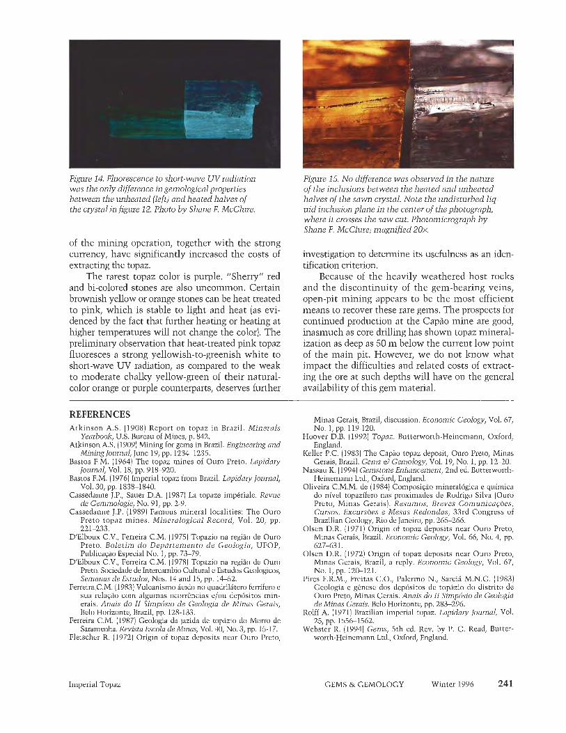
Figure 14. Fluorescence to short-wave UV radiation was the only difference in gemological properties between the unheated (left) and heated halves of the crystal in figure 12. Photo b y Shane F. McClure.
of the mining operation, together with the strong currency, have significantly increased the costs of extracting the topaz.
The rarest topaz color is purple. "Sherry" red and bi-colored stones are also uncommon. Certain brownish yellow or orange stones can be heat treated to pink, which is stable to light and heat (as evi- denced by the fact that further heating or heating at higher temperatures will not change the color). The preliminary observation that heat-treated pink topaz fluoresces a strong yellowish-to-greenish white to short-wave W radiation, as compared to the weak to moderate chalky yellow-green of their natural- color orange or purple counterparts, deserves further
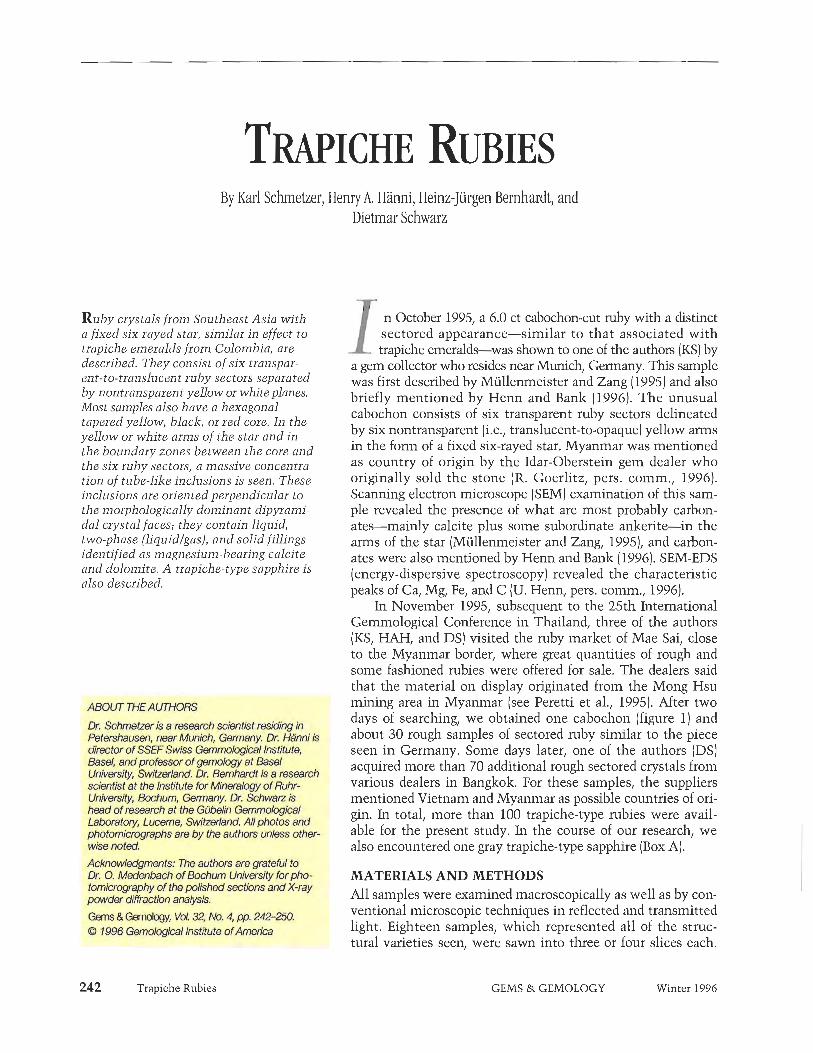
Figure 15. No difference was observed in the nature of the inclusions between the heated and unheated halves of the sawn crystal. Note the undisturbed liq- uid inclusion plane i n the center of the photograph, where i t crosses the saw cut. Photomicrograph b y Shane F. McClure; magnified 20x.
investigation to determine its usefulness as an iden- tification criterion.
Because of the heavily weathered host rocks and the discontinuity of the gem-bearing veins, open-pit mining appears to be the most efficient means to recover these rare gems. The prospects for continued production at the Cap50 mine are good, inasmuch as core drilling has shown topaz mineral- ization as deep as 50 m below the current low point of the main pit. However, we do not know what impact the difficulties and related costs of extract- ing the ore at such depths will have on the general availability of this gem material.
REFERENCES Atlzinson A.S. (1908) Report on topaz in Brazil. Minerals
Yearbook, U.S. Bureau of Mines, p. 842. Atkinson A.S. (1909) Mining for gems in Brazil. Engineering and
Mining Journal, June 19, pp. 1234-1235. Bastos F.M. (1964) The topaz mines of Ouro Preto. Lapidary
Journal, Vol. 18, pp. 918-920. Bastos F.M. (1976) Imperial topaz from Brazil. Lapidary Journal,
Vol. 30, pp. 1838-1840. Cassedanne J.P., Sauer DA. (1987) La topaze imperiale. Revue
de Gemmologie, No. 91, pp. 2-9. Cassedanne J.P. (1989) Famous mineral localities: The Ouro
Preto topaz mines. Mineralogical Record, Vol. 20, pp. 22 1-233.
DIElboux C.V., Ferreira C.M. (1975) Topazio na regiiio de Our0 Preto. Boletim d o Departemento de Geologia, UFOP, PublicaHo Especial No. 1, pp. 73-79.
DIElboux C.V., Ferreira C.M. (1978) Topazio na regiiio de Ouro Preto. Sociedade de Intercambio Cultural e Estudos Geologicos, Semanas de Estudos, Nos. 14 and 15, pp. 14-52.
Ferreira.C.M. (1983) Vulcanismo icido no quadrilitero ferrifero e sua relac30 corn alguinas ocorrt5ncias e/ou depositos min- erais. Anais do I1 Simpbsio de Geologia de Minas Gerais, Belo Horizonte, Brazil, pp. 128-133.
Ferreira C.M. (1987) Geologia da jazida de topizio do Morro de Saran~enha. Revista Escola de Minas, Vol. 40, No. 3, pp. 15-17.
Fleischer R. (1972) Origin of topaz deposits near Ouro Preto,
Minas Gerais, Brazil, discussion. Economic Geology, Vol. 67, NO, 1, pp. 119-120.
Hoover D.B. (1992) Topaz. Butterworth-Heinemann, Oxford, England.
Keller PC, (1983) The Capiio topaz deposit, Ouro Preto, Minas Gerais, Brazil. Gems el Gemology, Vol. 19, No. 1, pp. 12-20.
Nassau K. (1994) Gen~stone Enhancement, 2nd ed. Butterworth- Heinemann Ltd., Oxford, England.
Oliveira C.M.M. de (1984) Composi@o mineral6gica e quimica do nivel topazifero nas proximades de Rodrigo Silva (Ouro Preto, Minas Gerais). Resumes, Breves Comunica@es, Cursos, Excurs6es e Mesas Redondas, 33rd Congress of Brazilian Geology, Rio de Janeiro, pp. 265-266.
Olsen D.R. (1971) Origin of topaz deposits near Ouro Preto, Minas Gerais, Brazil. Economic Geology, Vol. 66, No. 4, pp. 627-63 1.
Olsen D.R. (1972) Origin of topaz deposits near Ouro Preto, Minas Gerais, Brazil, a reply. Economic Geology, Vol. 67, No. 1, pp. 120-121.
Pires F.R.M., Freitas C.O., Palermo N., Sarcih M.N.G. (1983) Geologia e genese dos depositos de tophzio do distrito de Ouro Preto, Minas Gerais. Anais do 11 Simpbsio de Geologia de Minus Gerais. Belo Horizonte, pp. 283-296.
Rolff A. (1971) Brazilian imperial topaz. Lapidary Journal, Vol. 25, pp. 1556-1562.
Webster R. (1994) Gems, 5th ed. Rev. by P. G. Read, Butter- worth-Heinemann Ltd., Oxford, England.
Imperial Topaz GEMS & GEMOLOGY Winter 1996
By Karl Schmetzer, Heniy A, Hanni, Heinz-Jurgen Bernhardt, and Dietmar Schwarz
R u b y crystals from Southeast Asia with a fixed six-rayed star, similar in effect to trapiche emeralds from Colombia, are described. They consist of six trunspar- ent-to-translncent ruby sectors separated by nontransparent yellow or white planes. Most samples also have a hexagonal tapered yellow, black, or red core. In the yellow or white arms of the star and in the boundary zones between the core and the six ruby sectors, a massive concentra- tion of tube-like inclusions is seen. These inclusions are oriented perpendicular to the morphologically dominant dipyrami- dal crystal faces; they contain liquid, two-phase (liquid/gas), and solid fillings identified as magnesium-bearing calcite and dolomite. A trapiche-type sapphire is also described.
ABOUT THE AUTHORS
Dr. Schrnetzeris a research scientist residing in Petemhausen, near Munich, Germany. Dr. HSnni ls director of SSEFSwiss Gemmotofifical Institute. Basel, and profiassor of gemology at Basel University, Swtearland. Dr. Bernhardt is a research scientist at the Institute for Mineralogy of Ruhr- University, &hum, Germany, Dr. Schwarz is head of research at the GQbelln Gemmdoglcal Laboratory, income, Switzerland. All photos and photomicrographs are by the authors unless other- wise noted.
Acknowledgments: The authors are grateful to Dr. 0. Madonbach of Bochum University for pho- tomicrography of the poUshed sections and X-ray pwder diffraction analysis.
Gems & Gemolooy, Vat 32 No. 4, pp. 242-250. @ 7996 Gemological Institute of America
Trapiche Rubies
I n October 1995, a 6.0 ct cabochon-cut ruby with a distinct sectored appearance-similar to that associated with trapiche emeralds~was shown to one of the authors (KS) by
a gem collector who resides near Munich, Germany. This sample was first described by Miillenmeister and Zang (1995) and also briefly mentioned by Henn and Bank (1996). The unusual cabochon consists of six transparent ruby sectors delineated by six nontransparent (i.e., translucent-to-opaque) yellow arms in the form of a fixed six-rayed star. Myanmar was mentioned as country of origin by the Idar-Oberstein gem dealer who originally sold the stone (R. Goerlitz, pers. comm., 1996). Scanning electron microscope (SEM) examination of this sam- ple revealed the presence of what are most probably carbon- ates-mainly calcite plus some subordinate anlzerite-in the arms of the star (Mullenmeister and Zang, 19951, and carbon- ates were also mentioned by Henn and Bank (1996). SEM-EDS (energy-dispersive spectroscopy) revealed the characteristic peaks of Ca, Mg, Fe, and C (U. Henn, pers. comm., 1996).
In November 1995, subsequent to the 25th International Gemmological Conference in Thailand, three of the authors (KS, HAH, and DS) visited the ruby market of Mae Sai, close to the Myanmar border, where great quantities of rough and some fashioned rubies were offered for sale. The dealers said that the material on display originated from the Mong Hsu mining area in Myanmar (see Peretti et al., 1995). After two days of searching, we obtained one cabochon (figure 1) and about 30 rough samples of sectored ruby similar to the piece seen in Germany. Some days later, one of the authors (DS) acquired more than 70 additional rough sectored crystals from various dealers in Bangkok. For these samples, the suppliers mentioned Vietnam and Myanmar as possible countries of ori- gin. In total, more than 100 trapiche-type rubies were avail- able for the present study. In the course of our research, we also encountered one gray trapiche-type sapphire (Box A).
MATERIALS AND METHODS All samples were examined macroscopically as well as by con- ventional microscopic techniques in reflected and transmitted light. Eighteen samples, which represented all of the struc- tural varieties seen, were sawn into three or four slices each.
GEMS & GEMOLOGY Winter 1996

The slices were oriented three ways: (1) parallel to the c-axis and parallel to one of the arms of the six- rayed stars, (2) parallel to the c-axis and perpendicu- lar to one of the arms of the stars, or (3) perpendicu- lar to the c-axis. From these slices, we had polished slabs about 1.0-1.3 mm thick prepared for each of the 18 crystals, as well as 10 polished sections about 200 p thiclz and two approximately 20 pm polished thin sections. We cut an additional 30 pieces of rough in one direction and polished one side to view the internal structure.
We examined the polished slabs with a gemo- logical microscope, first with fiber-optic illumina- tion and then immersed in methylene iodide. The polished sections were examined with convention- al petrographic microscopes (Leitz and Zeiss).
To identify the solid inclusions, we used two microanalytical techniques: 10 of the polished 1. ~apiche-typerubia have heen seen ill slabs were examined by Raman sPectroscoPY with Southeast Asian gem markets. This 1.55 ct cabo- a Renishaw Raman microscope (see Hanni et al., chon was purchased in Mae Sai, Thailand, from 1996, for experimental details), and eight of the among material that was mined in Mong Hsu, approximately 200 pm thiclz polished sections were Myanmar. As is the case with trapiche emeralds, analyzed with a CAMECA Camebax SX 50 elec- the six-rayed star is fixed; that is, it does not move tron microprobe. when the stone or light source is moved.
For additional chemical characterization of the material forming the arms of the six-rayed stars and comparison with the chemistry of the host ruby, we submitted five natural crystal fragments and six polished slabs to energy-dispersive X-ray fluorescence (EDXRF) analysis using a Philips PV 9500 X-ray generator and detectors with a Spectrace TX-6100 system and software package. We used lead foils with specially prepared holes to restrict analysis to the ruby areas only (without any arm component) and to analyze areas that included part of a yellow arm and some adjacent ruby.
In addition, yellow, nontransparent (translu- cent-to-opaque), triangular areas with massive inclusions in the outer zones of two samples were examined both by electron microprobe and by X- ray diffraction analysis (using a conventional 57.3 mm diameter Gandolfi camera).
VISUAL APPEARANCE We first examined the samples with the unaided eye or a lox loupe. All of the rough samples were fragments of barrel-shaped crystals; they ranged from about 3 to 8 mm in diameter and from 3 to 10 mm in length. Some were water-worn, but others revealed a distinct striated surface structure on planes more or less parallel to the basal pinacoid (figure 2). About 20 of the crystal fragments had natural faces, all with a uniform habit consisting of
a single &pyramidal crystal form. These faces were inclined about 5' to the c-axis, which indicates that the dominant form is the hexagonal dipyramid ft) j 14 14 28 31, Three crystals had one or two addi- tional rhombohedra1 faces i (1 011).
The divided structure of the crystals was best seen in those polished slabs oriented perpen- dicular to the c-axis. In these hexagonal cross- sections, six red, transparent-to-translucent sec- tors were subdivided by the yellow- or white- appearing arms of a six-rayed star. In some crys- tals, the six arms (which, unlike typical asteriat- ed gems, are fixed-that is, they do not move when the stone or light source is moved) inter- sected at one small point, forming six triangular ruby sectors (figure 3). In many cases, however, the arms extended outward from a hexagonal central core (figure 4)) producing trapezoidal ruby areas. The cores of our study samples were usu- ally either opaque yellow or black (figure 5); in some cases, they were transparent red. We also saw thin yellow or (rarely) white zones, similar in color to the arms of the stars, in the bound- aries between the black or red cores and the six triangular ruby sectors (see, e.g., figures 4 and 5).
In some samples, only a small intersection point between the six yellow arms of the star was
1 rapiche Rubies GEMS & GEMOLOGY Winter 1996 243

Figure 2. The six-rayed (hexagonal) star in this 7.5- mm-diameter trapiche ruby separates the nzby into six triangular sectors. Note the surface striations oriented perpendicular to the six dipyramidal faces of this crystal, almost parallel to the basal pinacoid.
observed on both sides of a crystal fragment or pol- ished slab. In most cases, however, distinct cores were seen, revealing a pyramidal or tapered outline (figure 6). That is, the diameters of the red, yellow, or black cores varied between the two ends of the crystal fragments or between the two sides of the polished slabs (figures 3 and 5). In most cases, the cores or intersection points at both ends were the same color; however, we also saw a few barrel- shaped samples with different colors at either end.
In some samples, yellow, nontransparent, feath- ery structures extended outward from the dividing planes into the transparent ruby sectors, forming tri- angular areas of massive inclusions toward the edges of the crystals (figure 7). Occasionally, these zones had been weathered out (figure 8).
MICROSCOPIC EXAMINATION In transmitted light, the yellow or blaclz central cores, the six yellow arms of the stars, and the yel- low triangular areas appeared opaque (figure 9). We observed a series of parallel tube-like structures or striations extending outward from the cores or arms into the ruby sectors (figure 10). Where the ruby sectors were transparent, these structures were largely restricted to thin areas close to the central core and the arms (figure 1 l), although some tubes did run through the full transparent
244 Trapiche Rubies
Figme 3. In some of the trapiche nzbies, the six arms intersect at one small point, forming six trian- gular ruby sectors. This polished slab is about 4.2 m m in diameter.
sectors to the outer dipyramidal faces of the crystal. Those ruby sectors that were semi-transparent to translucent had more of the tube-like inclusions.
Examination of the polished sections revealed the same characteristic patterns noted above, with striations restricted to the arms and boundaries between the cores and transparent ruby sectors (fig- ure 12a, b, c) and a dense concentration of tubes in semi-transparent samples (figure 12e, f). In some samples, the six arms intersected in a small point, that is, without a core (figure 12a, e); others had a small transparent red (figure 12f), a small nontrans-
Figure 4. In many of the trapiche rubies, the arms radiate from a hexagonal central core, so the six ruby sectors are trapezoidal. This polished slab measures about 3.2 m m in diameter.
GEMS & GEMOLOGY Winter 1996

Figure 5. These trapiche ruby cross-sections illus- trate some of the different forms observed in the samples examined. The arms of the stars inter- sect in a small point (lower right) or extend out- ward from the corners of a hexagonal black (upper right and lower left) or yellow (upper left) core. The upper left sample (which measures about. 4,2 m m in diameter) is the other side of the slab shown in figure 3; note the size difference in the centers on the two sides.
parent yellow (figure 12c), or an opaque blaclz core (figure 12d). A few had large cores (figure 12b, d). Occasionally, the arms widened toward the edges of the crystals, often with evidence of weathering (figure 12d-f).
With higher magnification, using crossed polarizers, we resolved the striations as thin tubes (figure 13a) that were often filled with bire- fringent minerals. The arms of the six-rayed stars were formed by massive concentrations of such tubes, which were filled with birefringent miner- als (figure 13b), a liquid, or a liquid and gas (fig- ure 13c).
Examination of sections parallel to the c-axis revealed that the tube-like structures are not ori- ented exactly parallel to the basal plane of the corundum crystals, but rather show a slight incli- nation, about 5O (figure 14). This indicates that they are oriented perpendicular to the dominant dipyramidal faces co, which are inclined about 5' to the c-axis.
In.the blaclz or yellow cores of some of the pol- ished sections, we observed small birefringent min- eral inclusions in the form of tiny round spots ('fig- ure 13a). These probably represent cross-sections of tubes oriented perpendicular to the basal pinacoid, which means that the tube-like structures also run parallel to the c-axis in the cores of some samples.
Trapiche Rubies
Figure 6. This view, parallel to the c-axis of this 7.2-mm-diameter sample, illustrates the tapered core, which is mostly red but black at one end. Note the strong striations in the outer zones, away from the core.
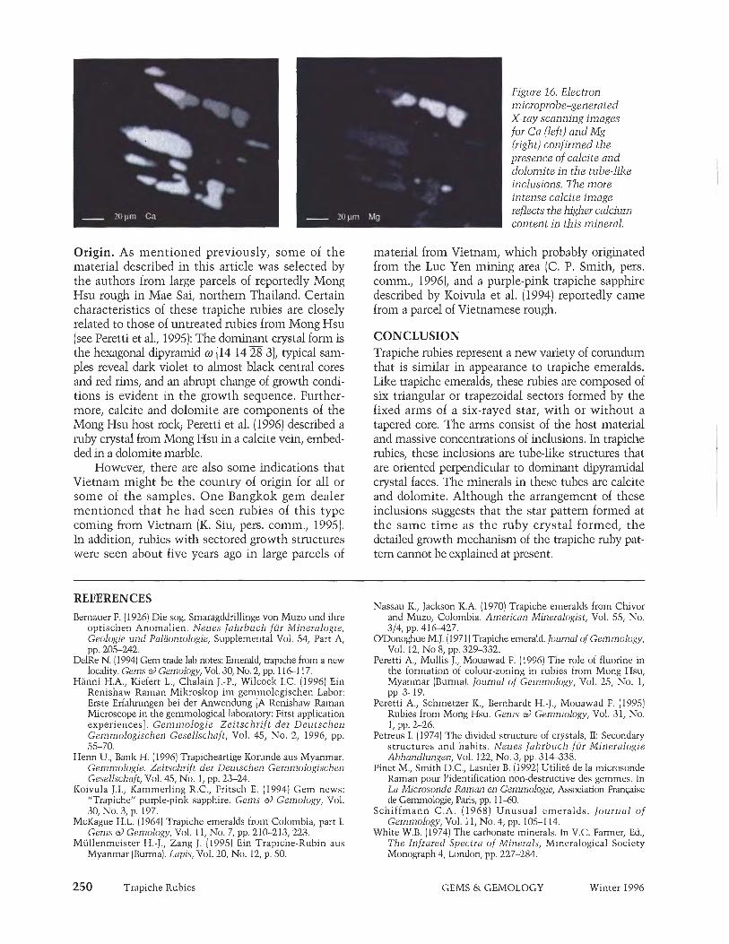
IDENTIFICATION OF THE MINERAL INCLUSIONS The birefringent mineral inclusions in the tube- like structures were analyzed independently by Raman spectroscopy and an electron microprobe. Two types of Rainan spectra were found repeatedly in all of the polished slabs (figure 15). These spectra were consistent with calcite and dolomite, as deter- mined by data in the literature (White, 1974; Pinet et al., 1992) and our own reference spectra.
Electron rnicroprobe analysis of the solids filling the tubes confirmed these results and provided some
Figure 7, The arms in this 4 x 6 m m trapiche ruby slab "feather out" and widen toward the outer edge of the crystal, almost completely absorbing one of the ruby sectors.
:- GEMS & GEMOLOGY Winter 1996

Figure 8. In some of the trapiche rubies in which the arms form triangular zones at the rim of the crystal, weathering has created re-entrant angles. This sample is approximately 7 x 8 m m .
additional chemical data. Two mineral phases were present in all of the samples examined (figure 16): a magnesium-bearing calcium-rich mineral (calcite) and a carbonate with higher magnesium and lower calci- um contents (dolomite). Quantitative chemical analy- ses gave a Mg:Ca ratio of 7:93 for the magnesium- bearing calcite (average of five analyses) and a Mg:Ca ratio of 49:51 for the dolomite (average of six analy- ses]. No iron was detected in either of these minerals.
Figure 9. When the slabs are examined with transmitted light, the cores and arms of the trapiche rubies appear opaque. The parallel stria- tions illustrated in figure 2 are clearly seen in this 3.5-mm-diameter cross-section. Immersion, crossed polarizers.
Trapiche Rubies
Figme 10. In reflected ngnt, this trapiche ruby slab reveals a series of parallel tube-like structures extending outward from each of the six arms of the star and from the dividing planes between the black core and the six trapezoidal red zones. Magnified Wx.
Because the tube-like structures that extended into the gem-quality ruby sectors were colorless, and the calcite and dolomite inclusions were iron- free, we concluded that the yellow color of the arms and some cores must be due to intense weathering and secondary iron staining of the cavities and tubes. This interpretation was supported by the presence of white arms in some (not deeply weath- ered) samples and by X-ray fluorescence analyses. In two samples for which we recorded distinct differ- ences, the iron signal in the XRF spectrum of the yellow arm was about four to five times stronger than the iron signal of the adjacent ruby sector (which contained fewer tube-like inclusions).
The massive, nontransparent, yellow triangular areas that broadened toward the outer rim in some samples consist of non-gem-quality corundum, according to X-ray powder diffraction and micro-
Figure 11. In those samples with transparent ruby sectors, most of the tube-like inclusions ended close to the arms or dividing planes with the core. Magnified 50x.
GEMS & GEMOLOGY Winter 1996

Figure 12. When viewed with transmitted light, the polished basal sections of the trapiche rubies clearly illustrate the different types of star-like structures: (a) the arms of the star in this 3-mm-diameter sample intersect in a small central point; (b) the arms in this 4-mm-diameter section extend outward from the corners of a yellow central core; (c) the arms m this 4.2-mm-diameter section extend outward from the corners of a small yellow central core, with evidence of weathering in the arms at the rim (re-entrant angles); id) the aims in this 3.2-mm-diameter sec- tion, which extend horn a black core, get thicker as they approach the outer rim of the crystal, with evidence of weathering at the rim; (e) weathering is more extensive in the arms of this 5-mm-diameter sample, which intersect in a small central point; and ( f ) the arms in this 4.5 x 5 m m sample extend outward from a red core, ending in par- tially weathered triangular structures at the rim of the crystal. Note the profusion of tube-like inclusions in the ruby sectors of semi-transparent samples e-f, as compared to the ruby sectors of transparent samples a-c. Polished sections a, b, and d-f are 200pm thick; sample c i s 20 ,um thick. Photomicrographs by 0. Medenbach.
probe analyses. In the samples we examined, these sectors contained a massive concentration of inclu- sions, apparently also accompanied by intense weathering and iron staining.
DISCUSSION Nomenclature. These ruby samples share a number of common structural features with trapiche emer- alds from Colombia, as described in the mineralog- ic and gemological literature (Bernauer, 1926; McKague, 1964; Schiffmann, 1968; Nassau and Jackson, 1970; O'Donoghue, 1971). In both mineral species, corundum and beryl, hexagonal single crys- tals are divided by included material into six distinct triangular or trapezoidal growth sectors, depending on the presence or absence of a central core. The arms of the six-rayed stars consist of the host (ruby
or emerald) with inclusions of other minerals: cal- cite and dolomite for ruby, albite for emerald. In both gem materials, the central core consists of the host mineral alone or of the host mineral plus inclusions (similar to the composition of the arms), and it is typically tapered.
Oriented striations (tube-like inclusions) occur both at the outline of the core and extending out- ward from the nontransparent arms into the trans- parent ruby or emerald sectors. In our ruby samples, these striations were oriented perpendicular to the dominant crystal form, that is, perpendicular to the hexagonal dipyramid a (14 14 28 31; in emerald, they are perpendicular to the first-order hexagonal prism m {1010].
In general, most of the structural characteristics that have been described for various trapiche einer-
Trapiche Rubies GEMS & GEMOLOGY Winter 1996 247

Figure 13. When the polished sections were viewed with higher magnification and crossed polarizers, i t became evident that the tube-like structures (a) were filled with birefringent minerals (b), or with a liquid or liquid and gas (c). The tiny round spots in the core of figure a are actually cross-sections of tubes that run per- pendicular to the basal pinacoid. Photomicrographs by 0. Medenbach; a = magnified lox, b = 40x, c = 40x.
aids from Colombia (see, e.g., Bemauer, 1926; Nassau and Jackson, 1970) were also found in the sectored rubies described in this article. Thus, it seems reason- able to apply the term trapiche not only to sectored emeralds from Colombia, but also to similarly sec- tored rubies regardless of geographic origin.
Formation Sequence. Discussions as to whether the structural features observed in Colombian trapiche emeralds are primary or secondary in origin (i.e., whether they formed during or after the formation of the host emerald) are ongoing (McKague, 1964; Nassau and Jackson, 1970; Petreus, 1974). However, the arrangement of the tube-like inclusions in our samples suggests that the sectored structure of the trapiche rubies described in this article is primary. Specifically, we believe that the red or black core formed first; then, a change in the growth environ-
Figure 14. In this view of a 5.9-mm-diameter trapiche ruby crystal parallel to the c-axis, the lube-like inclusions show a small inclination to the basal plane.
248 Trapiche Rubies
ment caused the massive formation of tube-like inclusions at the outer edge of the core. Subsequent to this event, new tube-like inclusions formed at the boundaries between the six dipyramidal growth sec- tors. In the direction along the c-axis, tube-like inclu-
Figure 15. On the basis of these Roman spectra (note that scales are different), the mineral inclusions in the tube-like structures were identified as (A) calcite and (B) dolomite. The lines at 414.8 and 749.7 cm-1 (not labeled) are assigned to the corundum host,
RAMAN SHIFT ( c m " )
GEMS & GEMOLOGY Winter 1996

from the state of Goihs, Bif DelRe (1994). The samples he hexagonal central co
ed by narrower green from the comers ,of the Brazilian-material ap "negative" of Colombian
Recently, trapiche corundum with a similar dipul^t. t k t h 9 . e of;&$, s r̂s .- in - the tr@che rubies "negative" appearance was seen in the gem market. d&re(C$'tliyflttlfcle5. Ten sapphires with a sectored structure were offered ~ i c r o ^ ~ o $ l ~ e@ri$@&jt&rffr&led yelhw- at the 1996 Base1 fair by a Berlin gem dealer. He had appearing min&r~'J&%lusi~t^\th$t'we^f,~c&~(ie~t~ated purchased several cabochons and one faceted trapiche in the areas confined by the dx Sfmt of the star. After sapphire in Myanmar in early 1996; at that time, he carefully r e p o l i s h the back of the sapphire cabo- was told that the samples originated from the Mong chon, we were able to Identify t h e inclusions by Hsu mining area (H.-J. Engelbrecht, pers. comm., SEM-EDS as phlogopite. These phlogopite inclusions, 1996). which were undoubtedly concentrated during crystal
A 6.59 ct trapiche sapphire was purchased by one growth, are responsible for the dark gray appearance of of the authors (HAH). It is whitish gray in color, with the arms of the star. six almost triangular white reflective (opaque) areas.
. . ' , , f , ^ ; f t à ˆ '  ¥ " J - 1 ' l l * > f l i i r f l ~ t t t y ^ ~ < . ^ ' i . ' f 7 ~ ^ , - , ^
~ ~ Ã ‡ . A ~ ,'~~fihishyf~tfwJP hf i~~tkf f . seofod reten-&-ia tfea.sfm tAai- ~ t ~ u t & ~ a ~ t f ~ a ~ s o a p M s a f
kt, A 4.59 mapiche sapphire is shown @xe 9 , the 1.35 ct ttapiche ruby cabochon /or coinpt~~on. . needles in the whitish reflecting growth Wnes,dr3.bri~
ented parallel to thegrowth planes, as indicated h w by the drms of the small six-rayed stars.
sions also formed in basal growth sectors, with an orientation perpendicular to the basal pinacoid. Where the basal faces were prominent, discrete cores with calcite and dolomite inclusions in tube-like structures formed; where the basal faces were small or absent, smaller cores or intersection points between the arms of the stars formed.
The appearance of the samples described in this article can be explained in terms of their rela- tive position in this trapiche ruby growth sequence: Samples with red and/or black cores on either end of the crystal fragments were grown at an earlier stage, and those with yellow cores or intersection points were grown in a later stage.
Trapiche Rubies GEMS & GEMOLOGY Winter 1996
Origin. As mentioned previously, some of the material described in this article was selected by the authors from large parcels of reportedly Mong Hsu rough in Mae Sai, northern Thailand. Certain characteristics of these trapiche rubies are closely related to those of untreated rubics from Mong Hsu (see Peretti et al., 1995): The dominant crystal form is the hexagonal dipyramid m (14 1428 31, typical sam- ples reveal dark violet to almost black central cores and red rims, and an abrupt change of growth condi- tions is evident in the growth sequence. Further- more, calcite and dolomite are components of the Mong Hsu host rock; Peretti et al. (1996) described a ruby crystal from Mong Hsu in a calcite vein, embed- ded in a dolomite marble.
However, there are also some indications that Vietnam might be the country of origin for all or some of the samples. One Bangkok gem dealer mentioned that he had seen rubies of this type coming from Vietnam (K. Siu, pers. comm., 1995). In addition, rubies with sectored growth structures were seen about five years ago in large parcels of
REFERENCES Bernauer F. (1926) Die sog. ~maragddrillinge von Muzo und ihre
optischen Anomalien. Neues Jahrbuch fui Mineralogie, Geologie und Palaontologie, Siipplemental Vol. 54, Part A, - . - - pp. 205-242.
DelRc N. 119941 Gem trade lab notes: Emerald. traniche from a new locality. ~ e h s ei} Gemology, Vol. 30, No. 2; pp.'l 16-1 17.
Hanni H.A., Kicfert L., Chalain J.-P., Wilcock I.C. (1996) Ein Rcnishaw Raman Mikroskop im gemmologischen Labor: Erste Erfahningen bei dcr Anwcndung [A Rcnishaw Raman Microscope in the gemmological laboratory: First application experiences]. Gen~mologie. Zeitschrift der Deutschen Gemmologischen Gesellschaft, Vol. 45, No. 2, 1996, pp. 55-70.
Henn U., Bank H. (1996) Trapicheartige Korunde aus Myanmar. Gemmologie. Zeitschrift der Deutschen Gemmologischen Gesellschaft, Vol. 45, No. 1, pp. 23-24.
Koivula J.I., Kammerling R.C., Fritsch E. (1994) Gem news: "Trapiche" purple-pink sapphire. Gems es) Gemology, Vol. 30, No. 3, p. 197.
McKague H.L. (1964) Trapiche emeralds from Colombia, part I. Gems a) Gemology, Vol. 11, No. 7, pp. 210-213,223.
Mullenmcister H.-J., Zang J , (1995) Ein Trapiche-Rubin aus Myanmar (Burma). Lapis, Vol. 20, No. 12, p. 50.
250 Trapiche Rubies
Figure 16. Election microprobe-generated X-ray scanning images for Ca (left) and Mg (right) confirmed the presence of calcite and dolomite in the tube-like inclusions. The more intense calcite image reflects the higher calcium content in this mineral.
material from Vietnam, which probably originated from the Luc Yen mining area (C. P. Smith, pers. comm., 1996), and a purple-pink trapiche sapphire described by Koivula et al. (1994) reportedly came from a parcel of Vietnamese rough.
CONCLUSION Trapiche rubies represent a new variety of corundum that is similar in appearance to trapiche emeralds. Like trapiche emeralds, these rubies are composed of six triangular or trapezoidal sectors formed by the fixed arms of a six-rayed star, with or without a tapered core. The arms consist of the host material and massive concentrations of inclusions. In trapiche rubies, these inclusions are tube-like structures that are oriented perpendicular to dominant dipyramidal crystal faces. The minerals in these tubes are calcite and dolomite. Although the arrangement of these inclusions suggests that the star pattern formed at the same time as the ruby crystal formed, the detailed growth mechanism of the trapiche ruby pat- tern cannot be explained at present.
Nassau K., Jackson K.A. (1970) Trapiche emeralds from Chivor and Muzo, Colombia. American Mineralogist, Vol. 55, No. 314, pp. 416-427.
O'Donoghue M.J. (1971) Trapiche emerald. Journal of Gemmology, Vol. 12, No 8, pp. 329-332.
Peretti A., Mullis J., Mouawad F. (1996) The role of fluorine in the formation of colour-zoning in rubies from Mong Hsu, Myanmar (Burma). Journal of Genamology, Vol. 25, No. 1, pp. 3-19.
Peretti A., Schmetzer K., Bernhardt H.-J,, Mouawad F. (1995) Rubies from Mong Hsu. Gems &> Gemmology, Vol. 31, No. - -. .
1, pp. 2-26. Petreus I. I19741 The divided structure of crystals, Ik Secondary . ,
structures and habits. Neues ~ a h r b u c h f i r ~inera lo&e Abhandlungen, Vol. 122, No. 3, pp. 314338.
Pinct M., Smith D.C., Lasnier B. (1992) Utilitk de la microsonde Raman pour l'idcntification non-destructive dcs gemmes. In La Microsonde Roman en Gemmologie, Association Franpisc de Gemmologie, Paris, pp. 1 1-60.
Schiffmann C.A. (1968) Unusual emeralds. Journal of Gemmology, Vol. 11, No. 4, pp. 105-1 14.
White W.B. (1974) The carbonate minerals. In V.C. Farmer, Ed., The Infrared Spectra of Minerals, Mineralogical Society Monograph 4, London, pp. 227-284.
GEMS & GEMOLOGY Winter 1996

Fall 1988 An Economic Review of Diamonds The Sapphires ot Penglai, Hainan Island, China Iridescenl Orlhoamphibole from Wyoming Detection of Treatment in Two Green Diamonds Spring 1989 The Sinkankas Library The Gujar Killi Emerald Deposit Beryl Gem Nodules from the Bananal Mine "Opalite:" Plastic Imitation Opal Summer 1989 Filled Diamonds Synlhetic Diamond Thin Films Grading the Hope Diamond Diamonds with Color-Zoned Pavilions Fall 1989 Polynesian Black Pearls The Capoeirana Emerald Deposil Brazil-Twinned Synthetic Quartz Thermal Alteration of Inclusions in Rutilated Topaz Chicken-Blood Stone from China Winter 1989 Emerald and Gold TÑ,ure of the Alocha
inge, Australia
nenecance Infrared Spectroscopy in Gemology Mildly Radioactive Rhinestones Sprlng 1990 Gem Localilies of the 1980s Gemstone Enhancement and Its Detection Synlhetic Gem Materials in Ihe 1980s New Technologies of the 1980s Jewelry of the 1980s Sprlng 1991 Age, Origin, and Emplacement of Diamonds Emeralds of Panjshir Valley, Afghanistan Summer 1991 Fracture Filling o l Emeralds: Opticon and "Oils' Emeralds from the Urat Mountains, USSR Treated Andamooka Matrix Opal Fall 1991 Rubies and Fancy Sapphires Irom Vietnam New Rubies Irom Morogoro, Tanzania Bohemian Garnet-Today Winter 1991 Marine Mining of Diamonds off Southern Africa Sunstone Labradorite from the Ponderosa Mine Nontraditional Gemstone Cutting Nontransparent "CZ" from Russia Spring 1992 Gem-Quality Green Zoisite Kilbourne Hole Peridot Fluid Inclusion Study of Querktaro Opal Natural-Color Nonconductive Gray-to-Blue Diamonds Peridot as an Interplanetary Gemstone Summer 1992 Gem Wealth of Tanzania Gamma-Ray Spectroscopy and Radioactivity Dyed Natural Corundum as a Ruby Imitation An Update on Sumitomo Synthetic Diamonds Fall 1992 Ruby and Sapphire Mining in Mogok Bleached and Polymer-Impregnated Jadeite Radiation-Induced Yellow-Green in Garnet Winter 1992 Determining the GoldContent of Jewelry Metals Diamond Sources and Production Sapphires from Changle, China Sprlng 1993 Queensland Boulder Opal Update on Diffusion-Treated Corundum:
Red and Other Colors A New Gem Beryl Locality: Luumaki, Finland De Beers Near Colorless-to-Blue Experimental
Gem-Quality Synthetic Diamonds Summer 1993 Flux-Grown Synthetic Red and Blue
Spinels Irom Russia Emeralds and Green Beryls of Upper Egypt Reactor-Irradiated Green Topaz
B A C K I S S U E S Fall 1993 Jewels of the Edwardians A Guide Map to the Gem Deposits of Sri Lanka Two Treated-Color Synthetic Red Diamonds Two Near-Colorless General Electric Type lla
Synthetic Diamond Crystals Winter 1993 Russian Gem-Quality Synthetic Yellow Diamonds Heat Treating Rock Creek (Montana) Sapphires Garnels from Allay, China Spring 1994 e,isa are still available The Anah, Ametrine Mine, b l i v i a Indaia Sapphire Deposits of Minas Gerais, Brazil Flux-Induced Fingerprints in Synthetic Ruby Summer 1994 Synthetic Rubies by Douros Emeralds from the Mananjary Region, Madagascar:
Internal Features Synthetic Forsterite and Synthetic Peridot Update on Mining Rubies and Fancy Sapphires in
Northern Vietnam Fall 1994 Filled Diamonds: Identification and Durability
*IS Spffng 1995 SwiM 1396 Inclusions of Nalive Copper and Tenorite in Cuprian-Elbaite Tourmaline, Paraiba, Brazil
Winter 1994 Color Gradina o l Colored Diamonds in the GIA
Gem ~ r a d e Laboratory Ruby and Sapphire from the Ural Mountains, Russia Gem Corundum in Alkali Basalt Sprlng 1995 Rubies from Mong Hsu The Yogo Sapphire Deposit
A visual Guide to the Identification of Filled Diamonds
Fall 1995 Gem-Quality Grossular-Andradite: A New Garnet
Irom Mali Sapphires from Southern Vietnam Ti-Sapphire": Czochralski-Pulled Synthetic Pink
Sapphire from Union Carbide Winter 1995 A History of Diamond Sources in Africa: Part 1 A Chart for the Separation of Natural and
Synthetic Diamonds Sprlng 1996 A History of Diamond Sources in Africa: Part II 1 ~emo lGca l Invcsligation of a New Type of Russian
Hvdrothermal Svnthetic Emerald ~ r o w l h Method and~rowth-Mated Properties of a New
Type of Russian Hydrothermal Synthetic Emerald Summer 1996
I Complete your back issues of Gems & Gemology NOW!
I U.S. Canada & Mexico Elsewhere Single Issues* $11.00 each $13.00 each $14.00 each
I Complete Volumes:' 1987,1991,1992,1993, 1994,1995,1996 $38.00 each $44.00 each $53.00 each Three-year set $105.00 each $120.00 each $155.00 each Five-year set $1 70.00 each $lSO.OO each $245.00 each 1
I '10% discount fw GIA Annual Fund donwsal \hi Booster's Circle level and above. I
TO ORDER: Call loll free (800) 421-7250, ext. 202 or (310) 829-2991, exl. 202
FAX (310) 453-4478 OR WRITE: G&G Subscriptions GIA P.O. Box 2110, Santa Monica, CA 90404 USA I
Sapphires from the Andranondambo Region, Madagascar
Russian Demantoid, Czar of the Garnet Family Opal from Shewa Province, Ethiopia Fall 1996 De Beers Natural versus Synthetic Diamond
Verification Instrumenis Analyzing Internal Growth Structures:
Identification of the Negative dPIane in Natural Ruby
Russian Flux-Grown Synthetic Alexandrite Winter 1996 Imperial Topaz Irom the Cap20 Mine,
Minas Gerais, Brazil Trapiche Rubies Identifying Black Opaque Gem Materials Enstatite, Cordierile, Kornerupine and Scapolite
from Embilipitiya, Sri Lanka Some Tanzanite Imitations
Some issues from the 1984-1987 volume years are also available. Please contact the Subscriptions Office for details.
O R D E R N O W !

By Mary L. Johnson, Shane F, McClure, and Dino G. DeGhionno
A m o n g the most difficult gems to identi- f y are those that are black and opaque (or nearly so). In general, any gem material can be opaque because o f inclusions, any black opaque material can be fashioned, and any porous material can be dyed. Thus, to identify a black opaque materi- al, every possible mineral, and many rocks and manufactured substances, must be considered. Microscopic appear- ance, refractive index, specific gravity, and other properties (such as magnetism or radioactivity) provide useful clues, but in most cases advanced identification techniques (X-ray diffraction, EDXRF spectroscopy) are necessary, and even these m a y not be conclusive. Black opaque pyroxenes, amphiboles, and spinel-group minerals are especially chal- lenging to identify.
, -3QUT THE AUTHORS
0 ne of the most challenging problems in gemology is that of determining the identity of a blaclz opaque gem inate-
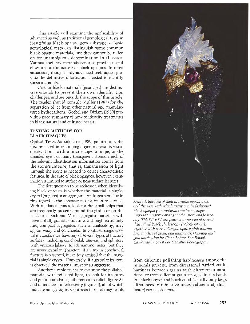
rial-hereafter called a "blaclz opaque." Such materials are a mainstay of the jewelry business, primarily as side stones, dec- orative elements in multi-stone mosaics, and in men's jewelry. Treated blaclz chalcedony ("black onyx"), black jade, and hematite traditionally have been the black opaques in greatest demand. As a variety of quartz, "black onyx" is probably the most familiar durable black opaque to lapidaries and gem cut- ters. Today, it is a popular medium for artistic carvings, many of which have been incorporated into fine jewelry (figure 1).
In recent years, various materials have been misrepresent- ed as "black onyx" or "black jade" to meet the trade's need for calibrated goods in high-volume markets. Members of the trade, in turn, have been sending samples to identification lab- oratories to ensure that the material has been properly repre- sented, so that they can sell it honestly. Usually, individual items sent to the GIA Gem Trade Laboratory (GIA GTL) for identification are representative of larger lots.
The purpose of this article is to provide a set of procedures by which to identify blaclz opaques (that is, materials that are black or almost blaclz and opaque or nearly so). The examples used are drawn primarily from our experience at GIA GTL. Not all black opaques are included here, as that is beyond the scope of a single article (and almost any gem material, by virtue of inclusions, can become a "black opaque"). Because so many materials-both common and exotic-may be used as black opaques, any set of identification procedures must take all pos- sibilities into consideration, not just a few. For example, black jade (nephrite or jadeite), "onyx," and hematite each have many imitations. Also, natural-color black diamonds may be confused with diamond imitations and treated-color diamonds. In the last few years, other black opaque materials have become increasingly available, such as the amphibole ferrohomblende (sold as blaclz "gem barlzevikite") and various spinel-group min- erals (figure 2). Still another challenging problem is that of iden- tifymg black opaque polycrystalline aggregates, or roclzs.
252 Black Opaque Gem Materials GEMS & GEMOLOGY Winter 1996
This article will examine the applicability of advanced as well as traditional gemological tests in identifying blaclz opaque gem substances. Basic gemological tests can distinguish some common blaclz opaque materials, but they cannot be relied on for unambiguous determination in all cases. Various ancillary methods can also provide useful clues about the nature of blaclz opaques. In most situations, though, only advanced techniques pro- vide the definitive information needed to identify these materials.
Certain blaclz materials (pearl, jet) are distinc- tive enough to present their own identification challenges, and are outside the scope of this article. The reader should consult Muller (1987) for the separation of jet from other natural and manufac- tured hydrocarbons; Goebel and Dirlam (1989) pro- vide a good summary of how to identify treatments in blaclz natural and cultured pearls.
TESTING METHODS FOR BLACK OPAQUES Optical Tests. As Liddicoat (1989) pointed out, the first test used in examining a gem material is visual observation-with a microscope, a loupe, or the unaided eye. For many transparent stones, much of the relevant identification information comes from the stone's interior; that is, transmission of light through the stone is needed to detect characteristic features. In the case of black opaques, however, exam- ination is limited to surface or near-surface features.
The first question to be addressed when identify- ing blaclz opaques is whether the material is single- crystal (or glass) or an aggregate. An important clue in this regard is the appearance of a fracture surface. With fashioned stones, look for the small chips that are frequently present around the girdle or on the back of cabochons. Most aggregate materials will have a dull, granular fracture, although extremely fine, compact aggregates, such as chalcedony, may appear waxy and conchoidal. In contrast, smgle-crys- tal materials may have any of several types of fracture surfaces (including conchoidal, uneven, and splintery with vitreous [glassy] to adamantine luster), but they are never granular. Therefore, if a vitreous conchoidal fracture is observed, it can be surmised that the mate- rial is single crystal. Conversely, if a granular fracture is observed, the material must be an aggregate.
Another simple test is to examine the polished material with reflected light, to look for fractures and grain boundaries, differences in relief (figure 3), and differences in reflectivity (figure 4), all of which indicate an aggregate. Contrasts in relief may result
Black Opaque Gem Materials
Figwe 1. Because of their dramatic appearance, and the ease with which many can be fashioned, black opaque gem materials are increasingly important in gem carvings and custom-made jew- elry. This 9.1 x 3.5 cm piece is composed of carved dmsy dyed blaclz chalcedony ("black onyx"), together with curved Oregon opal, a pink tourma- Line, mother-of-pearl, and diamonds. Carvings and gold fabrication by Glenn Lehrer, Sun Rafael, California; photo 0 Lee-Carraher Photography.
from different polishing hardnesses among the minerals present, from directional variations in hardness between grains with different orienta- tions, or from different grain sizes, as in the bands in "black onyx" and blaclz coral. Usually only large differences in refractive index values (and, thus, luster) can be observed.
GEMS & GEMOLOGY Winter 1996 253

have any type of luster and, in general, "higher" lusters (adamantine or metallic) correspond to high R.I. values. However, the apparent luster also may be affected by the quality of the polish on a fash- ioned gem.
In many cases, a blaclz opaque is not complete- ly black or opaque when a very thin region is viewed with a strong beam of light. Fiber-optic illu- mination in conjunction with magnification is ideal for this purpose (Koivula, 1982). Materials that have a blaclz appearance overall may look green, brown, red, blue, purple, gray, or the like, at these intensely lit thin edges. Sometimes growth structures are more visible in thin edges with strong transmitted light (figure 5) than with reflect-
-
ed light. Inclusions-such as gas bubbles in manu- Figure 2. These four opaque black stones, which rangefiom 1.88 to 13.63 CL, are hercynite (the two factured glass, and stretched gas bubbles and iron-
faceted stones) and magnetite, spinel-group miner- titanium oxide inclusions in natural volcanic glass 01s from the Bo Phloi region of Thailand. Photo by (obsidian; see, e.g., Gubelin and Koivula, 1986, p. Maha DeMaggio. 285)-may also be seen.
Some blaclz opaques display asterism (see, e.g., Kane, 1985 [sapphire]; Liddicoat, 1989 [diopside]; Koivula and Kammerling, 1990a [elzanite]; and Koivula et al., 1993b [irradiated quartz]), which can be an important consideration for identification. Chatoyancy is also seen occasionally (as in opal- Koivula and Kainmerling, 1990b). Both of these phe- nomena may be very weak, so strong pinpoint illu- mination also should be used to search for them.
In those cases where a material is blaclz because of inclusions, it often helps to try to determine which opaque material is responsible for coloring the unknown. Strong, low-angle oblique illumina- tion can be used, with magnification, to distinguish
If a microscope with an analyzer is available, a polarizer can be placed over the light source to obtain additional information. Anisotropic (uniaxi- al and biaxial) minerals have different refractive indices with different directions of polarized light, so the relative reflectivities of adjacent grains in an aggregate may change as the analyzer is rotated. Some highly reflective materials, such as the blaclz opaque minerals graphite and enargite, may show pleochroism in reflected light.
Observation of the sample's luster can be very important (again, see figure 2). Black opaques can
Figure 3. The contrast in relief seen in reflected l k h t in this dark green fuchsite (muscovite) mica is due to the presence of harder minerals, such as quartz, in this aggregate material. Photo by John I. Koivula; magnified 15x.
F i p e 4. The crystals in this sample of (manufac- tured) devitrified glass have a higher refractive index than the glass matrix, and so they reflect more light. Photomicrograph by John 1. Koivula; magnified 20x.
-.>;y,? 's' 'È!-.- -^ ., .: ft. t, ,
254 Black Opaque Gem Materials GEMS & GEMOLOGY Winter 1996

Figure 5. The banding in this "black onyx" cabo- chon is visible only on a very thin edge with strong fiber-optic illumination. The black chalcedony may have been a banded agate before being treated to produce the black color, or it could have been a chalcedony in which different layers selectively absorbed the coloring agent. Photomicrograph by John I. Koivula; magnified lox.
(for instance) between metallic iron oxides such as hematite, which appear reddish brown when lit from the side (figure 6), and titanium-bearing oxides such as ilmenite, which may appear white when lit from the side (because of surface alteration
Figure 6. The large hematite crystal in this quartz/magnetite/hematite cabochon looks red along a very thin edge; this red color is seen when the sample is examined with low-angle oblique illumination. Photomicrograph by John I. Koivula; magnified 1 5x.
into white so-called "leucoxene" [Deer et al., 1966, pp. 418-419], figure 7). Similarly, the opaque iron sulfides pyrite and pyrrhotite look brassy yellow in reflected light. This technique may also provide clues to the identity of a metallic-luster opaque.
Many other optical tests-such as observing the absorption spectrum, checking the optic sign, or looking for pleochroisin-are not feasible when testing blaclz opaques. (However, a reflected-light spectrum sometimes can be obtained from opaque samples: Kammerling et al., 1990.) Fortunately, the refractive index (or indices] of an opaque material still can be measured, although many materials, especially those with metallic lusters, have indices that are over the limits of a standard refractometer. Measurements should be taken from several places on a black opaque to check for multiple phases, or to determine if the material is doubly refractive. (As used here, "phases" refers only to solid phases, including distinct minerals, glass, plastic filler, etc.)
Other Physical Tests. Specific gravity can be extremely valuable for distinguishing among sin- gle-crystal materials, especially those with refrac- tive indices greater than 1.81. It is somewhat less reliable for aggregates and porous materials. Note that some materials give inconsistent specific gravity values, including the hercynite sample described below. Also, hydrostatic specific gravity measurements are less precise than, for example, refractive index measurements.
Figure 7. The ilmenite crystal in this black labradorite moonstone cabochon look's white when viewed with low-angle oblique illumination. Photomicrograph by John I. Koivula; magnified 20x.
Black Opaque Gem Materials GEMS & GEMOLOGY Winter 1996 255

Various other tests may be useful or even diag- nostic for certain materials. One common test is the observation of luminescence to long- or short-wave ultraviolet radiation. For example, some blaclz dia- monds fluoresce (Kammerling et al., 1990). Certain blaclz opaques, notably magnetite, are strongly attracted to magnets, as are some other spinel-group minerals (e.g., some hercynite: Johnson, 1994) and some hematite (Fryer et al., 1984). The streak test (scratching an inconspicuous comer of the sample across an unglazed porcelain plate) must be made carefully, as it is a destructive test, but it can provide distinctive information about certain materials. For example, the iron oxides-magnetite, hematite, and goethite-have black, red, and brown streaks, respectively. However, for aggregate materials with small grain sizes, the low-angle oblique illumination test mentioned above provides the same information as a streak test, without the risk of damaging the sample. Some low-luster materials, such as jet and plastics, react to the thermal reaction tester ("hot point") with diagnostic aromas.
Some materials are electrically conductive. These will give a positive response to an electrical conductometer-or even to an electrical multime- ter, which is available at most hardware stores. For example, a diagnostic test for manufactured yttri- um iron garnet, YIG, is that it does not conduct electricity (Kammerling et al., 1990). However, practically any mineral or rock can appear blaclz and opaque because of included graphite, which conducts electricity; consequently, such a material might give a positive response to an electrical con- ductivity test even though the host material is not electrically conductive.
Finally, a few materials are innately radioactive, such as pitchblende (uraninite; Fuhrbach, 1987) and dark green to blaclz-appearing elzanite (see, e.g., Kane, 1986a; "Gem News," 1987). Others are radioactive because of treatment, such as treated- color blaclz diamonds (see, e.g., Reinitz and Ashbaugh, 1992; Koivula et al., 1992~) . Radio- activity can be detected by means of handheld detectors [such as Geiger counters), by the fogging of photographic film (see, e.g., Fuhrbach, 1987), or with more sophisticated techniques (Ashbaugh, 19921.
In many cases, however, the information provid- ed by the tests described above will not be sufficient to identify a blaclz opaque (figure 8). For these pieces, more advanced testing, requiring more sophisticated equipment, is necessary.
Advanced Tests. Two advanced testing procedures will usually identify any unlznown blaclz opaque: X- ray powder diffraction analysis and energy-dispersive X-ray fluorescence (EDXRF) spectroscopy. In X-ray powder diffraction analysis, a minute amount of material is scraped from an inconspicuous spot on the sample. The resulting fine powder is attached to a small glass fiber "spindle" that is then mounted- along with a strip of X-ray sensitive film-in a spe- cial diffraction camera. Next, the sample is exposed to a strong beam of nearly monochromatic X-rays. The lattice of atoms (atomic structure) of the rotat- ing powdered sample causes the X-rays to be diffract- ed and expose arc-shaped regions on the film. By measuring the positions of these arcs, and estimat- ing their relative intensities, we can compare the X- ray powder diffraction pattern of the unknown mate- rial to diagnostic patterns for known materials. Because the powder sample is usually taken from only one or a few inconspicuous spots, care must be exercised to ensure that the area sampled is repre- sentative of die piece as a whole; this is especially difficult to guarantee for black aggregates.
In the second test, EDXRF spectroscopy, the entire sample is exposed to a strong X-ray source, which induces the elements in the sample to pro- duce (or "fluoresce") secondary X-rays. Because each element in the sample produces X-rays with a char- acteristic set of energy levels, the sample's chemical composition can be approximated by collecting and counting these X-rays. (Note that the elements with- in the sample absorb some or all of each other's emitted X-rays, as well as the source X-rays, and can cause one another to fluoresce; consequently, the quantitative or even semi-quantitative determina- tion of a sample's chemistry is much more compli- cated and, in general, requires appropriate and well- understood standards.)
The combination of X-ray powder diffraction and EDXRF analyses can usually determine both the structure and the chemistry of a sample. Frequently, this information is sufficient to identify the materi- al. However, we may not be able to separate miner- als that form groups or series (such as pyroxenes, amphiboles, and spinels) from other members of the same mineral group without further testing, such as the destructive or relatively inaccessible tests described below.
Petrographic and Other Geologic Tests (Not Recommended). Because of their destructive nature, certain standard geologic tests may be inap-
256 Black Opaque Gem Materials GEMS & GEMOLOGY Winter 1996

propriate for gemstones. The simplest of these is hardness testing, which requires scratching the sample one or more times. Detailed optical infor- mation about transparent minerals can be obtained from thin sections of that material-that is, slices with an ideal thickness of about 30 pm that have been polished on both sides. Many minerals that appear to be opaque at a thickness useful for jewel- ry are transparent or translucent in thin section; however, most customers do not want a represen- tative slice taken from their valuable gem!
Quantitative chemical techniques, such as electron microprobe analysis and scanning elec- tron microscopy-energy dispersive spectroscopy (SEM-EDS), require costly equipment, trained per- sonnel, and special sample preparation. Some other techniques, such as classical wet chemistry and the more modern gas chromatography/mass spectrometry (GC/MS), require complete destruc- tion of at least part of the sample. Other sophisti- cated tests-transmission electron microscopy, the "ion probe," single-crystal X-ray diffraction, and the like-require special preparation and/or some sample destruction in order to characterize a mineral completely. Consequently, such tests are usually inappropriate for isolated gem samples, although they may be required to characterize fully a new gem material.
SPECIFIC IDENTIFICATION EXAMPLES Several examples of black opaque materials that have been seen at GIA GTL are given in table 1, along with an abbreviated list of their properties and some references for further information. This list is certainly not complete, as any gem material may be blaclz and opaque if it contains a sufficient amount of dark inclusions (as is the case with black diamonds, for example). Furthermore, virtu- ally any dark-colored mineral that occurs in aggre- gates that are large and coherent enough can be fashioned into a cabochon, and any porous materi- al-such as calcite, dolomite, chalcedony, and the like-can be dyed blaclz (figure 9). Three especially challenging identification problems are discussed below: rocks (aggregate materials), pyroxene- and amphibole-group minerals, and black spinel-group minerals.
First Example: Aggregate Materials (Rocks). Inspection with reflected light provides the best clue to the appropriate procedure for identifying a blaclz opaque aggregate material. Are there differ-
Black Opaque Gem Materials
Figure 8. It would probably be necessary to use advanced testing methods, such as EDXRF spec- tioscopy and X-ray powder diffraction analysis, to determine which black material was used in this carving by Steve Walters. The carving is set with amethyst-citrine and diamonds in a con- vertible enhancer pin designed and manufac- tured by David W. Lisky and Christopher Engelken, of Christopher David Designs, Milwaukee, Wisconsin. Photo by John Parish.
ences in polishing hardness, relief, or reflectivity, indicating the presence of multiple phases or of multiple orientations of the same mineral phase? If so, what are the relative amounts of the phases pre- sent? (Relative amounts are determined by estimat- ing the proportion of different distinct areas, a stan- dard practice in sedimentary petrology.) In most cases, only phases comprising 25% or more of the sample are identified by GIA GTL.
Sometimes, the surface areas of individual phases in a rock are large enough so that individual R.I. values can be determined. The specific gravity, on the other hand, is an aggregate property: The specific gravity we determine for an aggregate sam- ple should agree with the weighted average of the S.G. values of the individual phases present. (If it does not, the material may be different below its surface-e.g., hollow, or zoned around a denser
GEMS & GEMOLOGY Winter 1996 257

TABLE 1. Gemological properties of some black opaque gem materiaha
Name R.I. S.G. Luster Comments References
Hematite 2.94-3.22
Not available
2.417
2.417
2.006-2.097
> 1.81
> 1.81
2.42
> 1.81; 1.835 1.77 1.765
1.885 1.740 1.760-1.78
1.702-1.728 1.675-1.701 1.65-1.67 1.622-1.655
4.95-5.16
5.2-10.0
3.52
3.52
6.99 4.84
4.35
5.20 4.40
3.83 3.93
3.84 About 3.72 3.989-4.0
3.20-3.35 3.33 3.20-3.34 3.15-3.2
Metallic Sometimes magnetic
Radioactive
Fryer el al. (1984), Liddicoat (1989), Webster (1994) Fuhrbach (1987), Liddicoat (1989), Kammerling et al. (1990) Liddicoat (1989), Crowningshield (1990), Kammerling el al. (1990), Johnson (1995)
Liddicoat (1989), Karnmerling el al. (1990), Koivula el al. (1992c), Reinitz and Ashbaugh (1992) Liddicoat (1989), Kammerling el al. (1990) Kammerling et al. (1995b), observation of same material (unpublished) Webster (1994)
Uraninile Submelaiiic, resinous
Diamond Adamantine
Irradiated "black diamond Adamantine Sometimes radioactive
Cassiterile Hausmannite
Adamantine lo vitreous
Adamantine
'Psilomelane" (Mn oxides)
Spinel Group Magnetite Hercynite Spinel Intermediate spinel-hercynite
Garnet Group Andradite garnet (melanile) Pyrope
Corundum (star sapphire)
Metallic to submelallic
Metallic
Subadamanfine Vilreous Subadamantine to vitreous
Magnetic Sometimes magnetic
Deer el al. (1966), Welch (1987), Ihis work Johnson (1994), Webster (1994), this work, Fryer el al. (1982), Hurwit(1984), Webster (1994) Koivula el al. (1993a)
Subadamanline to vitreous Vitreous Subadamantine
Liddicoal(1989) Fryer (1988)
Kane (1985), Liddicoat (19891, Webster (1994)
Pyroxene Group Augite
Diopside (star) Jadeite jade Tourmaline
'High' vitreous Vitreous Vitreous Vitreous
Hurwit (1988a) Liddicoat(1989), Webster (1994) Liddicoat(1989), Hargell(1990) Liddicoal(1989), Webster (1994)
Amphibole GI Ferrohornblende Nephrite jade Cummingtonite-grunerile Jet Ekanite Labradorite feldspar
Dyed chalcedony (black "onyx") Chalcedony with "Psilomelane"
Vilreous Vilreous
Crowningshield el al. (1994)
Liddicoat (1989), Webster (1994) Kane (1984)
Huwit (1985), Liddicoat (1989), Webster (1994) Kane (1986a) Liddicoat (1989), Webster (1994)
Koivula and Karnrnerling (1991), Webster (1994) Liddicoal(1989), Kammerling el al. (1990), Koivula and Karnrnerling (1989) Hurwit (1988b) Koivula et al. (1994), Webster (1994)
Liddicoat (1989), Webster (1994) Liddicoal(1989), Webstcr (1994) Koivula and Kammerling (1990b)
1.20-1.30
About 3.30 2.69
2.57-2.64 3.0-3.1
Resinous Vitreous Vitreous
Vitreous Metallic to submelallic
Radioactive Colored by inclusions
Banded
Coaled quartz Dolomite Black coral Obsidian
Opal (black opaque)
Rocks (aggregates) Dolomite/quartzile rock Basalt rock
Simulants Cubic zirconia
Silicon "Hematine" YIG (yttrium iron garnet) Barium sulfatelpolymer Plastics
Glass
Vilreous to dull Vitreous Resinous Vitreous
Vitreous
1.66 spot n.d?
Vitreous n.d.
Hargett (1991) Koivula and Kammerling (1989)
2.14
> 1.81
> 1.81 not available mid-1 50s 1.5-1.6 1.35-1.70
6.14-6.16
2.34 4.00-7.00 about 6 2.26,2.33
1.05-1.55 2.51-3.21
Adamantine Metallic Metallic Vitreous to submetallic n.d.
Vitreous to resinous
Kammcrling el al. (1991), Koivula et al. (1992b) Webster (1994), Johnson and Koivula (1996) Liddicoal(1989), Webster (1 994) Liddicoat(1989), Kammerling el al. (1990) Koivula elal. (1992a), Webster (1994) Liddicoat (1989), Webster (1994)
Gray Magnetic
Magnetic
Submetallic to vitreous Kane (1986c), Welch (1988), Liddicoat(1989), Moses (1989), Webster (1994)
a Arranged in approximate decreasing order ot refractive index. n.d.= not determined. -
258 Black Opaque Gem Materials GEMS & GEMOLOGY Winter 1996
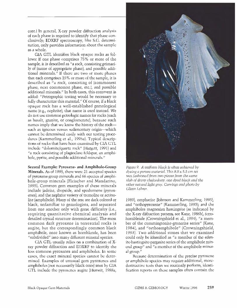
core.) In general, X-ray powder diffraction analysis of each phase is required to identify that phase con- clusively; EDXRF spectroscopy, like S.G. deterrni- nation, only provides information about the sample as a whole.
GIA GTL identifies black opaque roclzs as fol- lows: If one phase comprises 75% or more of the sample, it is described as "a roclz, consisting primari- ly of (name of appropriate phase), and possible addi- tional minerals." If there are two or more phases that each comprises 25% or more of the sample, it is described as "a roclz, consisting of (commonest phase, next commonest phase, etc.), and possible additional minerals." In both cases, this comment is added: "Petrographic testing would be necessary to fully characterize this material." Of course, if a blaclz opaque rock has a well-established gemological name (e.g., nephrite), that name is used instead. We do not use common petrologic names for roclzs (such as basalt, granite, or conglomerate), because such names imply that we know the history of the roclz- such as igneous versus sedimentary origin-which cannot be determined easily with our testing proce- dures (Kammerling et al., 1995a). Typical descrip- tions of roclzs that have been examined by GLA GTL include "dolomite/quartz rock" (Hargett, 1991) and "a rock consisting of plagioclase feldspar, an amphi- bole, pyrite, and possible additional minerals."
Second Example: Pyroxene- and Amphibole-Group Minerals. As of 1995, there were 21 accepted species of pyroxene-group minerals and 66 species of amphi- bole-group minerals (Fleischer and Mandarino, 1995). Common gem examples of these minerals include jadeite, diopside, and spodumene (pyrox- enes), and the nephrite variety of tremolite or actino- lite (amphibole). Many of the rest are dark colored or black, unfamiliar to gemologists, and separated from one another only with great difficulty (i.e., requiring quantitative chemical analysis and detailed crystal structure determination). The most common dark pyroxene in terrestrial roclzs is augite, but the correspondingly common blaclz amphibole, once known as hornblende, has been "subdivided" into many different mineral species.
GIA GTL usually relies on a combination of X- ray powder diffraction and EDXRF to identify the less common pyroxenes and amphiboles. In some cases, the exact mineral species cannot be deter- mined. Examples of unusual gem pyroxenes and amphiboles (not necessarily blaclz ones) seen by GIA GTL include the pyroxenes augite (Hurwit, 1988a,
Black Opaque Gem Materials
Figure 9. A uniform black is often achieved b y dyeing a porous material. This 8 . 0 ~ 5.5 c m set was fashioned from two pieces from the same slab of dnzsy chalcedony, one dyed blaclz and the other natural light gray. Carvings and photo b y Glenn Lehrer.
1989)) omphacite (Johnson and Kammerling, 1995), and "orthopyroxene" (Kammerling, 1993); and the amphiboles magnesian hastingsite (as indicated by the X-ray diffraction pattern; see Kane, 1986b), ferro- hornblende (Crowningshield et al., 1994), "a mem- ber of the cummingtonite-grunerite series" (Kane, 1984)) and "orthoamphibole" (Crowningshield, 1993). Two additional stones that we examined could only be identified as "a member of the eden- ite-hastingsite-pargasite series of the amphibole min- eral group" and "a member of the amphibole miner- al group."
Because determination of the precise pyroxene or amphibole species may require additional, more- destructive tests than we routinely perform, identi- fication reports on these samples often contain the
GEMS & GEMOLOGY Winter 1996 259

comment, "Petrographic testing would be neces- sary to fully characterize this material." This com- ment means that some of the "less appropriate" tests mentioned earlier would be needed to deter- mine the exact mineral species. Sometimes the comment, "Additional minerals may be present," is added as well.
Third Example: Black Opaque Spinel-Group Minerals. The blaclz members of the spinel mineral group present significant identification challenges, since the group contains 21 species (Fleischer and Mandarino, 1995), and many of these form com- plete solid solutions (similar to the garnets). However, only four of these end-members are important components of the black spinels that we have examined: spinel, hercynite, magnetite, and, to a much lesser extent, chromite (Fryer et al., 1982; Hurwit, 1984; Welch, 1987; Koiv~ila et al., 1993a; Johnson, 1994).
Magnetite and hercynite have refractive indices that are over the limit of the standard refractometer (that is, greater than 1.81)) but the R.I. of spinel can be measured, as can those of some of the interme- diates between spinel and hercynite. When measur- able, the R.I. of a spinel-group mineral provides an important clue about that mineral's identity.
For many of the blaclz spinels in our experi- ence, the measured specific gravity values (3.78-3.88 for five hercynites, including the two shown in figure 2, and 4.81-4.83 for the two mag- netites in figure 2) are lower than those predicted by Deer et al. (1966) from end-member composi- tions; however, other authors cite different S.G. values for hercynite (see, for instance, Phillips and Griffen, 1981). At least some of the hercynites that we have examined, and all of the magnetites, proved to be magnetic (e.g., Johnson, 1994).
The unit-cell spacing, a, is the fundamental distance between identical groups of atoms in the atomic structure of an isometric crystalline materi- al. In spinel-group minerals, this spacing can be measured by X-ray powder diffraction analysis, that is, by carefully measuring the spacings of arcs on the photographic film, with comparison to a stan- dard, preferably one mixed with the powdered unknown (an "internal" standard).
The general formula for the spinel group is AB204, where A and B are different cation ele- ments; spinel itself is Mg&04; and other spinel- group minerals contain Fe, Zn, Mn, Cr, V, Ni, Co,
Cu, Ti, and Ge. (Hercynite is Fe&04, magnetite is FeFe204, and chromite is FeCr204.) All of these elements (except oxygen) can be detected by the EDXRF instrumentation available at GIA GTL, although special attention must be paid to the lightest elements, Mg and Al, so that they are not overloolzed. EDXRF spectroscopy is a necessary step in identifying an unknown spinel-group min- eral, to make sure that no important component is overloolzed.
Deer et al. (1966, p. 431) provide a calibration chart for R.I., S.G., and a for six spinel-group miner- als, in the two series spinel-hercynite-magnesio- chroinite-chromite and spinel-hercynite-magnesio- ferrite-magnetite. We can identify a spinel-group mineral using this chart, our determined values of R.I. and a, and the EDXRF results for chemistry. (Because there is some debate as to the appropriate S.G. values to use, we do not include this informa- tion.) Black spinel-group minerals examined by GIA GTL include: spinel (Fryer e t al., 1982; Hurwit, 1984), hercynite (Johnson, 1994), mag- netite (Welch, 19871, and an intermediate between spinel and hercynite (Koivula et al., 1993a).
SUMMARY AND CONCLUSION Blaclz opaque gem materials present a special iden- tification problem, because many basic gemologi- cal identification techniques require the transmis- sion of light through the sample. In some cases, standard examination techniques are adequate to distinguish among common black opaques, at least to some minimum level of confidence. Certain modifications of these techniques, especially visual observation with reflected light, may provide addi- tional insights. Some specific but less routine tests (such as for magnetism) can also be very useful. By- now-routine advanced testing procedures, such as X-ray powder diffraction analysis and EDXRF spec- troscopy, give information that cannot be obtained by standard gemological testing alone. These advanced tests are usually required to positively identify most blaclz opaque materials. With black opaques, as with other gem materials, all of the information available should be used to identify a sample. Because they are blaclz and opaque, some uncertainty may remain about their identity even after advanced testing. In such cases, destructive testing techniques may be the only option for posi- tive identification.
260 Black Opaque Gem Materials GEMS & GEMOLOGY Winter 1996

REFERENCES Ashbaugh ni C.E. (1992) Gamma-ray spectroscopy to measure
radioactivity in gemstones. Gems ed Cemology, Vol. 28, No. 2, pp. 104-1 1 1 .
Crowningshield G.R. (19901 Gem trade lab notes: Diamond- Fancy black. Gems is) Gemology, Vol. 26, No. 3, p. 221.
Crowningshield G.R. (1993) Gem Trade lab notes: Iridescent orthoamphibole, "nuurnmite."Gems a) Gemology, Vol. 29, No. 4, p. 281.
Crowningshield G.R., Johnson M.L., Reinitz I. (1994) Gem trade lab notes: Iron-rich hornblende. Gems a) Gemology, Vol. 30, No. 3, pp. 186-187.
Deer W.A., Howie R.A., Zussman J. (1966) An Introduction to the Rock-Forming Minerals. Longman Group, London, 528 PP.
Fleischer M., Mandarino ).A. (19951 Glossary of Mineral Species 1995, 7th ed. Mineralogical Record, Tucson, Arizona, 280 PP.
Fryer C. (1988) Gem trade lab notes: Black pyrope garnet. Gems a) Gemology, Vol. 24, No. 2, pp. 116-1 17.
Flyer C., Crowningshield R.C., Hurwit K.N., Kane R.E. (19821 Gem trade lab notes: Spinel?. Gems e) Gemology, Vol. 18, No. 3, p. 173.
Fryer C., Crowningshield R., Hurwit K.N., Kane R.E. (1984) Gem trade lab notes: Hematite, magnetic. Gems &> Gemology, Vol. 20, No. 1, pp. 46-47.
Fuhrbach J. (19871 Editorial forum: Want to buy a "hot dia- mond"? Gems a) Gemology, Vol. 23, No. 2, p. l l l .
Gem news (1987) Large ekmite found. Gems a) Cemology, Vol. 23, No. 2, p. 123.
Goebel M., Dirlain D.M. (19891 Polynesian black pearls. Gems a> Gemology, Vol. 25, No. 3, pp. 130-148.
Cubelin E.J., Koivula J.I. (1986) Photoatlas of Inclusions in Gemstones. ABC Edition, Zurich, Switzerland, 532 pp.
Hargett D. (1990) Jadeite of Guatemala: A contemporary view. Gems ed Gemology, Vol. 26, No. 2, pp. 134-141.
Hargett D. (19911 Gem trade lab notes: Quartzite and dolomite bead. Gems d Gemology, Vol. 27, No. 4, p. 251.
Hurwit K.N. (1984) Gem trade lab notes: Black spinel. Gems a)
Gemology, Vol. 20, No. 2, p. 112. Hurwit K.N. (1985) Gem trade lab notes: Carved jet. Gems a)
Cemology, Vol. 21, No. 4, pp. 234-235. Hurwit K.N. (1988a) Gem trade lab notes: Augite, Chinese
"onyx." Gems a) Gemology, Vol. 24, No. 3, p. 170. Hurwit K.N. (1988b) Gem trade lab notes: Imitation dyed black
chalcedony beads. Gems a) Gemology, Vol. 24, No. 4, p. 241. Hurwit K.N. (19891 Gem trade lab notes: Transparent green
augite. Gems a) Gemology, Vol. 25, No. 1, p. 35. Johnson M.L. (1994) Gem trade lab notes: Magnetic hercyrute, a
warning about magnetic cards. Gems a) Gemology, Vol. 30, No. 1, p. 43.
Johnson M.L. (19951 Gem trade lab notes: Diamond-Fancy black, with iron. Gems a) Gemology, Vol. 31, No. 4, p. 266.
Johnson M.L., Kammerling R.C. (1995) Gem trade lab notes: Omphacite, rock carving. Gems a) Gemology, Vol. 31, No. 2, -. .
pp. 124-125. lohnson M.L.. Koivula 1.1. 119961 Gem news: "Drusv" silicon. a '
computer-industry by-product. Gems a) ~ e m o l h g ~ , Vol. 32, No. 2, pp. 138-139.
Kammerhg R.C. (1993) Gem trade lab notes: Orthopyroxene, a carved mask. Gems a) Gemology, Vol. 29, No. 4, p. 281.
Kammefling R.C., Kane R.E., Koivula J.I., McClure S. F. (1990) An investigation of a suite of black diamond jewelry. Gems a) Gemology, Vol. 26, No. 4, pp. 282-287.
Kammerling R.C., Koivula J.I., Kane R.E., Fritsch E., Muhlmeister S,, McClure S.F, (19911 An examination of non- transparent "CZ" from Russia. Gems &! Gemology, Vol. 276, No, 4, pp. 240-246,
Kammerling R., Johnson M.L., Moses M. (1995a) Describing aggregate gem materials. ICA Gazette, February 1995, p. 7.
Kamrnerling R.C., Koivula J.I., Johnson M.L., Fritsch E. (1995b) Gem news: "Drusy" hausmannite. Gems a) Gemology, Vol. 31, No. 3, pp. 207-208.
Kane R.E. (1984) Gem trade lab notes: Cun~mington.ite-gruner- ite, a series in the amphibole group. Gems a) Gemology, Vol. 20, No. 2, pp. 106-107,
Kane R.E. (1985) Gem trade lab notes: Corundum-Black star sapphire doublet. Cems a) Gemology, Vol. 21, No. 3, p. 171.
Kane R.E. (1986a) Gem trade lab notes: Ekanite, a markedly radioactive metamict gem. Gems ed Gemology, Vol. 22, No. 1, pp. 47-48.
Kane R.E. (1986b) Gem trade lab notes: Hornblende amphibole, magnesian hastingsite(?). Gems o) Gemology, Vol. 22, No. 1, p. 49.
Kane R.E. (1986~) Gem trade lab notes: Devitrified glass, cobalt- bearing. Gems a) Gemology, Vol. 22, No. 2, p. 108.
Koivula J.I. (1982) Pinpoint illumination: a controllable system of lighting for gem microscopy. Gems a) Gemology, Vol. 18, No. 2, pp. 83-86.
Koivula J.I., Kammerling R.C. (1989) Gem news: Psilomelane and basalt-novel black carving materials. Gems e4 Gemology, Vol. 25, No. 4, pp. 246-247.
Koivula J.I., Kammerling R.C. (1990a) Gem news: Star ekanite. Gems a) Gemology, Vol. 26, No. 1, p. 101.
Koivula J.1, Kammerling R.C. (1990b) Gem news: Black cat's- eye opal. Gems a) Gemology, Vol. 26, No. 4, p. 304.
Koivula J.I., Kammerling R. C. (1991) Gem news: Drusy gems in jewelry. Cems a) Gemology, Vol. 27, No. 1, p. 49.
Koivula J.I., Kammerling R.C., Fritsch E. (1992a) Gem news: Imitations of sugilite and other nontransparent gems. Gems a) Gemology, Vol. 28, No. 1, p. 66.
Koivula J.I., Karnmerling R.C., Fritsch E. (1992b) Gem news: Update on nontransparent CZ. Gems ed Gemology, Vol. 28, No. 2, p. 138.
Koivula J.I., Kammerling R.C., Fritsch E. (1992~) Gem news: More on irradiated "black" diamonds. Gems a) Gemology, Vol. 28, No. 4, pp. 276-277.
Koivula J.I., Kammerling R.C., Fritsch E. (1993a) Gem news: Black spinel from Mexico. Gems is) Cemology, Vol. 29, No. 3, pp. 212-213.
Koivula J.I., Karnmerling R.C., Fritsch E. (1993b) Gem news: Irradiated phenomenal quartz. Gems d Gemology, Vol. 29, No. 4, p. 288.
Koivula J.I., Kammerling R.C., Fritsch E. (1994) Gem news: "Zebra" stones from Australia. Gems a) Gemology, Vol. 30, No. 2, pp. 128-129.
Liddicoat R.T. Jr. (1989) Handbook of Gem Identification. Gemological Institute of America, Santa Monica, California, 364 pp.
Moses T. (19891 Gem trade lab notes: Chalcedony, imitation "black onyx." Gems a) Gemology, Vol. 25, No. 3, p. 171.
Muller H. (19871 Jet. Butterworth and Company, London, England, 149 pp.
Phillips W.R. and Griffen D.T. (19811 Optical Mineralogy. W. H. Freeman and Company, San Francisco, California, 677 pp.
Reinitz I., Ashbaugh 111 C.E. (19921 Gem trade lab notes: Diamond-Treated "black" diamond. Gems a) Gemology, Vol. 28, No. 2, pp. 124-125.
Webster R. (1994) Gems: Their Sources, Descriptions and Identification, 5th ed. Rev. by P.G. Read, Butterworth- Heinemann Ltd., Oxford, England, 1026 pp.
Welch C. (19871 Gem trade lab notes: Magnetite, lodestone. Gems a) Gemology, Vol. 23, No. 1, p. 45.
Welch C. (1988) Gem trade lab notes: Unusual glass. Gems a)
Gemology, Vol. 24, No. 2, p. 113.
Black Opaque Gem Materials GEMS & GEMOLOGY Winter 1996 26 1

AND SCAPOLITE WITH UNUSUAL PROPERTIES FROM EMBILIPITIYA, SRI ~ K A
By Pieter C. Zwaan
Gem-quality enstutite, cordierite, komerupine, and scapolite with unusu- al properties have been recovered from alluvial deposits in the region of Embilipitiya, Sri Lanka. The occurrence of almost pure, completely color- less, enstatite appears to be unique in the world. The cordierites are also usually colorless and nonpleochroic, The diversity in the properties of hornempine is mainly due to variations in the ratio of magnesium t o iron. The strong cccanary" yellow fluorescence of scapolites from this locality has not been observed in scapolites from other sources in Sri Lanka.
Since 1984, a group of mineralogists from Leiden, the l ether lands, have performed systematic field work in Sri Lanka. During the course of this research, they accumulated a representative collec- tion of rocks and minerals from that country.
ABOUT THE AUTHOR
Dr. Zwaan is emeritus professor of gemology (Ph. D. Mineralogy) at Leiden University, The Netherlands, and retired director of the Netherlands GemmotOgleal Laboratory, Leiden.
Acknowledgments: The author is much indebted to Dr. Charles â S. Arps of the National Museum of Natural History, Leiden, for his cooperation, years of fieldwork in Sri Lanka, and critical reading of the manuscript; and to E. GaminiZoysa, Mount Lavjnia, Sri G k a , for his valuable information and sucolv of material from different localfiies. The author also thanks k A J. Burke and W. J. Lusten- homer, of the Institute of Earth Sciences, Free University of Amsterdam, for the Raman spectroscopy and electron rnicro- probe analyses, respective&. D. van der Marel, of the National Museum technical staff, prepared the geologic map and pho- tographed the cut stones. The financial support of the Foundation Stichting Dr. Schijnnannfonds is gratefully acknowledged.
ta/land, October 1995. It was the last conference in wh, )bert C. Kammerling participated.
3ms & Gemology, Vol. 32, No. 4, pp. 262-269.
This article was developed In pert from a paper presented at the 25th Internatjonal Gemmological Conference in R a m Resort, Th toh Re
Gf
Although the well-known gem deposits of Ratnapura are still commercially the most important in Sri Lanka, other localities-such as Okkampitiya and Elahera-have also become interesting. Farther to the southeast, west of Kataragama, the Leiden researchers identified yet another locality of special mineralogic and gemological interest: Embilipitiya. Some gem minerals from this region were briefly described earlier (Zwaan, 1986). The present article discusses specific properties, and special aspects of these properties, for gem-quality enstatite, cordierite, komerupine, and scapolite (figure 1).
GEM MINERALS FROM EMBILIPITIYA Geoscientists have examined the geology, in par- ticular the petrology, of Sri Lanlzan basement rocks for many years. To date, however, there has been no clear explanation for the origins of most of the gem minerals. The best summary of the results obtained thus far are the recent article by Dissanayake and Rupasinghe (1995) and the book by Gunaratne and Dissanayake (1995). Most impor- tant gems are recovered from alluvial deposits; the few gem minerals that have been traced to their in situ sources are not commercially important (i.e., of cutting quality).
262 Notes &i New Techniques GEMS & GEMOLOGY Winter 1996

Embilipitiya is a small village about 70 lun (1.5 hours) by road southeast of Ratnapura. The village has a population of about 300 and is situated in a flat valley, about 25 km2, surrounded by distant hills. The region is tropical, with bothwet and dry seasons. Petrologically, it is in the Highland Complex, which contains Precambrian rocks of the metamorphic granulite facies (figure 2). Most of the gem deposits in Sri Lanka are located w i t h tiis complex.
A variety of gem-quality minerals are found in the Embilipitiya region, in an area of about 6 lzm2, primarily in alluvial deposits. Locals engage in small-scale primitive mining, digging narrow pits in the alluvium to a depth of 1.5-2 m (figure 3) to reach the gem-bearing gravels.
Of special interest, because of their unusual gemological properties, are scapolite and the Mg- rich minerals enstatite, cordierite, and komempine. Other gem minerals from this area include garnets, mainly alinandine, and spinels. Since our first trip in 1984, we have visited the area several times and obtained several hundred carats of these unusual minerals from local inhabitants. During the last five years, however, greater demand by cutters and decreasing production have made it difficult to obtain more samples. These minerals are found mostly as broken and corroded fragments, seldom as rounded pebbles. Komempine is the most com- mon of the four gems discussed in this article.
MATERIALS AND METHODS During fieldwork in 1984, the author and his col- leagues discovered Embilipitiya on the way from Ratnapura to gem localities in the southeast. At a lunch stop in the village, native people showed us dullam, pieces of rough gem minerals, that they were offering for sale. According to these villagers, the minerals came from the Embilipitiya area. At that time, we purchased about 3 kg of material, with a preponderance of colorless specimens. When we inspected these materials at the Netherlands Gemmological Laboratory in Leiden, we found that most of the samples were garnets and spinels, but we also identified a number of much less common gem minerals. During subsequent trips to Sri Lanka, we visited the village regularly and pur- chased almost every lot that was available.
Within the sample minerals were many frag- ments without crystal faces but with a recogriiz- able-though usually corroded-prismatic habit. Using a polarizing microscope, we were able to group hundreds of specimens by species. The
Notes & New Techniques
L c Figure 1. These f o w gem materials~cordierite (left, 3.75 ct), enstatite (top, 7.69 ct), scapolite (right, 3.08 ct), and kornerupine (bottom, 1.52 ct) -from the Embilipitiya area of Sri Lanka were found to have unusual gem properties. Photo by Shane F. McClz~re.
enstatites were selected for further study because of their range of color, from colorless to deep brown. About 20 rough specimens with a prismatic habit, approximately 10 to 30 mm long, were confirmed to be enstatite by their absorption spectra. In addi- tion, we examined 10 cut enstatites; the largest was a 10.67 ct colorless specimen.
From among the cordierites, we selected about 40 rough samples (15 to 20 mm each) for further study. Most of these were nearly colorless, but some were pale blue. They were easily distin- guished from the enstatites by their behavior in ethylene dibromide, in which the cordierites are almost invisible. Five cut stones, the largest of which was 3.75 ct, were available for examination.
A total of 56 rough kornerupines, varying in size from 5 to 20 mm and in weight from 0.91 ct (dark green) to 13.03 ct (dark brown), were selected for detailed study on the basis of their colors, inclu- sions, and physical properties (specimens with low and high refractive indices and specific gravities were represented). In addition, nine cut stones, the largest of which was 4.56 ct, were examined.
We examined 10 rough scapolites in detail. These were irregular, long prismatic, colorless-to- yellow pieces that ranged from 3.66 to 7.06 ct in weight and from 12 to 16 mm in their longest dimension. In addition, we studied seven cut scapo- lites from Embilipitiya, ranging from 0.80 ct (yel-
GEMS & GEMOLOGY Winter 1996

COLOMBO
figure a. m e village of Em bmpniya is m o m /u Km southeast of Ratnapma. It is in the Highland Com- plex, one of the main Precambrian geologic complex- es that comprise the island nation of Sri Lanka.
low) to 17.79 ct (colorless), and compared their properties to those of 17 cut scapolites from else- where in the world. Scapolite is the name given to an isomorphous series in which marialite and meionite are the Na-rich and Ca-rich end inem- bers, respectively. The intermediate members are usually indicated by their molecular percentage (mol. % ) of meionite.
The physical properties of all specimens were measured by standard gemological methods; for this purpose, two flat parallel faces were polished on each, thus forming a window. A Rayner refractome- ter with an yttrium aluminum garnet (YAG) prism was used to measure the refractive indices and bire- fringence. Specific gravity was determined with a hydrostatic balance.
The absorption spectra of all specimens were observed with both an Eickhorst handheld prism spectroscope and a Philips UV/ViS spectrometer (model PU 8730). X-ray powder diffraction pho- tographs were taken on representative samples of all gem minerals using a Debye-Scherrer camera on an Enraf-Nonius Diffractis 582 apparatus. Chemical analyses were performed on eight lzomer-
Notes & New Techniques
upines and six scapolites using an electron micro- probe (JEOL model JXA-8800M), with three to five analyses per sample and the results averaged. The inclusions, mainly in the lzoinerupines and scapo- lites, were identified with a laser Raman micro- spectrometer (Dilor S.A. model Microdil-28).
ENSTATITE The enstatites (Mg2Si206) varied from colorless to deep brown (figure 4). The colorless stones had exceptionally low refractive indices (as low as nn. =1.650 and 5=1.658] and specific gravities (as low as 3.194). Microprobe analysis of one colorless stone revealed that 99.4 mol.% of the magnesium sites in this stone were occupied by Mg. This com- position is very near the magnesium end-member of the enstatite-ferrosilite series. In fact, these were
Figure 3. The gems are recovered from shallow (about 1.5-2 m) alluvial pits in the Embilipitiya area. Photo hv Pieter C %an.
GEMS & GEMOLOGY Winter 1996

cordierites were very rich in Mg, up to 95 mol.% of the possible Mg sites. These confirm earlier micro-
Pigme 4. The high-Mg enstatites from Embilipitiya are among the purest natural enstatites known. The enstatites shown here were all part of the study sample; the colorless 10.67 ct oval in the center is 20.5 mm in longest dimension.
the purest natural enstatites ever examined at the Netherlands Gemmological Laboratory.
The sample specimens had a distinct to very strong narrow absorption band at 506 nm, due to fer- rous iron. The intensity of this band-which was present, though weak, even in the colorless stones- increased with the saturation of the brownish color, that is, with the Fe content. This absorption band was found to be characteristic of this material and, therefore, of diagnostic value. Most of the stones were relatively inclusion free, but some contained well-developed quartz crystals and two-phase inclu- sions. (For additional information on enstatites from this locality, see Zwaan, 1987.)
CORDIERITE We prefer the mineral name cordierite (MgT&- Si5018), instead of the gem names iohte or dichroite, for these colorless stones. All the crystals we exain- ined had a prismatic habit and were broken and cor- roded. We found little variation in their physical properties (refractive indices and specific gravity). Their refractive indices varied from 1.520 (lowest nJ to 1.541 (highest nJ, and they had an average specific gravity of 2.570. Pleochroism, usually a diagnostic property for cordierite, was not visible because of the lack of color. In most cordierites, iron (and other trace elements) can substitute for Mg, producing color. However, microprobe analyses showed that these
Notes & New Techniques
probe analyses of some specimens (Zwaan, 1986), which also showed relatively high Mg content.
Tothe unaided eye, when cut these cordierites (see, e.g., figure 5) strongly resembled colorless enstatites from the same locality. Immersion in ethylene dibromide, however, was sufficient to sepa- rate the two gem minerals easily, because the R.I. of the liquid is very similar to that of cordierite. In addition, the cordierites do not have the 506 nm absorption band in the blue that is found in enstatite. Liquid inclusions were common but not of any diagnostic value.
KORNERUPINE Although considered rare (see, e.g., Webster, 1994), hundreds of carats of komerupine [Mg4(AllFe+3)6- (Si,B)40:Ti(OH)] have been found in the Embilipitiya deposits (see also Zwaan, 1986, 1992). The author and colleagues saw large quantities of crystal frag- ments, varying from light brownish yellow, brown, or green to dark brown and dark grayish green (see, e.g., figure 6). Most of the specimens we examined were irregular in shape, because of fractures, and were corroded. Because we did not observe any crys- tal faces, we could not identify a typical habit. Note that chatoyant lzomerupine has not yet been reported from this area.
Specific gravity, ranging from 3.283 to 3.346, generally increased with color intensity (table 1).
Figire 5. Also very high in Mg, the cordierites from Embilipitiya strongly resemble colorless enstatites from this locality. Here, the largest cordierite weighs 3.75 ct and is 11.8 mm long.
GEMS & GEMOLOGY Winter 1996

Figure 6. Korl~erupine is usually considered a rare gem material. However, the author saw hundreds of carats of gem-quality lzornerupines in Embilipitiya, which ranged from light brownish yellow to the dark brown and dark green stones shown here (the 4.56 ct stone at the center top is 12.3 m m in longest dimension).
Light brown specimens rose in methylene iodide (S.G. = 3.32), while dark brown samples slowly sank in this liquid. This behavior in methylene iodide, together with the very strong pleochroisin in tones of greenish yellow and brownish yellow, appears to
be a reliable test for lzornerupine specific to this locality: Every green-to-brown strongly pleochroic gemstone from Embilipitiya that either rose or sank slowly in methylene iodide was lzomerupine. The refractive indices varied from the lowest r ~ , of 1.665 (for a light brown specimen) to the highest IL, of 1.690 (for a medium brown sample). The birefrin- gence, however, was usually 0.012.
Electron microprobe analyses of eight rough specimens, onto which one or two flat faces had been polished, indicated that the color intensity tended to be directly proportionate to the Fe content (again, see table 1). In general, pale lzomerupines have distinctly lower S.G.'s and R.I.'s than dark lzomerupines, but hue (that is, whether the stone is brown or green) has no effect on these properties.
Most of the Embilipitiya kornerupines we examined had interesting inclusions. The solid inclusions were identified by Raman spectroscopy. Well-crystallized black inclusions with a submetal- lie luster, seen in about 25 samples, were found to be rutile. One specimen also had red crystals of rutile (figure 7). Another common inclusion was colorless zircon, often with beautiful haloes in polarized light [figure 8). Rounded apatite crystals occurred frequently. However, elongated quartz
Sample
Properties A1 A 2 A 3 A 4 A 5 A 6 A 7 A 8
Color Light green
Weight (ct) 1.91
Oxides (wt .%) Sic2 31.87 TiOo 0.00 AlzOs 42.36 FezOo 1.85 Mgo 19.55 MnO 0.01 CaO 0.01 Na20 0.02
Totala 95.67
S.G. 3.288 "a 1.668
1.679 arefringence 0.01 1
Brown
3.17
31.36 0.05
44.75 2.51
18.49 0.02 0.01 0.03
97.22
3.283 1.669 1.681 0.012
Dark brown
13.03
30.23 0.10
44.71 6.61
15.82 0.05 0.01 0.02
97.55
3.346 1.672 1.685 0.01 3
Light brown
4.63
31.79 0.04
44.15 1.63
19.52 0.03 0.01 0.02
97.19
3.284 1.665 1.677 0.01 2
Green
3.39
30.98 0.05
43.37 5.40
17.21 0.07 0.01 0.02
97.11
3.337 1.678 1.687 0.009
Dark green
0.91
31.35 0.01
43.08 5.13
17.62 0.10 0.02 0.03
97.34
3.339 1.672 1.684 0.01 2
Medium brown
1.59
30.95 0.06
43.36 5.17
17.39 0.05 0.01 0.02
97.01
3.335 1.678 1.690 0.012
Green
1.79
31.51 0.04
42.05 4.60
17.80 0.05 0.02 0.02
96.09
3.332 1.668 1.680 0,012
a Note: The element B and the OH group could not be detected by the analytical method employed; hence, the totals are less than 100%.
Notes & New Techniques GEMS & GEMOLOGY Winter 1996

Figure 7. This Embilipitiya kornerupine specimen was observed to have red as well as black rutile crystals. Photomicrograph b y Pieter C. Zwaan; magnified 35x.
and tourmaline inclusions, observed parallel to one another , were seen only occasionally. Some lzomerupines revealed red-orange hexagonal plates of hematite. Many liquid and liquid-and-gas inclu- sions were also seen.
To summarize, kornempines from Embihpitiya can be easily identified by their strong pleochroism and their behavior in methylene iodide. Pale-colored specimens contained considerably less iron than dark-colored samples. There were no distinctive dif- ferences between the optical (R.I.) and physical (S.G.) properties of brown and green lzomerupines. Rutile appears to be a typical inclusion in lzomerupine from this locality.
S CAPOLITE T h e crystal fragments and faceted scapolites [Na4A13Si9024Cl-Ca4A16Si6024(COi,S04)] we examined were, for the most part, colorless to pale yellow (figure 9). We found that scapolites from Embilipitiya have mean refractive indices of n,. =
1.550 and no = 1.578, an average specific gravity of 2.694, and an average meionite content of 59.4 mol.% (table 2). They fluoresce a strong "canary" yellow to long-wave UV radiation, comparable to the well-known fluorescence of scapolite (wemer- ite) from Quebec, Canada. This UV reaction is gen- erally ascribed to sulfur (Webster, 1994).
Although most of these samples were remarlz- ably clean, some contained yellow needle-shaped crystals, which Raman spectroscopy identified as pyrrhotite (figure 10). Pyrrhotite inclusions in scapolite have been mentioned by others (Graziani and Giibelin, 1981; Giibelin and Koivula, 1986).
Notes & New Techniques
The pale yellow faceted scapolites from Embili- pitiya resemble some other gemstones from Sri Lanlza, such as citrine and feldspar. However, the scapolites can be differentiated readily by their strong yellow fluorescence and their much stronger birefringence (0.028).
X-ray diffraction patterns were obtained from six samples. As expected, these patterns were almost identical to one another and were character- istic of scapolites in general.
Electron microprobe analyses were made of two scapolites from Embilipitiya and four scapo- lites from other sources (one each from Palzistan and Brazil, and two from Pohorabawa, a small vil- lage in the Eheliyagoda area that is the major source of colorless scapolite in Sri Lanlza; figure 9). As evident in table 2, there were remarkable differ- ences in chlorine content, and thus the marialitel ineionite ratio, among the various scapolites.
Note also in table 2 that the one violet scapo- l i te has the lowest specific gravity, refractive indices, birefringence, and meionite content (7.66 mol.%). Previously, the scapolites from East Africa were thought to have the lowest meionite content (10.80 mol.%; Zwaan, 1979). However, the meion- ite content of this violet scapolite from Palzistan was even lower. The properties of Embilipitiya scapolite, on the other hand, are very similar to those reported for scapolite from Madagascar (n,. =
1.550, no = 1.571; S.G. = 2.686: Bank and Nuber,
Figure 8. In addition to the black rutile crystals, this Embilipitiya kornempine specimen revealed zircon crystals with haloes visible in polarized light. Zircon is a common inclusion in this material. Photomicrograph b y Pieter C. Zwaan; magnified 35x.
GEMS & GEMOLOGY Winter 1996

rigz~re 9. The study 07 &mbuipitiyo scapolites included stones from different localities. For example, the 1.58 ct violet triangle-cut scapolite is from Palzistan; the 3.01 ct yellow triangle cut is from Pohorabawa, Sri Lanlw, as is the 17.79 ct colorless emerald-CUL sLone; and the 3.08 ct yel- low marquise cut is from Embilipitiya.
1970; Zwaan and Arps, 1980J, although the latter do not have the strong yellow fluorescence.
DISCUSSION: SPECIAL ASPECTS OF THE EMBILIPITIYA GEM MINERALS All four of these gem materials are of special inter- est both mineralogically and gemologically. The enstatites have very uniform chemical composi- tions, which is consistent with the fact that they came froin a very limited area. The extremely high Mg content of these tnlly colorless enstatites has not been reported in gem enstatites from any other locality. It is of particular interest that all the enstatites from Embilipitiya can be readily identi- fied with a simple hand spectroscopel by the pres- ence of the 506 nm absorption band.
Like the enstatites! the cordierites also showed little variation in chemical composition from one sample to the next; they! too! were found to have a very high Mg content. The refractive indices and specific gravities of the specimens tested were extremely low. The complete lack of color in most of the sainples is exceptional for cordierite! as is the resulting absence of pleochroism.
Kornerupine occurs abundantly i; different tones of green and brown. The extremely strong
pleochroism, combined with a number of very interesting inclusions (in particular rutile! zircon! and apatite], malze it an attractive colIector's gem- stone.
The chemical composition of the Embilipitiya scapolites varied little from stone to stone. The strong "canary1' yellow fluorescence is a very inter- esting property that has not been found in other Sri Lanlzan scapolites (although it has been seen in scapolites from other localities).
The Mg-rich minerals in this area are probably derived from originally high-Mg roclzs that have been subjected to deep-seated high-grade metamor- phism. These are now represented by metamorphic roclzs (i.e.! metamorphic granulite facies of the Highland Cornple~)~ such as hypersthene gneisses (charnockite~)~ sillimanite-garnet gneisses (lzhon- dalites], biotite-garnet gneisses, and crystalline limestones (marbles).
Orthopyroxenes, particularly enstatite, are typ- ical minerals of the granulite facies [Deer et al.! 1978). They also occur together with cordierite in medium-pressureJhigh-teinperature metamor- phosed pelitic roclzs. Komerupine is a typical min- eral in high-grade regional and contact inetamor- phic roclis~ whereas scapolite is cominonly found in marbles! calcareous gneisses! and granulites. Petrologic studies have shown that many granulite facies roclzs elsewhere in the Highland/So~~thwest Complex are also Mg rich. However! those in the Embilipitiya area seem to be inordinately so.
The gem minerals in Einbilipitiya are mined from alluvial deposits. Strongly weathered gran- ulites that contain enstatite were encountered in the field. These might be the host rocks in which
Figure 10. Some of the Embilipitiya scapoli~es con- tained neede-like pyrrhotite crystals. Photo- ~nicrograph by Pieter C. Zwoon; magnified 35x.
Notes & New Techniques GEMS & GEMOLOGY Winter 1996

TABLE 2. Properties, including electron microprobe analyses, of six scapolites [Na4A13Si9024Cl-Ca4A16Si60~4(C03,S04)] from Sri Lanka and elsewhere,
Sample
Propeflies 1004 RGM 3,Ol 1006 98-5 R9 164299
Origin
Color Form
Weight (ct)
Oxides (wt.%) Si02 Ti02 A1203
Fe203 MnO MgO Na20 K20 CaO so3 F CI F=O Cl=O
Totala
Pakistan
Violet cut
1.58
62.1 1 0.05
19.07 0.06 0 0
12.06 1.45 1.87 0.1 1 0 3.92 0
-0.88
Braiil
Yellow Rough
19.88
55.96 0.02
22.83 0.07 0.03 0 9.55 I .24 6.88 0.03 0.01 3.44 0
-0.78
Pohorabawa Sri Lanka
Yellow Colorless cut cut
3.01 8.03
Meionite (mol.%) 7.66 27.07 30.92 32.51
S.G. 2.584 2.622 2.651 2.632 "E 1.533 1.540 1.544 1.542 no 1.540 1.557 1.560 1.559 Birefringence 0.007 0.01 7 0.01 6 0.01 7
Embilipitiya Sri Lanka
Yellow Yellow Cut Rough
1.13 4.47
-
aNote: The carbonate (C03) content of the samples could not be detected by the analytical method employed; hence, the totals are less than 100%.
the gem minerals originally formed. In other parts roclzs; with the exception of some almandine gar- of Sri Lanlza! gem ininerals that occurred in sec- nets, however! none of the ininerals found in sit11 ondary deposits were found in sit11 in basement thus far have been of gein quality.
REFERENCES Bank H., Nuber B. (19701 D ~ ~ r c h s i c h t i g e r Slcapolitl~ aus
~Mo~ambique. Zeitschrift der Deutsche~~ Ge~nrnologischen GeseUschab, Vol. 19, No. 2, pp. 47-54.
Deer W.A., Howie R.A., Z ~ ~ s s m a n J. (1978) Roclc-Forming Minerals, Vol. 2A Longlxm Group Ltd., London.
Dissanayalce C.B., Rupasu~ghe M.S. (19951 Classification of gem deposits of Sri Lanlca. Gwlogie en Milnbo~~w, Vol. 74, No. 1, pp. 79-88.
Graziani G., Gubelin E. (19811 Observations on some scapolites of central Tmzmia. Iournal of Gemmology, Vol. 17, No. 6, pp. 395405.
Gubelin E.J., I<oivula J.1. (19861 Photoatl(~s of Incl~~sions in Gemstones. ABC Edition, Zurich, Switzerland.
G ~ ~ n a r a t n e H.S., Dissanayalce C.B. (1995) Gems and Gem Deposits of Sri Lanlca. Publication of the National Gems and Jewellery Authority of Sri Lanka, Unigraphs (Pte] Ltd., Colombo, Sri Lanlca.
Webster R. (1994) Gems, Their Sources, Descriptions and Identification, 5th ed. Rev. by P. G. Read, B~~tterworth- Heincn~ann Ltd., Oxford, England.
Zwaan P.C. (1979) more data on violet gem scapolite, probably from Eastern Africa. Io~unal of Gemmology, Vol. 16, No. 7, pp. 44845 1.
Zwaan P.C. (19861 Gem minerals from the Embilipitiya and Kataragama areas in Sri Lanlca. Austr(11ian Gemmologist, Vol, 16, No, 2, pp. 3540 .
Zwaan P.C. (19871 Orthopyroxenes from the Embilipitiya area in Sri Lanlca. Schweiz~r i sche Mineralogische und P~trographiscl~e Mitteilungen, Vol. 67, pp. 1 19-125.
Zwaan P.C. (1992) La korn6mpine d'Embilipitiya, Sri Lanlca. R ~ W G de Gemmologie, No. I 10, pp. 5-6.
Zwaan P.C., Arps C.E.S. (19801 Properties of gem scapolites from different localities. Zeitschrjft der Deutschen Gemmo- logjschen GeseUschab, Vol. 29, No. 1-2, pp. 82-85.
Notes & New Techniques GEIMS & GEiMOLOGY Winter 1996 269

NOTESAND NEWTECHNIQUES
By Lore Kiefert and Susanne Tll. Schmidt
Since ut leust 1995, ~mrious w~ute~~iuls have been munufuctu~~ed t o imi- tute tunzunite in the fiem t~ude, These inclade two filusses) two dijfer- ent TAGS (yttrium uluminum fiurnet), und u synthetic corundum. These imitation tunzunites can be ~fieudiljl separated @om the nutu~pul &em by stundu~d fiemolofiicul tests, such as ~~fructive indices) opticul chu~pucte~, specij"cfir~uvity, UV-visible spectm, fluorescence, and the presence or absence of pleochroism. The ident$icutions were confi~med by chemicul unulysis and Rumun spectyoscopy.
Tamanitel the blue-to-violet gem variety of zoisitel was first dscovered in Tanzania in the 1960s [seel e.g.! Banlz et al.! 1967). It has increasingly gained pop- ularity as a result of its unusual color and the attrac- tiveness of the cut stones. Most of the material that enters the gem trade is act~~ally brown zoisite that has been heat treated to blue to violet (Nassa~~! 1994). Although a number of gem materials! includ- ing spinel and iolite! may resemble tamanitel m'mu- factured imitations of this material have also started to appear on the marlzet ( w e 1). These recent tCm- zanite imitations were mentioned by I<ammerhg et 4. [I9951 and in the ICA Gazette ["Gem newsl!! 1996). Johnson and Koivula (19961 describe one of the hnita- tions in more detail. However, rumors of Russian imi-
ABOUT THE AUTHORS
Dr. Kieferl is a research scientkt at the SSEF Swiss G e m m ~ i c a l Institute, F a l ~ t r a s s e 9, CH-4001 Basel, Switzerland. Dr. Schmidt is a research scientist at the MineraQisch-ht~aphisches lnstitut, Un/verdt+t Basel, CH-4056 Ba~d . AckmMgmmts: ?Re authors thark the Lannyte Co., Houston, Texas, for provMing sampI@ of Purple Coranite, and Rot B. Lasnier, of the University of Nantes, France, for the loan of the calcium phosphate glass. Dr. H. A. Htinni, of the SSEF Swiss Gemmological Institute, look the photos h rigures 7 and 2, provided c o m p a h material, 8nd contributed help- ful information. Mr T. Ostertag, Univmity of Freibuw, Germany, helped with the Raman sp&ctra. Special thanks to Mr. J-P. Chlain, of SSEE for reviewing the paper. A1 pho- tomicrogra~s are by the senior author.
Gems i% GXmokm, Vd. 3, No. 4, pp. 270-276. 0 1996 Gerndog/cal Institute of America
tations! and of efforts by Russian manufacturers to produce synthetic tamanitel were mentioned as early as 1991 (Koivula and Kamrnerhg, 1991).
The present article characterizes and provides identification criteria for some of the imitations that have recently appeared on the marlzet! includ- ing: (11 a doped heavy glass marlzeted under the trade name U.M. Tanzanic and produced by U.M. Science Companyl Glendale! California; (2) a YAG (yttrium aluminum garnet! a manufactured inateri- al) from Lannyte Companyl Houstonl Texas! which has been sold under the trade name Purple Coranite [figure 2); and (3 ) a synthetic corunduml also from Lannyte) marlzeted as Blue Coranite (fig- ure 1 ]. Also included are another glass and another YAG! with slightly different propertiesl that could be mistaken for tanzanite without gemological testing.
For the gemologistl these materials are easy to distinguish from tanzanite [see Box A) on the basis of specific gravity! refractive indices! optic charac- ter! internal features, and-to a lesser extent-their spectroscopic characteristics. To characterize these simulants fully, they were also analyzed chemical- ly and by Raman spectroscopy.
CHARACTERISTICS OF THE TAhTZANITE IMITATIONS Materials and Methods. The three imitations noted above-U.M. Tamanic1 Blue Coranitel and Purple Coranite-were readily available at the February 1996 Tucson gem shows. The authors purchased
270 Notes & New Techniques GEMS & GEMOLOGY Winter 1996

five faceted samples (0.59-2.89 ct) of the heavy glass (U.M. Tanzanic) and one 1.64 ct faceted syn- thetic corund~lm (Blue Coranite) for investigation purposes. In addition! the Lannyte Company lzindly supplied us with one rough (1 12.54 ct) and one 0.72 ct faceted piece of their YAG [Purple Coranite).
We also included in our study a 1.99 ct blue YAG from Russia and a 6.90 ct b l ~ s h purple Ca-phosphate glass (again, see figure 1). Although these two samples do differ somewhat in color from tanzanitel they are s ~ c i e n t l y close to be mistaken for it.
We determined refractive index! optic charac- ter! and specific gravity using standard geinological instrumentation and techniques, S.G. was deter- mined hydrostaticallyj the values reported are the average of at least two measurements per sample. Microscopic investigations were carried out with darkfield illumination ill air with a Geinolite binocular microscopel as well as with immersion in methylene iodide using a Nachet horizontal microscope. We tested the fluorescence to short- and long-wave UV radiation using a commercial gemological UV lamp! and we tested spectroscopic properties with a laboratory spectroscope (both instruments by System Eiclchorst).
Semi-quantitative chemical analyses of al l sam- ples were performed by energy-dispersive X-ray fluo- rescence (EDXM) spectroscopy on a Tracor Northem TN 5000 system. In addtion! q~iantitative chemical data for samples of the heavy glass (U.M. Tanzanic) and the synthetic corundum (Blue Coranite) were obtained using a JEOL 8600 electron inicroprobe. Raman spectroscopy was carried out on all samples with a Renishaw Raman System 1000 with a 25 mW air-cooled Argon Ion Laser (Omnichrome) lasing at 514 nm. Analysis times were between 60 and 100 sec- onds! dependmg on pealz intellsities and baclzground fluorescence; the spectral range covered was between 100 and 1900 cm-I (see also Hanni et al.! 1996).
U.M. Tanzanic. In their advertising brochure for this material! U.M. Science Co. claims that it was created by means of a high-temperature flux pro- cess. The material is described as a mix of amor- phous and microcrystalline phases! with a refrac- tive index of 1.66-1.67 and a specific gravity of 3.8-4.0. The brochure also lists a number of other properties! such as the absence of internal stress.
Gemologicul Properties. Our res~llts for the five faceted samples that we tested did not correspond to those published by the producer. The refractive indices varied between 1.600 and 1.605 (isotropic).
Figure 1. Several manz7factured materials have appeared on he gem marliet as tanzanite imita- tions. A number of these are shown here together wit11 two notz~rol tanzanites. From left to right: (top row) two tanzanites (3.03 ct ancl 5.89 ct) ancl a 6.90 ct sample of Ca-phosphate glass; {bottom row) a 1.64 ct synthetic corundum (caged Blue Coranite), a 1.99 C L synthetic garnet from Russia, and two samples (2.89 ct and 0.92 ct) of heavy glass (U.M.Tanzanic). Photo by H. A. Hanni,
The specific gravity ranged from 3.36 to 3.48. All specimens showed a poorly polished surface. Three of the five samples were virt~lally free of any inter- nal features! but two showed diffusel syrup-lilze flow structures. One of these two samples also had an irregularly shaped ~nillzy white incl~~sion! proba- bly a microcrystalline portion of the surrounding glass! with syrup-like flow structures (figure 3). The samples fluoresced chalky white to both short- and long-wave UV. They showed wealz absorption bands at approximately 490,550, and 590 nm.
Chemical Composition. We recorded an inhomo- geneity in chemical composition among the five samples (see table 1)) as well as within the samples. PbO contents between 38.5 and 51.2 wt.Yo were measured! which appear to be extremely high, The products of other heavy-glass man~~facturers only go up to 30 wt.Yo PbO (Swarogeml pers. comm., 1996). Perhaps U.M. Science Co. uses a light ele- ment such as B or Li in their glasses! neither of which is detectable using the microprobe or X-ray fluorescence units available to the authors. Si02 contents varied between 46.3 and 58.3 wt.Yo. The K20 content ranged between 2.3 and 3.2 wt.Yo, and the trace elements Mn and Fe were 0.084.14 wt.Yo and 0.03-0.07 wt.Yo, respectivelyl expressed as oxides (see table 1). For the two major elements, Si and Pb! Si02 and PbO concentrations within one
Notes & New Techniques GEMS & GEMOLOGY Winter 1996 271

Figure 2. Purple Coranite is u YAG manufactured by the Czochralski method that is marketed as a tanzanite imitation. T l ~ e unfashioned saction weighs 112.54 ct; the faceted piece is 0.72 ct. Photo by H, A. Hiinni.
stone varied by up to 7 wt."/~ (see table 1). A1203 levels were below the detection limits of the EDXRF apparatus and t11e microprobe.
The Raman spectra of this material showed broad pealzs at Raman shifts of 505! 777! 989! and 1064 cm-1 (figure 4)) as well as a considerable fluo- rescence to Argon laser light at 514 nm, as can often be observed in glass (Chemarin et al.! 1996; Sharma and Wangl 1996).
Coranite. The Lannyte Coinpany offered two tanzan- ite imitations: a YAG marlzeted as Purple Coranite and a synthetic corundum sold as Blue Coranite, The inanufacturer reported that both materials are grown using the Czochralslzi-puhg method.
Purple Corunite. The man~lfact~lrer's brochure reported that "the material is garnet based, with a hardness of nearly 9 and a refractive index of over 1.80, Our investigations confirmed that the R.I. is over 1-80! and we measured a specific gravity of 4-58!
Figme 3, Solid incl~lsions and a syrup-like flow structura are evident in this heavy glass manu- factured as a tanzanite imitation by U.M. Science Co, Immersion, magnified 35x
which is slightly higher than published data for YAG (e.g., Nassau! 1980). We saw no pleochroism in the samplesl but we did see some strain with the polar- iscope. Spectroscopic observation showed a broad band at approximately 540 nm.
The material fluoresced a strong challzy orange to short-wave UV radiation (figure 5). Similar data were obtained by Johnson and Koivula (1996) on a 7.07 ct tanzanite imitation purchased in 1995. EDXRF analysis of their specimen revealed yttri- um! aluminuml and europium; all three elements were found in the EDXRF analyses of our two pur- ple coranite samples.
The piece of unfaceted Purple Coranite was polished on one side for microscopic examination. However) we saw no bubbles or other internal fea- tures. Color zoning was apparent with the unaided eye: The core was a distinctly darlzer purple than the riml and the color faded gradually from core to rim. The orange fluorescence corresponded to the zoning! but inversely; that isl it was brightest near the colorless rim (figure 5). Microscopic examina- tion of the small faceted sample showed no growth
TABLE 1, @mi@ carpdon of glass imi tatb of lanzaniie iTcmn U.M. Science Co.
Oxide Sample la Sample 2b Sample 3a Sample 4a Sample 9 (vvt.%) (2.89 ct) (1 .&l ct) (0.92 ct) (0.76 ct) (0.59 ct)
Si02 51.26-58.30 51.39-55.77 50,2555.50 46.32-51 $55 46.49 -50.1 3
h o 2.87- 3.09 2.66- 3.15 2.89- 2.94 2.29- 2.84 2.29- 2.62 MflO 0.N- 0.10 0.08- 0,09 0.11 - 0.13 0.11 - 0,13 0,lO- 0.14 F a 0.04 * 0.002 0 . a - 0 . a 0.05+ 0.002 0.05- 0.07 0.04 - 0.05 PbO 38-47-4553 40.9f3-45.68 41.39-46.69 45.50-51.20 47.1 7-51.06
a Range of 3 mcroprcbe adyses. b Ran* of 4 micmpbe analyses,
Notes & New Techniques GEMS & GEMOLOGY Winter 1996

BOX A: CHARACTERISTICS OF TANZANITE, THE BLUE-TO-VIOLET VARIETY or r7nwnw-
Zoisite is an orthorhombic silicate mineral of the epi- dote group with the chemical formula: Ca2A13 Si30n(OH). Crystalline zoisite is found in various hues, including colorless, brown, yellow, blue, purple, green, and pink. Certain ornamental rocks, such as anyolite and sausserite, contain massive zoisite (Webster, 1975). The best known zoisite is the trans- parent blue-to-violet variety, tanzanite, a name first promoted by Tiffany and Company in the late 1960s.
The specific gravity of tanzanite is 3.35, with a hardness of 6-7 and refractive indices of na = 1.692, np = 1.693, and r+ = 1.700, with a birefringence of 0.008 (Webster, 1975). There is one perfect cleavage. Tanzanite typically exhibits trichroism of a = violet blue, b = violet, and c = brownish red, and it shows broad absorption bands at approximately 535 and 595 n m in the spectroscope (Gem Reference Guide, 1995). Tanzanite is inert to ultraviolet radiation (see, e.g., Malisa et al., 1986; Barot and Boehm, 1992).
Inclusions. A variety of inclusions have been identi- fied in tanzanite: calcite, gypsum, graphite, hematite, ilmenite, staurolite, rutile, titanite, xenotime, quartz, diopside and tremolite-actinolite, as well as fluid inclusions in healed "feathers" (Gubelin and Koivula, 1986; Malisa et al., 1986). As noted by Barot and Boehm (19921, however, faceted tanzanites are often flawless, because most inclusions are removed in the cutting process.
Chemical Composition. in iyo/, uanK et al. reported on strontium-containing blue zoisite crystals. In 1969, Hurlbut described this Tanzanian material more com- prehensively, published a chemical analysis, and attributed the blue color to its vanadium content: 0.03-0.44 wt.% V203. For more information about trace elements in tanzanite, refer to Schmetzer (1978; 1982), Barot and Boehrn (.1992), and Traber (1995).
Tanzanite also has a characteristic Raman spec- trum (figure A-I), so that it can be easily separated from simulants or imitations by Raman spectroscopy alone.
Figure A-1. This Raman spectrum of a tanzanite crys- tal loaned by Th. Ostertag, Freiburg, Germany, is con- sistent with the Raman spectra of other tamanites seen in the SSEF laboratory.
I I I I I I , 200 400 600 800 1000 1200 1400 1600 1800
RAMAN SHIFT (crn")
structures, but it did reveal small bubbles that were arranged in a circular pattern.
The Rainan spectra of the two Purple Coranite samples that we examined were very distinct, with a large number of peaks; some of these were at positions different from those in the Raman spectra of the other YAG investigated and described below (figure 6). Additional Raman studies of various col- ors of YAG showed that their peak intensities, and some peak positions, vary depending on the doping element. The Raman spectrum of a colorless YAG reproduced in Pinet et al. (1992) corresponds to that of the colorless YAG we analyzed.
Blue Coranite. The gemological properties of this synthetic corundum are consistent with the known properties for corundum and are similar to those reported by Kammerling et al. (1995) for compara- ble material. The 1.64 ct oval cut that we examined had a specific gravity of 4.02 Â 0.02 and refractive
indices of rig = 1.764, = 1.771, with a birefrin- gence of 0.007. In contrast to tanzanite, corundum is uniaxial, and this material showed much weaker pleochroism from purplish blue (ordinary ray) to light grayish blue (extraordinary ray). No lines were observed in the spectroscope. The sample was inert to both short- and long-wave UV radiation. Microscopic examination revealed no inclusions or growth features.
The chemical composition of this sample of Blue Coranite is similar to that of synthetic sap- phire, with few trace elements and no V or Ga. The only elements we detected were A1 (in a major amount) and Ti and Fe (each in the same amount, as trace elements). The Ti02 content varied slight- ly within the sample. The Raman spectrum of our sample of Blue Coranite corresponds to the Raman spectra of other corundum samples, with only weak peaks at 375, 416, 577, 644 and 750 cm-1 (see Pinet et al., 1992).
Notes &>. New Techniques GEMS GEMOLOGY Winter 1996 273

Figure 4. This Roman spectrum of the heavy-glass imitation of tanzanite illustrated in figure 3 shows broad peaks at Roman shifts of 505, 777,989, and 1064 cm-1.
YAG from Russia. A 1.99 ct round-brilliant-cut synthetic garnet from Russia (again, see figure 1) was available from the collection of Dr. H. A. Hanni, SSEF Swiss Gemmological Institute, who purchased it in 1991 from Mr. P. Hollenstein of Zurich, Switzerland. It had an S.G. of 4.56 and an R.I. over 1.80. As with the Purple Coranite, this sample showed no pleochroism, but we observed some strain with the polariscope. Also, it fluo- resced a moderate orange to short-wave UV radia- tion. We also saw minute inclusions on an irregular plane throughout the piece. They resembled the rutile particles frequently observed in corundum.
The chemical composition of this synthetic was found to be mainly Y and Al, with traces of Yb. The Raman spectrum differed distinctly from the one
Figure 5. This sample of Purple Coranite, a YAG manufactured to imitate tanzanite, fluoresced a strong, chalky orange to short-wave UV radiation. Note that the fluorescence is strongest near the near-colorless rim. Photo by L. Kiefert.
RUSSIAN BLUE YAQ
Figure 6. The Raman spectra of these two YAGs manufactured by the Lannyie Co, (top) and in Russia (bottom) differ from each other because of different trace elements, but neither resembles the spectrum of tanzanite.
obtained for Purple Coranite because of the different trace elements (again, see figure 6). We observed no characteristic spectrum in the spectroscope.
Ca-phosphate Glass. The mineral display at the University of Nantes contains a 6.90 ct bluish pur- ple brilliant-cut that was described to one of the authors by Dr. E. Fritsch, Nantes, as synthetic Ca-
I phosphate glass (again, see figure 1). It had been purchased several years earlier at a Tucson Gem Show. Testing revealed a refractive index of 1.537 for this isotropic material, a specific gravity of 2.64, and no reaction to either long- or short-wave ultra- violet radiation. In the spectroscope, we observed a moderate band in the yellow at 580 nm.
When viewed with the microscope, the piece revealed the irregular flow structures and large d number of bubbles frequently observed in glass [fig-
274 Notes &. New Techniques GEMS &. GEMOLOGY Winter 1996

Figure 7. Bubbles and devitrification crystals help identify this sample as a purple Ca-phosphate glass. Immersion, magnified 35x.
ures 7 and 8). Raman spectra of the bubbles gave no specific pealzs, which suggested that they might contain air. We observed devitrification effects around some of the bubbles, which had the appear- ance of needle-like crystals forming a radial pattern (figure 8). Various crystalline phases, such as that illustrated in figure 9, and an apparently ortho- rhombic crystal inclusion were also seen.
This sample was very similar in chemistry to apatite, with high amounts of Ca and P. We also recorded minor amounts of Mg, K, Fe, Co, Sr, and Zr, as well as traces of Zn.
The Raman spectrum showed a curve similar to that of other amorphous materials, with broad peaks (figure 10). The spectrum of the needle-like inclusions, however, had much more discrete pealzs, at the same position as the broad peaks from the larger sample; whereas the orthorhombic crys- tal revealed pealzs that were not observed in either the host or the crystallized areas around the bub-
Figure 9. Various crystal phases were seen in this Ca-vhosvhate slass. Magnified 1 OOx
Figure 8, Bubbles help show that this is a Ca-phos- phate glass. Note also the needle-like devitrifica- tion features on the large bubble. Magnified 1OOx.
bles. The Raman spectrum of apatite, however, is distinctly different (again, see figure 10).
The measured properties of this unusual stone contain no conclusive evidence as to the nature of the material, other than the fact that it is a Ca- phosphate glass.
Figure 10. Roman spectra of a purple Ca-phos- phate with glass structure (A), devitrification area (B), and crystal inclusions (C) help characterize this tanzanite imitation, which was chemically similar to apatite (D).
E'
a ./^L GLASSY AREAOF CA.PHOSPHATE
5 A I
2
i 'n
DEVITRIFICATION AREA
I I I , I I I
200 400 800 '800 1000 1200 1400 1600 18M - RAMAN SHIFT (an")
Notes 81 New Techniques GEMS & GEMOLOGY Winter 1996

TABLE 2. Gemological properties of tanzanite and some imitations.
Number Refractive Specific UV Trade name Identification of samples index Birefringence gravity fluorescence
Short-wave Long-wave
Tanzanite
U.M. Tanzania Purple Coranite
Blue Coranite
Russian YAG
None
Zoisite NAa 1.688-1.696 to 1,691-1.700
Heavy Pb-glass 5 1.600-1.605
YAG 2 > 1.80
Synthetic 1 1 ,764(nC,.1 .771(n0) corundum
YAG 1 > 1.80
Ca-phosphate glass 1 1.537
0.008 - 0.009 3.35 None None
Isotropic 3.36 - 3.48 Chalky white Chalky white
Isotropic 4.58 Strong chalky Weak, chalky OrWe orange
0.007 4.02 None None
Isotropic 4.56 Moderate None orange
Isotropic 2.64 None None
a Not applicable; properties quoted for tanzanite are from literature sources referenced in the text.
DISCUSSION AND CONCLUSIONS Various tanzanite imitations are gradually entering the gem market. Three of the most convincing in appearance are the heavy glass manufactured by U.M. Science Co. and marketed under the name U.M.Tanzanic, the YAG manufactured by the Lannyte Company (and marketed as Purple Coranite), and the synthetic corundum by Lannyte that is sold as Blue Coranite. For the gemologist, tanzanite irnita- tions are easily detectable because of differences in
refractive index and specific gravity (table 2). Pleochroism and optic character are also helpful, as tanzanite has a relatively strong pleochroism com- pared to the only other pleochroic imitation, Blue Coranite. However, Blue Coranite is uniaxial, where- as Tanzanite is biaxial. The tanzanite imitations can also be identified with a handheld spectroscope, pro- vided good spectra are obtained. Chemical composi- tion and Rainan spectra are useful to confirm that these are indeed imitations.
REFERENCES Bank H., Berdesinski W., Nuber B. (1967) Strontiumhaltiger
trichroitischer Zoisit von Edelsteinqualitat. Zeitschrift der Deutschen Gesellschafl fur Edelsteinlzunde, No. 61, pp. 27-29
Barot N.R., Boehm E.W. (19921 Gem-quality green zoisite. Gems o) Gemology, Vol. 28, No. 1, pp. 4-1 5.
Chemarin C., Panczer G., Champagnon B. (1996) Structural modifications in silica geoglasses related to their formation process. TERRA Abstracts, Abstract supplement No. 2 to TERRA Nova, Vol. 8, p. 14.
Gem news (1996). ICA Gazette, April 1996, pp. 4-5. Gem Reference Guide (19951 Gemological Institute of America,
Santa Monica, California. Gubelin E.J., Koivula J.I. (1986) Photoatlas of Inclusions in
Gemstones. ABC Edition, Zurich, Switzerland. Hiinni H.A., Kiefert L., Chalain J.-P., Wilcock I.C. (1996) Ein
Renishaw Raman Mikroskop im gemmologischen Labor: Erste Erfahrungen bei der Anwendung (A Renishaw Raman Microscope in the gemological laboratory: First experiences of application). Zeitschrift der De~itschen Gemrnologisc11en GeseOschaft, Vol. 45, No. 2, pp. 55-70.
Hurlbut C.S. (1969) Gem zoisite from Tanzania. American Mineralogist, Vol. 54, p. 702.
Johnson M.L., Koivula J.I. (1996) Gem news: A convincing tan- zanite substitute. Gems a) Gemology, Vol. 32, No. 2, pp. 138-139.
Kammerling R.C., Koivula J.I., Johnson M.L., Fritsch E. (1995) Gem news: Tanzanite-colored synthetic sapphire. Gems eJ Gemology, Vol. 31, No. 3, pp. 215-216.
Koivula J.I., Kammerling R.C. (1991 ) Gem news: More Soviet
synthetics. Gems a} Gemology, Vol. 27, No. 1, p. 55. Malisa E., Kinnunen K., Koljonen T. (1986) Notes on fluid inclu-
sions of vanadiferous zoisite (tanzanite) and green grossular in Merelani area, Northern Tanzania. Bu~lletin of the Geological Society of Finland, Vol. 58, No. 2, pp. 53-58.
Nassau I<. (1980) Gems Made by Man. Gemological Institute of America, Santa Monica, California,
Nassau K. (1994) Gemstone Enhancement, 2nd ed. Butterworth- Heinenlam, Oxford, England.
Pinet M., Smith D., Lasnier B. (19921 Utility de la microsonde Raman pour l'identification non destructive des gemmes. In La Microsonde Roman en Gemmologie, Revue de Gen~mologie, Association Francaise de Gemmologie, special edition, pp. 11-61.
Schmetzer K. (19781 Vanadium D l als Farbtrager bei natiirlichen Sililzaten und Oxiden--ein Beitrag zur Kristallchemie des Vanadiums. Ph.D. Dissertation, Universitat Heidelberg, Germany, p. 209-225.
Schmetzer K. (19821 Absorptionsspektroslzopie und Farbe von V^-haltigen naturlichen Oxiden und Sililzattx-ein Beitrag zur Kristallchemie des Vanadiums. Neues Jahrbzlch fin Mineralogie, Abhandlungen, Vol. 144, pp. 73-106.
Sharma S.K., Wang 2. (1996) A confocal micro-Raman system with oblique excitation for weakly scattering materials. TERRA Abstracts, Abstract supplement No. 2 to TERRA . - Nova, Vol. 8, p. 23.
Traber D. (1995) Eine mineralchemische und s~ektrosko~ische Studie an ~ b i s i t e n ails Merelani, ~ansani;. ~iplomirbei t , Mineralogisch-Petrographisches Institut der Universitat Basel, Switzerland.
Webster R. (1975) Gems: Their Sources, Descriptions and Identification. Butterworth & Co., London.
276 Notes & New Techniques GEMS & GEMOLOGY Winter 1996

1 G I A
LAB NOTES
Editor C. W. Fryer, GIA Gem Trade Laboratory
Contributing Editors
GM Gem Trade Laboratory, East Coast G. Robert Crowningshield Thomas Moses Ilene Reinitz
GIA Gem Trade Laboratory, West Coast Karin Hurwit Mary L. Johnson Shane F. McClure Cheryl Y. Wentzell
CORUNDUM and PLASTIC, A Surprising Assemblage
A charming dark-red Buddha stat- uette (figure 1) was submitted to the East Coast lab by a client who want- ed to know if it was artificially col- ored. When exposed to long-wave ultraviolet radiation in a darkened room, the statuette appeared splotchy orangy red, with some areas of slight- ly greenish blue (the ears, for exain- pie); on the back, just above the base, were two lines of what appeared to be raised Thai lettering that also fluo- resced greenish blue (figure 2). When we examined the item with a desk- model spectroscope, we saw a spec- trum typical of ruby in some areas but no spectrum at all in others.
Following these observations, we quickly turned to the microscope to determine better what material(s) we were dealing with. An examination of the most transparent area (the right arm) immediately showed the reason for the splotchy appearance to UV radi- ation: The Buddha was clearly made of a variegated-near-colorless to pink- matrix embedded with small pink-to- red fragments. The morphology of the fragments suggested that they were nat- ural corundum. Higher magnification showed fractures and fluid-filled inclu- sions in the fragments. Although the spectrum seen in some areas matched that of ruby, we could not determine if all the corundum chips had the prereq- uisite depth of color to be classified as ruby. Small globules and numerous gas bubbles in surface areas of the matrix (figure 3) indicated plastic. Therefore, we concluded that the piece was an assemblage consisting of natural corun- dum fragments in plastic.
Figure 1. Testing revealed that this Buddha statuette (about 41.60~21.40~16.20mm)isan assemblage of ruby and pmk corundum chips in plastic.
A Thai staff member informed us that the script was the name of a well- known Buddhist monk. The piece may have been a way of honoring him and providing a "good luck" talisman for its owners. In addition to the monk's name, the date "Sat 5" was present, which represents an important spiritual day for Buddhists.
It is intriguing to speculate how the piece was manufactured. Indica- tions are that it was made in a mold, since corundum could not be pol- ished while embedded in such soft material, and the raised script could not have been produced as it was except in a mold.
Figure 2. Two rows of raised Thai let- ters along the base, seen here under long-wave W, could only have been formed in this fashion if the stat- uette was produced in a mold.
Similar assemblages that we have seen include a necklace of barrel-shaped beads (amber fragments in plastic; Fall 1983 Lab Notes, pp. 171-172)) a drilled 'amber" tablet (Winter 1987 Lab Notes, p. 2321, and an imitation anyolite plaque (Gem News, this issue). Once again, we cannot say whether this statuette is a one of-a-kind piece or another mass- manufactured tourist bauble from some gem-producing country.
GRC andTM
Editor's note: The initials at the end of each item identity the contributing editorfs) who provided that item.
Gems & Gemology, Vol. 32, No. 4, pp. 277-281 @ 1996 Gemological Institute of America
Gem Trade Lab Notes GEMS & GEMOLOGY Winter 1996

Figure 3. With long-wave UVradi- ation, the individ~zal fragments and gas bubbles in the "matrix" are readily apparent on the base of the statuette shown in figure 1.
DIAMOND
Fracture Filled Although we have been aware of frac- ture-filled diamonds for many years, we continue to see new or different aspects to the appearance of filled dia- monds. In most cases, when a fracture- filled diamond is noteworthy, i t is because the flash effect is very subtle. Examples include a 0.88 ct heart- shaped brilliant with low-angle filled fractures (Summer 1993 Lab Notes, p. 123) and a 1.07 ct diamond in which the orange darkfield flash was masked by the stone's yellowish orange body color (Winter 1995 Lab Notes, pp. 266-267). However, a 2.03 ct heart- shaped brilliant seen in the West Coast laboratory was notable for the opposite reason: The flash effects were so pro- nounced that we discerned an addition- al feature.
One of our staff members (whose background includes petrology) keeps the analyzing filter (top polarizer) attached to the microscope. In general, flash-effect colors are more difficult to see with such an arrangement. How- ever, the flash-effect colors in this dia- mond were readily seen, even with the analyzing filter. In addition, and to our surprise, the flash colors changed as the polarizing filter was rotated, without any movement of the stone, in both darkfield and brightfield illumination (figure 4). For each polarization direc- tion, the darkfield colors continued to be the "subtractive" opposites of their respective bnghtfield colors. This reac- tion suggests that, for at least this
Gem Trade Lab Notes
stone, either the filler itself or optical effects resulting from the filling process behaved in an anisotropic fashion; that is, the flash colors acted as if the filler was pleochroic. Another possibility is that strain in this diamond caused the 'flash pleochroism."
Qualitative chemical analysis by energy-dispersive X-ray fluorescence (EDXRF) spectroscopy revealed lead, bromine, and thallium, the same trace elements identified in the filled dia- mond described in the aforementioned Winter 1995 Lab Note. Subsequent EDXRF examinations revealed thallium in many diamonds that we believe were treated by the Goldman Oved Diamond Company in the early 1990s (see R. C. Kammerling et al., "An Update on Filled Diamonds: Identification and Durability," Gems a) Gemology, Fall 1994, pp. 142^177). ML/
Unusual Cause of Uneven Color Distribution A 1.15 ct octagonal modified bnlliant- cut diamond submitted to the East Coast lab presented an unusual exam- ple of a colored diamond with uneven color distribution. In the color grading of colored diamonds, color distribution
is defined as the perceived evenness or unevenness of color seen in the dia- mond in its face-up position (J. King et al., "Color Grading of Colored Dia- monds in the GIA Gem Trade Labor- atory," Gems o) Gemology, Winter 1994, pp. 220-242). In most colored dia- monds, uneven color distribution is an effect of cutting (for instance, the tips of a marquise brilliant may show color, while the center of the stone may appear near-colorless), although occasionally the diamond is actually color zoned. In the case of this dia- mond, a large cleavage (and its reflec- tion) concentrated color in some areas, while other areas appeared nearly color- less. Consequently, the color distribu- tion of this stone was classified as ''uneven. "
The cleavage not only influenced the perceived distribution of color, but i t also affected the apparent strength of color. As we have noted before (Winter 1985 Lab Notes, p. 2341, in some cases the way a dia- mond is cut may intensify the color appearance. In this stone, the cleav- age performed a similar role, by inter- nal reflection and refraction (figure 5). When examined table-down, the dia-
Figure 4. By changing the direction of polarization of the light by 90' (below, left to right), the shift in darkfield (top) and brightfield (bot- tom) flash colors was readily seen in this filled fiacture in a 2.03 ct dia- mond. Magnified 20x.
GEMS & GEMOLOGY Winter 1996

Figure 5. There is n o foreign sub- stance in the cleavage of this 1.15 ct round brilliant-cut dia- mond; the presence of the cleav- age is enough to profoundly affect its face-up color. Magnified 1 Ox.
mond appeared to be in the W-Z range, that is, Light Yellow. Face up, however, the angle of the cleavage intensified the color so that i t appeared deeper than the "Z" master stone (figure 6). In accordance with GIA GTL policy to assess a diamond's color face up in such a situation (see p. 225 of the King et al. article cited above], this stone was graded Fancy Light Yellow.
Thomas Gelb
Figure 6. When viewed face up next to the "Z" master stone on the left, the 1.15 ct diamond in figure 5 appears more intense, which resulted in a Fancy Light Yellow color grade. The color appears to be concentrated in regions where the cleavage and its reflection are visible.
Gem Trade Lab Notes
SYNTHETIC EMERALD
The Fall 1985 issue of Gems &> Gem- ology carried a definitive article (by R. E. Kane and R. T. Liddicoati about the Biron hydrothermal synthetic emerald, at the time a challenging newcomer. GTL staff members on the East Coast have not seen many of these stones submitted for routine testing in recent years. However, our initial examination of a small, rela- tively inclusion-free marquise bril- liant mounted in a lady's cluster ring (figure 7) prompted another look at that enlightening article.
It is important to remember how much the properties of this product overlap those of some Colombian emeralds. For example, Biron hydro- thermal synthetic emeralds do not fluoresce to lone-wave ultraviolet radi- ation because of the quenching effect of vanadium; in natural emeralds, iron usually suppresses the fluorescence, Although the birefringence is lower, the refractive indices of this synthetic are very close to those of Colombian stones: np=1.569, no=1.573. The spe- cific gravity of 2.68-2.71 reported for the Biron synthetics in the Kane and Liddicoat article also does not help: This range almost exactly overlaps that of emeralds from Colombia. (In some other cases, S.G. provides a useful clue for separating unmounted stones.)
As for using magnification to help with the separation, some inclu- sions in these synthetics can be con- fused with those in natural stones. In this case, a very small, highly reflec-
Figure 7. The marquise brilliant (8.00 x 4.10 m m ) in this woman's closed-back cluster ring proved to be a hydrothermal synthetic emerald.
Figure 8. A t lower magnification, this gold-colored plate-like inclu- sion in the synthetic emerald in figure 7 resembled pyite, a min- eral found in some Colombian emeralds. Magnified 200x.
tive "crystal" was at first thought to be pyrite, a mineral seen in emeralds from some Colombian localities. With higher magnification (figure 8), how- ever, the yellowish inclusion was clearly not equant, but rather appeared platy-calling to mind the gold inclu- sions reportedly seen, albeit rarely, in the Biron synthetic. A few needle-like inclusions further confused the mat- ter, as they resembled the needles sometimes seen in natural stones. With the aid of fiber-optic illumina- tion, however, their true nature was revealed: a series of closely spaced lin- ear "dashes," not single solid needles. On further examination, we saw a two-phase "nail-head" spicule, similar to the one shown in figure 9, which is
Figure 9. A two-phase "nail- head" spicule, similar to this one, positively identified the stone in figure 7 as hydrother- mal synthetic emerald.
I
GEMS &. GEMOLOGY Winter 1996

Figure 10. A blister pearl (20 x 15 x 10 mm) can be seen at the top of this red abalone (Haliotus rufescens) shell.
typical of hydrothermal synthesis. For further corroboration, we had an infrared spectrum of the stone taken. As expected, the spectrum showed water peaks typical of hydrothermal growth. GRC and TM
PEARL, An Unusual Natural Abalone Blister
Irritants that find their way into a mol- lusk's shell can induce the animal to form calcareous concretions. If layers of nacre are then deposited on the sur- face of this concretion, it is properly called a pearl. The irritants can take many forms. They can be organic, even another living organism. Or they can be inorganic~sand, for example. The pearls formed from these concre- tions can be loose in the mollusk or attached to the shell. If attached to the shell, they are called blisters.
Our laboratories see many vari- eties of pearls. Although once rela- tively uncommon, abalone pearls are now appearing in the lab more fre- quently. Most natural abalone pearls seen in the trade are loose and irregu- lar in shape (see, e.g., Fall 1984 Lab Notes, p. 169). However, they can also occur as blister pearls, where they are a continuation of the inner nacreous part of the shell. Abalone blister pearls have been cultured, too, often with a specially shaped nucleus
to control the shape of the finished product. The blister is then cut from the shell and processed to make a "mabe," an assembled cultured blister pearl (see, e.g., Winter 1994 Lab Notes, p. 268; Spring 1996 Gem News, p. 55).
Recently, the East Coast lab examined a red abalone shell (Haliotus r~~fescens; figure 101, which presented an opportunity to view a sizable nat- ural blister pearl and the irritant that caused it. In this case, the irritant was a mollusk that made its home by burrowing into the outside of the shell (figure 11). It appears that the rate of pearl formation kept pace with the excavation of the burrow in the shell; the invader never reached the tissue of the abalone.
Nicholas DelRe
Imitation STAR RUBY
In recent years, the most convincing star-ruby imitations have been syn- thetic (or natural) rubies that were diffusion treated to induce the orient- ed rutile needle inclusions that pro- duce asterism (see, e.g., Fall 1985 Lab Notes, pp. 171-172). In these pieces, the star appears very close to the cabochon's surface. We have also seen assembled pieces where the star appears to come from deep within the cabochon. One such doublet, described in the Fall 1993 Lab Notes
Figure 11. The blister pearl shown in figure 10 was produced from the irritant provided by the bur- rowing of a small bivalve mol- lusk, the shell of which is seen here embedded in the outer sur- face of the abalone shell.
(p. 2051, had a nonasteriated synthetic ruby top and what was probably a poor-quality natural star ruby- sometimes called "mud rubyu-bot- tom. Last fall, the West Coast lab encountered a different type of assemblage imitating a star ruby that we had not seen in recent years.
The ring shown in figure 12 was set with a red oval double cabochon that measured 11.98 x 10.33 x 6.25 mm. Diaphaneity varied from trans- parent to opaque. It had a uniaxial optic figure, a spot R.I. of 1.76, and a typical ruby spectrum (when viewed with the desk-model spectroscope). It fluoresced medium chalky red to long-wave UV radiation, and medi- um-to-strong chalky red to short- wave UV. With magnification and immersion of the piece in methylene iodide, we discerned three distinct layers [figure 13): a top transparent- to-translucent layer with gas bubbles and curved striae, but no silk (syn- thetic ruby]; an opaque, highly reflec- tive middle layer; and a bottom layer that could not be characterized unless the cabochon was unmounted.
The asterism appeared to origi- nate from the middle, opaque layer. Although this material was difficult to test because the piece was mount- ed, we did reach one area which we indented with a sharp needle-as would be the case with a metallic foil. Close examination revealed that the foil had been scored with fine par- allel lines in three directions (figure 14)) producing the asterism. The con-
280 Gem Trade Lab Notes GEMS & GEMOLOGY Winter 1996

Figure 12. The starin this double cabochon (1 1.98 x 10.33 x6.25 m m ) occurs deep within the doublet.
elusion on our report stated that it was "an assemblage, consisting of a synthetic ruby top and an unidenti- fied back with a central engraved foil layer which produces the asterism of the material."
MLJ and SFM
Imitation SPINEL, An Unusual Assemblage
The jewelry trade first encountered a certain type of assembled stone, made to resemble either ruby or sapphire, in the early 1980s. Close observation of this material, even with the unaided eye, reveals a reflection from the join- ing plane, a strong indication that such a piece is an assemblage. When these stones are immersed in inethylene iodide, it is easily seen that the crowns are green and joined by colorless cement to the pavilions, which are either blue (for imitation sapphire] or red (for imitation ruby]. The iron line
Figure 13. When we examined the cabochon illustrated in figure 12 with 34xmagni/ication, we noted a shiny /oil layer in the middle of the assemblage.
Gem Trade Lab Notes
Figure 14. Three arrays of paral- lel lines, which appear to have been scored into the foil layer, produced the asterism in the assemblage shown in figure 12. Magnified 17x.
(or lines) in the absorption spectrum, as revealed with a handheld spectroscope, prove that the green crowns are natural sapphire. Typically, the blue pavilions are synthetic sapphire; the red pavil- ions are synthetic ruby, with an accom- panying ruby spectrum.
Over the years, we have seen a few variations in the composition of these types of assemblages. one had a color- less synthetic spinel crown and syn- thetic mby pavihon (Winter 1984 Lab Notes, pp. 231-232). Another had a green synthetic spinel crown and a blue synthetic sapphire back (Spring 1985 Lab Notes, pp. 46-47).
Last summer, the East Coast labo- ratory identified yet another variation of this genre, a 6.28 ct "stone" with a beautiful purplish red hue. Because the
Figure 15. Coarse polish lines (or scratches) on one of the surfaces that interfaces with the cement layer in this 6.28 ct assembled stone resemble needles that might be found in a natural ruby. Magnified 1 Ox.
crown's refractive index matched that of spinel, the piece was being offered as a natural spinel. Some coarse polish lines (figure 151, apparently in the join- ing plane, resembled the needles that are frequently seen in natural corun- dum, but not in natural spinel. How- ever, these lines were actually on the surface of the crown (or pavilion) at the interface with the cement layer; they could easily have misled someone into assuming the stone was natural.
Because we do not know the man- ufact~irer's intent, we cannot explain why this assemblage was made with a synthetic spinel crown instead of the usual natural green sapphire. However, the color of the crown (figure 16) does resemble that of the natural green
Figure 16. immersion revealed the green crown of the assembled stone in figure 15. Magnified 5x.
Australian sapphire crowns that are usu- ally used, so the manufacturer may have simply mistaken this material for green synthetic sapphire during the creation of this piece (the pavilion was synthetic ruby). Alternatively, inasmuch as the piece was being sold as natural spinel, this may have been an attempt ti imi- tate fine red Burmese spinel, which itself can have significant value.
GRC
PHOTO CREDITS Nicholas DeIRe supplied the pictures used in figures 1-3, 6-8, 10, 11, 15, and 16. The photomicrographs in figure 4 were taken by John I. Koivula. Figure 5 is by V. J, C m o . Figure 9 is courtesy of the Richard T. Liddicoat Libray and Information Center, photographer unknown. Figures 12-14 are by Shane F. McCfure.
GEMS & GEMOLOGY Winter 1996

GEM
DIAMONDS Diamonds from Kelsey Lake, Colorado. More than 1,000 carats of diamonds from the mine at Kelsey Lake, Colorado, were examined by Thomas Hunn of Thomas H u m Co., Grand Junction, Colorado, during his October 1996 visit to this newly active locality (see, e.g., figure 1). Mr. Hunn provided the following report based largely on his examination of these diamonds and on conversations with Howard Coopersmith, manager, North America, for Redaurum Ltd. of Toronto, Ontario, Canada, which owns the mine.
When the Kelsey Lake mine officially opened on June 1, 1996, it became the only active commercial dia- mond mine in the United States. (Although Crater of Diamonds State Park in Arkansas is well known as a pri- mary diamond occurrence in this country, we know of no significant commercial production from that source in recent years.) Mr. Coopersmith spent 20 years tracking indicator minerals and geologic clues to locate the heavi- ly weathered kimberlite pipes at Kelscy Lake. With approximately 16.9 million tons of ore reserve (that is,
Figure 1. These diamonds (3.28 ct total weight) are from Kelsey Lake, Colorado. Photo 0 1996 Thomas Hunn Co.
Editors Mary L. Johnson and John I. Koivula
Contributing Editors Din0 DeGhionno, Shane F. McClure,
GIA GTL, Carlsbad, California Henry A. Hanni, SSEF, Basel, Switzerland Karl Schmetzer, Petershausen, Germany
not proven but potential ore), and a monthly production of 2,000 to 4,000 carats, the mine has an estimated life of at least seven years.
The mix of "mine-run" (i.e., not specially selected) stones that Mr. Hunn examined contained both gem-quali- ty and industrial crystals. Of the more than 1,000 carats of rough diamonds that Mr. Hunn examined, 20%-25% were one carat and over, 50%-75% were 0.25 to 1 ct, and 10%-20% were smaller than 0.25 ct. Some crystals weighed more than 9.5 ct, and a 28.3 ct diamond from Kelsey Lake was sold recently (Rapaport Report, October 11, 1996, p. 5). Most of the crystals were dodecahedra or octahedra, with a few macles; well-shaped transparent octahedra ("glassies"), twinned crystals (interpenetrant and multiple twins), and modified dodecahedra were also present.
Figure 2. This 0.27 ct round brilliant (about 4.08 mm diameter x2.60mm deep) was cut from one of the "copper" brown diamond crystals found at Kelsey Lake, Colorado. Photo 0 1996 Thomas Hunn Co.
Gem News GEMS & GEMOLOGY Winter 1996

The crystals varied from near-colorless to very dark brown. Most were faint to light brown, but about 10%- 20% were in the near-colorless range. Only a very few crystals were yellow. Mr. Hunn noted a subtle "copper" tint in some of the brown crystals (figure 2).
With regard to clarity, about half the crystals were cuttable quality; this was true for stones of all sizes, shapes, and colors. "Feathers" (fractures or cleavages] and some inclusions were common throughout the rough; a garnet crystal was identified in one stone. Most crystals displayed fine growth striations on the surface.
Some of the Kelscy Lake diamonds already have been sold (again, see the previously mentioned Rapaport Report). Redaurum Ltd. is currently discussing (with var- ious firms) the possibility of bringing the mine's entire production onto the U.S. market.
Update on synthetic diamonds in jewelry. In the Summer 1996 Gem News section (pp. 128-1291, we presented infor- mation on the marketing of synthetic diamonds in jewel- ry. At that time, we believed that these synthetic dia- monds were being produced by the joint-venture company Tairus. Recently, Dr. Vitaly Efremov, deputy director of the United Institute of Geology, Geophysics and Mineralogy, Siberian Branch of the Russian Academy of Science, Novosibirsk, Russia, clarified this information. (Dr. Efren~ov also provided the technical information pre- sented in the Summer 1996 issue.) Although Tairus was involved in the early marketing of these synthetics, they actually were grown a t the United Institute in Novosibirsk. They are currently being marketed-through Superings, Los Angeles-as llSuperdimonds.lf
COLORED STONES AND ORGANIC MATERIALS - Natural "carvings." For several years now, we have wit- nessed the creation of increasingly attractive and detailed carvings in various gem materials, including beryl, tour- maline, and quartz. The gem artisans who carve these works of art are usually highly skilled, having labored many years to perfect their craft. Kevin Lane Smith of Tucson, Arizona, has shown a special ability to create pieces that blend nature with artistic vision.
His latest endeavors involve transparent gem crys- tals with picturesque natural etch or growth patterns that are works of art in themselves. When he locates such a crystal, Mr. Smith works the natural pattern into a fashioned gem, using cabochon or faceting methods, so that light hitting the stone casts shadows that enhance the effect of the pattern. One such natural scene in rock crystalquartz (figure 3) looks like the rugged crags of a mountain range. Most who saw this "carving" thought that the "mountain scene" had been artificially pro- duced. Perhaps the only immediate clue that i t is natural is the myriad of tiny red iron oxide diskettes that paint the concave surfaces of the "mountain range" a subtle red.
Gem News
Figure 3. The detailed mountain scene in this 48.7- mm-long fashioned rock crystal quartz by Kevin Lane Smith is the result of natural "carving." Photo b y Maha DeMaggio.
A number of other artists are incorporating the surface texture of natural crystal faces into the pavilions of their faceted stones. The distinctive optical effects that result make every stone unique. This past year, the editors have seen the unusual works of some talented Idar-Oberstein artisans, including the designers of the Philipp Becker Company, who work primarily in beryls (aquamarine, mor- ganite, and heliodor; figure 41, and Klaus Schafer.
Figure 4. To produce the unusual optics in these two faceted beryls, a 50.66 ct heliodor and a 67.51 ct aquamarine, artists of the Philipp Becker Company incorporated natural faces of the original crystals into the pavilions. Courtesy of James Alger Co., Manchester, New Hampshire; photo 0 Harold d Erica Van Pelt.
GEMS & GEMOLOGY Winter 1996 283

- I Figure 5. A new deposit in Nevada is the source for this 26.59 ct blue chalcedony cabochon, the polished nugget, and the piece of rough. Photo by Maha DeMagsio.
A new chalcedony locality. A new source for gem-quality blue-to-purple chalcedony was discovered in summer 1994 by geologist Chris Rose, of High Desert Gems and Minerals, Walker Lake, Nevada. The new deposit is located on Mount Airy, near the town of Austin in central Nevada.
Mining, which started in 1996, is currently done by hand. Initial exploration indicated that the site is a typi- cal geode ("thunder egg") deposit; in some areas, the chalcedony nodules are only four inches (10 cm) apart. Eight full claims have been staked out along a mile-long (1.6 km) zone that shows continuous mineralization. The thunder eggs removed so far are typically a few cen- timeters in diameter, but some nodules over a meter (as large as four feet) across have been found. To date, Mr. Rose has recovered approximately 150-200 kg (330-440 pounds) of nodules, approximately 20% of which con- tained gem-quality chalcedony. Minerals commonly associated with the chalcedony are calcite and the rock crystal and amethyst varieties of quartz.
Mr. Rose supplied some of the newly discovered material to lapidary Kevin Lane Smith, who provided the Gem News editors with three samples for examination: a large piece of rough, a 474 ct polished "nugget" and a 26.59 ct polished oval cabochon (figure 5).
The Mount Airy chalcedony ranges from a slightly grayish blue (the most common color) to a more purple hue, according to Mr. Rose. The finest specimens are translucent. Energy-dispersive X-ray fluorescence (EDXRF) spectroscopy of the cabochon in figure 5, per- formed by Sam Muhlmeister in GIA1s Research Department, provided no clue as to a possible coloring agent; only silicon and a very small trace of iron were detected. Even at 200x magnification, with pinpoint
284 Gem News
fiber-optic illumination, we could not resolve any inclu- sions in the cabochon. We saw only the fine fibrous tex- ture typical of chalcedony.
On the basis of his exploration activities to date, Mr. Rose believes that there are significant reserves of chal- cedony in this area.
Cat's-eye emerald from Colombia . . . Chatoyant emerald has been found at the historic Coscuez mine, in the Muzo region of Colombia. Ron Ringsrud, of the Constellation Colombian Emerald Company, Saratoga, California, supplied us with two pieces from this new source: a 22.21 ct oval double cabochon and a 6.75 ct piece of rough (figure 6). In the past, we have seen both cat's-eye and star emeralds from Santa Terezinha de GoiAs, Brazil (Gem News, Spring 1992, p. 60; Spring 1995, pp. 60-61; Fall 1995, p. 206); this is the first report of cat's-eye emerald from Coscuez.
The cabochon measured 18.97 x 15.80 x 11.20 mm. Its properties were consistent with those of natural emer- ald: spot R.I. of 1.58, doubly refractive optic character, typical emerald absorption spectrum when viewed with a desk-model spectroscope, and inert to both long- and short-wave UV radiation. The inclusions that caused the chatoyancy occurred as hazy linear "clouds." Irregular and conical black inclusions were seen at either end of the cabochon.
To date, according to Mr. Ringsrud, chatoyant emer- ald has been found in only one vein in the Coscuez mine,
Figure 6. Reportedly from Colombia's Coscuez mine, this cat's-eye emerald weighs 22.21 ct. The inset shows part of the chatoyant emerald rough; note the dark carbonaceous shale matrix on the upper and lower faces. Cabochon photo by Shane P. McClure; inset photo by Maha DeMaggio.
GEMS & GEMOLOGY Winter 1996

embedded in carbonaceous shale (figure 6, inset) rather than calcite. Discovered in 1996, this vein was depleted within a month. Most of the rough crystals were suitable for fashioning into flattened elongated cat's-eyes; the 22.21 ct stone shown here is the largest that was cut.
. . . And an update on emerald mining at the Muzo mine. On August 16, 1996, Gems e) Gemology editor Alice Keller and William Rohtert, manager of gemstones for the Kennecott Exploration Company, visited the historic Muzo emerald mine in the state of Boyaca, Colombia. They provided the following account of current activities there.
The Tecminas lease to mine at Muzo was renewed for 25 years in 1995. The three principals are Juan Beetar, Victor Carranza. and Miller Molina. Mr. Rohtert and Ms. Keller were accompanied on their trip to the mine, 100 lzm north of Bogota (about a half-hour ride by helicopter from a private airport on the outskirts of the capital), by Tecminas general manager German Bemal and geologist Adolpho Pacheco. They spoke with Mr. Beetar and Mr. Carranza at the mining complex in Muzo, and Mr. Carranza conducted the tour of the tunneling operation. At the mining complex, Mr. Carranza brought out a large group of fine emerald crystals-approximately 3,000 carats of rough-that represented the production from a particularly good day (figure 7).
The large mining complex has formal accommoda- tions for dozens of technical staff and has both television and cellular phone capabilities, with power supplied by on- site diesel generators. There is also a small mining village and trading area a few hundred meters down the valley.
Currently, 350 people are working the main Muzo mine, with all organized mining underground, although Tecrninas has helped approximately 1,000 local residents form cooperatives for small-scale surface mining and the re-processing of dump material throughout the Muzo area. Most of the tens of thousands of guaqueros (inde- pendent miners) who once worked the hills and streams at Muzo have moved on to Coscuez, about 10 km to the north, which is responsible for about 85% of Colombia's current emerald production. Tecminas has two conces- sions (85 ha and 115 ha) at Muzo and a 73 ha concession at Coscuez.
There are two major shafts at the main mine at Muzo-125 m deep and 95 m deep at the time of the August visit-approximately 1.5 lzm apart. The visitors descended through the 125 m shaft, which measures 5 m in diameter, in a dual-compartment skip (a small open elevator). This shaft is connected to the other, smaller shaft by an intricate network of reinforced tunnels, with a totallength of about 2,000-2,500 m. Every 3 m down is a new horizontal level. The mineralized (strike) zone in the district is 1 km wide by 15 km long, and trends north-south. The actual ore zone developed at the Muzo mine measures about 100 m wide by 3 km long. It is dis- continuous, so exploration is difficult. The miners look
Figure 7. William Rohtert, Juan Beetaz, and Victor Carranza (from left to right) examine approximately 3,000 carats of emeralds recently recovered from the historic Miizo mine, in Boyacd, Colombia. Photo by Alice S. IZeller.
for calcite veins and breccias, usually than 1 m thick, with evidence of green beryl and pyrite in the highly car- bonaceous black shale. When dolomite bodies are encountered, this signals the end of the emerald min- eralization zone.
Blasting is used to move the waste rock. An average of 30 tons of ore a day is shoveled into half-ton ore cars, which are pushed by hand through the tunnels and then taken to the surface in skips for processing in a trommel operation set up a few meters from one of the shafts. When a calcite vein is encountered while tunneling, the miners remove it with handheld pneumatic chisels in a careful and meticulous search for emeralds. About half the emeralds produced are collected by hand from under- ground, with the remainder recovered from the trommel at the surface. At the time of the visit, a large calcite vein was encountered toward the ceiling of one of the tunnels; four or five miners worlzed the heavy chisel in rapid suc- cession.
To date, the best stones have come from a small area on the surface called La Tequendama. According to Victor Carranza, efforts are now under way to tunnel under the Tequendama area. If the results are not satis- factory, they will return to open-pit mining in this region. Mr. Carranza believes that this is the richest "fringe" of the emerald area. Although the Muzo district has been mined for emeralds for more than a thousand years, Mr. Carranza believes that the reserves are excel- lent, and his company has identified a number of new mining prospects.
A second Sri Lankan locality for color-change garnet. Garnets that exhibit different colors in incandescent and
Gem News GEMS & GEMOLOGY Winter 1996

daylight-equivalent fluorescent light are known from at least three localities: Umba and Tunduru, Tanzania (Spring 1996 Gem News, pp. 58-59), and Embilipitiya, Sri Lanka. Similar garnets-with a color change strongly reminiscent of alexandrite chrysoberyl-have now been found at a second locality in Sri Lanka, according to Gordon Bleck of Radiance International, San Diego, California. This new locality, Athiliwewa, consists of about 25 acres (10.1 ha) of alluvium, approximately 1.5 miles (2.4 km) east of the Kirindi Oya (stream), near Telulla, in the Monaragala district of southern Sri Lanka. The gem-producing area lies about 4 lim east of highway A2 (between the 298 km and 302 lim signs), which runs between Tissamaharama and Wellawaya, the nearest large town. Because Athiliwewa is on government land, all mining there is illegal and so is usually done at night.
The garnets are found in alluvial gravels, usually less than a meter below the surface, although one productive hole was reported by Mr. Bleck to be about 3 m deep. The alluvial material, or ilium, differs from most Sri Lankan ilium in that it has less clay. The garnets were supposedly discovered by a vegetable farmer in the course of planting. Production as of May 1995 was estimated at 1 kg of gar- net rough. Mr. Blecli was told that the largest piece of rough weighed 173 ct, but he did not actually see it. He did see a 40 ct piece of rough (that was later fashioned into a 9.75 ct color-change garnet with a six-rayed star) and a particularly gemmy 32.9 ct piece. As with other color-change garnets that have similar colors (see Spring 1996 Gem News, p. 531, some buyers have mistaken this material for alexandrite.
Gemological properties were acquired for two oval stones, a 1.92 ct modified brilliant (figure 8) and a 0.82 ct mixed cut. In both, the color was evenly distributed and changed from gray-green (in daylight-equivalent fluores- cent light; grayish yellowish green in the 0.82 ct stone) to purple (in incandescent light; see again, figure 8). Both also had strong anomalous double refraction when viewed between crossed polarizers, and both were inert to long- and short-wave UV. The large stone had an R.I. of 1.760 and a specific gravity of 3.89; the smaller one had values of 1.768 and 3.91, respectively. Both showed similar absorp-
tion spectra with the desk-rnodel spectroscope435 nm cutoff; 460,480,504, and 520 nm lines; and a wide band at 573 nm-typical for color-change pyrope-spessartines. Visible with magnification were iridescent needle-like inclusions (roughly parallel in the larger stone), "finger- prints" (with a two-phase inclusion in the smaller stone), and fractures with iron-oxide stains. EDXRF analysis of these two garnets revealed Al, Si, Mg, Mn, and Fe, as well as lesser amounts of Ca, V, Y, Zr, and Ti.
It is interesting that the color change in these stones was much more obvious when viewed with transmitted light (an effect described by C. M. Stocliton in "Two Notable Color-Change Garnets," Summer 1982 Gems o) Gemology, pp. 100-101; see also "Pyrope-Spessartine Garnets with Unusual Color Behavior," D. V. Manson and C. M. Stockton, Winter 1984 Gems &> Gemology, pp. 200-207).
SYNTHETICS AND SIMULANTS - Composite tablets, imitation "anyolite." While in Thailand for the International Gemmological Conference in November 1995, the late Robert Kammerling acquired two inexpensive plaques, each representing a seated reli- gious figure (in pink) against a green background (figure 9). Mr. Kammerling suggested that the materials used in these plaques were meant to imitate so-called anyolite- ruby in a green-and-black zoisite matrix. Each plaque was tablet shaped; one measured 35.8 x 24.5 x 9.1 mm, and the other measured 35.3 x 24.3 x 9.5 mm. With mag- nification, we resolved individual grains of various colors (pink and near-colorless grains in the pink regions; near- colorless, yellow, blue, and green grains in the green regions) set in a pale yellow polymer. Bulk properties included an R.I. of 1.55 (for both tablets; a poor spot R.I. off the polymer filler material) and S.G.'s of 2.54 and 2.51, respectively. The pink regions fluoresced a weak, uneven red to long-wave UV radiation and a faint, uneven red to short-wave UV; the green regions were either inert or fluoresced a very faint bluish white to both short- and long-wave UV.
Both the pleochroism of the grains and the spectrum seen with the handheld spectroscope suggested that most
Figure 8. The color change in this 1.92 ct garnet from Athiliwewa, Sri Lanka, is apparent in different lighting condi- tions (left, daylight- equivalent fluorescent light; right, incandes- cent light). Photos by Maha DeMaggio.
Gem News GEMS & GEMOLOGY Winter 1996

--
Figure 9. This molded tablet resembles a ruby-in- zoisite ("anyolite") carving. Photo by Maha DeMaggio.
of the grains, in areas of both colors, were corundum. For example, the spectrum of the green area showed readily discernible bands at 450,460, and 470 nin. The polymer- like material in which the grains were set had many spherical gas bubbles [figure 10). In some of the rubylpink sapphire chips, we saw clouds of gas bubbles, which indicate synthetic origin. In fact, we could not identify any of the grains in the pink region as natural corundun~. Some grains in the green region showed evi- dence of heat treatment. Some yellow and near-colorless grains appeared to be of a different material, but we could not identify them by standard gemological testing.
UV-visible spectroscopy for the pink region of one plaque revealed a typical ruby spectrum, with an addi- tional absorption edge at about 400 nm, possibly caused by the polymer-like binder. The UV-visible spectrum of the green region showed a band at about 450 nrn (resolv- able into the 450, 460, and 470 nm peaks seen with the hand spectroscope by looking at the sharp edges) and broad bands centered at about 560 and 820 nm; the latter are related to Fe-Ti and Fe-Fe charge-transfer phenomena. Infrared spectra (between 6000 and 440 cm-1) resolved for both the pink and green regions of one tablet were virtu- ally identical to each other. They were consistent (in the range between 4000 and 1000 cm-1) with the polymer being a mixture of polymethyl methacrylate and alkyd resin [by comparison with the Hummel polymer library).
Bahamian beauties. One tourist now knows that the old adage "let the buyer beware" is more than an idle wam-
Gem News
ing. Susan Hendriclzson of Seattle, Washington, an expert and dealer in conch "pearls," was recently shown two beads that had been sold to a friend as conch "pearls." They were purchased during a visit to the Bahama Islands, off Florida's southeast coast.
As is typical with conch "pearls," the two beads were not perfectly spherical (figure 11). Their colors were also somewhat convincing: One (18.49 ct) was an intense shght- ly brownish red, and the other (18.89 ct) was somewhat more orange. However, the presence of eye-visible swirls in the beads-rather than the flame structure characteristic of conch "pearlsu-was immediate cause for suspicion.
Subsequent gemological testing revealed that each bead had a spot refractive index of 1.49 and a specific gravity of 2.52. When the beads were examined with lox magnification, small spherical gas bubbles were easily seen beneath the surface of each. All of these properties
Figure 10. With magnification, gas bubbles can be seen in the polymer matrix. The grains in the pink region appear to be synthetic ruby and synthetic pink sapphire; in the green region, some of the corundum grains show evidence of heat treatment. Photomicrograph by John I. Koiwia, magnified 5x,
GEMS & GEMOLOGY Winter 1996 287

Figure 11. These 14.2 m m (red) and 14.3 m m (orange) glass beads were sold in the Bahamas as conch "pearls. " Photo by Maha DeMaggio.
proved that the two beads were actually glass. On further investigation, Ms. Hendrickson learned
that such glass imitations were being sold as conch 'pearls" by local Bahamian fishermen. Apparently, at least some of the fishermen were convinced that these glass beads actually were conch "pearls." Ms. Hendriclzson was not able to find out who was manufac- turing and distributing the glass beads.
An ancient Tibetan pendant, represented as a meteorite. Nickel-iron meteorites have long been a source of iron metal, especially in times and regions that lacked easily processed or acquired iron. The Ahnigito meteorite, for example, now at the American Museum of Natural History in New York City, was used as a source of metal
by early Greenlanders. So it seemed plausible that a gray metal pendant in the form of a dog (figure 12) could be meteoritic iron that was fashioned in 15th century Tibet (as was represented to its newest owner).
Close examination of the piece, however, revealed various features that made such a composition improba- ble. Specifically, the piece looked brown in reflected light, not steel gray. In addition, it appeared to be devel- oping a green patina (figure 13), whereas nickel-iron meteorites alter to brown rust in most climates. On the other hand, the green patina could be residual polishing compound, and perhaps the metal surface was brown because it was only slightly rusty. Magnification (again, see figure 13) revealed no evidence of exsolution into a lattice of lath-like metal grains (Widmanstatten struc- turesÑSumme 1992 Gem News, p. 1331, but only cer- tain nickel-iron meteorites-those with the appropriate composition and cooling history-show these patterns. Etching with a caustic compound is usually needed to expose the pattern when it is present. Ultimately, EDXRF spectroscopy provided the definitive evidence: It revealed copper, tin, zinc, lead, only a trace of iron, and no nickel. Consequently, the material was a bronze (cop- per-tin) or brass (copper-zinc) alloy, but not a nickel-iron alloy.
University geology departments often are called on by members of the public to determine whether particular samples are meteorites; most of these unknown materials turn out to be "meteor-wrongs." Although this pendant is not a meteorite, it is an unusual and attractive piece. (For more on meteorites and jewelry, see Spring 1995 Gem News, pp. 61-63.) Its age, and whether it came from Tibet, are beyond the scope of our normal identification process.
Figure 12. The gray metal dog-shapedpendant, or Figure 13. With magnification, the metallic brooch in tolzcha, mound which this pin was built was repre- figure 12 appeared brown, with a greenish patina in seated to the owner as manufactured in 15th-century some regions. Chemical analysis confirmed that the Tibet from meteoritic iron. The brooch (40.7 x 30.9 material was not a nickel-iron meteorite but rather a mm) is courtesy of David Humphrey, Pacific copper alloy. Photomicrograph by John I. Koivula; Palisades, California; photo by Maha DeMagsio. magnified lox.
Gem News GEMS & GEMOLOGY Winter 1996

Figure 14. This 45.45 ct synthetic quartz was fashioned to emphasize the near-colorless seed plane. Stone courtesy of Paddie Harris, Santa Monica, California; photo by Maha DeMaggio.
Ail unusual fashioned "tricolor" synthetic quartz. Last summer, Gems d Gemology editor-in-chief Richard T. Liddicoat examined a particularly interesting example of a color-zoned synthetic quartz (figure 14). Most of the synthetic materials that we see have been fashioned in ways that disguise their manufactured origin, but this 45.45 ct emerald cut flaunted its synthetic nature: The near-colorless seed plane formed an obvious stripe through the center of the table. Thin bands of synthetic - yellow citrine flanked both sides of the stripe, and the balance of the sample was grayish green. The piece was acquired, already fashioned, in an inexpensive bag of mixed, predominantly rough material purchased at an auction. (For gemological information on a more typical- ly fashioned color-zoned-green and yellow-synthetic quartz, see Winter 1995 Lab Notes, pp. 268-269.)
Figure 15. The 4.01 ct modified brilliant pear shape and 3.84 ct triangular modified brilliant shown here are synthetic spinel triplets, imitat- ing tanzanite. Photo by Maha DeMaggio.
Another tanzanite imitation: Synthetic spinel triplets. So far, we have seen no examples of synthetic tanzanite, although rumors of its existence have circulated since the early 1990s. However, several tanzanite imitations were available at Tucson in February 1996, including synthetic sapphire; a compound of yttrium, aluminum, and europium; and glass (see the article by L. Kiefert and S. Th. Schmidt in this issue, pp. 270-276). Another imi- tation, marketed as "Tanzation," consisted of synthetic spinel triplets (figure 15). Max Schuster of Los Angeles gave two samples of this material to contributing editor Dino DeGhiomo for examination.
We recorded the following gemological properties for these triplets: color-violet; color distribution-even (face up), but all color was contained in the middle 'glue" layer; diaphaneity-transparent; optic character- weak anomalous double refraction, manifested as cross- hatching; color-filter reaction-red; refractive indices- 1.727 (top and bottom of larger sample), 1.729 (top and bottom of smaller sample); and specific gravity-3.73 (probably unimportant, as the imitation is likely to be successful only when mounted). Each triplet was inert to long-wave UV radiation, but the top and bottom layers fl~~oresced a strong chalky even greenish yellow to short- wave UV. The "glue" layer was inert to short-wave UV, but was a weak red transmitter to visible light ("trans- mission luminescence"). When viewed with a deslz- model spectroscope, each sample showed three diffuse absorption bands, the strongest at 570-590 nin and the other two at 520-540 nm and 630-650 nm.
With magnification, the "glue" layer in each piece was seen to extend from midway down the girdle into the upper pavilion (figure 16). This layer showed gas bub- bles, flow structure, and mottled color distribution, with the color occurring in "thready spots." EDXRF spec- troscopy revealed major Mg and A1 in the top and bottom layers-consistent with synthetic spinel; the middle layer also contained Si, Pb, Nd, and Co. Therefore, we believe that the "glue" in the middle is, in fact, a silicate
Figure 16. In profile, the bottom of the "glue" layer of the pear-shaped triplet in figure 15 can be seen as a faint line near the top of the pavilion. Photo by Maha DeMaggio.
Gem News GEMS & GEMOLOGY Winter 1996

.. - - -
-1*" rubies: rutile, apatite, muscovite, margarite, phengite, zircon, dolomite, calcite, labradorite, goethite, pyrite, chalcopyrite, and quartz. It is unusual to get good Raman spectra from sulfides [pyrite, chalcopyrite), and quartz was unexpected as an inclusion in ruby, as i t should react with corundum to form aluminosilicates. Prof. Dhamelincourt et al. also identified carbon dioxide as an inclusion in an African ruby, and lzaolinite and hydrocar- bons in fractures in a Burmese [Myanmar) ruby. Finally, they studied in extreme detail an inclusion in a diamond from Liaoning Province, China: The core of the inclusion was a pure forsterite [olivine] crystal that itself contained a microscopic diamond. The forsterite crystal was sur- rounded by silicate glass and black dendritic plates of graphite and inajorite garnet; calcite and a possible sul- fate mineral were also found in the inclusion.
Dr. Lore Kiefert, from the SSEF Swiss Gemmological Institute in Basel, reported that organic fracture fillings in filled emeralds can be recognized easily using Raman spectroscopy, as their Raman spectra are distinctive. In another study, grains of albite were detected as a minor rock component in a cabochon of deep violet jadeite jade.
Professors B. Lasnier, Fritsch, and Serge Lefrant from the University of Nantes identified-using Raman spec- troscopy and scanning electron microscope data-a spec- tacular "golf ball" inclusion in a heat-treated sapphire as the product of incongruent melting of zircon.
Another topic was the ability of Rainan spec- troscopy to characterize san~ples in geometrically obstructed environments. For instance, Prof. Lasnier demonstrated Raman spectroscopy's ability to document many small stones set in a large ring. Dr. Kiefert reported
, on identifying diamonds set in a watch face, testing through the transparent synthetic corundum watch
Figure 17. The bright red stones in this historic "glass." cross from Basel Cathedral were identified as Raman spectroscopy is now routinely used for die dyed red quartz by means of Raman spectroscopy, nondestructive identification of archaeological or muse- Photo by P. Partner, Museum for History, Basel, uin objects, the subject of another session. Using this Switzerland. technique, Prof. Lasnier characterized a neolithic bead
from Western Brittany as a sericitic mica; Dr. Kiefert identified red dyed quartz set in a crucifix from Basel
glass that owes its color to cobalt and neodymium chro- Cathedral (figure 17); and Prof. David Smith, from the mophores. The glass is fused to the synthetic spinel Paris Museum of Natural History, identified the compo- crown and pavilion layers. nent minerals of eclogite, gabbro, and jade rocks in pre-
Colombian Mexican artifacts. Prof. H. G. M. Edwards INSTRUMENTATION and co-workers at the University of Bradford [United International conference on Raman spectroscopy and geology. Prof. Emmanuel Fritsch of the Institut des Materiaux, University of Nantes, France, provided the following report on the Georaman 96 conference held in Nantes last June. The conference focused on applications of Raman spectroscopy to geologic objects. One session concentrated on applications to gemology, with many of the talks related to the identification of inclusions.
Prof. Paul Dhamelincourt and his colleagues from Villeneuve dlAscq, France, have identified-using Raman spectroscopy-the following inclusions in Vietnamese
Kingdom) identified cinnabar and lazurite pigments in wall paintings and frescoes.
Raman spectroscopy provides information about the vibrations of groups of atoms in a mineral or compound; because of this, it can be used to provide fundamental information on gem- and gem-related-minerals. Dr. Giancarlo Parodi and Prof. Smith used Raman spec- troscopy to describe the structures of the Fe3+ analog of beryl-the new mineral stoppaniite-and a lzimzeyite [titanium-zirconium-rich garnet), both from Latium, Italy. Prof. Smith also has interpreted systematics among the
Gem News GEMS & GEMOLOGY Winter 1996

Raman spectra of over 100 garnets from the pyrope-ban- dine-spessartine and grossular-andradite-uvarovite sub- groups. Dr. Shiv Sharma, of the University of Hawaii, Honolulu, Hawaii, obtained structural details of natural amphibole crystals. By using a spec$ technique called reso- nance Raman spectroscopy, Dr. Mildiail Osttooumov, from the St. Petersburg School of Mines, found that (S04)2- ionic groups may play an important role in the coloration of lazu- rite, the main component of lapis lazuli.
Instrument improvements were discussed as well. The use of the confocal technique has proved invaluable for obtaining the spectrum of an inclusion separate from that of the surrounding host. ear-infrared lasers and Fourier-transform spectrometers do not excite visible luminescence (sometimes called "transmission lumines- cence") and so are ideal for running highly fluorescent materials. Also, Dr. Sharrna and associates developed an oblique-illumination confocal microscope for the study of weakly (Raman) scattering materials, such as glasses.
The notion of a standard catalog of reliable Raman spectra is still being pursued, although at the time of the meeting too few individuals had subscribed to a proposed standard database that was unavoidably expensive. One additional difficulty is that Rainan peak intensities- though not positions~may vary with crystallographic ori- entation, which could lead to misidentifications by unin- formed users of such a catalog. Abstracts of the Georaman 96 conference are available as Terra Abstracts, Volume 8, supplement number 2 of Terra Nova, a Blackwell Science, London, journal. The next Georaman meeting is planned for 1999 in Valladolid, Spain.
MISCELLANEOUS Synthetic sapphire assemblage with a gold "fractal" design. In triplets, the material of greatest interest is often that in the interlayer. In opal triplets, for example, a frag- ile piece of opal with a desirable play-of-color is protected by a baclung material and a cover plate made of a durable transparent substance, such as quartz or glass (see Fall 1996 Lab Notes, pp. 209-210). Recently, the editors became aware of a different type of patterned assemblage (figure 18), in which a microscopically detailed design is etched on the back of a single crystal of Czochralslzi- pulled synthetic sapphire; the pattern is then deposited with gold. The front surface of the transparent crystal pro- tects the design. The piece is then "backed" by another material. This assemblage is by Refractal Designs, of Danvers, Massachusetts.
According to Refractal Designs president Rick Beclzer, a proprietary technique [patent pending) photo- lithographically deposits the 18k gold on a specially prepared surface. With this technique, extremely intri- cate designs can be created, some 10 times finer than a human hair.
A fractal is the physical representation of a very complicated mathematical pattern that is "infinitely self-iterating," according to Mr. Beclzer. In effect, design
Gem News
elements visible in the overall view are also seen in the components of that design (figure 191, at (in theory) any magnification level. Mr. Becker reports clearly visible detail in the pattern at 1 ,000~ with the proper instrumen- tation. Benoit Mandelbrot first, brought these patterns to general attention over a decade ago [see, for instance, The Fractal Geometry of Nature, W. H. Freeman and Co., New York, 1982)) but the complexity of the calculations made the representations very hard to produce until recently. Even with powerful desktop computers and special soft- ware, the generation of these images can take many hours.
The imaged crystal section is "R-plane" Czochral- ski-pulled synthetic sapphire from Union Carbide, according to Mr. Becker. We measured refractive indices consistent with this material (1.762-1.770). The blue background to the gold design is a metallic oxide that has also been deposited photolithographically. In the proto- type sample we examined, glass was used as the baclung material. Mr. Becker states that, in production runs, another piece of synthetic sapphire will be used. The
Figure 18. This 22-inm-diameter assemblage, called "Deep Sea Rising," consists of a piece of synthetic sapphire on one side of which a fractal design has been rendered in gold. The design is protected in the back by another slice of a transparent material, Courtesy of Refractal Designs; photo by Rudy DeLellis.
GEMS & GEMOLOGY Winter 1996

Figure 19. A t 1 l x magnification, individual compo- nents of the fractal design in the assemblage shown in figure 18 can be seen to mimic the overall pat- tern. Photomicrograph b y fohn I. Koivula.
company is also developing processes to deposit more than one color of metal simultaneously. The designs reportedly can be deposited on many jewelry materials.
Completely internal "carving" of glass. At two shows during the last year, one of the editors (MLJ) encountered an unusual method for carving isotropic materials. The 78.25 x 50.2 x 49.8 mm sculpture in figure 20 was made from optical-quality [manufactured] glass. The balloons, and the ground below them, are completely internal to the glass block; they are actually patterns of short dashes that have been carved by a laser.
Richard Sittinger of WonderWorlzs, Cambria, California, provided some information about this cutting style. The sculptures, called "Erokhin Laser Glass Crystals," are made in Russia. For each piece, the three- dimensional design is created using computer graphics; a computer-controlled "focus laser-ray apparatus" then produces tiny energy bursts at specified points within the optical glass block. The laser ray actually passes through the glass and affects only the spot on which it is focused, according to information provided by Mr. Sittinger. Additional designs were available at both shows, includ- ing abstract geometric figures and sailboats.
292 Gem News
Figure 20. The balloons in this glass block are completely contained by the surrounding glass; they were carved using a focused laser beam. Photo by Maha DeMaggio.
When examined with magnification and polarized light, the glass showed areas of strain in the regions of the laser-etched pattern elements (figure 21). We see no obvi- ous reason why this technique could not be applied to other isotropic substances, such as cubic zirconia-or even diamond.
Figure 21. When examined in polarized light, the areas of laser damage in the glass are seen to be sw- rounded b y small regions of strain. Photomicrograph by John I. Koivula; magnified 15x.
Â
GEMS & GEMOLOGY Winter 1996

T he Gemological Institute of America has created The Robert C. Kammerling Research Endowment in honor of Bob and the work he
cherished. The Endowment secures funds for qualified researchers world who are engaged in applied gemological research, and ensures the contin tion of Bob's unique and practical approach to gemology.
The following donors have contributed $100 or more to The Robert C. Kammerling Research Endowment for applied gemological research:
wide ua-
Anonymous Mr. George Bosshart Dr. G. V. Boukine Mr. William E. Boyajian Capalion Enterprise Ms. Neola Caveny Mr. George Crevoshay Mr. G. Robert Crowningshield Ms. Dona M. Dirlam Mr. Richard B. Drucker Mr. Robert A. Earnest Ms. Nanette Forester Dr. Neel K. French Dr. Edward J. Gubclin Gubelin Gemmological Laboratory Dr. Gary R. Hansen CIA Gem Trade Laboratory Patricia D. Harris and John Connelly Dr. Cornelius S. Hurlbut Ms. Karin Hurwit Dr. A. J. A. (Bram) Janse Jeweler's Circular-Keystone Mr. Alan Jobbins
Mr. Robert E. Kane Ms. Alice S. Keller Ms. Kathryn Kimmel Ms. Gina Latendresse Dr. Alfred A. Lcvinson Mr. Richard T. Liddicoat Mr. Philip Minsky Dr. Oded Navon Mr. David 0 . Ohlgisser Ms. Judith A. Osmer Mr. Mark Parisi and
Dr. Mary Johnson Ms. H. Ruth Patchick Ms. Anne Paul Dr. Frederick H. Pough Mr. Kenneth V. Scarratt Dr. and Mrs. John Sinkankas Stichting Nederlands
Edelsteen Laboratorium Mr. Tay Thye Sun Harold and Erica Van Pelt Ms. Merle White Mr. Raffaele Zancanella
The Robert C. Kammerling Research Endowment gives you the opportunity to help the Gemological Institute of America continue its efforts to secure the industry's future. Your commitment to the Endowment promotes the active advancement of gemological research, for the collective benefit of the industry.
For more information on how to support the Endowment, please contact Anna Lisa Johnston, GIA Associate Campaign Director. In the U.S. (800) 421-7250 ext. 274, outside the U.S. (3 10) 829-2991 ext. 274, fax (310) 829-2269, e-mail address [email protected].
Kamn~erling Research Endowment GEMS & GEMOLOGY Winter 1996

GEMSTONES OF AFGHANISTAN By Gary W. Bowersox and Bonita E. Chamberlin, 220 pp., iilus., publ. by Geoscience Press, Tucson, Arizona, 1995, US$60.00*.
While known for more than five mil- lennia as the producer of the world's best lapis lazuli, Afghanistan has recently emerged as a premier locality for a variety of other fine gemstones. Today more than ever before, gem- stones are playing a key role in Afghanistan's political development.
Bowersox and Chamberlin des- cribe the history and current status of each of five types of gem deposits: lapis lazuli, spinel, ruby and sapphire, emerald, and pegmatite gems. For each category, the historical back- ground establishes the role of the gems in the country's development, while generally excellent photography showcases the gems and the field areas. Maps are also included.
Following an introductory chap- ter on the general geography, geolo- gy, and tectonic setting, and a second on Afghanistan's history, each subse- quent chapter focuses on one gem category. The first is lapis lazuli, which is mined from remote moun- tains in the Hindu Kush Range, about 300 km northeast of Kabul. Access to the mines is along steep and dangerous pathways, and mining methods are still relatively primitive. In addition to Description and Location of the mines, the chapter on lapis lazuli includes Legends and Lore, Geology and Topography, Historic Notes, Illustrations of Lapis Use and Trade, Mining Methods, Production Figures, Properties, and extensive references.
This chapter format is then fol- lowed for each of the other gem cate- gories. For many years, spinel was mined in the far northeastern comer of Afghanistan, but there has been no recent production. This area was prob- ably the source for such famous stones as the 170 ct Black Prince's "ruby" (now known to be spinel) in the Imperial State Crown of Great Britain. In contrast, true ruby and sap- phire are mined today from white marble about 50 lzm southeast of Kabul, near Jegdalelz, and about 32 lun farther east, near Gandamalz. Current
production is limited, but the area has potential for more discoveries.
Emeralds of the Panjshir Valley are now world famous. Although noted in the literature as early as 250 years ago, there was apparently no ref- erence to these deposits between 1891-when they were briefly men- tioned in a report to the Geological Survey of India-and the 1970s. Today, more than 2,000 miners work nearly 100 mines under often-dangerous con- ditions. Although die geologic origin of these emeralds is not well understood, there appear to be many similarities with the Colombian deposits.
Gem-bearing pegmatites occur over a wide area in northeastern Afghan- istan, part of a 3,OOO-km-long chain of pegmatites along the Himalayas. Evi- dence of gem beryl in Badalzhshan archeoloeical sites indicates that these gems were discovered thousands of years ago. However, contemporary mining has been active only since the 1970s. Many fine gems will undoubtedly emerge from this region (21 pegmatite fields have already been discovered).
A final chapter outlines the potential for mineral development in Afghanistan, and an appendix lists locations of many deposits by latitude and longitude (a valuable feature).
The text is very readable and coherent. Nevertheless, some typo- graphical errors are apparent, such as the omission of the Cambrian period on the Geological Timetable (p. 151, and the incorrect age (600 million years rather than about 30 million years) assigned to the Oligocene Period (p.105). The book should be carefully edited for a second edition.
Because of the political upheaval that has plagued the country for many years, much of Afghanistan's mfras- tructure has been destroyed. Al- though gems are already being used by the mujahideen to finance their
struggle, mining could play a key role in rebuilding the country. Books such as this raise international awareness of the region. This book is also a nice addition to any gemologist's library.
F. A. COOK, Ph.D. University of Calgary
Calgary, Alberta, Canada
THE VOLCANIC EARTH By Lin Sutherland, 248 pp., illus., jmbl. by University of New South Wales Press, Sydney, NSW, Australia, 1995. US$45.95*
Volcanoes are the most visibly dra- matic, as well as one of the most destructive [they have killed about 100,000 people this century), of all geologic processes. Dr. Sutherland has used the eye-catching physical fea- tures of volcanoes, and melded them with modem scientific concepts such as plate tectonics and geochronology, to produce a superb book.
Although the main emphasis is on the southwest Pacific region (Australia and New Zealand), there are enough examples from other parts of the world [e.g., Hawaii, North America, Iceland) that readers from all countries will be able to relate to the material covered. Of particular inter- est to gemologists is the fact that important sources of some gems-dia- inond, sapphire, ruby, zircon, peridot, garnet, feldspar, and quartz-are directly or indirectly related to volcan- ism. These may be brought up from great depths by volcanic magmas (e.g., diamonds, pyrope), may form within crystallizing magmas [e.g., peridot], or may form as secondary minerals with- in cavities in the hardened rocks (e.g., amethyst). Later geologic processes, such as weathering and erosion, may result in secondary gem deposits. Succinct discussions of these volcanic gems are presented.
This book is superbly illustrated in color throughout. I recommend it highly for gemologists at all levels.
ALFRED A. LEVINSON, Ph.D. University of Calgary
Calgary, Alberta, Canada
*7his book is available for purchase through the GIA Bookstore, 1660 Stewart Street, Santa Monica, CA 90404. Telephone: (800) 421 -7250, ext. 282; outside the U.S. (310) 829-299 1, ext. 282. Fax: (3 10) 449- 1 161.
294 Book Reviews GEMS & GEMOLOGY Winter 1996

GEMOLOGICALAB'STRACTS
C. W. FRYER, EDITOR
REVIEW BOARDCharles E. Ashbaugh III Emmanuel Fritsch Mary L. Johnson Himiko NakaIsolope Producls Laboralories Universify ofNanles, France GIA Gem Trade Lab, Carlsbad Pacific Palisades, CaliforniaBurbank, California
A. A. Levinson Gary A. RoskinAnne M. Blumer Michael Gray
Universify ofCalgary European Gemological LaboraloryBloomington, lliinios Missoula, Montana
Calgary, Alberta, Canada Los Angeles, California
Andrew Christie Patricia A. S. Gray Loretta B. Loeb James E. ShigleyGIA, Santa Monica Missoula, Montana Visalia, California GIA, Carlsbad
Jo Ellen ColeCarol M. Stockton
Professor R. A. HowieElise B. Misiorowski Alexandria, Virginia
GIA, Santa Monica GIA, Santa MonicaRoyal Holloway Rolf Tatje
Maha DeMaggio University ofLondon Jana E. Miyahira Duisburg UniversityGIA Gem Trade Lab, Carlsbad United Kingdom GIA, Santa Monica Duisburg, Germany
COLORED STONES ANDORGANIC MATERIALS
Common and rare colourless gemstones. D. Kent, [oumalof Gemmology, Vol. 25, No.2, 1996, pp. 87-89.
David Kent describes and illustrates his collection of 46colorless faceted gem and collector minerals, which heuses in teaching gemology. A table provides useful R.I. andS.C. data for all 46 specimens-a bonus for gemologistscompiling their own databases. CMS
Compositional characteristics of sapphires from a new findin Madagascar. C. C. Milisenda and U. Helm, [ouituilof Gemmology, Vol. 25, No.3, 1996, pp. 177-184.
Blue sapphires from Andranondambo, Madagascar, havebeen well represented lately in the gemological literature.The first of two articles in this issue of [oumal ofGemmology succinctly reports on the regional geology,physical properties, and internal features of these sapphires. The spectrum of a single sample was typical of sapphires from high-grade metasedimentary sources, withFe3+ and Fe2+/Ti4+ charge transfer evident. Common mineral inclusions are apatite, carbonates, and (tentatively)mica. A more detailed study of solid inclusions is beingprepared. Color zoning, negative crystals, and fingerprinttype inclusions are also common. In general, Andranondambo sapphires resemble those from Burma and SriLanl<a. CMS
Emerald and green beryl from Central Nigeria. D. Schwarz,J. Kanis, and J. Kinnaird, [oumal of Gemmology, Vol.25, No.2, 1996, pp. 117-141.
Central Nigeria is relatively well known as a source ofaquamarine, but it also produces a limited quantity ofemeralds and green beryl. This in-depth study reveals thatthese beryls formed under unusual geologic conditionswhich resulted in a suite of mineral inclusions that appearsto be unique to emeralds and green beryls from this geographic region: albite, fluorides (fluorite, "boldyrevi te," andralstonite), F-silicate, Fe-rich mica, ilmenite, and monazite."Boldyrevite" and ralstonite have not been reported previously in emeralds from any locality. lnclusions were iden-
This section is designed to provide as complete a record as practical of the recent literature on gems and gemology. Articles areselected for abstracting solely at the discretion of the section editor and his reviewers, and space limitations may require that weinclude only those articles that we feel will be of greatest interestto our readership.
Inquiries for reprints of articles abstracted must be addressed tothe author or publisher of the original material.
The reviewer of each article is identified by his or her initials at theend of each abstract. Guest reviewers are identified by their fullnames. Opinions expressed in an abstract belong to the abstracter and in no way reflect the position of Gems & Gemology or GIA.
© 1996 Gemological Institute of America
Gemological Abstracts GEMS & GEMOLOGY Winter 1996 295

tified by SEM-EDS. Microprobe chemical analyses of the emeralds themselves revealed unusually low Mg and Na contents-another reflection of this material's environ- ment of formation. Unfortunate from a gemological view- point is the low-Cr, low-V, and high-Fe chemistry of these emeralds. Distinct Fe2+ absorptions in their optical spectra will distinguish these emeralds from Colombian material when inclusions do not. CMS
The genesis of emeralds and their host rocks from Swat, northwestern Pakistan: A stable isotope investiga- tion. M. Arif, A. E. Fallick, and C. J. Moon, Mineral- ium Deposits, Vol. 31, No. 4, 1996, pp. 255-268.
Emeralds in this area, locally associated with other Cr- bearing silicates (Cr-dravite and fuchsite mica), occur in veins with quartz, filling fractures in talc-inapesite and quartz-magnesite. The close spatial relationship and rela- tively narrow range of 8180 values suggest that all the investigated phases (emerald, quartz, fuchsite, and tourma- line) are genetically related. The 8D composition of chan- nel water (released at >800°C from emerald and that cal- culated for fuchsite and tourmaline are consistent with both magmatic and metamorphic origin, but a magmatic origin is favored. Relative to pegmatitic and other rnag- matic minerals from elsewhere, the 8180 values of the n ~ - erals all show a strong enrichment in ^O; the relatively high fluid 8180 [calc.) suggests extensive interaction with 180- rich crustal (n~etasedimentarvl rocks. The 813C and 8^O , r values of the mamesite are remarkably uniform; their rel- atively low values suggest formation at a high tern- perature. The COTbearing fluids responsible for the car- bonation were probably metamorphic in origin. RAH
Interesting gems from north-east Tasmania. B. Sweeney, Australian Gemmologist, Vol. 19, No. 6, 1996, pp. 264-267.
Author Boyd Sweeney describes and illustrates gems found by fossiclzers in northeast Tasmania, indicating the poten- tial for future production there. Significant gem minerals recovered to date include sapphire, ruby, spinel, zircon, topaz, quartz, chrysoberyl, and tourmaline. A 900 ct sap- phire was found in a paddock <it Weldborough; sapphire reportedly is more abundant in the Weld River area. According to the author, most Tasmanian sapphires are found as clean, conchoidal fragments of trigonal crystals. Faceting-quality sapphires range from near-colorless to cornflower blue. More common are dark green sapphires and sapphires of a very dark, "ink" blue. Opaque Tas- manian sapphires commonly are pale blue to dark brown.
Opaque spinel from this area is "steel" gray with a vit- reous luster and provides a good alternative to black onyx.
The author's discovery of a 2.95 ct ruby crystal helps disprove the misconception that there are no rubies in Tasmania. Zircon typically ranges from bright red to dark reddish brown, in sizes from 3 to 10 ct. Because the rough usuaUy is extensively fractured, only small fragments can be faceted. Paler Tasmanian zircons, however, yield fash-
ioned stones of good clarity and high dispersion. Much of the topaz from Weld River is found as colorless, waterworn pebbles. Only about 10% of all topaz rough occurs in pale shades of blue. The areas around Gladstone, Pioneer, South Mt. Cameron, and Derby are renowned for their yield of high-quality smoky quartz. Chrysobcryl is relatively com- mon in the Weld River alluvial gravels. Colors range from a satiny "creamy" green to "grass" green; some rough cuts good cat's-eyes. Black tourmaline, with some gem poten- tial, is also found in the rivers of northeast Tasmania.
MD
Mangatobangy: Amethyst-zepter aus Madagaskar (Manga- tobangy: Amethyst scepters from Madagascar). F. Pezotta, Lapis, Vol. 21, No. 9, 1996, pp. 3235 .
Madagascar has been an important source of amethyst, generally from numerous small mines. This article briefly describes one such locality near Mangatobangy, about 40 lzm (26 miles) west of Fianarantsoa. This mine is especial- ly interesting because it produces amethyst scepter crystals of extraordinary size and beauty. Most range from 5 to 20 cm (2-8 inches], but at least one was 60 cm (24 inches). They are usually well formed, with amethyst caps on n d k y to colorless quartz crystals; sometimes they are dou- bly terminated. Six color photos show the open-pit mining site and amethyst scepter specimens. R T
Mintabie opal Held. Australian Gold Gem &> Treasure, Vol. 11, No. 1, January 1996, pp. 68-72.
This article is a first-person report (strangely lacking a byline) on a visit to the mostly depleted opal fields at Mintabie, South Australia, 400 km by road south of Alice Springs. Techniques used there to mine opal are described.
The article is written in the breezy style of the maga- zine: for instance, "Like chooks on a con~post heap, we started to investigate the rubble, finding parts of the potch band and cracking them open with our geopiclzs." The claims are primarily strip-mined by bulldozer. On one claim visited, spotters behind the bulldozer had found table-top-sized seams of opal (showing green play-of-color, and worth eventually A~1s$350,000). The matrix sandstone is cemented together by strongly silicified bands of blue potch opal; jackhammers remove patches showing color. Some miners say that opal is often found in horizontal bands a few meters away from strong vertical faults. The Mintabie fields are due for a revival, as relocation of the air- field will free up many promising opal areas. MLJ
Ribbonstone. H. Bracewell, Australian Gold Gem o) Treasure, Vol. 11, No. 1, January 1996, p. 44.
Ribbonstone is a variety of chert, a cryptocrystalline quartz. It is distinguished by its pattern of fine banding: character- istic swirls that are partly concentric, the result of wide- spread cracking that was subsequently cemented by silica. This ornamental material is found at several localities in the Barkly Tableland of Australia's Northern Territory, with
296 Gemological Abstracts GEMS & GEMOLOGY Winter 1996

the best material coming from the Anthony's Lagoon area. The ribbonstone was formed as a large, sheet-like deposit along a subsurface water table, probably in Tertiary times; die banded nature reflects changing rates of silica deposi- tion with seasonal changes in g r o ~ l ~ d moisture. MLJ
Sapphir+"a funny place." P. O'Brien, Australian Gold Gem d Treasure, Vol. 11, No. 1, January 1996, pp. 22-25,58,60-61.
The town of Sapphire, in the Analue gemfields of Central Queensland, Australia, is colorfully profiled. Gem-quality sapphire was first found in the Analue district in 1878, and the Analue gemfields were officially named in 1902. By 1907, at least 500 people were digging at Analue: One cam- er shipped two tons of blue sapphires to the railhead that year. At that tune, sapphire of any color other than blue was discarded as worthless, so yellow, green, and parti-colored sapphires were left on the dumps for more recent collectors.
Several colorful characters populate the legends of Sapphire. Perhaps most notable was Darky Garnet, who entered the Analue district with all his worldly possessions in a wheelbarrow, and eventually left the same way-after winning and losing a couple of fortunes. (A 19 km wheel- barrow race, starting at the local pub, annually perpetuates his memory.) A resurgence of sapphire mining occurred fol- lowing World War II. Today the district supports both pro- fessional mining and tourism, includmg "fossicl~g," as amateur collecting is known in Australia. MLJ
Sapphires from Andranondambo area, Madagascar, L. Kiefert, K. Schmetzer, M. S. Krzemniclzi, H.-J. Bernhardt, and H. A. Hanni, Journal of Gemmology, Vol. 25, No. 3, 1996, pp. 185-209.
The second article on Andranondan~bo sapphires in this issue of Journal of Gemmology is more extensive, covering locality, geologic origin, crystallography, microscopic growth features, and detailed chemical and spectral analy- ses. The study collection contained more than 300 samples of both rough and cut stones that ranged from light to dark blue. Three types were distinguished on the basis of crystal habit: dipyramidal to barrel-shaped, prismatic, and tabular to short-prismatic dipyramidal. Internal growth features and color zoning are described in detail and illustrated extensively. Microscopic features and inclusions are dis- cussed by sapphire type. Many solid inclusions were idcn- tified by Raman spectroscopy: apatite, calcite, and spinel (type I); apatite accompanied by tubes (type II); and apatite, zircon, calcite, and three-phase inclusions (liquid and gaseous COi with diaspore needles; type in). Healed feath- ers were observed in types I and 11. Apatite and calcite crys- tals were often surrounded by rosette-like fissures in both heated and unheated type I samples, but only the heated specimens contained fissures with a dendritic pattern con- taining an unknown filler. EDXRF and electron micro- probe data document the chemical variability and zoning in this material. UV-visible spectra of samples before and after heat treatment reveal behavior similar to geuda-type
material from Sri Lanka. Infrared spectra reveal features typical of most corundum. CMS
Shape, structure and colours of Polynesian pearls. J.-P. Cuif, Y. Dauphin, C. Stoppa, and S. Beeck, Australian Gemologist, Vol. 19, No. 5, 1996, pp. 205-209 (trans- lated from Revue de G m o l o g i e , March 1993).
Described are the processes that determine nacre quality and color in bead-nucleated cultured pearls from Pinctada margaritifera, the black-lipped pearl oyster of French Polynesia. Biological researchers want to control these characteristics to achieve a greater yield of superior gem- quality cultured black pearls.
The successful insertion of the shell-bead nucleus and mantle graft into the oyster will affect the shape and color of the resultant cultured pearl. The nacre produced is the result of a change in function of the epithelial cells that secrete calcium carbonate from the mantle. Essentially, calcite secretions are replaced by aragonite during forma- tion. The final tile-like layer is caused by an ordered, rhyth- mic secretion of aragonite platelets; it yields the outer nacre that gives the cultured pearl its beauty and orient. The formation of "circle" pearls (undesirable cultured pearls that are typically of inhomogeneo~is color, with cir- cumferential depressions) is also discussed.
The periphery of the mantle has the greatest pigmen- tation for creating the color of the pearl, but it also has the highest concentration of calcite-secreting cells. The paler "inner" mantle tissue contains the cells necessary for arag- onite secretion and, thus, a thick nacre. The grafter risks producing cultured pearls with a thin nacre if he chooses to implant the darker epithelium that is restricted to the outer periphery of the mantle.
The authors analyzed the pigment of Polynesian cultured pearls using low-pressure chromatography, spectrophotomet- ric techniques, and reflectance spectroscopy. Reflectance spec- troscopy allows determination of statistical distributions of color in pearl populations. With such data, the influence on color distribution of factors such c i s changes in the geographic distribution of species or in production methods could be mon- itored-and possibly controlled.
Understanding nacre and color formation in pearls- and monitoring it with statistical techniques-is a sigmfi- cant step toward greater control in the production of high- er-quality Polynesian black cultured pearls.
Cheryl Y Wentzell
DIAMONDS Automation: Still the key word. C. Hourmo~izios,
Diamond International, January/February 1996, pp. 73-74, 76, 78-80.
Mr. Ho~irino~izios does an excellent job of explaining the technology and machinery now available in the diamond- cutting industry. He describes a diamond-cutting laser that is accurate to 0.4 microns, 40 times more precise than the previous model. The bruting machine can stop automati- cally when the stone reaches the preset diameter. There is
Gemological Abstracts GEMS & GEMOLOGY Winter 1996 297

a centering machine that plans, centers, and marks the stone. The blocking machine contains a diamond scade that has boosted production 50%-75% in some areas of India. The polishing machine can polish excellent to ideal cuts from two points up to 18 ct and has an accuracy to 2.5 microns. Finally, he describes a girdle-faceting machine that can polish from 40-200 girdle facets on rounds and fancies as small as 50 points to as large as 10 ct.
This technology did not exist 25 years ago. At that tinie, Sir P u p Oppenheimer said, "Automation can only be an addition to the diamond industry." Still, technologi- cal progress for the diamond-cutting industry has been slow compared to other industries. AMB
A crustal mineral in a mantle diamond. L. R. M. Daniels, J. J. Gurney, and B. Harte, Nature, January 11, 1996, pp. 153-156.
Recent studies of diamonds, their inclusions, and related rocks provide evidence for the origin of some of these materials in the Earth's crust. Much of this evidence is iso- topic, especially that involving isotopes of oxygen, sulfur, lead, and carbon. Tius article is the first report of staurolite, a metamorphic mineral that occurs in crustal rocks, as an inclusion in diamond.
The diamond that contained the staurolite inclusion came from the Dokolwayo kirnberlite, one of a series of kimberlites that erupted about 200 million years ago in what is now northeastern Swaziland. Included minerals in Dokolwayo diamonds are consistent in composition and abundance with inclusions in diamonds from lhberlites- except for the relatively high contents of coesite, a high- pressure polymorph of The diamond studied was over 3 mm in its largest dimension; the staurolite inclusion, a discrete crystal about 0.08 mm, was completely enclosed by die diamond, with no cracks leading to it. Graphite was also included in the diamond; the diamond's carbon iso- topic signature, S^C value of -5.0 per mil, was typical for mantle carbon without a crustal isotopic component.
Staurolite is a metamorphic silicate mineral that is typically found in metamorphosed sediments of clay-rich composition, derived from continental sources, which crystallized under conditions of moderate temperature and moderate-to-high pressure within the crust. However, other Dokolwayo diamonds contain silica (coesite) inclu- sions, and staurolite is not stable at the pressure-tempera- ture conditions at which diamonds form if silica is present. (Magnesium-rich staurolites are stable at higher pressures and temperatures, but this specimen did not contain suffi- cient magnesium for tllls to be a factor.) The authors sug- gest that this inclusion formed under crustal conditions, before or during subduction of its source material; the stau- rolite survived as a relict grain in the diamond because it was not in contact with any silica phases. Unfortunately, die inclusion has been misplaced, precluding further study.
MLJ
The diamond mining quarries of East Siberia as a factor affecting surficial water quality. V. N. Borisov, S. V.
Alexeev, and V. A. Pleshe~enlzova~ in Y. K. Kharalza and 0. V. Chudaev, Eds., Water-Rock Interaction, Proceedings of the 8th International Symposium on Water-Rock Interaction, WRI 8, Vladivostok, Russia, August 1995, published by A. A. Ballzeina, Rotterdam, Netherlands, pp. 863-866.
The development of diamond mines in the arctic climate of Siberia, which has been ongoing since about 1960, has resulted in severe environmental nroblems. The nature and extent of these problems are only now being discussed openly in the scientific literature. This particular paper describes the effect that mining of the Udachnaya kimber- lite pipe has had on the quality of surficial waters, specifi- cally in the Markha River system. The same conditions are present at other mining locations, such as Mirny and Ailchal. The detrimental environmental effects are caused by:
1. High chloride brines: When the open-pit mine at Udachnaya penetrated the permafrost (permanently frozen zone), which occurs here at depths of 150-600 in, highly saline groundwaters began to infiltrate the pit. These brines came from porous and permeable sedimentary rocks sur- rounding the pit. The brines are predominantly of the chlo- ride type, with associated Ca, Na, Mg, and K. Especially seri- ous, however, are the dangerously high levels of bromide they contain.
At first, the brines were discharged directly into the river system or released onto the surface. Because these practices had such a negative impact on the environment, in recent years the brines have been injected into the per- mafrost zones. However, this latter method has potential problems. For example, the brines may leak onto the sur- face, again leading to severe pollution.
2. T a h g s : The material that remains after diamond recovery generally includes finely ground lumberlite and chemicals from the separation process, which are stored in tailings piles on the surface. These piles are very suscepti- ble to leaching by water from rain and snow, a process that adds more undesirable chemical phases to surface waters. Leakages from the tailings have been recorded for more than 10 years.
3. Spoil heaps from stripped rocks: Open-pit mining requires the removal (stripping) of large volumes of rock surrounding the pipe. These rocks are placed in spoil heaps that typically cover huge areas and are over 80 in high. Most of the stripped rocks are sedimentary (e.g., linie- stones), and so are readily leached, adding more undesirable chemical constituents to the environment. In addition, these rocks have been used to build roads and for other industrial purposes, resulting in a "ubiquitous salting of [the] soil landscape."
Using background (baseline) values for chemical parame- ters in the Markha River system that were taken in 1979-1980, the authors show that pollution now can be detected as far as 700 km downstream from the Udachnaya mine!
[Abstracter's Note: Environmental pollution of the type described in this paper will not occur in the proposed diamond mining area of Canada's Northwest Territories,
298 Gemological Abstracts GEMS & GEMOLOGY Winter 1996
although the climate there is similar. In this area, the lcim- berhte pipes have intruded into metamorphic roclcs that contain no brines or easily leacl~able~sedimentary roclcs.]
AAL
Greenland: Country supplement ~dvettisement Supplement to Mining Journal, London, March 1, 1996,24 pp.
The discovery of diamond-bearing kiniberlites in Canada's Northwest Territories has led to a reappraisal of similar rocks in Greenland. Some 500 potentially diainondiferous occurrences have been found in Precambrian rocks in West Greenland, between GOON and 72ON (west of the perina- nent ice cap). Diamond indicator minerals have been found in stream sediments within the West Greenland Archaean craton, and microdiamonds have been found in kimberlite dike swarms at both the northern and southern margins of the craton. Exploration is proceeding on die Bjnrnesund, Inglefield Land, Itillii, I<obberminebugt, Narsarsuaq, and Qaqortoq properties/license areas. MLJ
Magnesite-bearing inclusion assemblage in natural dia- mond. A. Wan& J. D. Fastens, H. 0. A. Meyer, and M. L. Dele-Duboi, Earth and Planetary Science Letters, Vol. 141, No. 1 4 , 1996, pp. 293306.
An inclusion in a diamond from the Finsch lumberlite pipe, South Africa, contains a euhedral rhombohedron-shaped crystal (-30 urn) of niagnesite coexisting with several idiomorphic olivine ( F o ~ ~ ) grains (-80 pm); many tiny anatase particles (-2-5 pni) and microcrystallites of dia- mond (<1 pm) and disordered graphite are attached to tlie surface of the niagnesite grain. The occurrence of this syn- genetic n~ultiphase inclusion assemblage-that is, a group of many minerals that formed at the same time, before or during the growth of tlie host-in a natural dianiond pro- vides unambiguous evidence for the existence in the Earth's mantle of magnesite, which some have suggested is a niajor carbon reservoir in most of the mantle.
This inclusion assemblage suggests that two reactions involving the decomposition of the carbonates in mantle peridotite may occur during decompression. The P-T-fOi conditions defined by die inclusion are represented by the intersection of the graphite-diamond transition curve with the enstatite-magnesite-olivine-diamond buffer; they repre- sent the highest oxidation state under which a mantle dia- mond can be stabilized in a peridotite environment. This indicates that the conditions for diamond formation are very limited if carbonates are major carbon sources for dia- monds. It also suggests that, given the extremely low rela- tive amounts of free oxygen inferred for portions of the Earth's mantle, carbonates may surely occur in peridotites, and much of tlie carbon in the mantle may be locked in reduced phases. RAH
NWT Diamonds Project. Report of the Environmental Assessment Panel. Canadian Environmental Assess- ment Agency, Hull, Quebec, June 1996,88 pp.
BHP/Dia Met proposes to establish a dianiond mine to draw ore from five kimberlite pipes in the Lac de Gras area
of the Northwest Territories. They plan to use a combina- tion of open-pit and underground methods over a 25-year period. The extremely fragile environment in the Canadian Arctic has been a major concern with respect to this project. Further, there are economic, social, and cultural factors that require consideration (e.g., die welfare of aboriginal peoples).
hi December 1994, a four-member federal environ- mental assessment panel was appointed to review all aspects of the proposed project, as documented by BHP in a series of impact statements. The panel's report, published in this volume, concludes that the "environmental effects of the Project are largely predictable and mitigable." Further, the potential economic benefits from tlie project are large, and the socio-cultural effects are likely to be both positive and negative (the latter to be addressed in the future). Thus, the panel advocates approval by the Canadian government provided 28 recommendations are met.
The recommendations cover a wide spectrum of sub- jects, including: quick and equitable settlement of aborigi- nal land claims; annual reports on monitored air and water quality; detailed monitoring of caribou herds and bird pop- ulations; cash compensation for the loss of fish habitat; monitoring of socio-economic conditions and trends result- ing from the project; and establishment by the government of procedures for diamond evaluation before full production starts.
This report, which has been approved by tlie federal cabinet, is a major step toward establishing a significant diamond-mining industry in Canada. The first rough dia- monds may appear as soon as late 1998 (annual production is projected to reach 5,000,000 carats within five years). It also sets environmental, cultural, and sociological stan- dards for future diamond-mining projects in northern Canada. AAL
Review of diamond resources in South Australia. I. J. Townsend, B. J. Moms, and M. G. Farrand, Australian Gemmologist, Vol. 19, No. 5, 1996, pp. 233-236.
Although most people associate Australian diamonds with the highly productive Argyle lamproite pipe found in 1979 in the northwest of tlie country, diamonds were found much earlier in the east, in 1851 in New South Wales, and in the south, in 1859 in South Australia. In both cases. tlie diamonds were alluvial and were found by gold panners. This paper briefly describes the original (in the Echunga Goldfield) and all subsequent South Australian diamond occurrences, none of which is economic.
Occurrences in which 100-200 stones have been reported include the Echunga Goldfield (the largest record- ed stone weighed 5.25 ct), the Springfield Basin, and kiln- berlite dikes near Eurelia. The dikes are the only proven pri- mary source of diamonds: 140 nucrodiamonds have been recovered from them.
Ten other localities, widely scattered throughout South Australia, have yielded up to 10 stones from gravels (e.g., gold placers), soils, or occasionally from rocks of recy- cled secondary origin (e.g., sandstone). A number of zones with kimberlitic indicator minerals have been defined.
Gemological Abstracts GEMS & GEMOLOGY Winter 1996 299

All of these diamond occurrences are o n - o r adjacent to-the Gawler Craton, so this part of South Australia has the basic geologic requirement for primary diamond deposits. Accordingly, extensive exploration has been undertaken in recent years. Despite the use of various tech- niques (such as aeromagnetic geophysical surveys), no eco- nomic deposits have been reported. A A L
Subduction model for the origin of some diamonds in the Phanerozoic of eastern New South Wales. L. M. Barron, S. R. Lishmund, G, M. Oakes, B. J. Barron, and F. L. Sutherland, Australian Journal of Earth Sciences, Vol. 43, No. 3, 1996, pp. 257-267.
Eastern New South Wales, Australia, has produced more than 500,000 carats of alluvial diamonds, mostly from the Copeton-Bingara area. A local source is indicated by the diamonds' distinct character and lack of surface damage; their S^C values and suite of mineral inclusions are unhke those in diamonds from conventional diamond-producing areas, and their Phanerozoic setting is more than 1,000 km from the nearest craton. A subduction model has been developed to explain the origin and geology of these dia- monds. This model involves prolonged subduction of mature oceanic crust, resulting in the development of a low-temperature metamorphic window into the diamond stability field within the downgoing slab, at half the depth required by conventional models.
The diamonds are preserved at depth by the ter~nina- tion of subduction, and they are brought to the surface by overriding lithospheric plates or by entrainment in suitable magmas. The type of diamond formed depends on the orig- inal source rock. This model predicts that the New South Wales diamonds are young (Phanerozoic) and that their fea- tures, age, associated minerals, and geographic distribution relate to the New South Wales tectonic provinces. The subduction diamond model extends the range of indicator minerals to include corundum and Na-bearing garnet, with a new series of carrier magmas (basanite, nephelinite, leucitite]. RAH
World diamond production increases in '95. R. Shor, Jewelers' Circular-Keystone, Vol. 167, No. 10, October 1996, pp. 104-108.
World diamond production in 1995 increased by less than 1% over 1994, to a total of 107.9 million carats (Met). Australia is still the world's largest producer by volume (40.8 Mct), followed by Zaire (20 Mct), Botswana (16.8 Mct), Russia (12.5 Mct), and South Africa (10.1 Met). These five countries accounted for 92.8% by weight of the world's 1995 diamond production. Angola and Namibia produced about 1.8% and 1.2% of the world's diamonds, respective- ly; and about a dozen other countries, mainly in West Africa and South America, shared the remaining 4.2%. By value, the top-producing countries were (in decreasing order): Botswana, Russia, South Africa, Angola, Zaire, Namibia, and Australia.
This paper gives the current status of mining and pro- duction from all important diamond-producing countries,
including selected details on production from important mines, recent changes in mine recovery operations, insight into exploration activities, and plans of certain companies for specific mines (e.g., De Beers is converting the present- ly dormant Mwadui [Williamson] mine in Tanzania into a smaller, low-cost operation). Of particular interest is the fact that the Argyle mine stopped recovering stones small- er than 1.5 mm, resulting in a production decrease of 3 Mct but a revenue increase of $15 million.
Mention is made of a small diamond mine now oper- ating at Kelsey Lake, Colorado [see also the Gem News entry in this issue, pp. 282-2831! the first hi the United States since the 1930s, when the Prairie Creek pipe near Murfreesboro, Arkansas, ceased commercial operation. According to Mr. Shor, the Kelsey Lake mine is projected to produce 100,000 to 150,000 carats per year, which will barely register in future worldwide statistics. However, a mine in the Northwest Territories of Canada, expected to be fully operational in 1998, is projected to produce about 2% of the world's diamonds in the near future. AA L
INSTRUMENTS AND TECHNIQUES The Adamas Advantage Gem Identification Kit 1.2e-a
review. P. G. Read, Journal of Gemmology, Vol. 25, NO. 3, 1996, pp. 219-224.
The author of one well-known gem identification software package reviews a competitor's product: the Adainas Advantage Gem Identification Kit (version 1.2e for Microsoft Windows). This subset of the larger Adainas Advantage program is relatively easy to use and, with 524 gem materials in its database, will satisfy the needs of most gemologists. Following a description of how to use the pro- gram, Mr. Read points out a few of its shortcomings. Although these "bugs" are not sufficient to deter potential buyers, users should be aware of them. The article closes with recommendations for improvements and cm address where the kit can be obtained. CMS
The Bailey light source. T. Linton, R. Beattie, and G. Brown, Australian Gemmologist, Vol. 19, No. 6, 1996, pp. 250-25 1.
The Bailey light source, a refractometer light source devel- oped and manufactured by South Australian gemologist Tony Bailey, was assessed by the Brisbane Instrument Evaluation Committee. Mr. Bailey came up with the idea in 1992, in response to the high cost of conventional sodi- um-vapor light sources. The Bailey product has three com- ponents: an LED light source (with six LEDs), a stained wooden base, and alternative sources of power (either a 240-volt power pack or a nine-volt dry-cell battery).
Although the mean wavelength of 580 nm that is emitted by the LEDs closely approximates the sodium- vapor emission doublet, the measured luminance of the six LEDs was only 18% that of the sodium lamp. The accura- cy of the Bailey light source was evaluated on a Topcon refractometer; no significant differences were seen in the refractive indices determined by either the Bailey or the
300 Gemological Abstracts GEMS & GEMOLOGY Winter 1996

sodium-vapor light source, However, the Bailey light source, with its essentially horizontally directed yellow light beam, relies on the output of its six LEDs, rather than any slight increase in their angle of emission, to produce a bright scale. A significant increase in the brightness of the scale can be accomplished by either altering the orienta- tion of the LEDs a few degrees upward or by placing the wooden base on an angle.
The evaluation committee found the Bailey device to be a very economical alternative light source for the gem refracton~eter. However, the committee suggested that a more efficient array of LEDs be considered when this light is used with refractometers of Rayner or Topcon design, which produce brighter scales in their eyepieces when light is angled upward into their light-emitting portals. M D
Fade testing: Has the time come? K. Nassau, Jewelers' Ciradar-Keystone, Vol. 167, No. 9, September 1996, pp. 116-117.
This article, by noted author Kurt Nassau, raises an interest- ing question: Should fade testing be used routinely to check the permanence of color in certain gem materials? Although fade testing can be slightly destructive, sometimes it is need- ed to complete a difficult gemological identification.
Here, Dr. Nassau concentrates primarily on yellow- orange-brown sapphires and yellow-orange-brown topaz. A technique for fade testing is described that permits the rapid and reliable identification of rapid-fading color in some gem species. A simple, inexpensive fade testing appa- ratus is also described. It can be built and used easily-with negligible effect on colorfast or slowly fading material. A good suggestion by the author is that a person submitting material for a fade test should sign a disclaimer aclmowl- edging the potential for color loss. P C
JEWELRY HISTORY Antique Cameos. 0. Y. Neverov, World of Stone, No. 7,
1995, pp. 29-32. h this interesting article, more technical than many, the author proves to be extremely knowledgeable about antique cameos.
Antique cameos are distinctive works in miniature. Long known as exquisite, impeccable jewels, many ancient cameos served a different purpose: They were tributes to the strength and power of a worshipped sovereign, his military accon~plishments, and his dynasty-subjects very different from those used in today's cameos. In some ways, die art of canieo carving was similar to that of making m e d a l s ~ i t served to document the life of an important figure.
The author discusses minerals used during specific time periods and provides historical background on some
well-preserved cameos. There is also information on the cameo carvers themselves.
For those fascinated with cameos (for pleasure or pro- fession), this article is essential, AMB
Roots of the turquoise trade. S. E. Thompson, Lapidary /o~~rnal, Vol. 50, No. 3, June 1996, pp. 57-60.
A thousand years ago, turquoise was "so valuable, a man could carry on his back a fortune in turquoise. It was like carrying your weight in diamonds."
So says Dr. GarmanHarbottle, who-along with Phil C. Weigand-has spent 20 years piecing together turquoise trade routes between central Mexico and what is today tlie American southwest in an effort to discover where the Mesoamericans got their turquoise. By using an essential- ly nondestructive technique called neutron-activation analysis, Dr. Harbottle and Mr. Weigand have shown that samples recovered from individual mines have specific chemical profiles, and that these profiles are different for different mines. With this information, the two have been able to analyze samples from ancient Mexican artifacts and match the profiles to those of known samples from specif- ic mines in the southwest.
The historical perspectives are fascinating, as are the conclusions attained through analytical means. Far from being a purely scientific report, die article conjures images of resolute ancients. To think of tlie hardships endured a thousand years ago to travel hundreds of miles in search of a blue stone makes one realize how much humans are will- ing to sacrifice for objects of wealth or worship. IM
PRECIOUS METALS Improved wear resistance with electroplated gold contain-
ing diamond dispersions. A. R. Zielonlza, Gold Technology, No. 16, July 1995, pp 16-20
This article reports the results of a research project funded by the World Gold Council and conducted by the Precious Metals Research Institute in Germany. Electroplating gold with a conventional cyanide gold electrolyte containing a fine homogeneous diamond power improved (as one would expect] the wear resistance of gold jewelry. Almost all of the relevant details of the electrolyte composition and the oper- ation are presented: Auruna 556 electrolyte, 0.25 micron dia- mond powder, deposition rate of 0.5 microns per minute, turbulent flow, and so on. The diamond content of the elec- trolyte-and, therefore, the electroplated layers-was varied up to 200 grams per liter and 2.76%, respectively. Wear tests using "grinding" paper showed that wear resistance increased three-fold over that of pure gold, inaldng the fin- ished product almost twice as resistant to wear as copper but still only one-fifth as resistant as pure nickel. The additional cost to coat a ring would be a matter of pennies. CEA
Gemological Abstracts GEMS & GEMOLOGY Winter 1996

NUMBERS 1-4
SUBJECT INDEX
This index gives the first author (in parentheses), the issue, and the first page (plus "ff") of the article in which the indexed subject occurs. For Gem News (GN), Gem Trade Lab Notes (GTLN), and Letters (Let) sections, inclusive pages are given for the subject item. The reader is referred to the Author Index at the end of this section for the full title and the coauthors, where appropriate, of the articles cited. The pages covered by each issue are as follows: Spring (1-78)) Summer (79-152), Fall (153-230), Winter (231308).
A Abalone
pearls-[GTLN)Sp96:47-49, W96:280; cultured (GN] Sp96:55-56
Alexandrite synthetic flux-grown from Russia
(Schmetzer)F96:186ff Alexandrite effect, see Color change Alloy
copper, represented as meteorite (GN] W96:288
Amphibole group black opaque gem materials
(Johnson)W96:252ff Andradite
clemantoid from Russia (Phillips)Su96: 100ff
Andranonclambo, see Madagascar Anorthosite, see Rock Anyolite, see Rock Aragonite
resembling nephrite (GTLN)F96:204
Archeology, see Gemology Assembled stones
beryl triplets resembling Paraiba tourmaline (GN)Sp96:59
cultured blister pearls (GTLN]F96:210
opalÑdoublet from Brazil (GNJSu96: 136-137; and glass assemblage (GTLN)F96:209-210
ruby-ruby doublet (GTLN)Sp96:49-50
star ruby simulant (GTLN)W96:280-28 1
synthetic sapphire-and-gold assem- blage (GNJW96:291-292
synthetic spinel-and-glass triplets "Tanzation"] imitating tanzan- ite (GN)W96:289-290
synthetic spinel-synthetic ruby doublet (GTLN)W96:281
triplet imitating emerald
(GTLN)Sp96:44-45 see also Composite materials
Asterism induced in sapphires
(GN]Su96:136-138 simulated by engraved foil
(GTLN)W96:280-28 1 Australia
quartz and magnesite rock from (GN)F96:217
B Bahamas
source of glass imitation conch "pearls" (GN]W96:287-288
Beryl heliodor from Tajikistan
(GN)Sp96:53-54 morganite with "iris" effect
(GN)Su96: 132-133 triplets resembling Paraiba tour-
maline (GN]Sp96:59 see also Emerald
Black opaque gem materials identification of
(Johnson)W96:252ff Book reviews
The Art of Jewelry Design, Vol. I (Galli and Giambelli) Su96: 142
Collectible Beads: A Universal Aesthetic (Liu) Su96: 142
Colorado Roclzhounding, A Guide to Minerals, Gemstones, and Fossils [Voynick] Sp96:69
The Dealer's Book of Gems 01 Diamonds (Sevdermish and Mashiah] F96:223
Gemstones of Afghanistan (Bowersox and Chamberlin] W96:294
Mineral Books (Mineralogical Record, special issue) Sp96:69
New Frontiers in Diamonds-The Mining Revolution (Duval,
Green, and Louthean) F96:223-224
The Peking Diamonds (Read) Sp96:68-69
Photographing Minerals, Fossils and Lapidary Materials (Scovil] F96:224
Synthetic Diamond: Emerging CVD Science and Technology (Spear and Dismukes, Eds.) Sp96:68
The Volcanic Earth (Sutherland) W96:294
Brazil opal doublets from
(GN)Su96: 136-137 topaz from the Cap50 mine, Minas
Gerais (GN]F96:2 19-220; (Sauer)W96:232ff
Calcareous concretions, see Pearl Calcite, see Jade simulants Cap50 mine, see Brazil Carborundum, see Moissanite Carving, see Lapidary arts Ca thoclol~~minescence
of diamond, characteristic patterns (GN)Sp96:60
see also Fluorescence Cat's-eye, see Chatoyancy Cause of color, see Color, cause of Chalcedony
blue-to-purple, from Nevada (GN]W96:284
green-and-white jasper from Mexico (GN)Su96: 132-133
porous chrysocolla-colored (GN)Su96: 129-130
"Challenge," see Gems &> Gemology Chatoyancy
in clinochlore from Russia (GNjSu96: 130
in diopside from southern India
302 Annual Index GEMS & GEMOLOGY Winter 1996

(CNjSu96: 130-13 1 in emerald from Colombia
(CN)W96:284-285 Chemical composition
of flux-grown synthetic alexandrite from Russia (SchmetzcrjF96: 186ff -
of korncrupine from Sri Lanka (Zwaan)W96:262ff
of opal from Shewa, Ethiopia (TohnsonISu96: 112ff
of 'sapphire from Andranondambo, Madagascar lSchwarzlSu96:80ff
of scapolite from ~razi l , ' Pakistan, and Sri Lanka (Zwaan)W96:262ff
of tanzanite simulants (Kiefert)W96:270ff
see also specific gem materials China
diamond-in-matrix specimens from (CN)Sp96:52
Chlorite, see Rock Chrysoberyl
vanadium-bearing green (CN)F96:2 15-2 16
see also Alexandrite Chrysoprase
"lemon," see Rock Clinochlore
cat's-eye, from Russia (GN)Su96: 130
Coating of cubic zirconia ["Tavalitel']
(GN)Su96: 139-140 of quartz (GN)F96:220-221
Colombia cat's-eye emerald from Coscuez
(GN)W96:284-285 emerald mining at Muzo
(GN)W96:285 Color, cause of
in demantoid from Russia (Phillips)Su96: 100ff
in enstatite, cordierite, kornerupine, and scapolite from Embilipitiya, Sri Lanka (Zwaan)W96:262ff
Color change in alexandrite from the Tunduru
region, Tanzania (GN)Sp96:58-59
in pyrope-spessartine garnet (CN)Sp96:53, W96:285-286
in synthetic alexandrite from Russia (Schmetzer)F96:186ff
Color zoning in sapphire-(CTLN)Su96: 126;
from Madagascar (Schwarz) Su96:SOff
in synthetic quartz (GN)W96:289 Colorado, see United States Composite materials
corundum-and-plastic (CTLN)W96:277-278
dyed calcite-and-plastic imitation of jadeite (CN)Su96:137-138
faceted quartz construct
(CN)Sp96:59 synthetic corundum-and-polymer,
imitating ruby-in-zoisite (GN)W96:286-287
see also Assembled stones Conch "pearl"
glass simulant of (CN)W96:287-288
Cordierite from Sri Lanka (Zwaan)W96:262ff
'Coranite" "Blue," see Corundum, synthetic ' Purple," see YAG
Corundum, see Rock, Ruby, Sapphire Corundum, synthetic
"Blue Coranite," tanzanite simu- lant (Kiefert)W96:270ff
Coscuez, see Colombia "Crystal Mesh," see Glass Crystallography
of corundum (C.P. Smith) F96: 170ff
of flux-grown synthetic alexandrite (Schmetzer)F96: 186ff
see also Growth structure analysis Cubic zirconia
imitation diamond crystals (GTLN)F96:205
with optical coating ["Tavalite"] (GN)Su96: 139-140
Cutting Edge awards ACTA, for 1996 (CN)Su96:139-140
D De Beers
history of formation and marlzet- ing (Janse)Sp96:2ff
instruments to detect synthetic diamonds (Welbourn)F96:156ff
Demantoid, see Andradite Diamond
cathodol~~~ninescence "finger- prints" (GN)Sp96:60
from Colorado (GN)W96:282-283 fracturing due to strain
(CTLN)F96:205-206 history of sources in Africa
(JansejSp96:Zff; (LetjF96: 155 in matrix, from China and Russia
(CN)Sp96:52 origin, history of theories
(Janse)Sp96:2ff production in Africa
(Janse)Sp96:2ff Diamond, colored
black (Johnson)W96:252ff color intensified by cleavage
(GTLN)W96:278-279 and near-colorless from the same
rough (CTLN)F96:204-205 pinkish orange
(CTLN)F96:206-207 synthetic yellow
(GTLN)Sp96:4445 treated pink-to-purple
(CTLN)F96:207-208 Diamond, cuts and cutting of
Buddha (GN)F96:215 Mogul, drilled, tablet-cut, and
engraved tablet-cut (GTLN)Su96: 122-123
Diamond, inclusions in fluorescent phantom crystal
(GN)Su96: 128 Diamond simulants
cubic zirconia imitation of crystals (GTLN)F96:205
synthetic moissanite (CN) Sp96152-53
Diamond, synthetic Chatham "white" (GN)F96:214 comments on "A chart for the sep-
aration of natural from synthetic diamonds" (Let)Sp96:63
De Beers detection instruments (Welbourn)F96: 156ff
magnetic properties of (Let)Sp96:63, F96: 154
in the marketplace (CN)Sp96:52 from Russia-(GTLN)Sp96:44; in
jewelry (GN)Su96: 128-129, mar- keted as "Superdimonds" (CN)W96:283
Diamond treatment fracture filling (GTLN)W96:278
Diffusion treatment, see Treatment Diopside
cat's-eye, from southern India (GNjSu96: 130-13 1
chrome, from Siberia (CN)F96:216 Donors
"Robert C. Kammerling Research Endowment" W96:293
"Thank You, Donors" Su96: 141 Doublet, see Assembled stones Drusy gems
black "onyx" (Johnson)W96:252ff silicon produced for the computer
industry (CN)Su96:138-139 Durability
of natural and polymer-impregnat- ed jadeite (GN)Sp96:6 1-62
of opal from Ethiopia (Johnson)Su96: 1 12ff
see also Stability
E Editorials
'to Honor of Robert C. Kammerling" (Boyajian)W96:23 1
"Madagascar: Making Its Mark" (Liddicoat)Su96:79
"Opening Pandora's Black BOX" (Liddicoat)F96: 153
'The Quintessential Gemologist, Robert C. Kammerhne, 1947-1996" (Keller)Sp96: 1
Electron microprobe analysis of flux-grown synthetic alexandrite
(Schmetzer)F96: 186ff
Annual Index GEMS & GEMOLOGY Winter 1996 303

of inclusions in trapiche rubies (Schmetzer)W96:242ff
of kornerupine and scapolite from Sri Lanka (Zwaan)W96:262ff
of "U.M. Tanzania," glass imita- tion of tanzanite (Kiefert) W96:270ff
Embilipitiya, see Sri Lanka Emerald
cat's-eye, from Colombia (GN)W96:284-285
earrings with natural and imita- tion (GTLN)Sp96:44-46
infrared spectroscopy of, to sepa- rate from synthetic (GN)Sp96:62
mining at Muzo (GN]W96:285 Emerald simulant
triplet with quartz top in earring (GTLN)Sp96:44-46
Emerald, synthetic hydrothermal-growth-related
properties (Schmetzer)Sp96:40ffj inclusions in (GTLN)W96:279-280; from Russia (Koivula)Sp96:32ff
infrared spectroscopy of, to sepa- rate from natural [GN)Sp96:62
Enstatite from Sri Lanka (Zwaan)W96:262ff
Ethiopia opal from Shewa province
(Johnson)Su96: 1 12ff
Feldspar (anorthite, plagioclase), see Rock
Fluorescence of diamond-natural and synthetic
(Welbourn)F96:156ff; synthetic (GTLN)Sp96:44-45
of dyed synthetic ruby (GTLN)Sp96:49-50
of heat-treated and natural-color Imperial topaz (Sauer)W96:232ff
of scapolite from Sri Lanka (Zwaan)W96:262ff
see also Cathodoluininescence Foil backing, see Glass Fracture filling
of diamond, with unusually strong flash effect (GTLN)W96:278
G Garnet
color-change pyrope-spessartine- (GN)Sp96:53; from Sri Lanka (GNJW96:285-286
pale brownish pink pyrope (GTLN)Su96: 124-125
pyrope-alinandine from Mozambique (GN)Su96:13 1
spessartine from Namibia (GN]Sp96:57-58; from Pakistan (GNJF96:218-219
see also Grossular, Rock Gemology
of archeological gem materials- Roman (GN)Sp96:62; Visigoth (GN)Sp96:61-62; using Raman spectroscopy (GN)Sp96:290-29 1
identification of black opaque gem materials (Johnson)W96:252ff
Gems d Gemology "Challenge"-Sp96~66-67 "Challenge Winners1'-F96:203 "Most Valuable Article Award"
(Keller)Sp96:6465 Ghana
history of diamond sources in (Janse)Sp96:2ff
Glass black opaque (Johnson)W96:252ff Ca-phosphate, as a tanzanite simu-
lant (Kiefert)W96:27Off as a conch "pearl" simulant
(GN)W96:287-288 fiber-optic, imitating tiger's-eye
(GN]F96:222 foil-backed, in flexible fabric
"Crystal Mesh"] (GN)F96:221-222
internal laser carving of (GN)W96:292
see also Assembled stones, Jade siin~ilants
Grossular periclase resembling
(GTLN]Sp96:48-49 see also Rock
Growth structure analysis in diamond [synthetic and natural]
(Welbourn)F96: 156ff in hydrothermal synthetic emerald
(Schmetzer)Sp96:40ff in flux-grown synthetic alexan-
drite (SchmetzerJF96:186ff introduction to, with example in
ruby (C.P. Smith)F96: 170ff Guinea
history of diamond sources in (Janse)Sp96:2ff
Heat treatment of sapphire from Madagascar
(Schwarz)Su96:80ff of Imperial topaz (Sauer)W96:232ff
Heliodor, see Beryl Herderite
unusually large (GTLN)F96:208 History
of diamond mining and production in Africa (Janse)Sp96:2ff
"Hollandine," see Spessartine
I "Imperial" topaz, see Topaz Inclusions
of calcite and dolomite in trapiche ruby (Schinetzer)W96:242ff
of copper in jadeite (GTLN)Sp96:46-47
natural-looking, in flame-fusion synthetic sapphire (GTLN) F96:211-212
of phlogopite in trapiche sapphire (Schmetzer)W96:242ff
in quartz (GN)Sp96:60-61 rutile needles creating unusual star
pattern in sapphire (GTLN)Su96: 126-27
thread-like, in assembled cultured blister pearls (GTLN)F96:210
see also Diamond, inclusions in; Growth structure analysis
India cat's-eye diopside from
(GNJSu96: 130-13 1 Instruments
De Beers DiamondSureThl and DiamondViewrh' to separate nat- ural from synthetic diamonds (Welbourn)F96: 156ff
in identification of black opaque gem materials (Johnson)W96:252ff
modified stone holder (SmithJF96: 170ff
Raman spectroscopy conference (GN]W96:290-29 1
Infrared spectroscopy, see Spectroscopy, infrared
Internal growth structure analysis, see Growth structure analysis
Iridescence in morganite (GN)Su96:132-133
Ivory Coast history of diamond sources in
(Janse)Sp96:2ff
Jade sim~ilant anorthosi teluvarovite rock
(GNJSu96: 129 aragonite simulating nephrite
(GTLN)F96:204 composite of dyed calcite and plas-
tic (GN)Su96: 137-138 devitrifiecl glass (GTLN)Su96: 123
Jadeite antique carving
(GTLN)F96:208-209 with copper inclusions
(GTLN)Sp96:46-47 durability of natural and polynier-
impregnated (GN]Sp96:61-62 color filter reaction of
(GTLNJSu96: 123
304 Annual Index GEMS & GEMOLOGY Winter 1996

repaired carving (GTLN)Sp96:46-48
from Russia (GN)Su96: 13 1-132 Jasper, see Chalcedony Jewel~y
bronze/brass Tibetan pendant - (GN]W96:288
brooch with interchangeable stick- pins (GN)F96:222
faceted tanzanite beads (GN)Sp96:57-58
flexible fabric with foil-backed glass (GN)F96:22 1-222
K Kammerling, Robert C.
Obituary (KellerJSp96:l In honor of (Boyajian)W96:231
Kanchanaburi, see Thailand "Kashmirine," see Garnet Kelsey Lake, see United States Kenya
"golden" tourmaline from (GNJSu96:135-137
Kornerupine from Sri Lanka (Zwaan)W96:262ff
L Laos
ruby and sapphire from (GN)Sp96:61
Lapidary arts AGTA Cutting Edge awards
(GN)Su96: 139-140 etched gold fractal design on syn-
thetic sapphire (GN)W96:29 1-292
faceted construct of quartz (GN)Sp96:59
internal laser carving of glass (GN)W96:292
natural crystal faces on faceted stones (GNJW96:283
Liberia history of diamond sources in
(JanseJSp96:2ff Luminescence, see Fluorescence
M Madagascar
rubies from (GN)Su96:133-135 sapphires from Andranondambo
(Schwarz)Su96:8Off; (GN)F96:217-218
Magnesite, see Rock Magnetism
in black opaque gem materials (Johnson]W96:252ff
in synthetic diamond (Let]Sp96:63 Magnetite, see Spinel group Malachite
polymer-impregnated (GN]Sp96:59
"Mandarin" garnet, see Spessartine Meteorite
Tibetan pendant represented as (GN)W96:288
Mexico green-and-white jasper from
(GNjSu96: 132-133 "leopard opal" from
(GN)Sp96:54-55 "Treasures of Mexico" exhibit at
the Houston Museum of Natural Science [CNJSp96:62
Microscopy, see Growth structure analysis, Inclusions, Instruments
Minas Gerais, see Brazil Mining
of demantoid in Russia (PhillipsJSu96: 100ff
of diamond in Africa (Janse)Sp96:2ff
of emerald in Colombia (GN)W96:285
of gem materials at Tunduru, Tanzania (GN)Sp96:58-59
of opal in Ethiopia (Johnson\Su96:ll2ff
of sapphire-in Madagascar (Schwarz)Su96:80ff, (GN)F96:217-218; in Montana (CN)Sp96:60; in Thailand (CN)Su96: 134-135
of Imperial topaz in Brazil (GN)F96:219-220, (Sauer]W96:232ff
of tanzanite in Tanzania (GN)Su96:135-136
Moissanite synthetic silicon carbide, as a dia-
mond sinlulant (GN)Sp96:52-53 Montana, see United States Morganite, see Beryl Mozambique
pyrope-almandine from (GNJSu96: 13 1
Museums and gem collections gem exhibits in the United States
(GN)Su96:140 Myanma Gems Museum
(GN)Su96:131-132 synthetics exhibit a t the Sorbonne
(GN)Su96: 140 "Treasures of Mexico" exhibit a t
the Houston Museum of Natural Science (GN)Sp96:62
Muzo, see Colombia Myanmar
gem museum (GNJSu96: 13 1-132 trapiche ruby and sapphire report-
edly from [Schmetzer)W96:242ff
N Namibia
spessartine from (GN)Sp96:56-57 Nephrite
aragonite sin~ulant of (GTLN)F96:204
Nevada, see United States
0 Obituary
Kammerling, Robert C. (KellerjSp96: 1
Opal assemblage with glass top
(GTLN)F96:209-2 10 doublets from Brazil
(GN)Su96: 136-137 from Ethiopia (Johnson]Su96: 1 12ff "leopard," from Mexico
(GN)Sp96:54-55
P Pakistan
spessartine from (GN)F96:218-219 Pearl
abalone-(GTLNJSp96:4749; blis- ter (GTLN)W96:280
coated to conceal drill holes (GTLN)Sp96:47-49
drop-shaped hollow, with conchi- olin (GTLN)Su96: 123-124
from Scotland (GNJSu96: 133-134 X-radiography of
(GTLN)Su96: 123-124 "Pearl," conch, see Conch "pcarl" Pearl, cultured
abalone (GN)Sp96:55-56 assembled blister (GTLN)F96:210
Periclase resembling near-colorless grossular
(GTLN)Sp96:48-49 Plagioclase, see Rock Plastic, see Assembled stones, Jade
simulant Pyrope, see Garnet Pyrope-almandine, see Garnet Pyrope-spessartine, sec Garnet Pyroxene group
black opaque gem materials (fohnson)W96:252ff
see also Diopside, Enstatitc, Jadeite
Q Quartz
coated (GNJF96:220-221 dyed to simulate ruby
(GTLNJSp96:49-50 faceted construct (GN)Sp96:59 glass imitation of tiger's-eye
(GN)F96:222 green (GTLN)F96:210-211 inclusions in (CN)Sp96:60-61 infrared spectroscopy of natural
versus synthetic (GN)Sp96:62 synthetic tricolor (CN]W96:289
Annual Index GEMS & GEMOLOGY Winter 1996 305

see also Rock Quench crackling, see Treatment
Raman spectroscopy, see Spectroscopy, Raman
Rock anorthosite with uvarovite
(GN)Su96: 129 anyolite (ruby in zoisite) simulant
(CN)W96:286-287 black opaque (Johnson)W96:252ff grossular and chlorite, resembling
impregnated jadeite (GTLN)Sp96:4547
plagioclase and corundum, from Sri Lanka (GN)F96:216-217
quartz and n~agnesite ("lemon chrysoprase"), from Australia (GN)F96:217
Ruby doublet (CTLN)Sp96:49-50 growth-related features in
(Smith)F96: 170ff infrared spectroscopy of Thai
(GN)Sp96:62 from Laos (GN)Sp96:61 from Madagascar
(GN)Su96: 133-135 trapiche (Schmetzer)W96:242ff
Ruby sin~ulant assemblage with synthetic ruby
top and foil middle layer engraved with a star (CTLN)W96:280-28 1
corundum-and-plastic statue [GTLN)W96:277-278
corundum-and-polymer tablet (GN)W96:286-287
dyecl quartz (GTLN)Sp96:49-50 Ruby, synthetic
polishing marks on (GTLN)Sp96:50
quench-crackled (CTLN)Su96: 125-1 26
Russian Federation (includes Russia, Siberia, Yalzutia, Tajikistan, Ukraine, Uzbekistan) alexandrite, flux-grown synthetic
from (Schmetzer)F96:186ff cat's-eye clinochlore from Russia
(GN)Su96: 130 chrome diopside from Siberia
(GN)F96:2 16 demantoid from Russia
(Phillips)Su96: 100ff diamond-in-matrix specimen from
(GN)Sp96:50 diamond, synthetic from-
(GTLN)Sp96:4445; from Tairus, in jewelry (GN)Su96:128-129, W96:283
emerald, hydrothermal synthetic from (Koivula)Sp96:32ff;
(Schmetzer)Sp96:40ff heliodor from Tajikistan
(GN)Sp96:53-54 jadeite from Russia
(GN)Su96:131-132
s Sapphire
color-zoned, resembling doublet [CTLN)Su96: 126
with diffusion-induced star (GN)Su96: 136-138
diffusion-treated and quench- crackled (GTLN)F96:211-212
heat-treated and dyed (GTLN)F96:2 1 1-21 2
infrared spectroscopy of Thai (GN)Sp96:62
from Laos (GN)Sp96:6 1 from Madagascar
(Schwarz)Su96:80ff, (GN)F96:217-218
from Montana (CN)Sp96:60 purplish pink with rounded facet
junctions (CTLN)Sp96:50-51 star pattern of needles in
(GTLN)Su96:126-127 from Thailand (GN)Su96:134-135 trapiche (Schmetzer)W96:242ff
Sapphire, synthetic assemblage, with gold fractal
design (GN)W96:291-292 flame-fusion, with natural-appear-
ing inclusions (GTLN)F96:2 I 1-212
green (GTLN)Sp96:5 1 Scapolite
from Sri Lanka (Zwaan)W96:262ff Scotland
pearls from (CN)Su96:133-134 Shewa Province, see Ethiopia Siberia, see Russian Federation Sierra Leone
history of diamond sources in (Janse)Sp96:2ff
Silicon drusy, produced for the computer
industry (GN)Su96:138-139 Silicon carbide, see Moissanite Simulants, see specific gem materials Spectrometry, energy-dispersive X-ray
fluorescence [EDXRFI to identify black opaque gem
materials (Johnson)W96:252ff of Russian flux-grown synthetic
alexandrite (Schmetzer)F96: 186ff of sapphires from Madagascar
(Schwarz)Su96:80ff see also specific gem materials
Spectroscopy, infrared of emerald-natural versus syn-
thetic (GN)Sp96:62; Russian hydrothermal synthetic (Koivula)Sp96:32ff
of natural versus synthetic quartz (CN)Sp96:62
of ruby and sapphire from Thailand (CN)Sp96:62
see also Spectroscopy, ultraviolet- visible; specific gem materials
Spectroscopy, Raman Ceoraman 96 conference
(CN)W96:290-291 of tanzanite and tanzanite simu-
lants (Kiefert)W96:270ff of trapiche ruby
(Schmetzer]W96:242ff Spectroscopy, ultraviolet-visible
of demantoid from Russia (Phillips)Su96: 100ff
of hydrothermal synthetic emerald (Koivula)Sp96:32ff
of sapphire from Madagascar (Schwarz)Su96:80ff
see also specific gem materials Spessartine
from Namibia ["Hollandine," "Mandarin"] (GN)Sp96:56-57
from Pakistan ["Kashmirine"] (CN)F96:2 18-21 9
Spinel and spinel group black opaque gem materials
(Johnson)W96:252ff synthetic spincl-and-glass triplets
imitating tanzanite (GN)W96:289-290
synthetic spinel-synthetic ruby doublet imitating spinel (CTLN)W96:28 1
Sri Lanka color-change garnet from
(CN)W96:285-286 enstatite, cordierite, kornenipine,
and scapolite from Embilipitiya (Zwaan)W96:262ff
plagioclase and corundun~ rock from (GN)F96:216-217
taaffeite from (CN)Sp96:57 Stability
of color in heat-treated Imperial topaz (Sauer)W96:232ff
see also Durability Star, see Asterism Stone holder, see Instruments "Superdirnond," see Diamond, synthetic
T Taaffeite
from Sri Lanka (GN)Sp96:57 Tajikistan, see Russian Federation Tanzania
chrysoberyl with vanadium from Tunduru (GN)F96:215-216
gems from Tuncluru (GN)Sp96:58-59
history of diamond sources in (Janse)Sp96:2ff
tanzanite mining in Merelani
306 Annual Index GEMS & GEMOLOGY Winter 1996

(GN)Su96: 135-136 Tanzanite
beads (GN)Sp96:57-58 mining update [GN)Su96: 135-136 simulants-xonvincing substitute
(GN]Su96:138-139; glasses . ["U.M. Tanzanic," Ca-phos- phate], synthetic corundum ["Blue Coranite"], and YAG ["Purple Coranite"] (Kiefert) W96:270ff; synthetic spinel-and- glass triplets ["Tanzation"] (GN)W96:289-290
"Tanzation," see Assembled stones, Tanzanite
"Tavalite," see Cubic zirconia Thailand
infrared spectroscopy of ruby and sapphire from (GN)Sp96:62
sapphire mining in Kanchanaburi (GNjSu96: 134-135
Topaz Imperial, from Brazil
(GN)F96:219-220; (Sauer)W96:232ff
Tourmaline beryl triplets resembling Paraiba
(GN)Sp96:59 "golden," from Kenya
(GN)Su96: 135-137 Trapiche, see Ruby, Sapphire Treatment
of malachite with polymer (GN)Sp96:59
of sapphire-by diffusion and
quench crackling (GTLN]F96:212; with heat and dye (GTLN)F96:211-212; to induce star (GN)Su96:136-138
of synthetic ruby by quench crack- ling (GTLN]Su96:125-126
see also Diamond treatment, Heat treatment
Tunduru, see Tanzania
Ukraine, see Russian Federation Ultraviolet lun~incsccnce, see
Fluorescence Ultraviolet-visible spectroscopy, see
Spectroscopy, ultraviolet-visible 'U.M. Tanzanic," see Tanzanite United States
chalcedony- from Arizona, porous (GN]Su96:129-130; from Nevada (GN)W96:284
diamond from Kelsey Lake, Colorado (GN)W96:282-283
sapphire from Montana (GN)Sp96:60
Uvarovite, see Rock
Vietnam gems from (GN)F96:220 trapiche ruby and sapphire report-
edly from (Schmetzer)W96:242ff
w West Africa
History of diamond sources in (Janse]Sp96:2ff
x X-radiography
of abalone pearl(GTLN)Sp96:4749 of pearls(GTLN)Su96: 123-1 24
X-ray diffraction analysis to identify black opaque gem
materials (Johnson)W96:252ff
Y YAG
"Purple Coranite" tanzanite sirnu- lant (Kiefert]W96:270ff
Russian, simulating tanzanite (Kiefert)W96:270ff
z Zoning
growth- in flux-grown synthetic alexandrite from Russia (Schmetzer)F96:186ff; in ruby (Smith)F96:170ffj in natural and synthetic diamonds [Welboum) F96:156ffj in synthetic emerald from Russia (Schmetzer)Sp96:40ff
trapiche-type, in ruby from Myanmar (Schmetzer]W96:242ff
Annual Index GEMS & GEMOLOGY Winter 1996 307

AUTHOR INDEX
This index lists, in alphabetical order, the names of authors of all articles that appeared in the four issues of Volume 32 of Gems e) Gemology, together with the inclusive page numbers and the specific isssue (in parentheses). Full citation is provided under the first author only, with reference made from joint authors.
B Bernhardt H.-J., see Schmetzer K. Boyajian W.E.: In Honor of Robert C.
Kammerling, 23 1 (Winter)
c Cooper M., see Welbourn C.M.
D DeGhionno D.G., see Johnson M.L.
and Koivula J.I.
F Fritsch E., see Koivula J.I.
H Hanni H.A., see Schmetzer K.
J Janse A.J.A.: A History of Diamond
Sources in Africa: Part n, 2 3 0 (Spring)
Johnson M.L., Kammerling R.C., DeGhionno D.G., Koivula J.I.: Opal from Shewa Province, Ethiopia, 1 12-120 (Summer)
Johnson M.L., McClure S.F., DeGhionno D.G.: Some Gemological Challenges in Identifying Black Opaque Gem Materials, 252-261 (Winter)
Johnson M.L., see also Koivula J.I.
K Karnmerling R.C., see Koivula J.I. and
Johnson M.L. Icanis J., see Schwarz D. Keller A.S.:
The Gems d Gemology Most
Valuable Award, 64-65 (Spring) The Quintessential Gemologist:
Robert C. Kanlmerling, 1947-1996, 1 (Spring)
see also Sauer D.A. Kiefert L., Schmidt S.T.: Some
Tanzanite Imitations, 270-276 [Winter)
Koivula J.I., Kammerling R.C., DeGhionno D.G., Reinitz l., Fritsch E., Johnson M.L.: Gemological Investigation of a New Type of Russian Hydrothermal Synthetic Emerald, 32-39 (Spring)
Koivula J.I,, see also Jolmson M.L.
L Liddicoat R.T.
Madagascar: Making Its Mark, 79 (Summer]
Opening Pandora's Black Box, 153 (Fall)
M McClure S.F., see Johnson M.L. and
Saner D.A. Medenbach 0,) see Schmetzer I<.
P Peretti A., see Schmetzer I<. Petsch E.J., see Schwarz D. Phillips W.R., Talantsev AS,: Russian
Demantoid, Czar of the Garnet Family, 100-1 11 (Summer)
R Reinitz I., see Koivula J.1,
s Sauer D.A., Keller AS., McClure S.F.:
An Update on Imperial Topaz from the Cap20 Mine, Minas Genus, Brazil, 232-241 (Winter)
Schmetzer K.: Growth Method and Growth-Related Properties of a New Type of Russian Hydro- thermal Synthetic Emerald, 40-43 (Spring)
Schmetzer l<., Hiinni H.A., Bernhardt H-1.. Schwarz D.: Traoiche Rubies, 242-250 (Winter) A
Schmetzcr K., Peretti A,, Medenbach O., Bernhardt H-J.: Russian Flux- Grown Synthetic Alexandrite, 186-202 (Fall)
Schmidt S.T., see Kiefert L. Schwarz D., Petsch E.J,, Kanis J.: Sapphires from the Andranondan~bo
Region, Madagascar, 80-99 (Summer)
Schwarz D., see also Schn~etzer I<. Smith C.P.: Introduction to analyzing
internal growth structures: Identifi- cation of the Negative d Plane in Natural Ruby, 170-184 (Fall)
Spear P.M., see Welbourn C.M.
T Talcintsev AS., see Phillips W.R.
w Welbourn C.M., Cooper M., Spear
P.M.: De Beers Natural versus Synthetic Diamond Verification Instruments, 156-169 (Fall)
z Zwaan P.C.: Enstatite, Cordierite,
Kornerupine, and Scapolite with Unusual Properties from Embili- pitiya, Sri Lanka, 262-269 (Winter]
VOTE N O W A N D WIN! Tell us which three 1996 articles you found most valuable. Mark them in order of prefer- ence: (1) first, (2) second, (3) third, and you could win a five-year subscription to Gems e ̂Gemology from our random drawing. See the insert card in this issue for your ballot, fill it out completely (including your name and address-all ballots are strictly confidential], and make sure it arrives no later than March 18, 1997. Remember, mark only three articles for the entire year.
VOTE TODAY!
308 Annual Index GEMS & GEMOLOGY Winter 1996