wedel 2009 air sacs
TRANSCRIPT

8/11/2019 Wedel 2009 Air Sacs
http://slidepdf.com/reader/full/wedel-2009-air-sacs 1/18
A Journal of Integrative Biology
Evidence for Bird-Like Air Sacs in Saurischian DinosaursMATHEW JOHN WEDEL1,2,3
1University of California Museum of Paleontology, Berkeley, California 2College of Osteopathic Medicine of the Pacific, Western University of HealthSciences, Pomona, California3College of Podiatric Medicine, Western University of Health Sciences, Pomona,California
ABSTRACT Among extant tetrapods, pneumatic postcranial bones are only present in birds,and they are osteological correlates of the diverticular lungs and pulmonary air sacs. The presence of postcranial pneumaticity in sauropod and theropod dinosaurs suggests that some form of air sacsystem was also present in the dinosaurian ancestors of birds. In particular, anatomical andevolutionary patterns of pneumatization in nonavian saurischian dinosaurs are diagnostic forspecific air sacs, including the cervical, clavicular, and abdominal air sacs. Pneumatic hiatuses aregaps in the pneumatization of the vertebral column and indicate pneumatization from multiplesources. A pneumatic hiatus in Haplocanthosaurus provides additional support for the presence of abdominal air sacs in sauropods. The origins of postcranial pneumaticity in archosaurs are enigmaticbecause the earliest putative traces of pneumaticity are difficult to distinguish from skeletal imprintsof other soft tissues. Nevertheless, several lines of evidence suggest that air sac-driven lung ventilation was primitive for Saurischia. J. Exp. Zool. 311A, 2009. r 2009 Wiley-Liss, Inc.
How to cite this article: Wedel MJ. 2009. Evidence for bird-like air sacs in Saurischiandinosaurs. J. Exp. Zool. 311A:[page range].
Among extant tetrapods, air-filled bones posteriorto the skull are present only in birds. Postcranialskeletal pneumaticity (PSP) is also present in mostsaurischian dinosaurs and pterosaurs and wasrecognized in these animals from very earlydiscoveries (Owen, 1857; Seeley, 1870). After a century of infrequent study (Janensch, ’47), PSP infossil archosaurs has received increasing attentionin the past decade and a half (Britt, ’97; Britt et al.,’98; Christiansen and Bonde, 2000; Wedel et al.,2000; Bonde and Christiansen, 2003; Wedel,2003a,b, 2005, 2006, 2007; O’Connor and Claessens,
2005; O’Connor, 2006; Schwarz and Fritsch, 2006;Schwarz et al., 2007; Sereno et al., 2008).
Pneumaticity in saurischian dinosaurs is parti-cularly interesting in light of the evolutionaryhistory and fate of the group. One lineage of saurischians, sauropodomorphs, gave rise to thelargest terrestrial animals of all time. The otherlineage, theropods, produced such well-knowncarnivores as Velociraptor and Tyrannosaurusduring the Mesozoic Era, and today is representedby 10,000 or more species of birds. Postcranialpneumaticity may have facilitated the evolution of
unprecedented body sizes in saurischians bylightening the postcranial skeleton. It also servesas the osteological footprint of the respiratorysystem. If nonavian saurischians had lungs and airsacs like those of birds, they might have enjoyedsome of the same physiological advantages asbirds.
My goals in this article are to present newevidence for bird-like air sacs in dinosaurs,especially sauropods, and to discuss the origin of the avian lung–air sac system in a comparativeframework.
CT protocols follow Wedel et al. (2000).
Published online in Wiley InterScience (www.interscience.wiley.com). DOI: 10.1002/jez.513
Received 31 January 2008; Revised 11 October 2008; Accepted 3November 2008
Grant sponsors: Jurassic Foundation; Sigma Xi; The Departmentof Integrative Biology at the University of California, Berkeley; TheUniversity of California Museum of Paleontology; The UCMP Dorisand Samuel P. Welles Fund.
Correspondence to: Mathew John Wedel, Department of Anatomy, Western University of Health Sciences, 309 E. Second Street, Pomona,CA 91766-1854. E-mail: [email protected]
2009 WILEY-LISS, INC.
JOURNAL OF EXPERIMENTAL ZOOLOGY 311A (2008)

8/11/2019 Wedel 2009 Air Sacs
http://slidepdf.com/reader/full/wedel-2009-air-sacs 2/18
Institutional abbreviations
AMNH, American Museum of Natural History,New York, USA; BYU, Earth Sciences Museum,Brigham Young University, Provo, USA; CCG,Chengdu College of Geology, Chengdu, China; CM,
Carnegie Museum of Natural History, Pittsburgh,USA; FMNH, Field Museum of Natural History,Chicago, USA; HM, Humbolt Museum Fur Nat-urkunde, Berlin, Germany; MAL, Malawi Depart-ment of Antiquities Collection, Lilongwe andNguludi, Malawi; OMNH, Oklahoma Museum of Natural History, Norman, USA; UCMP, Univer-sity of California Museum of Paleontology, Berke-ley, USA.
AVIAN RESPIRATION AND POSTCRANIALPNEUMATICITY
Birds are unique among extant animals inhaving a respiratory system that uses flow-through ventilation with air as the respiratorymedium. The lungs of birds consist of minute,parallel tubes called parabronchi that are sur-rounded by dense networks of even smallerpassages called air capillaries (Duncker, ’71). Airis forced through the parabronchi on both inspira-tion and expiration by air sacs, which are attachedto the lungs both anteriorly and posteriorly. Thelung may be divided into a paleopulmo, which ispresent in all birds, and in which airflow is
unidirectional, and a neopulmo with bidirectionalairflow, which is variably developed among avianclades and absent in some (e.g., Dromaius,Spheniscidae) (Duncker, ’71). Blood flow in thevascular capillaries of the lungs is at right anglesto the direction of airflow in the parabronchi(Scheid, ’79). This combination of flow-throughventilation (uni- and bidirectional) and cross-current exchange allows birds to extract up to160% more oxygen from the air than mammalscan (i.e., up to 260% of mammalian levels; Brownet al., ’97).
The lungs and air sacs of birds also give rise to a network of blind, air-filled tubes of epitheliumknown as pneumatic diverticula. These diverticula may be present throughout the body, among theviscera, between muscles, and under the skin(King, ’66; Duncker, ’71). Some diverticula invadethe bones of the postcranial skeleton. The marrowof these bones is resorbed and replaced with airspaces, the dense trabeculae are reorganized intoan interconnected network of larger air cells, andthe walls of the bones typically become thinner asthe inner layers of bone are resorbed (Bremer,
’40a,b). These changes reduce the density of thepneumatic bones, which in birds are typically onlyhalf as dense as apneumatic bones (see Appendix).Pneumatization of the postcranial skeleton is anepiphenomenon of the formation of lung and air
sac diverticula. Gas exchange outside of the lungsaccounts for less than 5% of the total respiratorygas exchange, and takes place mostly in theposterior air sacs (Magnussen et al., ’76). Thesedata suggest that essentially no respiratory gasexchange takes place in the pneumatic bones.
In extant birds, the cervical and anterior dorsalvertebrae are pneumatized by diverticula of cervical air sacs; the sternum, pectoral girdle,and humeri are pneumatized by diverticula of theclavicular air sacs; dorsal vertebrae and ribsadjacent to the lungs are pneumatized by diverti-cula of the lungs; sternal ribs are pneumatized by
diverticula of the anterior thoracic air sacs; andposterior dorsal, synsacral, and caudal vertebrae,femora, and pelvic girdle elements are pneuma-tized by diverticula of abdominal air sacs(Duncker, ’71; O’Connor, 2006). Distal limbelements are pneumatized by subcutaneous diver-ticula (O’Connor, 2006). Not all elements arepneumatic in all taxa, and in some birds PSP isentirely absent (e.g., Apteryx, Gavia; Owen, 1841;Gier, ’52).
The relationships among the different parts of the pulmonary system and their respective skele-
tal ‘‘domains’’ are invariant in all birds thathave been studied to date. Diverticula of cervicalair sacs never pneumatize elements posterior tothe middle of the dorsal series, diverticula of abdominal air sacs never penetrate anterior to themiddle of the dorsal series, and cervical andsynsacral vertebrae are never pneumatized bydiverticula of the lungs (O’Connor and Claessens,2005; O’Connor, 2006; contra Sereno et al., 2008).The invariant relationships among components of the respiratory system and the regions of theskeleton that they pneumatize form the basis forinferences about the pulmonary anatomy of extinct taxa.
Is postcranial pneumaticity informative?
Farmer (2006, p 91–92) argued that PSP doesnot provide information about the structure of therespiratory system:
‘‘Without integrating functional data into thestudy, the most that can be inferred from post-cranial pneumaticity in extinct animals is that, aspointed out by Owen (1857), the pneumatized
M.J. WEDEL2
J. Exp. Zool.

8/11/2019 Wedel 2009 Air Sacs
http://slidepdf.com/reader/full/wedel-2009-air-sacs 3/18
bones received parts of the lung in the living animaly Because pneumaticity has no knownfunctional role in ventilation or thermoregulationor metabolic rates, its usefulness as a hard-partcorrelate for lung structure and metabolism is,
unfortunately, questionable.’’Inferences based on the presence of PSP andinferences based on the distribution of PSP mustbe distinguished. If all that is known about a postcranial element is that it is pneumatic, thenFarmer is correct in stating that the most that canbe concluded is that it was connected to therespiratory system. (The thermoregulatory func-tion of pneumaticity discussed by Seeley (1870)has been demonstrated for cranial pneumaticity(Warncke and Stork, ’77) but has not beenexperimentally tested for PSP (Witmer, ’97).)However, the inference of cervical and abdominal
air sacs in nonavian dinosaurs does not dependsimply on the existence of PSP. Rather, theseinferences are based on patterns of PSP that arediagnostic for specific air sacs. Similarly, thepaleobiological implications of PSP are not basedon its mere presence, but rather on the probablecapabilities of the air sac system, of which PSP isan indicator.
These inferences are asymmetric; the presenceof PSP in a fossil taxon shows that diverticula of lungs or air sacs were present, but the absence of PSP does not mean that air sacs were absent.
Similarly, pneumatization of the dorsal vertebraeshows that the lungs were attached (O’Connor2006), but dorsally attached lungs can be presentwithout pneumatizing the vertebral column(Perry, 2001).
POSTCRANIAL PNEUMATICITY AND THE AIR SACS OF NONAVIAN DINOSAURS
Contributions of this study in the context of previous work
The study of soft tissues in fossil taxa dependson the ability to recognize osteological correlatesof those soft tissues, and to infer likely ancestralstates using phylogenetic bracketing. This mode of phylogenetic inference was developed by Bryantand Russell (’92) and Witmer (’95, ’97).
The most important works to date on PSPin nonavian dinosaurs and birds are those of O’Connor and Claessens (2005) and O’Connor(2006). The major contributions of these studieshave been (1) to establish the relationships among pulmonary components and regions of the skele-ton in birds, (2) to show that patterns of PSP
present in nonavian theropods enable the infer-ence of the presence and relative positions of thelungs, cervical air sacs, and abdominal air sacs inthese animals, and (3) to show that most nonaviantheropods had all of the pulmonary components
necessary for flow-through lung ventilation.In this article, I build on the results of O’Connorand Claessens (2005) and O’Connor (2006). Inparticular, this study focuses on the evidence forair sacs in sauropodomorph dinosaurs. Sauropo-domorpha is the sister group to Theropoda withinSaurischia, and sauropodomorphs are the onlyother clade of dinosaurs with extensive PSP(although PSP is also present and extensive inpterosaurs, and absent in most basal sauropodo-morphs). Documenting the origin and evolution of PSP in sauropodomorphs provides new informa-tion that helps to phylogenetically constrain
inferences about the evolution of PSP andthe presence of pulmonary air sacs in Saurischia (Fig. 1).
Some authors have posited the presence of an airsac system in sauropodomorphs based on physio-logical modeling (Daniels and Pratt, ’92; Paladinoet al., ’97; Perry and Reuter, ’99). Although suchmodels can explore the utility of air sac breathing in dinosaurs, the question of whether air sacs wereactually present must be decided on anatomicalgrounds.
Evidence for cervical air sacsThe cervical vertebrae of almost all sauropods and
theropods have invasive pneumatic features (Britt,’97; Wedel et al., 2000; Wedel, 2003a,b; O’Connorand Claessens, 2005; O’Connor, 2006; Schwarz andFritsch, 2006; Schwarz et al., 2007). Furthermore,putative PSP in basal sauropodomorphs and ther-opods is confined to cervical vertebrae that are notadjacent to the lungs, so the pneumatic diverticula that pneumatized the neck must have originatedfrom another source. The only hypothesis that isconsistent with the available evidence is that the
cervical vertebrae of sauropodomorphs and ther-opods were pneumatized by diverticula of cervicalair sacs (Wedel, 2006).
Evidence for clavicular air sacs
In extant birds, diverticula of the clavicular airsac pneumatize the furcula, sternum, pectoralgirdle elements, and humerus (Duncker, ’71;O’Connor, 2006). Pneumatization of any of these elements in a nonavian dinosaur suggeststhat a clavicular air sac was present. Pneumatic
AIR SACS 3
J. Exp. Zool.
8/11/2019 Wedel 2009 Air Sacs
http://slidepdf.com/reader/full/wedel-2009-air-sacs 4/18
furculae are present in the dromaeosaur Buitrer-aptor (Makovicky et al., 2005) and in the basaltetanuran Aerosteon (Sereno et al., 2008). This
suggests that either the clavicular air sac evolvedat or before the diversification of tetanurantheropods, or that several clades evolved clavicularair sacs in parallel.
A broken humerus of the basal tyrannosauroid Eotyrannus shows several large, irregular cham-bers. Although the form of the chambers isreminiscent of pneumatic internal structure, theproximal part of the humerus is crushed and it isnot clear if any pneumatic foramina are present(Hutt et al., 2001). Hollow appendicular elements,
presumably filled with marrow in life, are ubiqui-tious in theropods (Gauthier, ’86; Colbert, ’89), soit is not surprising that the humerus of
Eotyrannus is hollow. The question is, are thechambers pneumatic or not? From the evidence inhand, both hypotheses are viable. If Eotyrannushad a pneumatic humerus, it was the only knownnonavian dinosaur that did because pneumatichumeri have not been reported in taxa that arecloser to the origin of birds (e.g., Caudipteryx,Sinornithosaurus: Ji et al., ’98; Xu et al., ’99).Those facts weigh against the pneumatic inter-pretation for Eotyrannus, but it cannot be ruledout on the basis of available evidence. New and
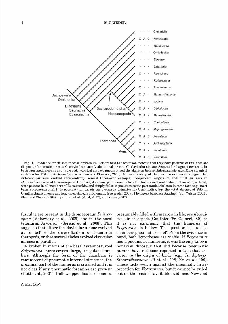
Fig. 1. Evidence for air sacs in fossil archosaurs. Letters next to each taxon indicate that they have patterns of PSP that arediagnostic for certain air sacs: C, cervical air sacs; A, abdominal air sacs; Cl, clavicular air sacs. See text for diagnostic criteria. In
both sauropodomorphs and theropods, cervical air sacs pneumatized the skeleton before abdominal air sacs. Morphologicalevidence for PSP in Archaeopteryx is equivocal (O’Connor, 2006). A naıve reading of the fossil record would suggest thatdifferent air sacs evolved independently several times—for example, independent origins of abdominal air sacs in Mamenchisaurus and Neosauropoda. However, it is more parsimonious to infer that cervical and abdominal air sacs, at least,were present in all members of Eusaurischia, and simply failed to pneumatize the postcranial skeleton in some taxa (e.g., mostbasal sauropomorphs). It is possible that an air sac system is primitive for Ornithodira, but the total absence of PSP inOrnithischia, a diverse and long-lived clade, is problematic (see Wedel, 2007). Phylogeny based on Gauthier (’86), Wilson (2002),Zhou and Zhang (2002), Upchurch et al. (2004, 2007), and Yates (2007).
M.J. WEDEL4
J. Exp. Zool.

8/11/2019 Wedel 2009 Air Sacs
http://slidepdf.com/reader/full/wedel-2009-air-sacs 5/18
better specimens will be required to resolve thisproblem.
No other pneumatic furculae, sterna, pectoralgirdle elements, or humeri have been reported innonavian dinosaurs.
Evidence for abdominal air sacs I. Pneumatization of the posterior vertebral
column
Wedel et al. (2000) hypothesized that pneuma-tization of postdorsal vertebrae in nonaviandinosaurs implied the presence of abdominal airsacs. The posterior dorsal vertebrae and synsa-crum of birds are pneumatized by diverticula of the abdominal air sacs (Duncker, ’71). The poster-ior dorsal, sacral, and anterior caudal vertebraeare pneumatic in many sauropods (Table 1) and
nonavian theropods. Therefore, it seems likelythat these taxa had abdominal air sacs. All of thosestatements are still accurate, but further work hasclarified several important points. (For the sake of simplicity, the term ‘‘posterior compartment’’ isused herein to refer to the portions of thevertebral column that are pneumatized by diver-ticula of the abdominal air sacs.)
The crucial inference, that posterior compart-ment PSP implies the presence of abdominal air
sacs, has been obscured by inconsistency inpublished accounts. Diverticula of the cervical airsacs, lungs, and abdominal air sacs have beendescribed as anastomosing to produce a contin-uous network of diverticula that spans most or all
of the vertebral column (Muller, ’08, p 377; Cover,’53, p 241). In some older accounts (e.g., Cover,’53), the entire network of vertebral diverticula iscalled an extension of the cervical diverticulum,and so some authors have inferred that diverticula of the cervical air sacs alone can pneumatize theentire vertebral column (King, ’75; Britt et al., ’98;Sereno et al., 2008). If that were true, then itwould not be possible to infer the presence of abdominal air sacs based on postdorsal PSP (Brittet al., ’98; Sereno et al., 2008).
The uncertainties and contrary claims listedabove have been dispelled or falsified by O’Connor
and Claessens (2005) and O’Connor (2006).Diverticula of the cervical vertebrae never passfarther posteriorly than the middle dorsal verteb-rae, diverticula of the lungs pneumatize only thedorsal vertebrae and vertebral ribs immediatelyadjacent to the lungs, and the posterior compart-ment is only pneumatized by diverticula of abdominal air sacs. These relationships werefound to be invariant in more than 200 individualbirds representing 19 extant higher clades, andthey support the hypothesis that posterior com-partment PSP in sauropods and theropods indi-
cates the presence of abdominal air sacs. No otherhypothesis is consistent with known patterns of pneumatization in extant tetrapods.
Examples
In theropods, pneumatization of the posteriorcompartment is present in at least some taxa in Abelisauroidea, Spinosauroidea, Allosauroidea,Tyrannosauroidea, Oviraptorosauria, and Dro-maeosauridae (O’Connor and Claessens, 2005).
Among sauropods, posterior compartment pneu-maticity was recognized very early. Marsh (1878,1879, 1881, 1884) described and figured cavities inthe sacral vertebrae of many sauropods, and heconsidered hollow sacral vertebrae a diagnosticcharacter of Sauropoda. Pneumatic caudal verteb-rae were first illustrated by Marsh (1890) for Barosaurus. Osborn (1899), Hatcher (’01), andGilmore (’32) also illustrated pneumatic caudalvertebrae in Diplodocus. Although Marsh de-scribed the sacral vertebrae of Morosaurus(Camarasaurus) as hollow, the evidence for sacralpneumaticity in Camarasaurus is equivocal
TABLE 1. Vertebral pneumaticity in the posterior compart-
ment in sauropods
Taxon PD S AC MC Source or specimen
Omeisaurus
tianfuensis
X – – – He et al. (’88)
Mamenchisaurus
youngi
X – – – Pi et al. (’96)
M. hochuanensis X – – – CCG V 20401
Apatosaurus louisae X X – – CM 3018
Apatosaurus sp. na na X na OMNH 1436
Diplodocus carnegii X X X X CM 84
Barosaurus lentus X X X X AMNH 6341
Camarasaurus
supremus
X – – – AMNH 5761
Camarasaurus lewisi X – – – BYU 9047 Haplocanthosaurus
priscus
X X X – CM 572, 879
Brachiosaurus
altithorax
X X – na FMNH P 25701
Brachiosaurus brancai X X X X HM Fund no
Euhelopus zdanskyi X X na n a Wiman (’29)
Malawisaurus dixeyi X X X – MAL holotype
series
An X indicates that pneumaticity is present, a dash indicates that it is
absent, and na (not applicable) indicates that the elements in question
are not preserved. Abbreviations: PD, posterior dorsal vertebrae; S,sacral vertebrae; AC, anterior caudal vertebrae; MC, middle caudal
vertebrae.
AIR SACS 5
J. Exp. Zool.

8/11/2019 Wedel 2009 Air Sacs
http://slidepdf.com/reader/full/wedel-2009-air-sacs 6/18
(McIntosh et al., ’96). In C . lewisi (BYU 9047), thesacral vertebrae have large lateral fossae but noforamina, and the internal structure of thevertebrae is composed of apneumatic spongiosa (personal observation). However, unequivocally
pneumatic sacral vertebrae are present in Bra- chiosaurus altithorax (Riggs, ’04) and B. brancai(Janensch, ’50). Pneumatic fossar are present inthe caudal vertebrae of B. brancai HM ‘‘Fund no’’and will be described elsewhere. In Malawisaurusdixeyi, the neural spine of the first caudal vertebra is pneumatic but the centrum is not (Fig. 2).Posterior compartment pneumaticity is, therefore,present in both lineages of Neosauropoda (Diplodocoidea and Macronaria) and in somenonneosauropods, such as Mamenchisaurus youngi (Pi et al., ’96).
Evidence for abdominal air sacs II. Pneumatization of the pelvic girdle and
hindlimb
Pneumatization of the pelvic girdle and hin-dlimb in birds is accomplished by diverticula of the
abdominal air sacs (Muller, ’07; Cover, ’53; King,’66, ’75; Duncker, ’71; Hogg, ’84a,b; Bezuidenhoutet al., ’99; O’Connor and Claessens, 2005;O’Connor, 2006). Pneumatization of the pelvicgirdle and hindlimb elements in nonavian dino-saurs would be further evidence for the presenceof abdominal air sacs.
Examples
Among sauropods, large chambers have beenreported in the ilia of the diplodocoid Amazon- saurus (Carvalho et al., 2003), the titanosaur
Sonidosaurus (Xu et al., 2006), and the salt-asaurine titanosaurs Saltasaurus and Neuquen- saurus (Powell, ’92; Sanz et al., ’99). Frompublished descriptions, these internal chambersappear to have the same morphology as those inthe pneumatic vertebrae of sauropods, and someauthors (e.g., Carvalho et al., 2003; Xu et al., 2006)have interpreted the chambers as pneumatic.However, the case would be stronger if thepathways by which the air got into the bones wereknown. Pneumatization cannot take place andpneumatic chambers cannot persist without a
patent (open) foramen (Ojala, ’57; Witmer, ’97).The case for appendicular pneumaticity in saur-opods would be strengthened by the discovery of pneumatic foramina on the outside of the ilium, ora series of chambers connecting the ilium to thesacral vertebrae.
Keeping that caveat in mind, there is no strong reason to doubt that the chambers reported in theilia of the sauropods listed above are pneumatic.Compelling evidence of sacral pneumaticity inboth Diplodocoidea and Macronaria (the sisterclade to Diplodocoidea, and a clade that contains,among other taxa, the Titanosauria) alreadyexists. Iliac chambers are so far only found inclades in which sacral pneumatization is known,so the phylogenetic distribution of iliac chambersis consistent with the hypothesis that they arepneumatic. At the very least, broken specimens(e.g., Camarasaurus BYU 9047, Jensen, ’88)demonstrate that ilium chambers are absent inmost sauropods, so the presence of iliac chambersis a derived character that was independentlyacquired in Diplodocoidea and Macronaria in taxa for which sacral pneumaticity was also present.
Fig. 2. MAL-200, an anterior caudal vertebra of Malawi- saurus dixeyi. ( A) The vertebra in left lateral view showing theposition of CT slices. (B–D) CT cross sections. Matrix waserased from the internal chambers using Photoshop 5.5.Pneumatic foramina on the neural arch and spine areconnected to a network of internal chambers, but the centrumis apneumatic.
M.J. WEDEL6
J. Exp. Zool.
8/11/2019 Wedel 2009 Air Sacs
http://slidepdf.com/reader/full/wedel-2009-air-sacs 7/18
In theropods, the ilium of the basal tetanuran Aerosteon is highly pneumatic, with numerousforamina that communicate with an extensivecomplex of internal chambers (Sereno et al., 2008). A large foramen is present in the proximal femur
of the oviraptorosaur Shixinggia (Lu and Zhang,2005). In its size and location, this foramen issimilar to pneumatic foramina in the femora of extant birds. It would be helpful to know whatconnections, if any, this foramen shares withspaces inside the femur. If this foramen ispneumatic, then femoral pneumaticity evolvedindependently within oviraptorosaurs and birds(although femoral diverticula may have beenpresent in the common ancestor of both clades).Note that Maryanska et al. (2002) recoveredoviraptorosaurs as basal birds, but this has notbeen supported by subsequent phylogenetic ana-
lyses using larger datasets (e.g., Hwang et al.,2004; Senter, 2007).
Evidence for abdominal air sacs III. Recapitulatory development of PSP in
extant birds
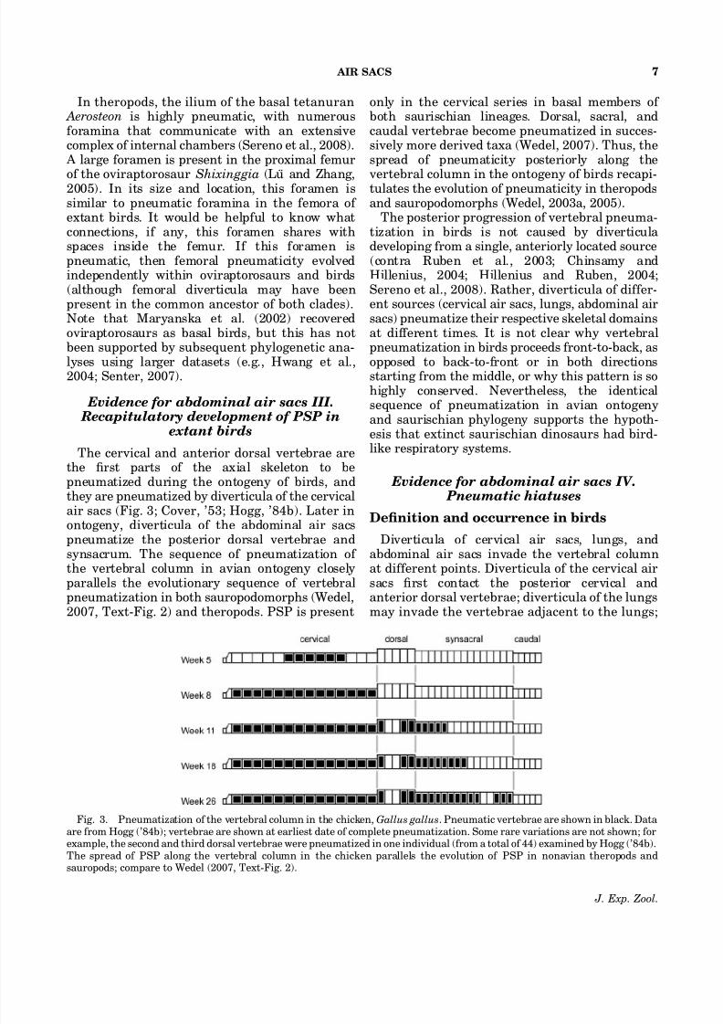
The cervical and anterior dorsal vertebrae arethe first parts of the axial skeleton to bepneumatized during the ontogeny of birds, andthey are pneumatized by diverticula of the cervicalair sacs (Fig. 3; Cover, ’53; Hogg, ’84b). Later inontogeny, diverticula of the abdominal air sacs
pneumatize the posterior dorsal vertebrae andsynsacrum. The sequence of pneumatization of the vertebral column in avian ontogeny closelyparallels the evolutionary sequence of vertebralpneumatization in both sauropodomorphs (Wedel,2007, Text-Fig. 2) and theropods. PSP is present
only in the cervical series in basal members of both saurischian lineages. Dorsal, sacral, andcaudal vertebrae become pneumatized in succes-sively more derived taxa (Wedel, 2007). Thus, thespread of pneumaticity posteriorly along the
vertebral column in the ontogeny of birds recapi-tulates the evolution of pneumaticity in theropodsand sauropodomorphs (Wedel, 2003a, 2005).
The posterior progression of vertebral pneuma-tization in birds is not caused by diverticula developing from a single, anteriorly located source(contra Ruben et al., 2003; Chinsamy andHillenius, 2004; Hillenius and Ruben, 2004;Sereno et al., 2008). Rather, diverticula of differ-ent sources (cervical air sacs, lungs, abdominal airsacs) pneumatize their respective skeletal domainsat different times. It is not clear why vertebralpneumatization in birds proceeds front-to-back, as
opposed to back-to-front or in both directionsstarting from the middle, or why this pattern is sohighly conserved. Nevertheless, the identicalsequence of pneumatization in avian ontogenyand saurischian phylogeny supports the hypoth-esis that extinct saurischian dinosaurs had bird-like respiratory systems.
Evidence for abdominal air sacs IV. Pneumatic hiatuses
Definition and occurrence in birds
Diverticula of cervical air sacs, lungs, andabdominal air sacs invade the vertebral columnat different points. Diverticula of the cervical airsacs first contact the posterior cervical andanterior dorsal vertebrae; diverticula of the lungsmay invade the vertebrae adjacent to the lungs;
Fig. 3. Pneumatization of the vertebral column in the chicken, Gallus gallus. Pneumatic vertebrae are shown in black. Data are from Hogg (’84b); vertebrae are shown at earliest date of complete pneumatization. Some rare variations are not shown; forexample, the second and third dorsal vertebrae were pneumatized in one individual (from a total of 44) examined by Hogg (’84b).The spread of PSP along the vertebral column in the chicken parallels the evolution of PSP in nonavian theropods andsauropods; compare to Wedel (2007, Text-Fig. 2).
AIR SACS 7
J. Exp. Zool.
8/11/2019 Wedel 2009 Air Sacs
http://slidepdf.com/reader/full/wedel-2009-air-sacs 8/18
and diverticula of the abdominal air sacs mayinvade the synsacrum at several points (King, ’57;Duncker, ’71; O’Connor, 2006). Paravertebraldiverticula derived from these sources may growalong the column until they contact each other
and anastomose. The growth and anastomosis of the paravertebral diverticula may produce anuninterrupted pattern of vertebral pneumatiza-tion, so that every vertebra from the secondor third cervical back to the free caudals ispneumatic.
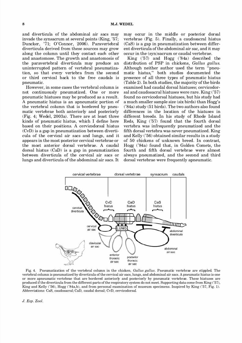
However, in some cases the vertebral column isnot continuously pneumatized. One or morepneumatic hiatuses may be produced as a result. A pneumatic hiatus is an apneumatic portion of the vertebral column that is bordered by pneu-matic vertebrae both anteriorly and posteriorly(Fig. 4; Wedel, 2003a). There are at least three
kinds of pneumatic hiatus, which I define herebased on their positions. A cervicodorsal hiatus(CvD) is a gap in pneumatization between diverti-cula of the cervical air sacs and lungs, and itappears in the most posterior cervical vertebrae orthe most anterior dorsal vertebrae. A caudaldorsal hiatus (CaD) is a gap in pneumatizationbetween diverticula of the cervical air sacs orlungs and diverticula of the abdominal air sacs. It
may occur in the middle or posterior dorsalvertebrae (Fig. 5). Finally, a caudosacral hiatus(CaS) is a gap in pneumatization between differ-ent diverticula of the abdominal air sac, and it mayoccur in the (syn)sacrum or caudal vertebrae.
King (’57) and Hogg (’84a) described thedistribution of PSP in chickens, Gallus gallus. Although neither author used the term ‘‘pneu-matic hiatus,’’ both studies documented thepresence of all three types of pneumatic hiatus(Table 2). In both studies, the majority of the birdsexamined had caudal dorsal hiatuses; cervicodor-sal and caudosacral hiatuses were rare. King (’57)found no cervicodorsal hiatuses, but his study hada much smaller sample size (six birds) than Hogg’s(’84a) study (51 birds). The two authors also founddifferences in the location of the hiatuses indifferent breeds. In his study of Rhode Island
Reds, King (’57) found that the fourth dorsalvertebra was infrequently pneumatized and thefifth dorsal vertebra was never pneumatized. King and Kelly (’56) obtained similar results in a studyof 50 chickens of unknown breed. In contrast,Hogg (’84a) found that, in Golden Comets, thefourth and fifth dorsal vertebrae were almostalways pneumatized, and the second and thirddorsal vertebrae were frequently apneumatic.
Fig. 4. Pneumatization of the vertebral column in the chicken, Gallus gallus. Pneumatic vertebrae are stippled. Thevertebral column is pneumatized by diverticula of the cervical air sacs, lungs, and abdominal air sacs. A pneumatic hiatus is oneor more apneumatic vertebrae that are bordered anteriorly and posteriorly by pneumatic vertebrae. These hiatuses areproduced if the diverticula from the different parts of the respiratory system do not meet. Supporting data come from King (’57),King and Kelly (’56), Hogg (’84a,b), and from personal examination of museum specimens. Inspired by King (’57, Fig. 1). Abbreviations: CaS, caudosacral; CaD, caudal dorsal; CvD, cervicodorsal.
M.J. WEDEL8
J. Exp. Zool.

8/11/2019 Wedel 2009 Air Sacs
http://slidepdf.com/reader/full/wedel-2009-air-sacs 9/18
Each type of pneumatic hiatus is informative.The most anterior dorsal vertebrae may bepneumatized by diverticula of cervical air sacs or
by diverticula of the lungs. Based on osteologicalevidence alone, it is impossible to determinewhether diverticula of the cervical air sacs orlungs were involved if the posterior cervical andanterior dorsal vertebrae are all pneumatic. How-ever, if a cervicodorsal hiatus is present, then theanterior dorsal vertebrae posterior to the hiatuswere most likely pneumatized by diverticula of thelung. Similarly, vertebrae posterior to a caudaldorsal or sacrocaudal hiatus were most likelypneumatized by diverticula of abdominal air sacs.
If it is possible for a single diverticulum to span a pneumatic hiatus, then vertebrae on either side of a pneumatic hiatus would not have to be pneuma-tized by independent sources of diverticula, andpneumatic hiatuses would have little or nodiagnostic value. I am not aware of any suchcases, and it seems unlikely that such a develop-mental anomaly would occur bilaterally. Diverti-cula on the left and right side of the vertebralcolumn are derived from contralateral lungs andair sacs, and they presumably develop indepen-dently. Nevertheless, the inferences drawn below
can potentially be falsified if future studies showthat it is possible for a single diverticulum to spana pneumatic hiatus.
Importance
If the development of pneumaticity in nonaviandinosaurs followed that of birds, then pneumatiza-tion of the cervical, dorsal, and sacral vertebrae insome sauropods and theropods shows that theyhad both cervical and abdominal air sacs—and,therefore, all of the components necessary forflow-through lung ventilation (O’Connor andClaessens, 2005). However, this inference onlyholds if the development of pneumaticity innonavian dinosaurs followed that of birds. Although diverticula of the cervical air sacs orlungs never pneumatize the posterior compart-ment in extant birds, the possibility remains as a hypothesis. The inference that abdominal air sacswere present in sauropods and nonavian thero-pods is already robust (see above); it would bestronger still if pneumatic hiatuses occur in thesegroups because the vertebrae posterior to the
Fig. 5. A pneumatic hiatus in a chicken. The notarium of UCMP 119225 is composed of four vertebrae. The threeanterior vertebrae are pneumatic, but the fourth is not. ( A)The specimen in left lateral view under normal lighting. (B)The specimen lit from behind to show the pneumatic(translucent) and apneumatic (opaque) regions. (C) A microCT slice through a pneumatic vertebra. (D) A micro CT slicethrough the apneumatic vertebra. Note the density of thetrabeculae in D compared to C. The anterior synsacral vertebraeof this individual are pneumatic. The apneumatic vertebra isbordered anteriorly and posteriorly by pneumatic vertebrae,and constitutes a caudal dorsal pneumatic hiatus.
TABLE 2. Frequencies of pneumatic hiatuses in two breeds of
chickens
Pneumatic hiatuses
CvD CaD CaS
Rhode Island Red
Males (1) 0/0% 1/100% 0/0%
Females (5) 0/0% 5/100% 2/40%
Total (6) 0/0% 6/100% 2/33%
Golden Comet
Males (3) 1/33% 2/67% 0/0%
Females (48) 3/6% 34/71% 2/4%
Total (51) 4/8% 36/71% 2/4%
Data on Rhode Island Reds from King (’57); data on Golden Comets
from Hogg (’84a). See text for descriptions of different types of
hiatuses. Abbreviations: CaS, caudosacral; CaD, caudal dorsal; CvD,
cervicodorsal.
AIR SACS 9
J. Exp. Zool.

8/11/2019 Wedel 2009 Air Sacs
http://slidepdf.com/reader/full/wedel-2009-air-sacs 10/18
hiatus would have to have been pneumatizedindependently, by diverticula of abdominal airsacs (Wedel, 2003a). One such hiatus is present inthe sauropod Haplocanthosaurus.
Example: Haplocanthosaurus
Haplocanthosaurus is a sauropod from theUpper Jurassic Morrison Formation of westernNorth America (Hatcher, ’03). In phylogeneticanalyses it has been recovered as a eusauropodbasal to Neosauropoda (Upchurch, ’98; Rauhutet al., 2005), the most basal diplodocoid (Wilson,2002, Fig. 13A), the most basal macronarian(Wilson and Sereno, ’98), or a macronarian morederived than Camarasaurus (Upchurch et al.,2004).
The CM 879 specimen of Haplocanthosaurus hasa mostly complete vertebral column. All of thepreserved cervical and dorsal centra have promi-nent lateral cavities that penetrate to a medianseptum (Fig. 6). A CT scan of a dorsal vertebra of the CM 572 specimen of Haplocanthosaurus showsthat the lateral fossae do not invade the condyle orthe ventral half of the centrum and that they areonly partially bounded by a distinct lip of bone.Fossae in the dorsal vertebrae of CM 879 have the
same morphology. In both specimens, the ventralmargins of the lateral fossae are more clearlydelimited than the dorsal margins, so that thefossae open dorsolaterally.
The sacrum of CM 879 is incomplete, and
includes five coossified spines and the fourth andfifth sacral centra. The sacral neural spines haveno distinct pneumatic fossae or foramina. The onlywell-developed laminae are the spinodiapophyseallaminae, which are present only in the first threespines (Fig. 7). In particular, the fossae present onthe neural arch and spine of the first caudalvertebra (described below) are absent in the sacralneural spines.
The fourth sacral vertebra of CM 879 isasymmetrically pneumatized. The right side of the centrum bears a large, distinct fossa thatextends upward underneath the facet for the
sacral rib. This fossa is 78 mm long, 33 mm tall,and 27mm deep. The fossa differs from theapneumatic fossae of extant crocodylians in being proportionally larger and deeper and in having a distinct margin. The dorsal margin is much more
Fig. 6. Pneumatization of the presacral vertebrae in Haplocanthosaurus. ( A) X-ray image of a posterior cervicalvertebra of CM 879 in right lateral view. (B) A CT slicethrough the same vertebra. (C) X-ray image of an anteriordorsal vertebra of CM 572 in left lateral view. (D) X-ray imageof the same vertebra in anterior view. All of the preservedpresacral vertebrae of both specimens have large, sharp-lippedfossae that penetrate to a narrow median septum.
Fig. 7. A pneumatic hiatus in a sauropod dinosaur. Thepreserved portions of the sacrum (S1–S5) and anterior caudalvertebrae (Ca1–Ca3) of Haplocanthosaurus CM 879 are shownin right lateral (top) and left lateral (bottom) views. All of thepreserved cervical and dorsal vertebrae have large, distinctfossae. Distinct fossae are also present on the right sides of thefourth sacral and first caudal vertebrae, and on the left side of the first caudal. The left side of the fourth sacral and bothsides of the fifth sacral are waisted but lack distinct fossae (seetext for discussion), and constitute a caudosacral pneumatichiatus.
M.J. WEDEL10
J. Exp. Zool.

8/11/2019 Wedel 2009 Air Sacs
http://slidepdf.com/reader/full/wedel-2009-air-sacs 11/18
pronounced than the ventral, so in cross sectionthe fossa is similar to the fossae of the dorsalvertebrae, only flipped upside down. The left sideof the centrum is strongly waisted (i.e., narrowerin the middle than at either end) but has no
distinct fossa below the articular surface for thesacral rib.On both sides of the centrum are smaller cavities
above the sacral rib facets (Fig. 8). On each side,this space is bounded ventrally and anteriorly bythe sacral rib facet, posteriorly by the rim of thecotyle, and dorsally by a bony lamina. Each spaceis also divided into anterodorsal and posteroven-tral compartments by an accessory lamina. Theanterodorsal compartments on both sides consistof shallow fossae only a few millimeters deep. Theposteroventral fossae are much deeper. On the leftside, the posteroventral fossa is conical and 32 mm
deep. The fossa on the right side is similar in sizeand shape, but it still contains some matrix so itsdepth cannot be determined. Hatcher (’03,Figs. 15 and 20) illustrated both the lateral anddorsal fossae on the right side of the centrum.
The fifth sacral vertebra has no evidence of pneumaticity. The sides of the centrum areshallowly waisted, but there are no invasive fossaeanywhere on the element. The facets for the sacralribs are more dorsally extensive than in the fourthsacral and cover the area occupied by the dorsalfossae in the preceding vertebra. It is possible that
the large sacral rib facets simply left no room forthe dorsal fossae to form. However, the sacral ribfacets are no more ventrally extensive than thoseof the fourth sacral. In other words, there is roomfor lateral fossae on the sides of the centrum, but
the fossae are not present.The first caudal vertebra has deep, distinctfossae on both the centrum (Fig. 7) and the neuralspine (Fig. 9). The lateral fossae of the centrumare similar in size and form to the fossa on theright side of the centrum of sacral vertebra four.The fossa on the right side of the centrum is69 mm long, 41 mm tall, and 31 mm deep. Like theright-hand fossa of the fourth sacral, it extendsupward under the attachment of the transverseprocess and the dorsal margin is more sharplydelineated than the ventral. The fossa on the leftside of the centrum is 54 mm long, 29 mm tall, and
mostly filled with matrix, so its depth cannot bedetermined.
The neural spine fossae of the first caudal are alllocated just posterior to the prezygapophyses. Thevertebra lacks a true intraprezygapophyseal lami-na. Instead, a low rampart of bone connects theprezygapophyseal rami below and behind theprezygapophyses. A bilobate fossa is situatedbehind this bony rampart at the base of theprespinal ligament scar. The fossa is 17 mm long,16 mm wide, and at least 17 mm deep. The fossa istoo deep and too narrow to accept the postzyga-
pophyses of the preceding vertebra; it is verticallyoriented, unlike the pits left on the neural spinesof many bird vertebrae by the interspinousligaments; and it is smooth, unlike the rugoseinterspinous ligament scar just dorsal to it. Thesecharacteristics suggest that the fossa is not relatedto the interspinous ligaments or to the zygapo-physeal articulations. Rather, the form of the fossa is similar to that of the dorsal fossae on the fourth
Fig. 8. The fourth and fifth sacral centra of Haplocantho- saurus CM 879. Above, the centrum of the fourth sacralvertebra in right posterolateral ( A), right lateral (B), leftlateral (C), and left posterolateral (D) views. Below, thecentrum of the fifth sacral vertebra in right lateral ( E) and leftlateral (F) views. The centrum of S4 has dorsolateral fossae onboth sides and a lateral fossa on the right side. The left side of S4 is waisted but lacks a lateral fossa. The centrum of S5 iswaisted and lacks both lateral and dorsolateral fossae. Abbreviations: dlf, dorsolateral fossa; lf, lateral fossa.
Fig. 9. Anterior caudal vertebrae of HaplocanthosaurusCM 879 in dorsal view. ( A) The first caudal vertebra. (B) Thesecond caudal vertebra.
AIR SACS 11
J. Exp. Zool.

8/11/2019 Wedel 2009 Air Sacs
http://slidepdf.com/reader/full/wedel-2009-air-sacs 12/18
sacral vertebra, and the location of the fossa behind the prezygapophyses is similar to theposition of pneumatic fossae and foramina in thevertebrae of birds (Wedel, 2007, Text-Fig. 9).
In dorsal view, with anterior to the top, the
prezygapophyseal laminae of the first caudalvertebra resemble the letter M. The middle legsof the M are formed by short spinoprezygapophy-seal laminae that converge on the lower portion of the neural spine. The bilobate fossa described inthe preceding paragraph sits just above theconvergence of the middle legs. The lateral legsof the M are formed by posterolaterally directedlaminae that connect the prezygapophyses to thetransverse processes. On the right side of thevertebra, these laminae are poorly developed, andthere is only a shallow depression between theright spinoprezygapophyseal lamina and the pos-
terolateral lamina. On the left side, the laminaeare much more pronounced and the same space isoccupied by a prominent fossa at least 19 mmdeep.
None of the fossae present in the first caudalvertebra are present in the second. The lateralfaces of the centra are shallowly waisted but haveno distinct fossae. Two small nutrient foramina are present on the right side of the centrum, bothless than 2 mm in diameter. Most of the rightprezygapophysis is missing. The remainder of theright prezygapophysis, the left prezygapophysis,
and the neural spine form three sides of a rectangular trough. This trough does not extendposteriorly or ventrally past the margin of theright prezygapophysis. It is no larger or deeperthan needed to accept the postzygapophyses of thefirst caudal vertebra.
In summary, the fourth sacral and first caudalvertebrae have a variety of large, distinct fossaethat compare well to those found on the dorsalvertebrae, and to pneumatic features in thevertebrae of birds. These fossae are compelling
evidence of pneumaticity. These fossae are absentin the centra and neural spines of the fifth sacraland second caudal vertebra. The apneumatic fifthsacral vertebra is bordered anteriorly and poster-iorly by pneumatic vertebrae, and constitutes a caudosacral pneumatic hiatus. The first caudalvertebra was, therefore, most likely pneumatizedby diverticula of abdominal air sacs (Fig. 10).
DISCUSSION
Shared developmental pathways and the
origin(s) of air sacs and PSPUntil now, the hypothesis that nonavian
saurischian dinosaurs had cervical and abdominalair sacs has been supported by the presence of pneumaticity in the parts of the skeleton that arepneumatized by those air sacs in extant birds. Theevidence presented above shows that PSP innonavian dinosaurs also shared some aspects of development with PSP in birds. In basal theropodsand sauropodomorphs, PSP is present only in thecervical vertebrae (if at all). This shows thatdiverticula of the cervical air sacs must have
developed anteriorly from the thorax before theypneumatized the skeleton, just as in extant birds.The pneumatic hiatus in Haplocanthosaurussuggests that vertebral diverticula developed frommore than one part of the abdominal air sacs. Theparallel between the evolution of PSP in nonavian
Fig. 10. The air sacs of Haplocanthosaurus. Preserved elements of CM 879 are shown in right lateral view. The cervical andanterior dorsal vertebrae were pneumatized by diverticula of cervical air sacs (green). Middle dorsal vertebrae werepneumatized by diverticula of the lung (red). Diverticula of the abdominal air sac (blue) pneumatized the posterior dorsal,sacral, and first caudal vertebrae. Other air sacs may have been present (gray), but their presence is not detectable from thepreserved elements.
M.J. WEDEL12
J. Exp. Zool.

8/11/2019 Wedel 2009 Air Sacs
http://slidepdf.com/reader/full/wedel-2009-air-sacs 13/18
dinosaurs and its development in birds alsosuggests that similar generative mechanisms wereresponsible.
Evidence for PSP in Archaeopteryx is equivocal(contra Britt et al., ’98; Christiansen and Bonde,
2000; see O’Connor, 2006; Mayr et al., 2007).Foramina in the vertebrae and pelvic elements arenot clearly pneumatic. Some may be vascularforamina, and some may be breaks in the speci-mens (O’Connor, 2006). Furthermore, radio-graphs of at least one specimen show that thevertebrae are dense and solidly constructed, whichis more consistent with apneumatic bone thanwith pneumaticity (Mayr et al., 2007). However,PSP was present in other basal birds (e.g., Ichthyornis, Jeholornis; Marsh, 1880; Zhou andZhang, 2003), and many other basal birds havehumeral and vertebral fossae that may have been
pneumatic (Sanz et al., ’95). Therefore, it is notclear if PSP in nonavian theropods is taxicallyhomologous (sensu Patterson, ’82) with that of birds. On the basis of currently available evidence,the absence of unequivocal PSP in Archaeopteryxcould represent a loss in that taxon alone.
Archaeopteryx is not the only lacuna in thephylogenetic distribution of PSP in Saurischia.Sauropodomorphs evolved PSP independentlyfrom theropods; PSP is absent in the most basalknown sauropodomorph (Saturnalia; Langeret al., ’99) and absent or, at least, not clearly
present in most basal sauropodomorphs (Wedel,2007). Furthermore, posterior compartment pneu-maticity evolved independently in diplodocoids,titanosauriforms, ceratosaurians, and coeluro-saurians. The skeletal traces of abdominal air sacsare not phylogenetically continuous throughoutSaurischia.
Although PSP does not have a continuousphylogenetic distribution in Saurischia, it seemsto have been produced by similar developmentalpathways in sauropodomorphs, nonavian thero-pods, and birds, and none of the gaps in thephylogenetic distribution of pneumatic charactersare very large. It is possible that both cervical andabdominal air sacs were present in the ancestralsaurischian, but did not pneumatize the postcra-nial skeleton in some descendants of that ances-tor—for example, most basal sauropodomorphs. A similar situation exists in extant birds, all of whichhave an air sac system even though PSP has beenlost in some clades.
On the other hand, air sacs (and not just PSP)may have evolved independently in sauropodo-morphs and theropods. Sac-like regions of the lung
are present in many sauropsids (Wolf, ’33; Perry,’83, ’98), and these suggest some level plasticity insauropsid pulmonary systems (O’Connor andClaessens, 2005). If air sacs were absent in theancestral saurischian, they must have evolved in
both lineages very soon after the divergence of theropods and sauropodomorphs because PSP ispresent in early representatives of both clades.The ancestral saurischian must have had at leastthe potential to evolve air sacs, but this potentialmay have been realized independently in ther-opods and sauropodomorphs. However, the bal-ance of the evidence suggests that air sacs areprimitive for Saurischia.
The origin of flow-through lung ventilation
Flow-through lung ventilation like that of birds
minimally requires four things: (1) lungs thatfunction as tubes rather than sacs; (2) air sacsanterior to the lungs; (3) air sacs posterior to thelungs; and (4) a musculoskeletal system capable of driving ventilation. Evidence from the fossilrecord, particularly PSP, can extend the under-standing of the origin of this system, but it maynot be sufficient to pinpoint the origin of avian-style lung ventilation.
Fossil evidence and phylogenetic inferencessuggest that all of the components necessary forflow-through lung ventilation were present in
basal saurischians. But the hypothesis that bird-like lung ventilation was common to all saur-ischians comes with two important caveats. Thefirst is obvious: at least for now, there is no way of knowing the path of inspired air in nonaviandinosaurs. The fossil evidence can only show thatsaurischians had the anatomical componentsnecessary for flow-through lung ventilation. It ispossible that basal saurischians had air sacs butnot flow-through ventilation. However, in croco-dilians adjacent lung chambers are often con-nected by small foramina, which allow thepossibility of extra-bronchial airflow (Perry, ’88,’91). The presence of these foramina in crocodi-lians together with the parabronchi of birds showsthat some form of extra-bronchial airflow isprobably primitive for archosaurs. Nevertheless,the detailed internal structures and airflowpatterns of extinct archosaurs are not wellconstrained by available evidence.
The other caveat extends uncertainty in theopposite direction. PSP is present in pterosaurs(Bonde and Christiansen, 2003; O’Connor, 2006)but absent in the closest outgroups of Saurischia,
AIR SACS 13
J. Exp. Zool.

8/11/2019 Wedel 2009 Air Sacs
http://slidepdf.com/reader/full/wedel-2009-air-sacs 14/18
which are Ornithischia and nondinosauriandinosauromorphs. Avian development demon-strates that the air sac system and its diverticula are present before they leave skeletal traces (Locyand Larsell, ’16a,b; Hogg, ’84a,b), and that the
fully avian air sac system can be present withoutproducing PSP, as in loons and penguins. There-fore, the possibility exists that an air sac system,and possibly even flow-through lung ventilation,evolved earlier in archosaurs and only becamedetectable in the fossil record when it startedleaving diagnostic traces in the skeletons of basalsaurischians and pterosaurs.
The enigmatic origin of postcranial pneumaticity
The evolution of PSP in saurischians is marked
by trends in the invasiveness and physical scale of the pneumatic traces. Pneumatic fossae in thevertebrae of basal sauropods and theropods areantecedent to the simple, camerate (large-cham-bered) vertebrae of more derived taxa that, inturn, give way to the complex, camellate (small-chambered) vertebrae of the most derived taxa inboth lineages. In sauropods, at least, there wasalso a trend toward increasing pneumatization of individual elements, so that the pneumatic ver-tebrae of most sauropods were only about half asdense as the apneumatic vertebrae of their basal
sauropodomorph ancestors (see Appendix). Thistrend may also be present in theropods, althoughit has not yet been documented.
If progressively more basal taxa are examined inthe quest to find the origin of PSP, the problem isnot that evidence of PSP disappears entirely. It isthat the shallow, unbounded fossae of basaldinosaurs are no longer diagnostic for pneumati-city (Wedel, 2007). Similar fossae are present inthe vertebrae of many tetrapods, and they may beassociated with many soft tissues, including muscles, cartilage, and fat (O’Connor, 2006).
One potential step forward is to search forcriteria that would distinguish pneumatic fossaefrom those associated with other soft tissues. Suchcriteria might be present in the microscopicsurface texture of pneumatic bones, or in theirhistology, which has been little studied except fora few brief treatments (e.g., Reid, ’96; Woodward,2005). Perhaps the ancestors of Saurischia had airsacs and diverticula but the skeletal traces of therespiratory system are so faint or so nondiagnosticthat they have not been recognized. Perhaps theyhad air sacs but no diverticula, or no air sacs at all.
There is no guarantee that diagnostic criteria forpneumatic fossae exist to be found, or that, if found, they will be present in the outgroups toSaurischia. Nevertheless, to improve the under-standing of the origin of the avian air sac system,
that is where and how they should be sought.
CONCLUSION: SHIFTING THE NULLHYPOTHESIS OF SAURISCHIAN
RESPIRATION
In an article on the antorbital fenestra of archosaurs, Witmer (’87) found that the fenestra probably housed an air sac rather than a muscle ora gland. Witmer argued that because the air sachypothesis was the best supported of the three, itshould become the null hypothesis for the soft-tissue contents of the antorbital fenestra.
A similar turning point is at hand for hypothesesof saurischian respiration. As they diverged from a common ancestor, the linear ancestors of birdsand crocodylians must have passed throughfunctionally intermediate stages. Nonavian dino-saurs did not necessarily have the same pulmo-nary anatomy as crocodylians or extant birds. Ashypotheses of pulmonary anatomy in dinosaurs,‘‘croc lungs’’ versus ‘‘bird lungs’’ is a falsedichotomy. It is more informative to identify thederived features that nonavian dinosaurs sharewith their extant relatives, and to determine the
hierarchical distribution of these characters inarchosaurian phylogeny. The pulmonary anatomyof nonavian saurischians may not have beenexactly like that of extant birds, but the evidencediscussed above suggests that both cervical andabdominal air sacs were present in all saur-ischians. Some form of air sac-driven lung ventila-tion should be considered the null hypothesis forsaurischians.
Some previous studies assumed that sauropodsand other saurischians had lungs like those of crocodylians (Hengst and Rigby, ’94; Hengst et al.,’96; Ruben et al., ’97, ’99, 2003) or turtles (Spotila et al., ’91). Others have located the origin of the airsac system in the first coelurosaurian theropods,without entertaining the possibility that thesystem might have originated much earlier(Farmer, 2006). It is not beyond the bounds of possibility that sauropods had turtle lungs or thatair sac-driven lung ventilation originated incoelurosaurs, but neither of those hypotheses isconsistent with the abundant evidence for cervicaland abdominal air sacs in all saurischians, andthey can no longer be regarded as null.
M.J. WEDEL14
J. Exp. Zool.

8/11/2019 Wedel 2009 Air Sacs
http://slidepdf.com/reader/full/wedel-2009-air-sacs 15/18
ACKNOWLEDGMENT
This work was completed as part of a doctoraldissertation in the Department of IntegrativeBiology, University of California, Berkeley. Ithank my advisors, K. Padian and W. A. Clemens,and the members of my dissertation committee, J. Gerhart, F. C. Howell, D. Wake, and M. Wake,for timely assistance and timeless advice. Thiswork would have been impossible without thehospitality and helpfulness of many curators,collections managers, and other colleagues, espe-cially P. Barrett, S. Chapman, A. Henrici,M. Lamanna, L. Jacobs, D. Winkler, E. Gomani,R. Cifelli, N. Czaplewski, and J. Person. I amgrateful to G. Maier for a translation of Janensch(’47). I am grateful to J. Harris, P. O’Connor, andtwo anonymous reviewers for comments that
substantially improved this article. This is Uni-versity of California Museum of PaleontologyContribution No. 1987.
APPENDIX
The method of calculating the relative densitiesof pneumatic and apneumatic bones is given here.The thickness of the walls of tubular bones istypically expressed as the variable K , which is theinner diameter of the bone (r) divided by the outer
diameter ( R). For a large sample (4150) of avianlong bones, Cubo and Casinos (2000) reportedaverage K values of 0.65 for apneumatic bones and0.77 for pneumatic bones. For tubular bones, theamount of the cross-sectional area that is notoccupied by bone can be found by taking thesquare of K (pr2 / p R2
5 r2 / R25 K 2). For the apneu-
matic bones, this is 0.652 or 0.42, and for thepneumatic bones it is 0.772 or 0.59. This meansthat on average, the cross-sectional area of a pneumatic element is 59% air and 41% bone, andthe cross-sectional area of an apneumatic elementis 42% marrow and 58% bone. The specific gravityof marrow is 0.95 (Currey and Alexander, ’85), andthat of avian compact bone is 1.8 (Spector, ’56).The mass of air is negligible. On average, thedensity of pneumatic avian long bones is1.80.415 0.74 g/cm3, and the density of apneu-matic avian long bones is (1.80.58)1(0.950.42)51.4 g/cm3. It may seem surprising that pneumatic bones that differ from apneumaticbones by 10% of K are only half as dense. However,the cross-sectional geometry of the bones isproportional to the square of K , and the diaphyses
of apneumatic bones are filled with marrow thatcontributes much of the mass of the elements.
Pneumatic sauropod vertebrae were, on avaer-age, about 60% air by volume (Wedel, 2005;Schwarz and Fritsch, 2006). Assuming that the
compact bone of sauropods was as dense as that of birds, the average density of their pneumaticvertebrae was 1.80.450.72 g/cm3—about thesame as that of pneumatic long bones in birds. Although quantitative comparisons are lacking,the apneumatic vertebrae of basal sauropodo-morphs are similar in cortical thickness andinternal structure to those of large extant mam-mals (personal observation), and were probablyabout as dense. In Giraffa and Syncerus thedensity of the vertebrae is 1.3–1.4 g/cm3 (vanSchalkwyk et al., 2004), very close to the meanfor apneumatic bird bones calculated above. If the
vertebrae of basal sauropodomorphs were equallydense, then pneumatization of sauropod vertebraeand avian long bones both produce the samedensity reduction, about 50%.
LITERATURE CITED
Bezuidenhout AJ, Groenewald HB, Soley JT. 1999. Ananatomical study of the respiratory air sacs in ostriches.Onderstepoort J Vet Res 66:317–325.
Bonde N, Christiansen P. 2003. The detailed anatomy of Rhamphorhynchus: axial pneumaticity and its implications.Geol Soc Lond Spec Publ 217:217–232.
Bremer JL. 1940a. The pneumatization of the head of thecommon fowl. J Morphol 67:143–157.
Bremer JL. 1940b. The pneumatization of the humerus in thecommon fowl and the associated activity of theelin. Anat Rec77:197–211.
Britt BB. 1997. Postcranial pneumaticity. In: Currie PJ,Padian K, editors. The encyclopedia of dinosaurs. San Diego,CA: Academic Press. p 590–593.
Britt BB, Makovicky PJ, Gauthier J, Bonde N. 1998.Postcranial pneumatization in Archaeopteryx. Nature395:374–376.
Brown RE, Brain JD, Wang N. 1997. The avian respiratorysystem: a unique model for studies of respiratory toxicosisand for monitoring air quality. Environ Health Perspect105:188–200.
Bryant HN, Russell AP. 1992. The role of phylogeneticanalysis in the inference of unpreserved attributes of extincttaxa. Philos Trans R Soc Lond B 337:405–418.
Carvalho IS, Avilla LS, Salgado L. 2003. Amazonsaurusmaranhensis gen. et sp. nov. (Sauropoda, Diplodocoidea)from the Lower Cretaceous (Aptian-Albian) of Brazil. CretRes 24:697–713.
Chinsamy A, Hillenius WJ. 2004. Physiology of nonaviandinosaurs. In: Weishampel DB, Dodson P, Osmolska H,editors. The dinosauria, 2nd edition. Berkeley, CA:University of California Press. p 643–659.
Christiansen P, Bonde N. 2000. Axial and appendicularpneumaticity in Archaeopteryx. Proc R Soc Lond B267:2501–2505.
AIR SACS 15
J. Exp. Zool.

8/11/2019 Wedel 2009 Air Sacs
http://slidepdf.com/reader/full/wedel-2009-air-sacs 16/18
Colbert EH. 1989. The Triassic dinosaur Coelophysis. MusNorth Ariz Bull 57:1–60.
Cover MS. 1953. Gross and microscopic anatomy of therespiratory system of the turkey. III. The air sacs. Am J Vet Res 14:239–245.
Cubo J, Casinos A. 2000. Incidence and mechanical signifi-
cance of pneumatization in the long bones of birds. Zool JLinn Soc 130:499–510.Currey JD, Alexander RM. 1985. The thickness of the walls of
tubular bones. J Zool (Lond) 206:453–468.Daniels CB, Pratt J. 1992. Breathing in long-necked dino-
saurs: did the sauropods have bird lungs? Comp BiochemPhysiol 101A:43–46.
Duncker HR. 1971. The lung air sac system of birds. Adv AnatEmbryol Cell Biol 45:1–171.
Farmer CG. 2006. On the origin of avian air sacs. RespirPhysiol Neurobiol 154:89–106.
Gauthier J. 1986. Saurischian monophyly and the origin of birds. Calif Acad Sci Mem 8:1–55.
Gier HT. 1952. The air sacs of the loon. The Auk 69:40–49.Gilmore CW. 1932. On a newly mounted skeleton of
Diplodocus in the United States National Museum. ProcUSA Natl Mus 81:1–21.Hatcher JB. 1901. Diplodocus (Marsh): its osteology, taxon-
omy, and probable habits, with a restoration of the skeleton.Mem Carn Mus 1:1–63.
Hatcher JB. 1903. Osteology of Haplocanthosaurus, with a description of a new species, and remarks on the probablehabits of the Sauropoda, and the age and origin of Atlantosaurus beds. Mem Carn Mus 2:1–72.
He X, Li K, Cai K. 1988. The middle Jurassic dinosaur fauna from Dashanpu, Zigong, China, Vol. IV. Sauropod dinosaurs(2): Omeisaurus tianfuensis. Chengdu, China: SichuanPublishing House of Science and Technology.
Hengst R, Rigby Jr JK. 1994. Apatosaurus as a means of understanding dinosaur respiration. In: Rosenberg GD,
Wolberg, DL, editors. DinoFest. The Paleontological SocietySpecial Publication 7. Knoxville, TN: University of Tennes-see Press. p 199–211.
Hengst RA, Rigby Jr JK, Landis GP, Sloan RL. 1996.Biological consequences of Mesozoic atmospheres: respira-tory adaptations and functional range of Apatosaurus. In:MacLeod N, Keller G, editors. Cretaceous-Tertiary massextinctions: biotic and environmental changes. New York,NY: W.W. Norton & Company. p 327–347.
Hillenius WJ, Ruben JA. 2004. The evolution of endothermyin terrestrial vertebrates: who? When? Why? PhysiolBiochem Zool 77:1019–1042.
Hogg DA. 1984a. The distribution of pneumatisation in theskeleton of the adult domestic fowl. J Anat 138:617–629.
Hogg DA. 1984b. The development of pneumatisation in the
postcranial skeleton of the domestic fowl. J Anat139:105–113.
Hutt S, Naish D, Martill DM, Barker MJ, Newberry P. 2001. A
preliminary account of a new tyrannosauroid theropod from
the Wessex formation (Early Cretaceous) of southern
England. Cret Res 22:227–242.
Hwang SH, Norell MA, Ji Q, Gao K. 2004. A large
compsognathid from the Early Cretaceous Yixian formation
of China. J Syst Palaeontol 2:13–30.
Janensch W. 1947. Pneumatizitat bei Wirbeln von Sauropoden
und anderen Saurischien. Palaeontographica Suppl 7 3:1–25. Janensch W. 1950. Die Wirbelsaule von Brachiosaurus
brancai. Palaeontographica Suppl 7 3:27–93.
Jensen JA. 1988. A fourth new sauropod dinosaur from theUpper Jurassic of the Colorado plateau and sauropodbipedalism. Great Bas Nat 48:121–145.
Ji Q, Currie PJ, Norell MA, Ji SA. 1998. Two feathereddinosaurs from northeastern China. Nature 393:753–761.
King AS. 1957. The aerated bones of Gallus domesticus. Acta
Anat 31:220–230.King AS. 1966. Structural and functional aspects of the avianlungs and air sacs. Int Rev Gen Exp Zool 2:171–267.
King AS. 1975. Aves respiratory system. In: Getty R, editor.Sisson and Grossman’s anatomy of the domestic animals,5th edition, Vol. 2. Philadelphia, PA: W.B. Saunders.p 1883–1918.
King AS, Kelly DF. 1956. The aerated bones of Gallusdomesticus: the fifth thoracic vertebra and sternal ribs. Br Vet J 112:279–283.
Langer MC, Abdala F, Richter M, Benton MJ. 1999. A sauropodomorph dinosaur from the Upper Triassic(Carnian) of southern Brazil. C R Acad Sci Paris Sci TerrPlan 329:511–517.
Locy WA, Larsell O. 1916a. The embryology of the bird’s lung
based on observations of the domestic fowl. Part I. Am J Anat 19:447–504.Locy WA, Larsell O. 1916b. The embryology of the bird’s lung
based on observations of the domestic fowl. Part II. Am J Anat 20:1–44.
Lu JC, Zhang BK. 2005. A new oviraptorid (Theropod:Oviraptorosauria) from the Upper Cretaceous of theNanxiong Basin, Guangdong Province of southern China. Acta Pal Sin 44:412–422.
Magnussen H, Willmer H, Scheid P. 1976. Gas exchange in airsacs: contribution to respiratory gas exchange in ducks.Respir Physiol 26:129–146.
Makovicky PJ, Apesteguıa S, Agnolın FL. 2005. The earliestdromaeosaurid theropod from South America. Nature437:1007–1011.
Marsh OC. 1878. Principal characters of American Jurassicdinosaurs. Part I. Am J Sci 3rd Ser 16:411–416.
Marsh OC. 1879. Principal characters of American Jurassicdinosaurs. Part II. Am J Sci 3rd Ser 17:86–92.
Marsh OC. 1880. Odontornithes: a monograph on the extinct
toothed birds of North America. Yale Univ Peab Mus Mem
1:1–201.Marsh OC. 1881. Principal characters of American Jurassic
dinosaurs. Part V. Am J Sci 3rd Ser 21:417–423.Marsh OC. 1884. Principal characters of American Jurassic
dinosaurs. Part VII. On the Diplodocidae, a new family of the Sauropoda. Am J Sci 3rd Ser 27:161–167.
Marsh OC. 1890. Description of new dinosaurian reptiles. Am J Sci 3rd Ser 39:81–86.
Maryanska T, Osmolska H, Wolsam M. 2002. Avialian status
for Oviraptorosauria. Acta Palaeontol Pol 47:97–116.Mayr G, Pohl B, Hartman S, Peters DS. 2007. The tenth
skeletal specimen of Archaeopteryx. Zool J Linn Soc
149:97–116.McIntosh JS, Miles CA, Cloward KC, Parker JR. 1996. A new
nearly complete skeleton of Camarasaurus. Bull Gunma Mus Nat Hist 1:1–87.
Muller B. 1908. The air-sacs of the pigeon. Smiths Misc Coll50:365–420.
O’Connor PM. 2006. Postcranial pneumaticity: an evaluationof soft-tissue influences on the postcranial skeleton and thereconstruction of pulmonary anatomy in archosaurs. J Morphol 267:1199–1226.
M.J. WEDEL16
J. Exp. Zool.
8/11/2019 Wedel 2009 Air Sacs
http://slidepdf.com/reader/full/wedel-2009-air-sacs 17/18
O’Connor PM, Claessens LPAM. 2005. Basic avian pulmonarydesign and flow-through ventilation in nonavian theropoddinosaurs. Nature 436:253–256.
Ojala L. 1957. Pneumatization of the bone and environmentalfactors: experimental studies on chick humerus. Acta Otolaryngol Suppl 133:1–28.
Osborn HF. 1899. A skeleton of Diplodocus. Mem Am Mus NatHist 1:191–214.Owen R. 1841. On the anatomy of the Southern Apteryx
( Apteryx australis, Shaw). Trans Zool Soc Lond 2:257–301.Owen R. 1857. Monograph on the fossil Reptilia of the
Wealden and Purbeck formations. Part III. Dinosauria ( Megalosaurus). Pal Soc Monogr 9:1–26.
Paladino FV, Spotila JR, Dodson P. 1997. A blueprint forgiants: modeling the physiology of large dinosaurs. In:Farlow JO, Brett-Surman MK, editors. The completedinosaur. Bloomington, IL: Indiana University Press.p 491–504.
Patterson C. 1982. Morphological characters and homology.In: Joysey KA, Friday AE, editors. Problems of phylogeneticreconstruction. New York, NY: Academic Press. p 21–74.
Perry SF. 1983. Reptilian lungs: functional anatomy andevolution. Adv Anat Embryol Cell Biol 79:1–81.Perry SF. 1988. Functional morphology of the lungs of the
Nile crocodile, Crocodylus niloticus: non-respiratory para-meters. J Exp Biol 134:99–117.
Perry SF. 1991. Gas exchange strategies in reptiles and theorigin of the avian lung. In: Wood SC, Weber RE, Hargens AR,Millard RW, editors. Physiological adaptations in vertebrates:respiration, circulation, and metabolism. Boca Raton, FL:CRC Press. p 149–168.
Perry SF. 1998. Lungs: comparative anatomy, functionalmorphology, and evolution. In: Gans C, Gaunt AS, editors.Biology of the reptilia, Vol. 19. Ithaca, NY: Society for theStudy of Amphibians and Reptilians. p 1–92.
Perry SF. 2001. Functional morphology of the reptilian
and avian respiratory systems and its implications fortheropod dinosaurs. In: Gauthier J, Gall LF, editors. Newperspectives on the origin and early evolution of birds.Proceedings of the international symposium in honor of John H. Ostrom. New Haven, CT: Special Publication of the Peabody Museum of Natural History Yale University.p 429–441.
Perry SF, Reuter C. 1999. Hypothetical lung structureof Brachiosaurus (Dinosauria:Sauropoda) based on func-tional constraints. Mitt Mus Naturkd Berl Geowiss Reihe2:75–79.
Pi L, Ou Y, Ye Y. 1996. A new species of sauropod from Zigong,Sichuan, Mamenchisaurus youngi. Papers on GeosciencesContributed to the 30th International Geological Congress:87–91 (in Chinese).
Powell JE. 1992. Osteologia de Saltasaurus loricatus (Saur-opoda Titanosauridae) del Cretacico Superior del noroeste Argentino. In: Sanz JL, Buscalioni AD, editors. LosDinosaurios y Su Entorno Biotico: Actas del Segundo Cursode Paleontologia in Cuenca. Cuena, Argentina: Instituto Juan de Valdes. p 165–230.
Rauhut OWM, Remes K, Fechner R, Gladera G, Puerta P.
2005. Discovery of a short-necked sauropod dinosaur from
the Late Jurassic period of Patagonia. Nature 435:670–672.Reid REH. 1996. Bone histology of the Cleveland-Lloyd
dinosaurs and of dinosaurs in general, Part 1: introductionto bony tissues. BYU Geol Stud 41:25–72.
Riggs ES. 1904. Structure and relationships of the opistho-coelian dinosaurs, part II: the Brachiosauridae. FieldColumbia Mus Publ Geol 2:229–247.
Ruben JA, Jones TD, Geist NR, Hillenius WJ. 1997. Lung structure and ventilation in theropod dinosaurs and earlybirds. Science 278:1267–1270.
Ruben JA, Dal Sasso C, Geist NR, Hillenius WJ, Jones TD,Signore M. 1999. New evidence for pulmonary function andmetabolic physiology of theropod dinosaurs. Science283:514–516.
Ruben JA, Jones TD, Geist NR. 2003. Respiratory andreproductive paleophysiology of dinosaurs and early birds.Physiol Biochem Zool 76:141–164.
Sanz JL, Chiappe LM, Buscalioni AD. 1995. The osteology of Concornis lacustris (Aves: Enantiornithes) from the LowerCretaceous of Spain and a reexamination of its phylogeneticrelationships. Am Mus Nov 3133:1–23.
Sanz JL, Powell JE, Le Loeuff J, Martınez R, Pereda-Suberbiola X. 1999. Sauropod remains from the UpperCretaceous of Lano (northcentral Spain). Titanosaur phylo-genetic relationships. Estud Mus Cienc Nat A ´lava
14:235–255.Scheid P. 1979. Mechanisms of gas exchange in bird lungs. RevPhysiol Biochem Pharmcol 86:137–186.
Schwarz D, Fritsch G. 2006. Pneumatic structures in thecervical vertebrae of the Late Jurassic Tendaguru sauropods Brachiosaurus brancai and Dicraeosaurus. Eclog Geol Helv99:65–78.
Schwarz D, Frey E, Meyer CA. 2007. Pneumaticity andsoft-tissue reconstructions in the neck of diplodocidand dicraeosaurid sauropods. Acta Palaeontol Pol 52:167–188.
Seeley HG. 1870. On Ornithopsis, a gigantic animal of thepterodactyle kind from the Wealden. Ann Mag Nat Hist Ser4 5:279–283.
Senter P. 2007. A new look at the phylogeny of Coelurosauria
(Dinosauria: Theropoda). J Syst Palaeontol 5:429–463.Sereno PC, Martinez RN, Wilson JA, Varricchio DJ, Alcober OA,
Larsson HCE. 2008. Evidence for avian intrathoracic air sacs ina new predatory dinosaur from Argentina. PLoS ONE 3:e3303. DOI: 10.1371/journal.pone.0003303.
Spector WS. 1956. Handbook of biological data. Philadelphia,PA: WB Saunders.
Spotila JR, O’Connor MP, Dodson P, Paladino FV. 1991. Hotand cold running dinosaurs: body size, metabolism, andmigration. Mod Geol 16: 203–227.
Upchurch P. 1998. The phylogenetic relationships of sauropoddinosaurs. Zool J Linn Soc 124:43–103.
Upchurch P, Barrett PM, Dodson P. 2004. Sauropoda. In: Weishampel DB, Dodson P, Osmolska H, editors. Thedinosauria, 2nd edition. Berkeley, CA: University of
California Press. p 259–324.Upchurch P, Barrett PM, Galton PM. 2007. A phylogenetic
analysis of basal sauropodomorph relationships: implica-tions for the origin of sauropod dinosaurs. Spec PapPalaeontol 77:57–90.
van Schalkwyk OL, Skinner JD, Mitchell G. 2004. A comparison of the bone density and morphology of giraffe(Giraffa camelopardalis) and buffalo (Syncerus caffer)skeletons. J Zool 264:309–315.
Warncke G, Stork HG. 1977. Biostatische und thermoregula-
torische Funktion der Sandwich-Strukturen in der Scha ¨-
deldecke der Vogel. Zool Anz 199:251–257.
AIR SACS 17
J. Exp. Zool.
8/11/2019 Wedel 2009 Air Sacs
http://slidepdf.com/reader/full/wedel-2009-air-sacs 18/18
Wedel MJ. 2003a. Vertebral pneumaticity, air sacs, and thephysiology of sauropod dinosaurs. Paleobiology 29:243–255.
Wedel MJ. 2003b. The evolution of vertebral pneumaticity insauropod dinosaurs. J Vert Palaeontol 23:344–357.
Wedel MJ. 2005. Postcranial skeletal pneumaticity in sauropods
and its implications for mass estimates. In: Wilson JA, Curry-Rogers K, editors. The sauropods: evolution and paleobiology.Berkeley, CA: University of California Press. p 201–228
Wedel MJ. 2006. Origin of postcranial skeletal pneumaticity indinosaurs. Integr Zool 2:80–85.
Wedel MJ. 2007. What pneumaticity tells us about ‘‘prosaur-opods,’’ and vice versa. Spec Pap Palaeontol 77:207–222.
Wedel MJ, Cifelli RL, Sanders RK. 2000. Osteology, paleobiol-ogy, and relationships of the sauropod dinosaur Sauropo- seidon. Acta Palaeontol Pol 45:343–388.
Wilson JA. 2002. Sauropod dinosaur phylogeny: critique andcladistic analysis. Zool J Linn Soc 136: 217–276.
Wilson JA, Sereno PC. 1998. Early evolution and higher-levelphylogeny of sauropod dinosaurs. Soc Vert Palaeontol Mem5:1–68.
Wiman C. 1929. Die Kriede-Dinosaurier aus Shantung.Palaeontol Sin 6:1–67. Witmer LM. 1987. The nature of the antorbital fossa of
archosaurs: shifting the null hypothesis. In: Currie PJ,Koster EH, editors. Fourth symposium on mesozoic terres-trial ecosystems, short papers. Occasional paper no. 3.Drumheller, Canada: Royal Tyrrell Museum of Paleontol-ogy. p 230–235.
Witmer LM. 1987. The extant phylogenetic bracket and theimportance of reconstructing soft tissues in fossils.. In:Thomason SS, editor. Functional morphology in vertebratepaleonotology.. Cambridge, United Kingdom: CambridgeUniversity Press. p 19–33.
Witmer LM. 1997. The evolution of the antorbital cavity of
archosaurs: a study in soft-tissue reconstruction in the fossilrecord with an analysis of the function of pneumaticity. Soc Vert Palaeontol Mem 3:1–73.
Wolf S. 1933. Zur Kenntnis von Bau und Funktion derReptilienlunge. Zoologische Jahrbucher Abteilung Anato-mie und Ontogenie der Tiere 57:139–190.
Woodward H. 2005. Bone histology of the titanosauridsauropod Alamosaurus sanjuanensis from the Javelina Formation, Texas. J Vert Palaeontol 25:132A.
Xu X, Zhou Z, Wang XL, Wu XC. 1999. A dromaeosauriddinosaur with a filamentous integument from the YixianFormation of China. Nature 401:262–266.
Xu X, Zhang X, Tan Q, Zhao X, Tan L. 2006. A newtitanosaurian sauropod from Late Cretaceous of NeiMongol, China. Acta Geol Sin 80:20–26.
Yates AM. 2007. The first complete skull of the Triassicdinosaur Melanorosaurus Haughton (Sauropodomorpha: Anchisauria). Spec Pap Palaeontol 77:9–55.
Zhou Z, Zhang F. 2002. A long-tailed, seed-eating bird fromthe Early Cretaceous of China. Nature 418:405–409.
Zhou Z, Zhang F. 2003. Jeholornis compared to Archaeopteryx,with a new understanding of the earliest avian evolution.Naturwissenschaften 90:220–225.
M.J. WEDEL18
J. Exp. Zool.