· web viewfsai (2008): food safety authority of ireland abbey court, lower abbey street, dublin i...
TRANSCRIPT

Researcher 2017;9(9) http://www.sciencepub.net/researcher
Review on assessment of bovine tuberculosis and its associated risk factors for human health
Aklilu Biru, Misgana Duguma, Chala Mohammed, and Awel Hussein
Wollega University, College of Health and Medical Sciences, School of Veterinary Medicine, EthiopiaCorresponding Author: Dr. Chala Mohammed, Wollega University, School of Veterinary Medicine,
P.O. Box: 395, Ethiopia. Tel: [email protected]
Abstract: Bovine tuberculosis (TB) is caused by the bacterium Mycobacterium bovis (M. bovis) and it is a worldwide problem. The genus Mycobacterium comprises more many species of mycobacterium that occur in the environment and are rarely associated with disease in humans or animals. Transmission of M. bovis can occur between animals, from animals to humans and vice versa and rarely, between humans. Transmission to humans is mostly through air droplets, consumption of unpasteurized milk and milk products and raw and improper cooked meat. Risk groups are elders, children, immunocompromised individuals, dairy workers and butchers and vulnerable people gathered together like prisons; homeless shelters and medical providers. Clinical symptoms may include chest pain and a prolonged cough producing sputum in humans. It is difficult to diagnose TB particularly in the early stages. The test used for TB in humans is similar in mechanism and function to the skin test used on cattle. Microscopic examination is an important laboratory diagnosis. Immunoassays are used for the confirmation of tuberculin screening skin test. Molecular diagnostic techniques are very important to identify the strain of the species. Treatment of TB should be early and the drugs should be taken properly. BTB can be controlled by test-and-slaughter or test-and-segregation methods. Affected herds must be re-tested periodically to eliminate cattle that may shed the organism and tuberculin test is generally used. Animals that have been in contact with reactors should be traced back. Raw milk and meat consumption is very common in Guto Gidda District especially in rural kebeles and sharing the same shelter with cattle. Human tuberculosis can prevented through consumption of pasteurized milk and milk products and proper cooked meat.[Aklilu Biru, Misgana Duguma, Chala Mohammed, and Awel Hussein. Review on assessment of bovine tuberculosis and its associated risk factors for human health. Researcher 2017;9(9):59-71]. ISSN 1553-9865 (print); ISSN 2163-8950 (online). http://www.sciencepub.net/researcher. 9. doi:10.7537/marsrsj090917.0 9 .
Key words: Bovine, Human, Transmission, Tuberculosis
1. IntroductionBovine tuberculosis is an infectious disease
caused by M. bovis that affects cattle, other domesticated animals and certain free or captive wildlife species. It is usually characterized by formation of nodular granulomas known as tubercles. Although commonly defined as a chronic debilitating disease, BTB can occasionally assume a more progressive course. Any body tissue can be affected, but lesions are most frequently observed in the lymph nodes (particularly of the head and thorax), lungs, intestines, liver, spleen, pleura, and peritoneum. Mycobacterium bovis is a zoonotic organism and should be treated as a risk/hazard organism with appropriate precautions to prevent human infection occurring [1].
It should be noted that other members of the M. tuberculosis complex, previously considered to be M. bovis, have been accepted as new species despite identical 16s RNA sequences and over 99.9% identity of their genome sequences. These include M. caprae. In some countries considered to be a primary pathogen of goats and M. pinnipedii, a pathogen of fur seals and sea lions. These two new species are known to be
zoonotic. In central Europe, M. caprae has been identified as a common cause of bovine tuberculosis. The link between animal and human tuberculosis has long always been known to be strong which demonstrated the cross adaptability of the tubercle bacilli from one species to another to cause disease; pointing out the danger that tuberculosis could be transmitted from animals to humans [2].
Bovine tuberculosis is becoming increasingly important due to the susceptibility of humans to the disease caused by M. bovis and there is increasing evidence that M. bovis infections may be much more significant than generally considered [3]. The direct correlation between M. bovis infection in cattle and the disease in the human population has been well documented in developed countries, whereas scanty information is available from developing countries and this lack of data according to [4], relates to its perception as an animal disease, with the health problems relating to the HIV/AIDS and human tuberculosis given a greater priority. Infection due to M. bovis, which is the principal agent of zoonotic tuberculosis, was once a major problem in developed countries but following eradication programs
1

Researcher 2017;9(9) http://www.sciencepub.net/researcher
involving test and slaughter policy and milk pasteurization, the incidence drastically reduced. However, the infection currently poses a major concern in the human population in developing countries, as humans and animals are sharing the same microenvironment and dwelling premises, especially in rural areas [3].
The genus Mycobacterium comprises more than 70 species. Many species of mycobacteria occur in the environment and are rarely associated with disease in humans or animals. A number of species of mycobacteria are important pathogens of animals or humans. Human tuberculosis is chiefly associated with infection with the species Mycobacterium tuberculosis, although M. africanum is also important in some regions. Tuberculosis in bovines and many other animal species is primarily associated with infection with Mycobacterium bovis. M. tuberculosis, M. bovis and M. africanum together with Mycobacterium microti (associated with infection of rodents) form a very closely related phylogenetic group and may be referred to collectively as the M. tuberculosis complex (MTBC). Human infection with members of the MTBC produces an indistinguishable clinical picture and the individual species cannot be distinguished from each other based on microscopic examination of stained tissues or other clinical specimens. Determination of which species is responsible for infection in a particular case normally requires culture of the microorganism in the laboratory [5].
However, M. bovis is the most universal pathogen among mycobacteria and affects many vertebrate animals of all age groups including humans although, cattle, goats and pigs are found to be most susceptible, while sheep and horses are showing a high natural resistance [6]. Both Mycobacterium bovis and Mycobacterium tuberculosis occur in countries on every continent and across the economic spectrum, with recurring outbreaks in the last decade. BTB has been significantly widely distributed throughout the world and it has been a cause for great economic loss in animal production [7].
It is estimated that approximately 85% of the cattle population and 82% of the human population of Africa are in areas where bovine tuberculosis surveillance and control activities are often inadequate or unavailable, therefore, many epidemiologic and public health aspects of the infection remain largely unknown. The emergence of drug resistant strains of Mycobacterium species, the rise and synergism of HIV/AIDS infection with tuberculosis, poverty, and neglect of tuberculosis control programs have further complicated this disease current situation in Africa [8], [9].
Therefore the objectives of this paper are:
To overview the epidemiology of human tuberculosis and its associated risk factors.
To review data on the trends of current bovine tuberculosis and its public health importance in Guto Gida district.
2. Bovine TuberculosisBovine tuberculosis primarily affects cattle
especially in Africa. However, infection in other farm and domestic animals are sometimes reported [10]. Mycobacterium bovis has a broad host range as the principal cause of TB in free-living wildlife, captive wildlife, domestic livestock, and non-human primates. Wild ruminants and carnivores, such as African buffalo, lion, cheetah, greater kudu, leopard, warthog, and eland, can be infected [11]. Most of the time BTB transmission was considered as passing from livestock to wildlife. In wild ruminants, the disease has been documented worldwide, and lesions and symptoms are very similar to those of domestic ruminants [12].
Depending on the susceptibility of a species to, and on the prevalence of BTB, these animals could act as reservoirs or spill-over hosts to other species. In Africa, TB infection in humans is principally caused by Mycobacterium tuberculosis. However, human TB of animal origin caused by M. bovis is becoming increasingly prevalent in developing countries due to the lack of both control and diagnostic measures and pasteurization of milk. TB is a major opportunistic infection in HIV-infected persons, and the World Health Organization (WHO) estimated that 70% (6 million) of the people co-infected with TB and HIV live in Sub-Saharan Africa [13].
The prevalence, incidence, and deaths caused by TB, reported in 2002 by WHO indicated that every year, there are 8-10 million new cases of TB reported, and 2-3 million deaths attributed to TB, but the exact percentage of TB that may be caused by M. bovis is not known. Global prevalence of human TB due to M. bovis is estimated at 3.1% of all human TB cases, of which 2.1% are pulmonary infections, and 9.4% extra pulmonary. However, the proportion of M. bovis in Africa and within the TB–HIV complex is unknown. Tuberculosis of both the Mycobacterium bovis and Mycobacterium tuberculosis types can be spread not only through inhalation of infective mucus droplets but also through consumption of raw milk and milk products and raw and improper cooked meat. The natural history of zoonotic tuberculosis has been best studied in cattle, although the progression and outcome of infections are probably similar in most species of animal used for food production [14].2.1. Etiology and Susceptibility
2.1.1. Taxonomy of mycobacteriumIdentification of mycobacterium began in 1800s
with the discovery of tubercle-bacilli originally named
2
Researcher 2017;9(9) http://www.sciencepub.net/researcher
“Bacterium tuberculosis” and the leprosy bacillus originally named “Bacillus leprae”. The classification of mycobacterium began in 1896 and the genus mycobacterium included many species. Classification was based on one or more characters or patterns of characters which all members of a group have and other members of group do not have. The definition characteristics of mycobacterium were based on morphology, rod-shaped, acid-alcohol fastness. Additional criteria were Gram staining, growth rate, penicillin resistance, mole% G+C, mycolic acid profiles and arylsulfatase production. Currently, the minimum standards for including species in the genus mycobacterium are acid-alcohol fastness, the presence of mycolic acid containing 60-90% carbons, which are
cleaved to C22 to C26 fatty acid methyl esters by pyrolysis, and mole% G+C of 61-71% [15].
Bacteria of the M. tuberculosis complex are aerobic, non-motile, non–spore-forming, slow-growing, acid-fast bacilli. Because they are slow growing, isolation of the bacteria can require 3 to 8 weeks of incubation. Results of experimental studies indicate that the strain of the organism, dose of the organism, route of inoculation, and prevailing conditions for growth of the organism may influence the time required to produce disease. The natural and acquired immune response mechanisms of a host are often successful in limiting proliferation of tubercle bacilli and the development of progressive disease [16].
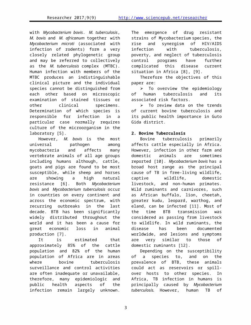
Fig.1: Appearance of Mycobacterium tuberculosis under electron micrograph [17].
The susceptibility of certain animal species to different types of tubercle bacilli is variable. Nonhuman primates, swine, cats, and dogs are susceptible to M. tuberculosis. Ruminants are quite resistant to infection by M. tuberculosis; however, this organism may induce responses to M. bovis purified protein derivative (PPD) tuberculin. Other mycobacterial infections including those involving M. avium complex (M. avium-M. intracellulare-M. scrofulaceum), M. kansasii, M. fortuitum, and M. avium subsp paratuberculosis may induce skin sensitivity to tuberculin, but do not usually induce progressive pulmonary disease in cattle and other animals. Cattle are susceptible to M. bovis, yet they
are comparatively resistant to infection with M. tuberculosis [18].
Moreover, laboratory animals such as guinea pigs and rabbits are susceptible to M. bovis, whereas chickens are resistant to that organism. This difference in observed susceptibilities may be associated with differences in body temperature. In Ethiopia, M. bovis was found to be a cause for tuberculous lymphadenitis in 17.1% of 29 human tuberculosis cases [19]. Also in Ethiopia, [3] reported that 16.7% of 42 human isolates were identified as M. bovis. These findings show that the role of M. bovis in causing human tuberculosis seemed to be significantly important [17].
3
Researcher 2017;9(9) http://www.sciencepub.net/researcher
2.2. EpidemiologyBovine tuberculosis caused by M. bovis is a
zoonosis in which both natural and anthropogenic movement of animals has influenced the epidemiology. In many parts of the world, badgers, brush-tail opossums, wild boars, deer and other wildlife species constitute a wildlife reservoir of the pathogen. Thus, the natural movement of these reservoir animals increases the spread of the disease to domestic animals and thereby, its public health impact. M. bovis infection is transmissible from cattle to humans directly by erogenous route and through direct contact with material contaminated with nose and mouth secretions from an infected herd of cattle [20].
Research findings revealed that at risk, individuals are persons in contact with potentially infected animals such as veterinarians, abattoir workers, meat inspectors, autopsy personnel, farmers, milkers, animal keepers [21]. Also, in the United Kingdom, disease in humans from M. bovis has
occurred in no more than twenty-five cases a year for the last five years [2].
Several mammalian species are known to be susceptible to infection with M bovis, including hoofed mammals (Artiodactylae and Perissodactylae), marsupials, carnivores, primates, pinnipeds, lagomorphs, rodents, and other species. In addition to mammals, some avian species are also susceptible to infection, including parrot like birds (Psittacciformes), rock doves, and North American crows. Humans are also susceptible to M. bovis, and there are numerous instances of human infection resulting from contact with infected animals [22]. The disease still spread, as it can today, through females transferring it to their young through their colostrum and milk, as well as through random contact of uninfected herbivores with infected mucus discharge, urine and feces spilled onto forage and browse, and, in the case of carnivores, through the eating of infected herbivores [23].
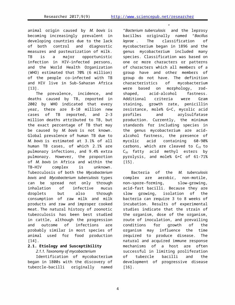
Fig 2: Epidemiology of tuberculosis
3. Zoonotic Importance Of Bovine TuberculosisHuman tuberculosis due to M. bovis has become
very rare in countries with pasteurized milk and bovine tuberculosis eradication programs. However, this disease continues to be reported from areas where bovine disease is poorly controlled. The incidence is higher in farmers, abattoir workers and others who work with cattle. In addition, humans can be infected by exposure to other species; documented infections have occurred from goats, seals, farmed elk and a rhinoceros. Some human infections are asymptomatic. In other cases, localized or disseminated disease can develop either soon after infection [24].
The role of BTB causing tuberculosis in humans has not been studied adequately. However, very few studies have indicated the isolation of the causal agent of BTB from humans in Ethiopia. With respect to this
out of 85 sputum samples taken from 28 dairy farm workers and 57 tuberculous patients where, 48 samples were positive for acid fast bacilli, of which 14 (29.2%) were niacin negative indicating M. bovis and 34 (70.2%) M. tuberculosis isolates. With a similar scenario, demonstrated that, out of 87 sputum and 21 fine needle aspiration (FNA) human samples, 42 mycobacteria species were identified by culture, of which, 7 (16.3%) and 31 (73.8%) were found as M. bovis and M. tuberculosis, respectively [25].
Localized disease can affect the lymph nodes, skin, bones and joints, genitourinary system, meninges or respiratory system. Cervical lymphadenopathy (scrofula), which primarily affects the tonsillar and pre-auricular lymph nodes, was once a very common form of tuberculosis in children who drank infected milk. In some cases, these lymph nodes rupture and
4
Researcher 2017;9(9) http://www.sciencepub.net/researcher
drain to the skin; chronic skin disease (lupus vulgaris) may occasionally result. Humans infected through the skin can develop localized skin disease (“butcher’s wart”), a form usually thought to be benign and self-limiting. Pulmonary disease is more common in people with reactivated infections than initially. The symptoms may include fever, cough, chest pain, cavitation and hemoptysis. Genitourinary disease can result in kidney failure [24].
When bacteria of the MTBC gain access to tissues they proliferate locally. Human tuberculosis (HTB) of animal origin (zoonotic TB) is an important public health concern in developing countries like Ethiopia. Recently [3] reported the range of prevalence of human TB in different consecutive years in Ethiopia (Table 1).
Table 1: Human tuberculosis case findings in EthiopiaYears Pulmonary smear cases extra pulmonary cases all new casesPositive % negative % No. %20002001200220032004Total
2810434473382913737441 430179 672
33.137.235.634.234.234.8
30 33328 99432 19732 89737 119161 540
35.731.329.930.130.731.3
26542293123713839 07642 477174 545
31.231.634.535.735.133.8
84 97992779107 626109 347121 026515 757
Source: [3]
3.1. Source of Infection and Modes of Transmissions
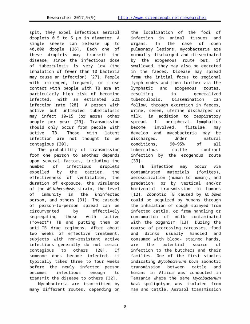
When people with active pulmonary TB cough, sneeze, speak, sing, or spit, they expel infectious aerosol droplets 0.5 to 5 µm in diameter. A single sneeze can release up to 40,000 drople [26]. Each one of these droplets may transmit the disease, since the infectious dose of tuberculosis is very low (the inhalation of fewer than 10 bacteria may cause an infection) [27]. People with prolonged, frequent, or close contact with people with TB are at particularly high risk of becoming infected, with an estimated 22% infection rate [28]. A person with active but untreated tuberculosis may infect 10–15 (or more) other people per year [29]. Transmission should only occur from people with active TB. Those with latent infection are not thought to be contagious [30].
The probability of transmission from one person to another depends upon several factors, including the number of infectious droplets expelled by the carrier, the effectiveness of ventilation, the duration of exposure, the virulence of the M. tuberculosis strain, the level of immunity in the uninfected person, and others [31]. The cascade of person-to-person spread can be circumvented by effectively segregating those with active ("overt") TB and putting them on anti-TB drug regimens. After about two weeks of effective treatment, subjects with non-resistant active infections generally do not remain contagious to others [28]. If someone does become infected, it typically takes three to four weeks before the newly infected person
becomes infectious enough to transmit the disease to others [32].
Mycobacteria are transmitted by many different routes, depending on the localization of the foci of infection in animal tissues and organs. In the case of open pulmonary lesions, mycobacteria are normally discharged and disseminated by the erogenous route but, if swallowed, they may also be excreted in the faeces. Disease may spread from the initial focus to regional lymph nodes and then further via the lymphatic and erogenous routes, resulting in generalized tuberculosis. Dissemination can follow, through excretion in faeces, urine, semen, uterine discharges or milk, in addition to respiratory spread. If peripheral lymphatics become involved, fistulae may develop and mycobacteria may be discharged. Under natural conditions, 90-95% of all tuberculous cattle contract infection by the erogenous route [33].
TB infection may occur via contaminated materials (fomites), aerosolization (human to human), and predation, or by vertical and/or horizontal transmission in humans [12]. Zoonotic TB caused by M. bovis could be acquired by humans through the inhalation of cough sprayed from infected cattle, or from handling or consumption of milk contaminated with the organism [13]. During the course of processing carcasses, food and drinks usually handled and consumed with blood- stained hands, are the potential source of infection to the butchers and their families. One of the first studies indicating Mycobacterium bovis zoonotic transmission between cattle and humans in Africa was conducted in
5
Researcher 2017;9(9) http://www.sciencepub.net/researcher
Tanzania where the same Mycobacterium bovis spoligotype was isolated from man and cattle. Aerosol transmission of bovine tuberculosis to humans continues to occur among meat industry and slaughter house workers in regions where infection is still prevalent in cattle [34].. 3.2. Risk factors
A number of factors make people more susceptible to TB infections. The most important risk factor globally is HIV; 13% of all TB cases are infected by the virus [35]. This is a particular problem in Africa, where rates of HIV are high [36]. Tuberculosis is closely linked to both overcrowding and malnutrition, making it one of the principal diseases of poverty [37]. Those at high risk thus include people who inject illicit drugs, inhabitants and employees of locales where vulnerable people gather (e.g. prisons and homeless shelters), medically underprivileged and resource-poor communities, high-risk ethnic minorities, children in close contact with high-risk category patients and health care providers serving these clients. Chronic lung disease is another significant risk factor with silicosis increasing the risk about 30 folds. Those who smoke cigarettes have nearly twice the risk of TB than non-smokers [38].
Other disease states can also increase the risk of developing tuberculosis, including alcoholism [37] and diabetes mellitus (threefold increase). There is also a genetic susceptibility [39] for which overall importance is still undefined [37]. At present, worldwide over one million children are infected with tuberculosis (TB) and 630,000 by HIV annually [40], [41]. While TB alone is responsible for over 250,000 deaths every year, HIV is projected to cause more than 56,000 deaths worldwide annually [42].
Co-infection with both organisms, also described as "cursed duet" or "deadly duo", is becoming an increasing global emergency especially in the sub-Saharan Africa, where between 10% and 60% of the children are co-infected. HIV infection has been known to weaken the immune system by depleting the CD4 cell counts and in turn giving room for opportunistic infections. TB itself can also lower the CD4 cell count in children and exacerbate the immunodeficiency caused by HIV infection. Children infected with HIV have 50 times greater risk of developing primary progressive TB in a given year from severe immune suppression of young age and HIV infection. Those with co-infection also have a higher case fatality rate [43], [44]. Studies of TB co-infection with HIV in children have been reported in both industrialized [45] and non-industrialized countries, with proportions ranging from 5.5% in United Kingdom, to 17% in Peru [43].
3.2.1. Pathogenesis
About 90% of those infected with M. tuberculosis have asymptomatic, latent TB infections (sometimes called LTBI) [46] with only a 10% lifetime chance that the latent infection will progress to overt, active tuberculous disease [47]. In those with HIV, the risk of developing active TB increases to nearly 10% a year. If effective treatment is not given, the death rate for active TB cases is up to 66%. TB infection begins when the mycobacteria reach the pulmonary alveoli, where they invade and replicate within endosomes of alveolar macrophages. The primary site of infection in the lungs, known as the "Ghon focus", is generally located in either the upper part of the lower lobe, or the lower part of the upper lobe [30].. All parts of the body can be affected by the disease, though for unknown reasons it rarely affects the heart, skeletal muscles, pancreas, or thyroid [48].
Tuberculosis is classified as one of the granulomatous inflammatory diseases. Macrophages, T lymphocytes, B lymphocytes, and fibroblasts are among the cells that aggregate to form granulomas, with lymphocytes surrounding the infected macrophages. The granuloma prevents dissemination of the mycobacteria and provides a local environment for interaction of cells of the immune system. Bacteria inside the granuloma can become dormant, resulting in latent infection. Another feature of the granulomas is the development of abnormal cell death (necrosis) in the center of tubercles. To the naked eye, this has the texture of soft, white cheese and is termed caseous necrosis [49].
If TB bacteria gain entry to the bloodstream from an area of damaged tissue, they can spread throughout the body and set up many foci of infection, all appearing as tiny, white tubercles in the tissues. This severe form of TB disease, most common in young children and those with HIV, is called miliary tuberculosis [50]. People with this disseminated TB have a high fatality rate even with treatment (about 30%) [51], [52]. Tissue destruction and necrosis are often balanced by healing and fibrosis. Affected tissue is replaced by scarring and cavities filled with caseous necrotic material. During active disease, some of these cavities are joined to the air passages bronchi and this material can be coughed up. It contains living bacteria, and so can spread the infection [49].
3.2.2. Clinical signs and symptomsThe main symptoms of variants and stages of
tuberculosis are given [53], with many symptoms overlapping with other variants, while others are more (but not entirely) specific for certain variants. Multiple variants may be present simultaneously. About 5–10% of those without HIV, infected with tuberculosis, develop active disease during their lifetimes In contrast, 30% of those co-infected with HIV develop active disease [54]. Tuberculosis may infect any part
6

Researcher 2017;9(9) http://www.sciencepub.net/researcher
of the body, but most commonly occurs in the lungs (known as pulmonary tuberculosis) [17].
Extra pulmonary TB occurs when tuberculosis develops outside of the lungs. Extra pulmonary TB may co-exist with pulmonary TB as well. General signs and symptoms include fever, chills, night sweats, loss of appetite, weight loss, and fatigue and significant finger clubbing may also occur [54]. If a tuberculosis infection does become active (pulmonary form), it most commonly involves the lungs in about 90% of cases. Symptoms may include chest pain and a prolonged cough producing sputum. About 25% of people may not have any symptoms (i.e. they remain "asymptomatic") Occasionally, people may cough up blood in small amounts, and in very rare cases the infection may erode into the pulmonary artery, resulting in massive bleeding (Rasmussen's aneurysm) [37].
Tuberculosis may become a chronic illness and cause extensive scarring in the upper lobes of the lungs. The upper lung lobes are more frequently affected by tuberculosis than the lower ones [17]. The reason for this difference is not entirely clear. It may be due either to better air flow, [30] or to poor lymph drainage within the upper lungs [17]. In 15–20% of active (extra pulmonary) cases, the infection spreads outside the respiratory organs, causing other kinds of TB. These are collectively denoted as "extra pulmonary tuberculosis. Extra pulmonary TB occurs more commonly in immunosuppressed persons and young children. In those with HIV this occurs in more than 50% of cases [55].
Notable extra pulmonary infection sites include the pleura (in tuberculous pleurisy), the central nervous system (in tuberculous meningitis), the lymphatic system (in scrofula of the neck), the genitourinary system (in urogenital tuberculosis), and the bones and joints (in Pott's disease of the spine), among others. When it spreads to the bones, it is also known as "osseous tuberculosis"[56] a form of osteomyelitis [30]. A potentially more serious, widespread form of TB is called "disseminated" TB, commonly known as miliary tuberculosis [17]. Miliary TB makes up about 10% of extra pulmonary cases [51].
3.2.3. DiagnosisClinical diagnosis of tuberculosis is usually
possible only after the disease has reached an advanced stage and, with the exception of miliary tuberculosis, is dependent on the site of lesions. At the time of diagnosis, most infected animals are shedding bacilli and are a source of infection for other animals. Ante mortem evaluations are a critical component of tuberculosis control programs throughout the world. At this time, one of the most reliable and practical
methods of diagnosis (albeit tentative) in domestic animals is assessment via the tuberculin skin test [57].
In live cattle, tuberculosis is usually diagnosed in the field with the tuberculin skin test. In this test, tuberculin is injected intradermal; a positive test is indicated by a delayed hypersensitivity reaction (swelling). The tuberculin test can be performed using bovine tuberculin alone, or as a comparative test that distinguishes reactions to M. bovis from reactions to environmental mycobacteria. The U.S. uses the caudal fold (bovine tuberculin) test for the preliminary screening of cattle; reactors are re-tested with the comparative cervical test [18].
Animals infected with mycobacteria are allergic to the proteins contained in tuberculin and develop characteristic delayed type hypersensitivity reactions when exposed to those proteins. The deposition of tuberculin intradermal in the deep layers of the skin usually elicits a local reaction characterized by inflammation and swelling in infected animals, whereas such reactions at the injection site fail to develop in uninfected animals. The sensitivity and specificity of the intradermal test often depend on the field conditions, prevalence of infection, and other factors [58]. Tuberculosis can be difficult to diagnose based only on the clinical signs. In developed countries, few infections become symptomatic; most are diagnosed by routine testing or found at the slaughterhouse. In cervids, tuberculosis should be considered in the differential diagnosis when abscesses of unknown etiology are found [57].
Laboratory test: A presumptive diagnosis can also be made by histopathology and/or the microscopic demonstration of acid-fast bacilli. Direct smears from clinical samples or tissues may be stained with the Ziehl/Neelsen stain, a fluorescent acid-fast stain or immunoperoxidase techniques. Following Ziehl-Neelsen test, Acid fast bacilli appeared pinkish in a bluish background and tuberculous lesions can be observed more in slaughtered animals [58]. (Figure 1).
The diagnosis is confirmed by the isolation of M. bovis on selective culture media. Mycobacteria grow slowly, and cultures are incubated for eight weeks; growth usually becomes visible in 3 to 6 weeks. The identity of the organism can be confirmed with biochemical tests and culture characteristics, or polymerase chain reaction (PCR) assays. Genetic fingerprinting techniques (e.g. spoligotyping) can distinguish different strains of M. bovis. Animal inoculation is rarely done, but may be necessary if the histopathology suggests tuberculosis and cultures are negative. All procedures for bacterial culture should be done in a biological safety cabinet, as the bacteria may survive in heat-fixed smears or become aerosolized during specimen preparation [17].
7
Researcher 2017;9(9) http://www.sciencepub.net/researcher
Other assays are typically used as ancillary tests to the tuberculin test. Enzyme-linked immunosorbent assays (ELISAs) measure antibody titers to M. bovis. However, tests of humoral immunity are generally of limited utility in cattle, because titers are inconsistent and rise only in the late stages of infection. In deer, titers can rise earlier in the course of disease, and may be more predictable. Radiographs are used for diagnosis in dogs and cats, together with culture [57].
Diagnosing active tuberculosis based merely on signs and symptoms is difficult, [59] as is diagnosing the disease in those who are immunosuppressed. A diagnosis of TB should, however, be considered in those with signs of lung disease or constitutional symptoms lasting longer than two weeks. A chest X-ray and multiple sputum cultures for acid-fast bacilli are typically part of the initial evaluation [60]. Interferon-γ release assays (IGRA) and tuberculin skin tests are of little use in the developing world [61], [62].
A definitive diagnosis of TB is made by identifying M. tuberculosis in a clinical sample (e.g. sputum, pus, or a tissue biopsy). However, the difficult culture process for this slow-growing organism can take two to six weeks for blood or sputum culture. Thus, treatment is often begun before cultures are confirmed. Nucleic acid amplification tests and adenosine deaminase testing may allow rapid diagnosis of TB, [59]. These tests, however, are not routinely recommended, as they rarely alter how a person is treated [63]. Blood tests to detect antibodies are not specific or sensitive, so they are not recommended [64]. The tuberculin skin test is often used to screen people at high risk for TB [60]. Those who have been previously immunized may have a false-positive test result. Interferon gamma release assays (IGRAs), on a blood sample, are recommended in those who are positive to the Mantoux test [65].
Figure 3: Pinkish Acid- fast bacilli in a bluish background and tuberculous lesions observed in animals [57].
3.2.4. Treatment Treatment of TB uses antibiotics to kill the
bacteria. Effective TB treatment is difficult, due to the unusual structure and chemical composition of the mycobacterial cell wall, which hinders the entry of drugs and makes many antibiotics ineffective. The two antibiotics most commonly used are isoniazid and rifampicin, and treatments can be prolonged (months). Latent TB treatment usually employs a single antibiotic, while active TB disease is best treated with
combinations of several antibiotics to reduce the risk of the bacteria developing antibiotic resistance [37].
People with latent infections are also treated to prevent them from progressing to active TB disease later in life. Directly observed therapy, i.e. having a health care provider watch the person take their medications, is recommended by the WHO in an effort to reduce the number of people not appropriately taking antibiotics [66]. The evidence to support this practice over people simply taking their medications independently is poor [67]. The only way to get rid of
8

Researcher 2017;9(9) http://www.sciencepub.net/researcher
tuberculosis is by taking TB medicines. Even if you feel better after a few weeks on the TB medicines, it does not mean all the TB germs are dead. Treating TB takes months. Staying on your medicine the way you are supposed to is the only way to cure TB [31]. 3.3. Prevention and control
Bovine tuberculosis can be controlled by test-and-slaughter or test-and-segregation methods. Affected herds are re-tested periodically to eliminate cattle that may shed the organism; the tuberculin test is generally used. Infected herds are usually quarantined, and animals that have been in contact with reactors are traced. Only test-and-slaughter techniques are guaranteed to eradicate tuberculosis from domesticated animals. Once eradication is nearly complete, slaughter surveillance, with tracing of infected animals, may be a more efficient use of resources. Sanitation and disinfection may reduce the spread of the agent within the herd. M. bovis is relatively resistant to disinfectants and requires long contact times for inactivation [67]. Tuberculosis prevention and control efforts primarily rely on the vaccination of infants and the detection and appropriate treatment of active cases. The World Health Organization has achieved some success with improved treatment regimens, and a small decrease in case numbers [37].
Effective disinfectants include 5% phenol, iodine solutions with a high concentration of available iodine, glutaraldehyde and formaldehyde. In environments with low concentrations of organic material, 1% sodium hypochlorite with a long contact time is also effective. M. bovis is also susceptible to moist heat of 121°C (250°F) for a minimum of 15 minutes. Rodent control may also be advisable on affected farms; meadow voles and house mice can be infected experimentally, and voles shed M. bovis in feces. The basic strategies required for control and elimination of bovine tuberculosis are well known and well defined. However, because of financial constraints, scarcity of trained professionals, lack of political will, as well as the underestimation of the importance of zoonotic tuberculosis in both the animal and public health sectors by national governments and donor agencies, control measures are not applied or are applied inadequately in most developing countries [67].
M. bovis is resistant to pyrazinamide, which is widely used in the treatment of infections caused by M. tuberculosis Complex in humans. Cattle should not be treated at all and as such farm animals with tuberculosis must be slaughtered (culled). In the case of cattle, a tuberculin test should be performed in the course of the twelve months prior to presentation for slaughter. Milk should be pasteurized or effectively treated with heat prior to human consumption or further processing, as this is the generally agreed
critical and effective control measure to prevent transmission of zoonotic tuberculosis through milk. The tuberculosis bacteria are killed when meat is cooked and when milk is pasteurized, hence these products are safe to eat in the unlikely event that products inadvertently gained access to the food chain [68].
3.3.1. Current Tuberculosis Vaccine Advances in mycobacterial molecular genetics,
which have culminated in the establishment of the genome sequence of Mycobacterium tuberculosis H37, make it possible to generate a vast new repertoire of potential tuberculosis vaccine candidates. Two broad strategies can be envisaged. Following the paradigm of BCG vaccine, live attenuated mycobacterial vaccines can be constructed with the aim of presenting the immune system with a challenge as close as possible to that of the natural infection but without the pathological sequelae associated with the virulent bacteria [26].
Although BCG vaccine arose as the result of spontaneous deletion of fragments of the mycobacterial genome including genes encoding important antigenic determinants [69], it is now possible to use the tools of gene replacement and transpose on mutagenesis to allow precise inactivation of individual genetic loci. This strategy has been applied successfully in the design of live vaccines for the prevention of other bacterial infections. A second general approach to vaccination is to boost the immune response to selected antigenic determinants that are delivered in the form of subunit vaccines. Such vaccines have advantages over live mycobacteria with respect to safety and quality control. Several strategies are available for delivery of subunit vaccines. For example, promising results have been obtained in experimental models of tuberculosis by immunization with purified mycobacterial proteins mixed with appropriate adjuvants [71].
The only currently available vaccine as of 2011 is bacillus Calmette–Guérin (BCG) which, while it is effective against disseminated disease in childhood, confers inconsistent protection against contracting pulmonary TB. Nevertheless, it is the most widely used vaccine worldwide, with more than 90% of all children being vaccinated [37]. However, the immunity it induces decreases after about ten years. As tuberculosis is uncommon in most of Canada, the United Kingdom, and the United States, BCG is only administered to people at high risk [72]. Part of the reasoning arguing against the use of the vaccine is that it makes the tuberculin skin test falsely positive, and therefore, of no use in screening [31].
4. Conclusions And Recommendations
9
Researcher 2017;9(9) http://www.sciencepub.net/researcher
Tuberculosis is a common deadly infectious disease and is important zoonotic disease and caused by M. bovis that affects a wide range of animals in the world including human. Most farmers have close contact and share the same shelter in the District with their livestock which expose them to the disease. The disease can transmit to human by direct contact with the infected animals, drinking unpasteurized milk and consumption of uncooked meat. Contamination from human to human of M. bovis is usually considered as a rare event. But in the context of high HIV prevalence and the presence of many risk factors, the epidemiology of the disease in the human host could be changed and human-to-human transmission could be more common. BTB remains to be a great concern due to the susceptibility of humans to the disease. This will go a long way in helping to assess the risk posed by the disease to both livestock and humans particularly the high risk groups (herdsmen, butchers, abattoir workers, cattle traders; vulnerable people gathered together like prisons and homeless shelters). From this it can be said that bovine tuberculosis is endemic in many countries of the world. The infections mainly take place by drinking raw milk and meat that occurs particularly in extra-pulmonary form, in the cervical lymphadenitis form.
In line with the above conclusion the following points are recommended:
The sale of unpasteurized milk intended for human consumption, originating from all farm animals, and consumption of uncooked meat and meat product should be prohibited in the area.
Animal and human tuberculosis surveillance needs to be established or strengthened in most in the District, taking zoonotic aspects into consideration.
Diagnostic methods which are able to differentiate between M. bovis and M. tuberculosis infection in humans should always be applied when feasible.
As much as possible it is essential to establish areas or BTB free farms and conducting a bi-annual testing.
National and regional cooperation should be strengthened to combat the spread and prevalence of bovine and human tuberculosis in the country.
Education is the most suitable and effective tool for prevention where infrastructure and technologies for the commercialization of safe milk are insufficient or non-existent.
References1. Aranaz A, Cousins D., Mateos A. & Dominiguez
l. (2003): Elevation of Mycobacterium tuberculosis subsp. caprae Aranaz. 1999 to species rank as Mycobacterium caprae comb.
nov., sp. nov. Int. J. Syst. Evol. Microbiol., 53: 1785– 1789.
2. Davies, PDO (2006). Tuberculosis in humans and animals: are we a threat to each other? J.R.Soc.Med., 99: 539-540.
3. Shitaye, JE, Tsegaye W. and Pavlik I. (2007): Bovine tuberculosis infection in animal and human populations in Ethiopia: A review. Vet. Med., 52: Pp 317-332.
4. Bolognesi, N. (2007): TB or not TB: The threat of bovine tuberculosis. http://www.scidev.ne /en/ features/tb-or-not-tb-thethreat-of-bovinetuberculosis.
5. FSAI (2008): Food Safety Authority of Ireland Abbey Court, Lower Abbey Street, Dublin I (Ghosh, editors-in-chief, Thoma M. Habermann and Amit K. (2008): Mayo Clinic internal medicine : concise text book Rochester, M N: Mayo Clinic Scientific Press. http//:www.fsai.ie). p 789.
6. Thoen, Charles; Philip Lobue; Isabel and DE Kantor (2006): Importance of Mycobacterium bovis as a zoonosis; Vet. Microbiol., 112: Pp 339- 345.
7. Amanfu, W. (2006): The situation of tuberculosis and tuberculosis control in animals of economic interest. Tuberculosis, 86: Pp 330–335.
8. World Health Organization (WHO) (2002): Summary o Tuberculosis. http://www.who.int /tb/ publication/gobalreport/en/. Accessed on 23rd
June, 20099. Ofukwu, RA (2008): Zoonotic Mycobacterium
species in fresh cow milk and fresh skimed, unpasteurised market milk (nono) in Makurdi, Nigeria: implications for public health. J. Anim. Plant. Sci., 1: 21-25.
10. Bernard, F., C. Vincent, L. Matthieu, R. David, and D. James (2005): Tuberculosis andbrucellosis prevalence survey on dairy cattle in Mbarara milk basin (Uganda). Prev. Vet. Medic., 67: 267–81.
11. Michel, A.L. (2002): Implications of tuberculosis in African wildlife and livestock. Ann. N. Y. Acad. Sci. 969: Pp 251–255.
12. Biet, F, M. L. Boschiroli, M.F. Thorel, and L.A. Guilloteau (2005): Zoonotic aspects of Mycobac terium bovis and Mycobacterium avium-intracellulare complex (MAC). Veterinary Research, 36: Pp 411–36.
13. Wedlock, D. N., M. A. Skinner, G. W. de Lisle, and B. M. Buddle (2002): Control of Mycobacterium bovis infections and the risk to human populations. Microbes and Infec., 4: 471–80.
14. LoBue, P. A., W. Betancourt, L. Cowan, L. Seli, C. Peter, and K. S. Moser (2004): Identification
10

Researcher 2017;9(9) http://www.sciencepub.net/researcher
of a familial cluster of pulmonary Mycobacterium bovis disease. Int. J. Tuberc., and Lung Dis., 8: 1142–46.
15. Shininninck, T.M and Good R.C. (1994): Eur.J.Microbiol. Infect. Dis., 13: 884-901.
16. Thoen, CO and Barletta RG (2004): Mycobacterium. In: Prescott JF, Songer G, Thoen CO, eds. Pathogenesis of bacterial infections in animals. 3rd ed. Ames, Iowa: Blackwell Publishi- ng, in press. Pp 46-87.
17. Dolin, Gerald L, Mandell, John E. Bennett and Raphael (2010): Mandell, Douglas, and Bennett's principles and practice of infectious diseases (7th
ed.). Philadelphia, PA: Churchill Livingstone/Elsevier. ISBN 978-0-443-06839-3 Chapter 250.
18. Thoen, CO and Garcia-Marin JF (2002): Mycobacterium. In: Compendium of animal production [CD-ROM]. Wallingford, Oxon,UK: CAB International Publishing Inc. p 86.
19. Kidane, D, Olobo JO, Habte A, Negesse Y, Aseffs A, Abate G, Yassin MA, Betreda K andHarboe M (2002): Identification of the causativeorganism of tuberculosis lymphadenitis in Ethiopia by PCR. J. Clin. Microbiol., 40: 4230-4234.
20. Beals, FT (2007). The Risk of Bovine Tuberculosis from Raw Milk Consumption with A Focus on Michigan in Wise Traditions in Food, Farming and the Healing Arts, the quarterly magazine of the Weston A.
21. Della, Rua-Domenech R. (2006): Human Mycobacterium bovis infection in the United Kingdom: Incidence, risks, control measures and review of the zoonotic aspects of bovine tuberculosis. Tubercul. 86: Pp 77- 109.
22. Wilkins, MJ, Bartlett PC and Frawley B. (2001): Mycobacterium bovis (bovine TB) exposure as a recreational risk for hunters: results of a Michigan Hunter Survey, Veterinary Record Int. J. Tuberc. Lung Dis., and 7:Vet. Record, 166: 306 and 1001–1009.
23. Van der Burgt, G. (2010): Mycobacterium bovis causing clinical disease in adult sheep. Vet. Record, 166: p 306
24. Evans, JT, Smith EG, Banerjee A, Smith RM, Dale J, Innes JA, Hunt D, Tweddell A, Wood A, Anderson C, Hewinson RG, Smith NH, Hawkey PM and Sonnenberg P. (2007): Cluster of human tuberculosis caused by Mycobacterium bovis: evidence for person-to-person transmission in the UK. Lancet, 369: Pp 1236-8.
25. Regassa, A. (2005): Study on Mycobacterium bovis in animals and human in and around Fiche, North Shewa zone, Ethiopia. [MSc. Thesis.]
Faculty of Veterinary Medicine, Addis Ababa University, Debre-Zeit, Ethiopia.
26. Cole, E and Cook C. (1998): "Characterization of infectious aerosols in health care facilities: aid to effective engineering controls and preventive strategies". Am. J. Infect. Control, 26: 453–64.
27. Nicas, M, Nazar off WW and Hubbard A. (2005): "Toward understanding the risk of secondary air borne infection: emission of respirable pathogens". J. Occup. Environ. Hyg., 2: 143– 54.
28. Ahmed, N. and Hasnain, S. (2011): "Molecular epidemiology of tuberculosis in India: Moving forward with a systems biology approach".J.Tuberc., 91: 407–3.
29. World Health Organization (WHO) (2010): http://www.who.int/ media centre/ factsheets/f s104 /en/ index.html. Retrieved 26 July, 2011.
30. Kumar, V, Abbas AK, Fausto N and Mitchell RN (2007): Robbins Basic Pathology (8th ed.). Saunders Elsevier. ISBN 978-1-4160-2973-1 Pp 516–522.
31. Centers for Disease Controland Prevention (CDC), Division of Tuberculosis Elimination. (2000): "Core Curriculum on Tuberculosis: What the Clinician Should Know". updated August 2003
32. Mayo Clinic (2006):”Causes of Tuberculosis” "http://www.mayoclinic.com/health/tuberculosis/ DS00372/DSECTION=3. Retrieved 19 October 2007.
33. Blood, D.C. & Radostits O.M. (1989): Diseases caused by bacteria - IV. In Veterinary medicine, 7th Ed. Baillière Tindall, London, Pp 710-740.
34. HILTY, M. (2006): Molecular Epidemiology of Mycobacteria: Development and Refinement of Innovative Molecular Typing tools to study Mycobacterial Infections; Inaugural Dissertation of a PhD Thesis Hlavsa, M. C., P. K. Moonan, L. Cowan, T. R. and Navin, J.S, 2006. p 98.
35. World Health Organization (WHO) (2011): "Global tuberculosis control surveillance, planning a nd financing WHO Report". http://www.who.int/tb/publications/global_report/en/index.h ml. Retrieved 13 October, 2011. "Global lung health: the colliding epidemics of tuberculosis, tobacco smoking, HIV and COPD.” The European respiratory J., official J. of the European, 71: 93-98.
36. Chaisson, RE; Martinson, NA (13 March 2008): "Tuberculosis in Africa--combating an HIV- driven crisis". The New England J.Medic., 358: 108992.
37. Lawn, SD and Zumla AI (2011): "Tuberculosis". Lancet, 378: 57–72
11

Researcher 2017;9(9) http://www.sciencepub.net/researcher
38. Van Zyl Smit, RN; Pai, M, Yew, WW, Leung, CC, Zumla, A, Bateman, ED. and Dheda, K. (2010): "Global lung health: the colliding epidemics of tuberculosis, tobacco smoking, HIV and COPD.” The European respiratory J., official J. of the European.71: 93-98.
39. Möller, M. and Hoal, EG (2010): "Current findings, challenges and novel approaches in human genetic susceptibility to tuberculosis". Tuberc. (Edinburgh, Scotland), 90: 71–83.
40. Cotton, MF, Schaaf HS, Hesselling AC and Madhi SA (2004): HIV and Childhood Tuberculosis, the way forward. Int. J. Tuberc. Lung Dis., 8: 675-82.
41. United Nations Agency for International Development United Nations Agency for International Development (UNAIDS) (2007): Report o the Global AIDS Epidermic. Joint United Nations Programme on HIV/ AIDS (UNAIDS) 2007.
42. Raminez- Cardich, ME, Kawai V, Oberhelman RA, Bautista CT, Castillo ME. And Gilman RH. (2006): Clinical Correlates of TB co-infection in HIV infected hospital lixed in Peru. Int. J. Infect Dis., 10: 278-81.
43. Palme, IB, Gudetta B, Bruchfeld J, Muhe L. and Giesecke J. (2002): Impact of human immunodeficiency virus infection on clinical presentation, treatment, outcome and survival in a cohort of Ethiopian children with tuberculosis. Paediatr. Infec. Dis., J. 21: 1053-61.
44. Cohen, JM, Whittaker E, Walter S, Lyall H, Tndor-Williams G and Kampmann B. Presentation, (2008): Diagnosis and Management of Tuberculosis in HIV infected Children in the UK HIV Med., 9: Pp 277-84.
45. Warrell, ed. by D. J. Weatherall... [4. + 5. ed.] ed. by David A. (2005). Sections 1 - 10. (4th.ed., Paper back. ed.). Oxford [u.a.]: Oxford Univ. Press. p 560.
46. Skolnik and Richard (2011): Global health 101 (2nd ed. ed.). Burlington, MA: Jones & Bartlett Learning. p 253.
47. Arch, G. Mainous III, Claire Pomeroy, (2009): Management of antimicrobials in infectious e (Totowa, N.J.: Humana. ISBN 978-1-60327-238-4. 2nd rev. ed. ed.). p 74.
48. Agarwal, R., Malhotra, P., Awasthi, A., Kakkar, N. and Gupta, D. (2005): "Tuberculous dilated cardio-myopathy: an under-recognized entity?"BMC Infect. Dis., 5: 29.
49. Grosset, J. (2003): "Mycobacterium tuberculosis in the Extracellular Compartment: an Underemated Adversary". Antimicrob Agents Chemother
.http//www.pubmedcentral.nih.gov/articlerender.fcgi?tool=pmcentrez & artid, 47: p 833–6.
50. Anthony Harries (2005): TB/HIV a Clinical Manual. (2nd ed.). Geneva: World Organization. ISB N 978-92-4-154634-8. p 75.
51. Ghosh, Thomas M. Habermann and Amit K. (2008): Mayo Clinic internal medicine : concise textbook. Rochester, MN: Mayo Clinic Scientific Press. p 789.
52. Jacob, JT; Mehta, AK and Leonard MK (2009): "Acute forms of tuberculosis in adults.” The American. J. medici., 122: 12-7.
53. Schiffman, G. (2009): "Tuberculosis Symptoms". eMedicine Health. http://www.emedicine heal th.com/ tuberculosis/ page3_em.htm.
54. Peter, G. Gibson; section editors, Michael and Abramson (2005): Evidence base respiratorymedi cine (1st publ. ed.). Oxford: Blackwell. p 321.
55. Golden, MP and Vikram HR (2005): "Extrapulmonary tuberculosis: an overview". American Fam. Physician, 72: Pp 1761–8.
56. Kabra, [edited by] Vimlesh Seth, S.K. (2006): Essentials of tuberculosis in children (3rd. ed.). New Delhi: Jaypee Bros. Medical Publishers. p 249.
57. Palmer, MV and Waters WR (2006): Advances in bovine tuberculosis diagnosis and pathogenesis: what policy makers need to know? Vet. Microbiol., 112: 181-90
58. O’Reilly, LM. and Daborn CJ. (1995): The epidemiology of Mycobacterium bovis infection in animals and man: a review. Tuberc. Lung Dis., 76: Pp 1–46.
59. Bento, J, Silva, AS, Rodrigues, F, Duarte and R. (2011): "[Diagnostic tools in tuberculosis].” Acta medica portuguesa PMID 21672452. 24: Pp 145–54.
60. Escalante, P. (2009): "In the clinic. Tuberculosis.” Annals of internal medicine: ITC61-614; quiz ITV616. PMID 19487708.Ethiopian Journal of Animal Production, 1, Pp Evans, J. T., E. G. Smith, A. Banerjee, R. M. M. Smith, J. Dale, J. A. Innes, D. Hunt, et al. 2007. Cluster of human tuberculosis caused by Mycobacterium bovis: Evidence for person to person transmission in the UK. The Lancet during routine surveillance. Veterinary Record, 150, 163, 369: Pp 94–95 and Pp 1270–1276.
61. Metcalfe, JZ; Everett, CK, Steingart, KR, Cattamanchi, A, Huang, L, Hopewell, PC, and Pai M (2011): "Interferon-γ release assays for active pulmonary tuberculosis diagnosis in adults in low- and middle-income countries: systematic
12

Researcher 2017;9(9) http://www.sciencepub.net/researcher
review and meta-analysis.” The J.Infect. Dis., 204: S1120-9.
62. Sester, M; Sotgiu, G, Lange, C, Giehl, C, Girardi, E, Migliori, GB, Bossink, A, Dheda, K, Diel, R, Dominguez, J, Lipman, M, Nemeth, J, Ravn, P, Winkler, S, Huitric, E, Sandgren, and Manissero, D. (2011): "Interferonγ release assays for the diagnosis of active tuberculosis a systematic review and meta-analysis.". The European respiratory journal: official J. the. European Society for Clini. Resp. Physiology, 37: 100-11 .
63. National Institute for Health and Clinical Excellence (NIHCE) (2011): Clinical guideline: Tuberculosis. London, UK. p 117.
64. Steingart, KR; Flores, LL, Dendukuri, N, Schiller, I, Laal, S, Ramsay, A, Hopewell, PC. and Pai, M. (2011): "Commercial serological tests for the diagnosis of active pulmonary and extrapulmonary tuberculosis: an updated systematic review and meta-analysis." p 45.
65. Menzies, D; Al Jahdali, H, Al and Otaibi B. (2011): "Recent developments in treatment of latent tuberculosis infection.” The Indian J. medic. Research., 133: 257-66.
66. Arch, G. and III Mainous (2010). Management of Antimicrobials in Infectious Diseases: Impact of Antibiotic Resistance. Humana Press, p 69.
67. Centers for Disease Control (CDC) (2011): "Fact Sheets: The Difference Between Latent Infection and Active TB Disease".
http://www.cdc.gov/tb/publications/fact sheets/general/LT BIand Active TB. htm. Retrieved 26 July 2011.
68. Cosivi, O, Grange JM, Daborn CJ, Raviglione MC, Fujikura T, Cousins D, Robinson RA, Huchz ermeyer HF, DE Kantor I and Meslin FX (1998): Zoonotic tuberculosis due to Mycobact eri-um bovis in developing countries. Emerg. Infect. Dis., 4: 1-17.
69. Health Protection Agency (HPA) (2009): Reducing the risk of human M.bovi infection: Information for farmers. Bovine TB pages: February, 2010.
70. Cole, ST, Brosch R and Parkhill J. (1998): Deciphering the biology of Mycobacterium tuberculo sis from the complete genome sequence. Nature 1998; 393: Pp 537-44.
71. Behr, MA, Wilson MA and Gill WP (1999): Comparative genomics of BCG vaccines by whole- genome DNA microarray. Science; 284: Pp 1520-3.
72. Baldwin, SL, D'Souza C, Roberts AD (1998): Evaluation of new vaccines in the mouse and guinea pig model of tuberculosis. Infect. Immun., 66: Pp 2951-9.
73. Shane, H. (2011): "Tuberculosis vaccines: beyond bacille Calmette–Guérin". Philosophical transactions of the Royal Society of London. Series B, Biological sciences, 366: Pp 2782–9.
9/25/2017
13