water in actin polymerization
TRANSCRIPT

Water in Actin Polymerization
N. Fuller and R. P. RandDepartment of Biological Sciences, Brock University, St. Catharines, Ontario, Canada
ABSTRACT We have addressed the question whether water is part of the G- to F-actin polymerization reaction. Underosmotic stress, the critical concentration for G-Ca-ATP actin was reduced for six different osmolytes. These results areinterpreted as showing that reducing water activity favored the polymerized state. The magnitude of the effect correlated, thensaturated, with increasing MW of the osmolyte and suggested that up to 10–12 fewer water molecules were associated withactin when it polymerized. By contrast, osmotic effects were insignificant for Mg-ATP actin. The nucleotide binding site of theMg conformation is more closed than the Ca and more closely resembles the closed actin conformation in the polymerizedstate. These results suggest that the water may come from the cleft of the nucleotide binding site.
INTRODUCTION
Actin is a dynamic polymerizing protein, found in almost allcells, that serves cellular locomotion. Its crystal structureshave been determined recently (Chik et al., 1996; Kabsch etal., 1990; Lorenz et al., 1993) and its properties have beenextensively studied. Globular actin (G-actin) aggregatesinto two-stranded helical polymer filaments (F-actin) con-sisting of up to thousands of monomers. Under physiolog-ical conditions, this polymerization process can be influ-enced by many different actin-binding proteins, by divalentcations, and by intrinsic ATPase activity (Korn et al., 1987;Tobacman and Korn, 1982).
Actin polymerization itself was modeled (Oosawa andHigashi, 1967, Oosawa and Kasai, 1971) analogous to acrystallization process. According to this scheme, shownschematically in Fig. 1, there exists at equilibrium a criticalconcentration of monomers coexisting with the polymerswhere there is no further growth of polymer, i.e., the rate ofaddition of monomers equals the rate of loss of monomers.This model for polymerization has since been complicatedby the discovery that irreversible ATP hydrolysis is linkedto, but is not necessary for, the addition of monomers (Kornet al., 1987; Carlier, 1991). Futhermore, the role of ATPhydrolysis is dependent on the particular metal ion bound tothe nucleotide itself (Valentin-Ranc and Carlier, 1991).
In addition to the modification of actin polymerization byspecific ligands, it is also possible that a change in macro-molecular hydration occurs upon transition from one state toanother, in this case, from monomer to polymer (see Fig. 1).This would cause the polymerization reaction to be sensitiveto the surrounding water activity. In fact, Na and Timasheff(1981) have measured such an effect for an analogousprocess, polymerization of microtubules. Glycerol, as well
as DMSO, polyethylene glycol, and dextran, in high con-centrations, were shown to enhance tubulin self-assemblyinto microtubules. A detailed study (Na and Timasheff,1981) was undertaken to examine the glycerol effect interms of nonspecific thermodynamic interactions and, as aresult, the solvent additives were referred to as “thermody-namic boosters”.
A number of globular proteins have been shown to ex-clude neutral solutes close to their surfaces (Timasheff,1993), the phenomenon of preferential hydration. Manymacromolecules show strong mutual hydration repulsiveforces (Leikin et al., 1993). The osmotic stress of neutralsolutes that are excluded from more hydrated spaces hasbeen used to measure water’s participation in reaction equi-libria in a number of very different systems. These includeion channel opening and closing, oxygenation of hemoglo-bin, DNA/receptor binding, enzyme activity and substratebinding, and hydration forces between all classes of biolog-ical molecules including lipid, protein, nucleic acid, andsaccharide systems (Parsegian et al., 1995).
In this paper we investigate water’s participation in actinpolymerization. Early results of Kasai et al. (1965) suggestthat actin polymerization, in the absence of nucleotide anddivalent ion, is enhanced by high concentrations of sucrose.Using Mg-ATP actin, Drenckhahn and Pollard (1986)showed no change in monomer critical concentration insolutions containing sucrose, glycerol or ethylene glycol.Here we have examined the effects of six different solutemolecules on the polymerization of both Mg- and Ca-ATPactin, and interpret the results in terms of changes in actinhydration. We estimate a value for a change in hydrationupon binding of Ca-actin monomer to actin polymer.
MATERIALS AND METHODS
Actin was extracted from rabbit leg and back muscle as described byPardee and Spudich (1982). Sodium dodecyl sulfate gel electrophoresisrevealed a single band. Concentration was determined by absorption at 290nm using an extinction coefficient of 0.617 mg�1 ml cm�1 (Pantaloni et al.,1984).
Labeling with N-(1-Pyrenyl)iodoacetamide at the cys residue 373 wasaccomplished by a modification of the protocol of Kouyama and Mihashi
Received for publication 23 November 1998 and in final form 25 March1999.
Address reprint requests to Dr. R. P. Rand, Department of BiologicalSciences, Brock University, St. Catharines, Ontario L2S 3A1, Canada.Tel.: 905-688-5550; Fax: 905-688-1855; E-mail: [email protected].
© 1999 by the Biophysical Society
0006-3495/99/06/3261/06 $2.00
3261Biophysical Journal Volume 76 June 1999 3261–3266

(1981). The extraction protocol yielded about 22 ml G-actin, 3 mg/ml inG-buffer (5 mM Tris, 0.1 mM CaCl2, 0.2 mM ATP, and 0.01% NaN3).This was polymerized by adding 50 mM KCl, 2 mM MgCl2, and 1 mMATP. Immediately after these additions, excess label (1 ml of 2 mg/ml) inacetone was added. The mixture sat in the dark at room temperature for 2days. Centrifugation pelleted the polymer along with unreacted probe. Thepellet was homogenized and dialyzed against G-buffer overnight. Aftercentrifugation, the supernatant contained the labeled actin monomers. Theresulting actin was approximately 70% labeled as determined by absor-bance at 344 nm, using an extinction coefficient of 2.2 � 104 M�1 for thelabeled actin.
Fluorescence was monitored using a Perkin-Elmer LS50 LuminescenceSpectrometer (Perkin-Elmer, Foster City, CA). Excitation was at 365 nmand emission was 387 nm. Slits were 2.5 nm and 4 nm, respectively.Typical spectra were obtained for the monomer (G-actin) and polymer(F-actin) forms (Kouyama and Mihashi, 1981). The polymerization pro-cess, triggered by the addition of Mg or Ca in the presence of ATP, couldbe monitored by observing a dramatic increase in fluorescence at 387 nm.
Determination of critical concentrations
Two methods were used to determine the critical concentration of G-actinfor Mg or Ca. First, according to the traditional fluorescent method(Kouyama and Mihashi, 1981), various amounts of G-actin were added toequal volumes of buffer containing saturating levels of Mg or Ca. Sampleswere mixed well by repeated inversion and left to equilibrate at roomtemperature for 16–20 h (overnight). The equilibrium fluorescence wasmeasured as a function of total actin concentration and the critical con-centration determined by extrapolation to the actin concentration that giveszero polymer fluorescence.
This dilution method requires large amounts of actin for each determi-nation. Consequently, a second method was used. For each fluorescent-labeled actin preparation, Mg-actin polymer was produced and used tocalibrate fluorescence intensity with polymer concentration. This calibra-tion could be used because of the very small and well-known criticalconcentration of Mg-actin, which itself did not change in any of oursolutions, in agreement with results of Drenckhahn and Pollard (1986). Inaddition, our results showed that Ca and Mg polymer give identicalfluorescence (Fig. 3). Critical concentrations for Ca-ATP actin were thendetermined from the difference between the total actin added and theobserved polymer concentration.
We started with the Ca monomer. It has been shown (Carlier, 1991;Gershman et al., 1994) that Mg binds to actin with much lower affinity thanCa. The rapid polymerization rate on addition of 4 mM MgCl2, can give
polymers with a mixture of Ca-and Mg-bound actin, which Orlova et al.(1997) have shown to have two subsets of structure. In our conditions ofovernight incubation and extensive monomer cycling, one might expect aMg/Ca ratio close to that of the monomer, 9/1 (Gershman et al., 1994). Inany case, such heterogeneous polymers have critical concentrations as lowas those where polymerization began with Mg monomers (De La Cruz andPollard, 1995). Therefore, our measure of critical concentration would beunaffected by the cationic heterogeneity of the polymers and indeed givesthe same values as the classical dilution method.
Measurement of critical concentrations in variousosmotic stress solutions
Critical concentrations under osmotic stress, using the second methodabove, were measured using G-buffers containing polymerizing cation andvarious amounts of osmolytes. Osmolytes used in the study were sucrose(BDH, Inc., Toronto), D-glucose (BDH), glycerol (Sigma, St. Louis, MO),sorbitol (Sigma), glucopyranoside (Sigma), and trimethylamine N-oxide(TMAO) (Sigma). Osmotic solutions were prepared by adding measuredweight percents of osmolytes to prepared buffers already containing 4 mMMg or 6 mM Ca. Osmotic pressures � were measured using a vaporpressure osmometer (Wescor 5500, Mandel, Guelph, Ontario) or obtainedat http://aqueous.labs.brocku.ca/osfile.html. Actin samples were preparedby pipetting equal volumes of buffer or osmotic solution into cuvettes andadding the same volume of actin to each cuvette, to a final concentrationof 0.1 mg/ml actin. Samples were mixed well by repeated inversion and leftto equilibrate overnight at 22°C. Repeated trials on separate actin prepa-rations were carried out in the same manner. Although the final proportionof the actin that became fluorescently labeled varied slightly betweenpreparations, each actin preparation was internally controlled through its owncalibration.
The observed critical concentration can be equated to an equilibriumbinding or dissociation constant for the simplified reaction:
G.�New � F-actin(n)7 �New � F-actin(n�1)
where �New is the change in number of solute-excluded water moleculesassociated with an actin molecule when it joins the polymer, and G.�New
is the critical concentration Cc. Oosawa and Higashi (1967) have shownthat for a general model of helical or tubular polymerization, the bindingconstant, Kb � Cc
�1. Cc would then be the dissociation constant Kd. Themeasurement we are making, shown graphically in Fig. 1, is the value of�New.
The magnitude of the effect of osmotic pressure � on Kd (observedexperimentally as a change in critical concentration, from Cc0 to Ccs) willdepend on �New, and therefore, �New can be determined from the relation:
d�kT lnKd�
d�w� �New (1)
where the change in chemical potential of water d�w, is given by
�d�w � �w�d�osm (2)
We take �w, the molecular volume of water, as 30 Å3, and the molarity ofpure water as 55.6. For a complete derivation of these thermodynamicrelations see Parsegian et al. (1995).
RESULTS
Fig. 2 shows the degree of fluorescence or actin polymer-ization as it varies with Ca and Mg concentration. From this,4 mM Mg or 6 mM Ca was used in subsequent experimentsto measure critical concentrations.
Fig. 3 shows dilution curves for polymerization of ATPactin under various conditions. In buffer with saturating
FIGURE 1 Schematic of the osmotic stress technique applied to the G-Fequilibrium reaction of actin. The equilibrium is between G (monomer) andF (polymer) actin. Solutes that do not have access to aqueous compart-ments around either actin act osmotically on those compartments. Anydifference in the size of those compartments between G and F actins resultsin the solute osmotically driving the equilibrium to the more dehydratedstate.
3262 Biophysical Journal Volume 76 June 1999

levels of either MgCl2 or CaCl2, the resulting critical con-centrations, 0.01 mg/ml in MgCl2 and 0.04 mg/ml in CaCl2,agree with those measured previously (Brenner and Korn,1983; Detmers et al., 1981; Pantaloni et al., 1984). Note thatthe difference between the Mg and Ca fluorescence ismaintained over the range of actin concentrations shown.The fluorescence corresponds to the concentration of poly-merized actin, whether for Ca or Mg, and the determinedcritical concentrations of monomers add to give the knowntotal actin concentration. Furthermore, the dilution curvesfor Mg-ATP actin in the presence and absence of 36 wt%glucose, show no difference in the critical concentration ofthe Mg-ATP actin within the detection limits of our exper-iments. This allows us to calibrate the fluorescence withconcentration of polymerized actin, using a Mg sample.Ca-ATP actin, on the other hand, shows significantly dif-
ferent fluorescence levels in the presence of glucose (seebelow).
Fig. 4 shows the effect of increasing concentrations ofsucrose in the polymerizing buffer to which equal concen-trations of actin monomers are added. After equilibrationthere is little difference in fluorescence intensity (polymer-ization) for Mg-ATP actin at any concentration of sucrose.Similar results were obtained for Mg-ATP actin by Drenck-hahn and Pollard (1986), who showed no change in criticalconcentration for sucrose, glycerol, or ethylene glycol so-lutions. Ca-ATP actin, however, shows increasing levels ofpolymerization with increasing sucrose concentration. Crit-ical concentrations were calculated in these experiments bycomparing the fluorescence of Ca- and Mg-ATP actin intightly controlled samples as described in Materials andMethods.
Experiments similar to those shown in Fig. 4 were doneusing glucose, glycerol, TMAO, sorbitol, and glucopyrano-side, giving changes in critical concentrations for each ofthese solutes.
Our results would suggest that adding a neutral solute tothe polymerizing buffer for Ca-ATP actin changes the dis-sociation constant from Cc0 (critical concentration with noosmolyte) to Ccs (critical concentration with added os-molyte) and therefore changes the free energy of the actin-polymerizing reaction. In order to investigate further thisdifference in free energy, we plotted ln[critical concentra-tion], (equivalent to ln[dissociation constant]) versus theosmotic pressure of the polymerizing solutions according toEqs. 1 and 2. Data for the six different solute molecules areshown in Fig. 5. The observed dependence of ln[criticalconcentration] on osmotic pressure is consistent with theexclusion of these solute molecules being different for G-and F-actin (Garner and Rau, 1995; Parsegian et al., 1995).Raising the osmotic pressure favors the state with the lowervolume of solute-excluding water. The decrease in criticalconcentrations for all solutes shows that Ca-ATP G-actin
FIGURE 2 Fluorescence intensity reflecting F-actin concentration asit depends on divalent cation concentration. Open symbols representtwo different actin preparations. From this data we established thatusing 4 mM Mg and 6 mM Ca in polymerizing reactions would not belimiting in divalent cation concentration when determining critical mono-mer concentrations.
FIGURE 3 Dilution curves for the polymerization of ATP actin. Criticalconcentrations were determined by linear extrapolation to the x axis giving0.01 mg/ml and 0.04 mg/ml for Ca and Mg, respectively. These agree wellwith previous studies. Glucose and sucrose did not change the criticalconcentration for Mg-actin. Their effects using Ca-actin were explored insubsequent experiments.
FIGURE 4 Fluoresence intensity, reflecting F actin concentration, as it isaffected by added sucrose. Divalent cation concentrations, 4 mM Mg(squares) and 6 mM Ca (circles), were above the saturating levels shownin Fig. 2. Critical concentrations for each experiment were determined bythe difference between total actin concentration and polymerized actin asdescribed in the text. Mg-actin shows the independence of degree ofpolymerization as shown in Fig. 3. Ca-actin shows a dependence of degreeof polymerization on sucrose concentration.
Fuller and Rand Water in Actin Polymerization 3263
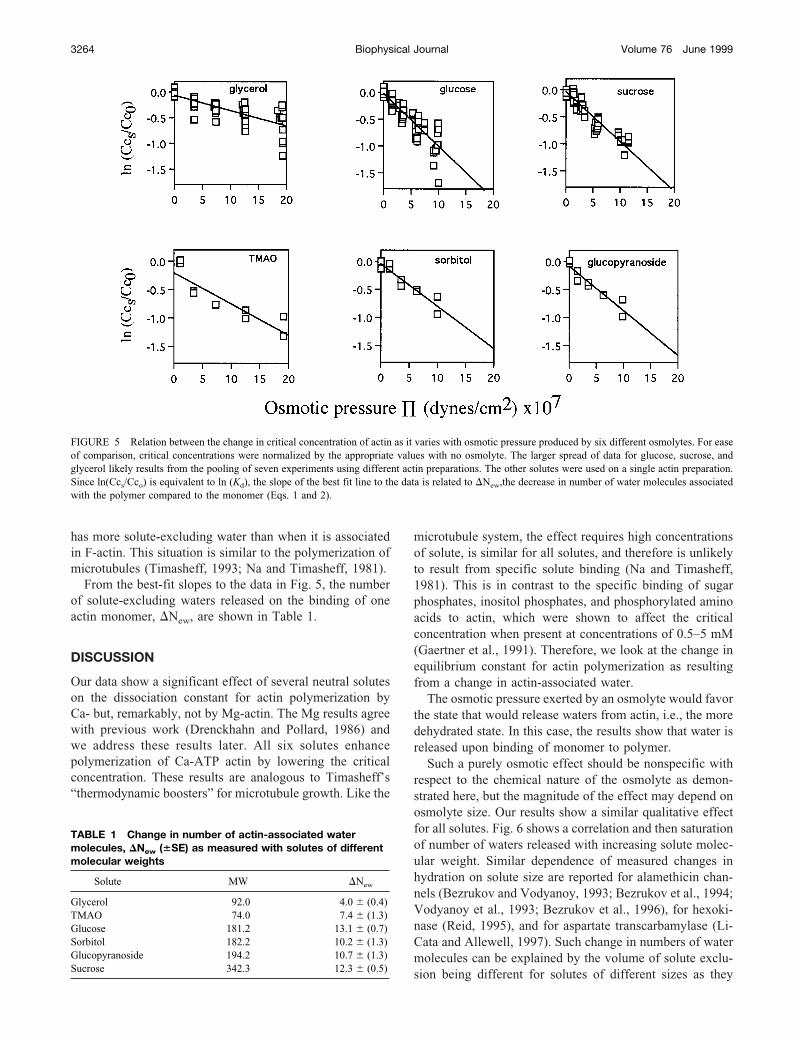
has more solute-excluding water than when it is associatedin F-actin. This situation is similar to the polymerization ofmicrotubules (Timasheff, 1993; Na and Timasheff, 1981).
From the best-fit slopes to the data in Fig. 5, the numberof solute-excluding waters released on the binding of oneactin monomer, �New, are shown in Table 1.
DISCUSSION
Our data show a significant effect of several neutral soluteson the dissociation constant for actin polymerization byCa- but, remarkably, not by Mg-actin. The Mg results agreewith previous work (Drenckhahn and Pollard, 1986) andwe address these results later. All six solutes enhancepolymerization of Ca-ATP actin by lowering the criticalconcentration. These results are analogous to Timasheff’s“thermodynamic boosters” for microtubule growth. Like the
microtubule system, the effect requires high concentrationsof solute, is similar for all solutes, and therefore is unlikelyto result from specific solute binding (Na and Timasheff,1981). This is in contrast to the specific binding of sugarphosphates, inositol phosphates, and phosphorylated aminoacids to actin, which were shown to affect the criticalconcentration when present at concentrations of 0.5–5 mM(Gaertner et al., 1991). Therefore, we look at the change inequilibrium constant for actin polymerization as resultingfrom a change in actin-associated water.
The osmotic pressure exerted by an osmolyte would favorthe state that would release waters from actin, i.e., the moredehydrated state. In this case, the results show that water isreleased upon binding of monomer to polymer.
Such a purely osmotic effect should be nonspecific withrespect to the chemical nature of the osmolyte as demon-strated here, but the magnitude of the effect may depend onosmolyte size. Our results show a similar qualitative effectfor all solutes. Fig. 6 shows a correlation and then saturationof number of waters released with increasing solute molec-ular weight. Similar dependence of measured changes inhydration on solute size are reported for alamethicin chan-nels (Bezrukov and Vodyanoy, 1993; Bezrukov et al., 1994;Vodyanoy et al., 1993; Bezrukov et al., 1996), for hexoki-nase (Reid, 1995), and for aspartate transcarbamylase (Li-Cata and Allewell, 1997). Such change in numbers of watermolecules can be explained by the volume of solute exclu-sion being different for solutes of different sizes as they
FIGURE 5 Relation between the change in critical concentration of actin as it varies with osmotic pressure produced by six different osmolytes. For easeof comparison, critical concentrations were normalized by the appropriate values with no osmolyte. The larger spread of data for glucose, sucrose, andglycerol likely results from the pooling of seven experiments using different actin preparations. The other solutes were used on a single actin preparation.Since ln(Ccs/Cco) is equivalent to ln (Kd), the slope of the best fit line to the data is related to �New,the decrease in number of water molecules associatedwith the polymer compared to the monomer (Eqs. 1 and 2).
TABLE 1 Change in number of actin-associated watermolecules, �New (�SE) as measured with solutes of differentmolecular weights
Solute MW �New
Glycerol 92.0 4.0 (0.4)TMAO 74.0 7.4 (1.3)Glucose 181.2 13.1 (0.7)Sorbitol 182.2 10.2 (1.3)Glucopyranoside 194.2 10.7 (1.3)Sucrose 342.3 12.3 (0.5)
3264 Biophysical Journal Volume 76 June 1999
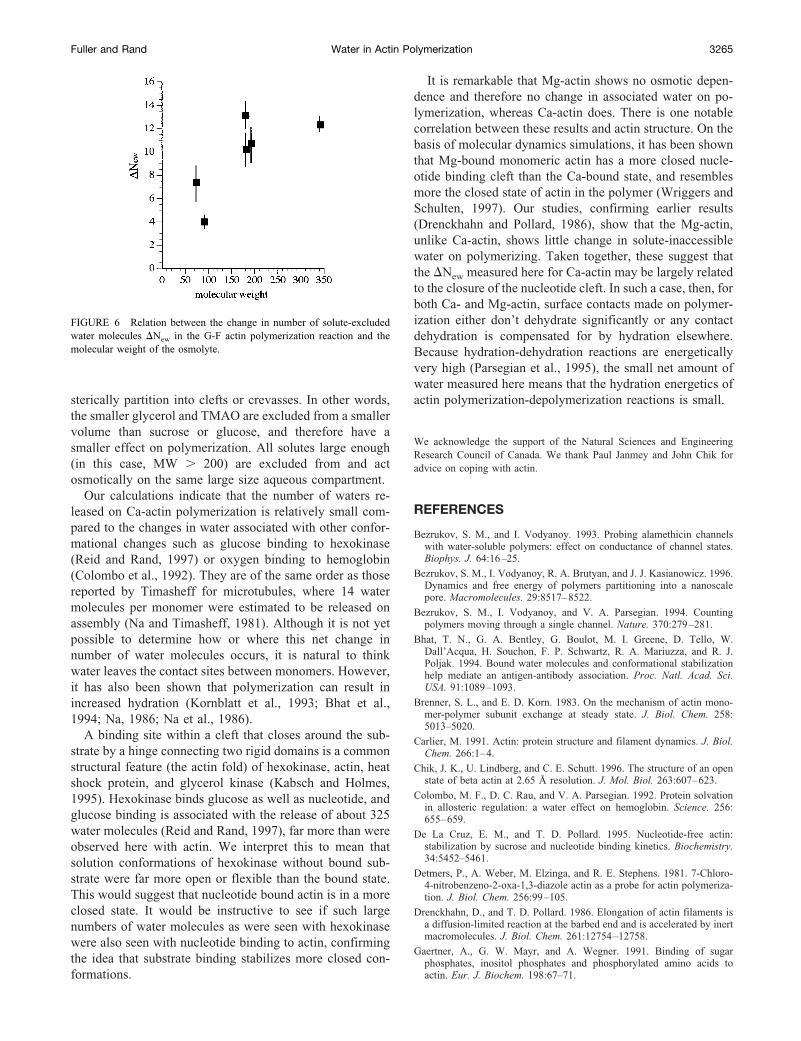
sterically partition into clefts or crevasses. In other words,the smaller glycerol and TMAO are excluded from a smallervolume than sucrose or glucose, and therefore have asmaller effect on polymerization. All solutes large enough(in this case, MW � 200) are excluded from and actosmotically on the same large size aqueous compartment.
Our calculations indicate that the number of waters re-leased on Ca-actin polymerization is relatively small com-pared to the changes in water associated with other confor-mational changes such as glucose binding to hexokinase(Reid and Rand, 1997) or oxygen binding to hemoglobin(Colombo et al., 1992). They are of the same order as thosereported by Timasheff for microtubules, where 14 watermolecules per monomer were estimated to be released onassembly (Na and Timasheff, 1981). Although it is not yetpossible to determine how or where this net change innumber of water molecules occurs, it is natural to thinkwater leaves the contact sites between monomers. However,it has also been shown that polymerization can result inincreased hydration (Kornblatt et al., 1993; Bhat et al.,1994; Na, 1986; Na et al., 1986).
A binding site within a cleft that closes around the sub-strate by a hinge connecting two rigid domains is a commonstructural feature (the actin fold) of hexokinase, actin, heatshock protein, and glycerol kinase (Kabsch and Holmes,1995). Hexokinase binds glucose as well as nucleotide, andglucose binding is associated with the release of about 325water molecules (Reid and Rand, 1997), far more than wereobserved here with actin. We interpret this to mean thatsolution conformations of hexokinase without bound sub-strate were far more open or flexible than the bound state.This would suggest that nucleotide bound actin is in a moreclosed state. It would be instructive to see if such largenumbers of water molecules as were seen with hexokinasewere also seen with nucleotide binding to actin, confirmingthe idea that substrate binding stabilizes more closed con-formations.
It is remarkable that Mg-actin shows no osmotic depen-dence and therefore no change in associated water on po-lymerization, whereas Ca-actin does. There is one notablecorrelation between these results and actin structure. On thebasis of molecular dynamics simulations, it has been shownthat Mg-bound monomeric actin has a more closed nucle-otide binding cleft than the Ca-bound state, and resemblesmore the closed state of actin in the polymer (Wriggers andSchulten, 1997). Our studies, confirming earlier results(Drenckhahn and Pollard, 1986), show that the Mg-actin,unlike Ca-actin, shows little change in solute-inaccessiblewater on polymerizing. Taken together, these suggest thatthe �New measured here for Ca-actin may be largely relatedto the closure of the nucleotide cleft. In such a case, then, forboth Ca- and Mg-actin, surface contacts made on polymer-ization either don’t dehydrate significantly or any contactdehydration is compensated for by hydration elsewhere.Because hydration-dehydration reactions are energeticallyvery high (Parsegian et al., 1995), the small net amount ofwater measured here means that the hydration energetics ofactin polymerization-depolymerization reactions is small.
We acknowledge the support of the Natural Sciences and EngineeringResearch Council of Canada. We thank Paul Janmey and John Chik foradvice on coping with actin.
REFERENCES
Bezrukov, S. M., and I. Vodyanoy. 1993. Probing alamethicin channelswith water-soluble polymers: effect on conductance of channel states.Biophys. J. 64:16–25.
Bezrukov, S. M., I. Vodyanoy, R. A. Brutyan, and J. J. Kasianowicz. 1996.Dynamics and free energy of polymers partitioning into a nanoscalepore. Macromolecules. 29:8517–8522.
Bezrukov, S. M., I. Vodyanoy, and V. A. Parsegian. 1994. Countingpolymers moving through a single channel. Nature. 370:279–281.
Bhat, T. N., G. A. Bentley, G. Boulot, M. I. Greene, D. Tello, W.Dall’Acqua, H. Souchon, F. P. Schwartz, R. A. Mariuzza, and R. J.Poljak. 1994. Bound water molecules and conformational stabilizationhelp mediate an antigen-antibody association. Proc. Natl. Acad. Sci.USA. 91:1089–1093.
Brenner, S. L., and E. D. Korn. 1983. On the mechanism of actin mono-mer-polymer subunit exchange at steady state. J. Biol. Chem. 258:5013–5020.
Carlier, M. 1991. Actin: protein structure and filament dynamics. J. Biol.Chem. 266:1–4.
Chik, J. K., U. Lindberg, and C. E. Schutt. 1996. The structure of an openstate of beta actin at 2.65 Å resolution. J. Mol. Biol. 263:607–623.
Colombo, M. F., D. C. Rau, and V. A. Parsegian. 1992. Protein solvationin allosteric regulation: a water effect on hemoglobin. Science. 256:655–659.
De La Cruz, E. M., and T. D. Pollard. 1995. Nucleotide-free actin:stabilization by sucrose and nucleotide binding kinetics. Biochemistry.34:5452–5461.
Detmers, P., A. Weber, M. Elzinga, and R. E. Stephens. 1981. 7-Chloro-4-nitrobenzeno-2-oxa-1,3-diazole actin as a probe for actin polymeriza-tion. J. Biol. Chem. 256:99–105.
Drenckhahn, D., and T. D. Pollard. 1986. Elongation of actin filaments isa diffusion-limited reaction at the barbed end and is accelerated by inertmacromolecules. J. Biol. Chem. 261:12754–12758.
Gaertner, A., G. W. Mayr, and A. Wegner. 1991. Binding of sugarphosphates, inositol phosphates and phosphorylated amino acids toactin. Eur. J. Biochem. 198:67–71.
FIGURE 6 Relation between the change in number of solute-excludedwater molecules �New in the G-F actin polymerization reaction and themolecular weight of the osmolyte.
Fuller and Rand Water in Actin Polymerization 3265
Garner, M. M., and D. C. Rau. 1995. Water release associated with specificbinding of gal repressor. EMBO J. 14:1257–1263.
Gershman, L. C., L. A. Selden, H. J. Kinosian, and J. E. Estes. 1994.Actin-bound nucleotide/divalent cation interactions. Adv. Exp. Med.Biol. 358:35–49.
Kabsch, W., and K. C. Holmes. 1995. The actin fold. FASEB J. 9:167–174.Kabsch, W., H. Mannherz, D. Suck, E. Pai, and K. C. Holmes. 1990.
Atomic structure of the actin: DNase complex. Nature. 347:37–44.Kasai, M., E. Nakano, and F. Oosawa. 1965. Polymerization of actin free
from nucleotides and divalent cations. Biochim. Biophys. Acta. 94:494–503.
Korn, E. D., M. Carlier, and D. Pantaloni. 1987. Actin polymerization andATP hydrolysis. Science. 238:638–644.
Kornblatt, J. A., M. J. Kornblatt, G. H. Bon Hoa, and A. G. Mauk. 1993.Responses of two protein-protein complexes to solvent stress: doeswater play a role at the interface? Biophys. J. 65:1059–1065.
Kouyama, T., and K. Mihashi. 1981. Fluorimetry study of N-(1-Pyrenyl)iodoacetamide-labelled F-actin. local structural change of actinprotomer both on polymerization and on binding of heavy meromyosin.Eur. J. Biochem. 114:33–38.
Leikin, S., V. A. Parsegian, D. C. Rau, and R. P. Rand. 1993. Hydrationforces. Ann. Rev. Phys. Chem. 44:369–395.
LiCata, V. J., and N. M. Allewell. 1997. Functionally linked hydrationchanges in E. coli aspartate transcarbamylase and its catalytic subunit.Biochemistry. 36:10161–10167.
Lorenz, M., D. Popp, and K. C. Holmes. 1993. Refinement of the F-actinmodel against X-ray fiber diffraction data by the use of a directedmutagen algorithm. J. Mol. Biol. 234:826–836.
Na, G. C. 1986. Interaction of calf skin collagen with glycerol: linkedfunction analysis. Biochemistry. 25:967–973.
Na, G. C., L. J. Butz, D. G. Bailey, and R. J. Carroll. 1986. In vitro collagenfibril assembly in glycerol solution: evidence for a helical cooperativemechanism involving microfibrils. Biochemistry. 25:958–966.
Na, G. C., and S. N. Timasheff. 1981. Interaction of calf brain tubulin withglycerol. J. Mol. Biol. 151:165–178.
Oosawa, F., and S. Higashi. 1967. Statistical thermodynamics of polymer-ization and polymorphism of protein. Prog. Theor. Biol. 1:79–164.
Oosawa, F., and M. Kasai. 1971. Actin. In Biological Macromolecules.Vol. 5, Subunits in Biological Systems. S. N. Timasheff and G. D.Fasman, editors. Marcel Dekker, New York. 261–322.
Orlova, A., X. Chen, P. A. Rubenstein, and E. H. Egelman. 1997. Modu-lation of yeast F-actin structure by a mutation in the nucleotide-bindingcleft. J. Mol. Biol. 271:235–243.
Pantaloni, D., M. Carlier, M. Coue, A. A. Lal, S. L. Brenner, and E. D.Korn. 1984. The critical concentration of actin in the presence of ATPincreases with the number concentration of filaments and approaches thecritical concentration of actin-ADP. J. Biol. Chem. 259:6274–6283.
Pardee, J. D., and J. A. Spudich. 1982. Purification of muscle actin. Meth.Cell Biol. 24:271–289.
Parsegian, V. A., R. P. Rand, and D. C. Rau. 1995. Macromolecules andwater: probing with osmotic stress. Methods Enzymol. 259:43–94.
Reid, C. 1995. The influence of water on the glucose affinity of hexoki-nase. Thesis. Biological Sciences. Brock University, St. Catharines,Canada. 162 pp.
Reid, C., and R. P. Rand. 1997. Probing protein hydration and conforma-tional states in solution. Biophys. J. 72:1022–1030.
Timasheff, S. N. 1993. The control of protein statility and association byweak interactions with water: how do solvents affect these processes?Ann. Rev. Biophys. Biomol. Struct. 22:67–97.
Tobacman, L. S., and E. D. Korn. 1982. The regulation of actin polymer-ization and the inhibition of monomeric actin ATPase activity by Acan-thamoeba profilin. J. Biol. Chem. 257:4166–4170.
Valentin-Ranc, C., and M. Carlier. 1991. Role of ATP-bound divalentmetal ion in the conformation and function of actin. J. Biol. Chem.266:7668–7675.
Vodyanoy, I., S. M. Bezrukov, and V. A. Parsegian. 1993. Probing ala-methicin channels with water-soluble polymers: size-modulated osmoticaction. Biophys. J. 65:2097–2105.
Wriggers, W., and K. Schulten. 1997. Stability and dynamics of G-actin:back-door water diffusion and behavior of a subdomain 3/4 loop. Bio-phys. J. 73:624–639.
3266 Biophysical Journal Volume 76 June 1999