volume 5, issue 3, 2016, 449 - 466 issn 2284-6808 letters
TRANSCRIPT

Page | 449
Magnetite nanostructures: a novel delivery system for enhanced antimicrobial therapy
Adnaik Rahul Shivaji 1,*, Gavarkar Pratibha Shivaji 1 , Mohite Shrinivas Krishna 1 1 Rajarambapu College of Pharmacy, Kasegaon, Tal Walwa, Dist. Sangli 415 404, MS, India
*corresponding author e-mail address: [email protected]
ABSTRACT Resistance of microorganisms for antibiotics is a serious and increasing public health problem in today’s world. Therefore novel approaches for controlling microbial infection are urgently needed for which nanomaterials can be a very promising approach. Inefficient delivery of antimicrobials results in inadequate therapeutic index and side effects. Nanostructured biomaterials, nanoparticles in particular, have unique physicochemical properties such as ultra small and controllable size, large surface area to mass ratio, high reactivity and functionalizable structure. These properties can be applied to facilitate the administration of antimicrobial drugs, thereby overcoming some of the limitations in traditional antimicrobial therapeutics. Iron oxide magnetic NPs (MNPs) have many advantages and are considered very promising drug carriers. Iron oxide MNPs have been used with various synthetic and natural drugs to inhibit the development of microorganism causing serious infectious diseases. This review highlights potential of MNPs in developing novel routes for fighting antimicrobial resistance. Keywords: antibiotics, infection, nanomaterials, antimicrobial, magnetic nanoparticles, magnetic field, microbial resistance.
1. CURRENT STATUS OF MICROBIAL INFECTIONS AND ANTIMICROBIAL AGENTS Microbial infections are still a major cause of morbidity
and mortality in the world. The effect on morbidity and mortality
due to infectious diseases is very noteworthy among the
population. Antimicrobial agents are substances that kill or inhibit
the growth of microorganisms. Many infectious diseases have
been overcome, since the discovery of antimicrobial drugs in the
1960s [1]. Typically, antimicrobials kill bacteria by binding to
some vital compounds of bacterial metabolism, leading to
inhibition of synthesis of functional biomolecules or impeding
normal cellular activities, e.g. β-lactams (penicillins and
cephalosporins) inhibit bacteria cell wall synthesis; tetracyclines,
macrolides, and clindamycin inhibit protein synthesis;
metronidazole and quinolones inhibit nucleic acid synthesis; and
sulphonamides and trimethoprim have an inhibitory effect on
enzyme synthesis. Antimicrobials such as penicillin are only
effective against a narrow range of bacteria, whereas others, like
ampicillin, eradicate a broad spectrum of both Gram-positive and
Gram-negative bacteria [2]. Despite the development of many
antimicrobial drugs, many infectious diseases remain difficult to
treat; may be due to difficulty in transport of drug through cell
membranes and they may have low activity inside the cells leading
to weak inhibitory or bactericidal effects on the intracellular
bacteria. In addition, antimicrobial toxicity to host tissues leads to
a significant restriction to their use. Aminoglycosides, for
instance, cause ototoxicity and nephrotoxicity and have to be
given in controlled dosages. Another major issue with
antimicrobials agents is the acquired resistance of infectious
microbes.
In spite of availability of existing potent antimicrobial
agents and other modern antibacterial means, microbial infections
are still a challenge. Presently available antimicrobial agents
clinically used today are having significant shortfalls, which
include weak antimicrobial activity, risk of microbial resistance
and difficulty in functioning in a dynamic environment.
Most of the currently used antimicrobial agents generally affect
three microbial targets: cell wall synthesis, translational
machinery, and DNA replication [3]. Unfortunately, bacterial
resistance may develop against these targets.
The mechanisms by which resistance can be developed
includes:
• Enzymes that modify or degrade the antibiotic such as �-
lactamases and aminoglycosides;
• Modification of cellular components such as cell wall as
seen in vancomycin resistance [3] and ribosomes in tetracyclines
resistance;
• Efflux pumps that provide multidrug resistance against
numerous antibiotics [3].
During the last few decades antimicrobial resistance has
increased due to the continuous use of antibacterial agents. The
resistance has resulted in genetic and biochemical modification of
microorganisms in order to ensure their bacterial survival and
multiplication. Antimicrobial resistance may be defined as a
means of genetic events leading to biochemical alterations in
bacterial genome. The biochemical changes resulting into
resistance can be point mutations or gene amplifications. Also,
usage of antibacterial drugs has increased the accumulation of
genetic material which codes for bacterial resistance which can be
transferred from one microbe to another resulting in multi-
resistant clones.
In antimicrobial resistant bacteria the drug molecules are
structurally distorted leading to prevention of binding of
antimicrobial agent by the following mechanisms:
• Blocking of entry of antimicrobial agent into bacterial cell;
• inactivation of antimicrobial agent (e.g. enzymatic
degradation);
Volume 5, Issue 3, 2016, 449 - 466 ISSN 2284-6808
Open Access Journal Received: 02.03.2016 / Revised: 06.07.2016 / Accepted: 15.08.2016 / Published on-line: 30.09.2016
Review Article
Letters in Applied NanoBioScience www.NanoBioLetters.com

Magnetite nanostructures: a novel delivery system for enhanced antimicrobial therapy
Page | 450
• Rapid exit of antimicrobial agent from the bacterial cell
through powerful efflux pumps before they bind to any site [4].
Thus, there is an increased concern regarding ever
increasing microbial infections in addition to drug resistant
bacterial strains. Moreover new antimicrobial agents are needed to
treat bacterial infections and/or to develop new drug moieties
which can be more effective than the currently used antibiotics.
This had leaded the way for the development of newer and
additional anti-microbial agents. In 2001 the WHO (World Health
Organization) global strategy for the control of Antimicrobial
Resistance [5] has provided a framework for reducing infectious
diseases, reducing spread of infections, upgrading their use and
access to antimicrobials. Since 1930s until now some new classes
of antimicrobial drugs have been discovered, but the antimicrobial
resistance still remains an issue to be solved yet. In many
conditions antimicrobials has turn out to be less effective which
has resulted in the usage of large concentration of antimicrobial
agents to treat life-threatening infections. However, antimicrobial
agents used in large concentration can be lethal for patients,
leading to severe side effects. In order to resolve such issues,
alternative antimicrobial drug delivery approaches have been
proposed. Therefore, one of the most promising antimicrobial
approach relies in the application of nanosized carriers which can
transport as well as control the release of antimicrobial agents [6].
Over the last few decades, the applications of
nanotechnology in medicine have been extensively explored for
effective drug delivery. Nanotechnology deals with the
understanding and control of matters in the 1- 100 nm range.
These nanosized materials, at such scale have unique
physicochemical properties including ultra small size, large
surface to mass ratio, high reactivity and unique interactions with
biological systems [7]. The pharmacokinetics and therapeutic
index of the drugs can be significantly improved in contrast to the
free drug counterparts by loading drugs into nanoparticles through
physical encapsulation, adsorption, or chemical conjugation.
nanoparticle-based drug delivery systems has advantages of
improved serum solubility of the drugs, extended systemic
circulation time, at a sustained and controlled release drugs, drug
delivery specifically to the targeted tissues and cells, and delivery
of multiple therapeutic agents to the same cells for combination
therapy [7, 8, 9]. Moreover, drug-loaded nanoparticles can enter
host cells through endocytosis and then release drug payloads to
treat microbes-induced intracellular infections. Therefore, a
number of nanoparticle-based drug delivery systems have been
approved for clinical uses to treat a variety of diseases and many
other therapeutic nanoparticle formulations are currently under
various stages of clinical tests [7, 10]. Hence, in the era of current
drug development, attention has been especially focused on new
and emerging nanoparticle-based systems for antimicrobial
chemotherapy.
Nanomaterials (NM) may be strategically advantageous as
active antibacterial groups since their surface area is exceedingly
large relative to their size. Nanosized particles may provide high
activity although only a small dose of the particles is used.
Consequently, NM could serve as an alternative to antibiotics to
control bacterial infections. Most of the resistance mechanisms
seen with antimicrobial agents fail by use of nanoparticles; since
nanoparticles act mainly by direct contact with the bacterial cell
wall, without the need to penetrate the cells. This raises the hope
that nanoparticles would be less prone than existing antibiotics to
promote resistant bacteria.
Figure 1. Discovery of Antimicrobial drugs from 1930s-2000s.
Generally, nanoparticles act by two major lethal pathways,
which occur simultaneously in many cases and often related to
each other:
(1) Disruption of membrane potential and integrity and
(2) Production of reactive oxygen species (ROS), also known
as oxygen-free radicals, the NM acting as nanocatalysts [11, 12,
13].
2. NANOTECHNOLOGY AS A TOOL FOR DRUG DELIVERY Nanobiotechnology is the science that investigates the
interactions between nanoscale materials and biological systems.
Nanotechnology is referred as the manipulation of matter with at
least one dimension sized from 1 to 100 nanometers.
Nanoparticles are typically defined as solids with less than
100 nm in all three dimensions. The nanoparticles exhibit new
thermal, mechanical, magnetic and optical properties such as small
size, large surface area to mass ratio, and high reactivity delivery,
that allow for their widespread application in biomedicine [14].
Most often, they are particles having diameters about 10 nm or
less similar to most biological molecules and structures [15].
Nanoparticles are composed of mostly inorganic materials or
organic (e.g. polymeric) materials. These materials may or may
not be biodegradable. The significance of such Nanomaterials is
ascribed to its characteristics such as size effects, magnetic and
electronic properties and the role played by surface phenomena as
their size is reduced [16]. Some nanoparticles commonly consist
of magnetic elements such as iron, nickel and cobalt and their
chemical compounds. Such nanoparticles can be synthesized and
modified with various chemical functional groups and conjugated
with biological molecules or structures, such as drugs of interest,
opening a wide range of potential applications in biomedicine
[14]. Metal and magnetic nanoparticles have been continuously
used and modified to enable their use as a drug delivery system.

Adnaik Rahul Shivaji, Gavarkar Pratibha Shivaji, Mohite Shrinivas Krishna
Page | 451
3. THE EVOLUTION OF MAGNETIC DRUG DELIVERY As described by Paul Ehrlich (1854-1915) if an agent
selectively targets a disease-causing organism, then a toxin for that
organism could be delivered with the agent of selectivity. Hence, a
‘magic bullet’ might be created which can be able to kill the
targeted organism exclusively [17]. Since then, various strategies
have been proposed to deliver a drug to the vicinity of an invading
organism.
In 1960, Freeman et al. proposed that magnetic
nanoparticles could be transported all the way through the vascular
system and can be concentrated in a specific body part with the
help of a magnetic field [18]. The use of magnetic microparticles
and nanoparticles has evolved since 1970s for the delivery of
antimicrobial agents. In 1976, Zimmermann and Pilwat used
magnetic erythrocytes for the cytotoxic drug delivery [20]. Widder
et al. studied the targeting of magnetic albumin microspheres of
doxorubicin in animal models [20]. In the 1980s, several
researchers utilized this concept for delivering drugs with
magnetic microcapsules and microspheres [21, 22, 23]. Hafeli et
al. prepared biodegradable poly (lactic acid) magnetite and the β-
emitter 90Y microspheres for targeted radiotherapy for
subcutaneous tumors [24, 25].
However, all these early approaches utilizing magnetic
drug delivery were of microsized systems. Magnetic nanoparticles
were first used in animal models by Lubbe et al [26]. In 1996, the
first Phase I clinical trial of epirubicin magnetic NPs was
conducted in advanced and unsuccessfully pretreated cancers
patients [27]. Although more than 50% of nanoparticles were
destroyed in liver in these trails. Afterwards, various scientists
have developed magnetic vectors and proved their potential
applications. Several other start-ups currently manufacture
magnetic microparticles and nanoparticles employed in magnetic
resonance imaging, magnetic fluid physiological condition, cell
sorting and targeting, bioseparation, sensing, enzyme
immobilization, immunoassays, and factor transfection and
detection systems.
Magnetic Propreties of Nanoparticles [28]
Magnetic nanoparticles for bioseparation are made of one
or more magnetic cores coated with matrix of polymers such as
silica or hydroxylapatite with terminal functionalized groups.
The magnetic core is usually made of either of magnetite
(Fe3O4) or maghemite (gamma Fe2O3) with superparamagnetic or
ferromagnetic properties.
Magnetic cores can be produced using magnetic ferrites,
such as cobalt ferrite or manganese ferrite.
Theory of Magnetic Drug Targeting [29]
For magnetically controlled drug targeting it is assumed
that magnetic nanocomposite particles having high saturation
magnetization could be used for potential application. These
particles have relatively magnetic properties along with discrete
randomly oriented magnetic moments. When the magnetic
particles are placed in the external magnetic field, their moments
rapidly rotate into the direction of the field and thus improve the
magnetic flux density. In order to manage the motion of such
particles within a circulating system, a magnetic force and a
hemodynamic drag force are combined to generate a total vectoral
force on the particles. The magnetic force of the external field
must be greater than the drag force or hydrodynamic force so as to
effectively overcome the influence of a fluid flow in order to
achieve the desired external magnetic field. Therefore, the
magnetic force on the magnetic particles will be governed by:
Where F is the magnetic force, m is the total magnetic
moment of the material in the microsphere, Δ is the gradient that is
assumed to be derived from characteristics of the field alone, and
the magnetic flux density- also known as the Bfield. Hence these
quantities influence to some degree to which an external magnetic
field may be used to internally guide particles in the body. The del
operator is defined for magnetic field distribution at xyz
directions.
It is eminent that the gradient of a scalar function at any
point is the maximum spatial change of the magnetic field. The
Bfield tends to align with the net magnetic moment of a particle in
a fixed direction while the gradient leads to a force so as to moves
the particles. The second factor characterizes the magnetic
properties of the particles. The magnetic moment of a material m,
is proportional to the applied magnetic field H, and the intrinsic
magnetic susceptibility of the material, χm.
The magnetic volume susceptibility for various materials
ranges from aluminium at 2.07 x 10⁻⁵ to magnetite 1.0 x 10⁶ 5.7 x
106 and as high as 106 for various ferromagnetic rare-earth
materials. The force which counteracts the magnetic force on the
particle in the fluid stream is due to the liquid flow (blood flow).
Stokes law governs the hemodynamic forces on a particle in the
liquid. The equation is given by:
Where, F is the drag force, is the viscosity of fluid, and is
the relative velocity of a spherical particle and r is the radius of the
particle.
Also, there are other variables for drug delivery together
with tissue c porosity, particle distribution, and allowable cell
damage caused by incompatible sphere size and variable blood
flow and body. An extremely porous tissue permits small particles
to be simply manipulated out of the blood stream and into tissue.
However, a comparatively tight tissue structure would need a lot
of magnetic field elicited force to drag the nanoparticles out of the
bloodstream, and such interfacial transport may additionally cause
damage to the tissue. As a result, the magnetic nanocomposite
particle size and external forces required for effective particle
manipulation are extremely dependent on the area in which drug
delivery is performed. Factors Affecting Magnetic Targeting of
Drug [30]:
1. Size of the particles in ferrofluid.
2. Surface characteristics of particles.
3. Concentration of the ferrofluid.

Magnetite nanostructures: a novel delivery system for enhanced antimicrobial therapy
Page | 452
4. Volume of the ferrofluid.
5. Reversibility and strength of drug/ferrofluid binding
(desorption characteristics).
6. Access to the organism (infusion route).
7. Duration or rate of injection/infusion.
8. Geometry, strength and duration of the magnetic field
application.
Limitations of Magnetic Drug Delivery [31, 32, 33]
The magnetic gradient decreases with the distance to the
target.
The geometry of the magnetic field is extremely necessary
and should be taken under consideration when coming up with a
magnetic targeting method because the magnetic carriers
accumulate at the target site similarly as throughout the cross
section from the surface supply to the depth marking the effective
field limit.
Once the magnetic flux is removed magnetic agglomeration
are often developed owing to the small size of NPs, a requisite for
superparamagnetism.
Limitations additionally arise in extrapolating applications
of MNPs from animal models to humans
The fates of magnetic carriers are insufficient and, in
several instances, there's inadequate characterization.
Magnetic targeting is an expensive, technical approach and
needs specialized manufacture and quality control system.
It desires specialized magnet for targeting, advanced
techniques for observation, and trained personnel to perform
procedures. Magnets should have relatively constant gradients, so
as to avoid focal over dosing with poisonous drug. A massive
fraction of magnetite, which is entrapped in carriers, is deposited
permanently in targeted tissue.
4. MAGNETIC NANOPARTICLES (MNPs) Nanoparticles consisting of ferromagnetic elements such as
iron, cobalt, nickel, or their oxides and alloys exhibiting magnetic
properties are called magnetic nanoparticles [34]. Elemental
manganese upon physicochemical treatment can display magnetic
behavior [35]. In brief, the magnetic properties of a material
replicate their magnetization arising from magnetic moments of
unpaired electrons. Since thermal fluctuations of magnetic
moments reverse their direction, some magnetic nanoparticles
display superparamagnetic properties. Such properties are termed
as the non-appearance of magnetic behavior when the magnetic
field is not present [36, 37].
Iron oxide nanoparticles of size having approximately 10
nm in diameter like maghemite (γ-Fe2O3) display a
superparamagnetic behavior (superparamagnetic iron oxide
nanoparticles called as SPIONs) leading to improved dispersive
properties in the nonappearance of a magnetic field [38]. Later on
these nanopartcles are guided to in order to accumulate at the site
of interest in presence of magnetic field. Such approach has
enormous importance in targeted drug delivery applications [36].
Hence, magnetic nanoparticles having smaller particle size,
consisting of iron oxide and display magnetic behavior only in the
existence of a magnetic field are termed as superparamagnetic iron
oxide nanoparticles (SPIONs) [36]. When the magnetic field is
removed permanent magnetic behavior of magnetic particles result
in destruction within the organism. Hence it is necessary to use
SPIONs in biomedical applications [39].
Iron oxide nanoparticles such as magnetite Fe3O4 or magnemite
Fe2O3 and gadolinium nanopartcles such as chelated organic
gadolinium complexes have capacity to separate into iron and
oxygen within the body. Due to this they are generally used as
contrast agents in MRI for biological applications, since they can
be safely eliminated and utilized in metabolic and oxygen
transport (academia) systems [36]. Magnetic nanoparticles also
exhibit low cytotoxicity and has been approved by the United
States FDA for clinical MRI applications (academia) [40-46].
Most of the magnetic nanoparticles used as targeted delivery
systems are chemically iron oxides (Table 2). Iron is essential to
every organism. In addition endogenous iron oxide nanoparticles
were also found in the human hippocampus [40, 41]. On the other
hand, iron oxide cause direct cellular toxicity due to the
production of reactive oxygen and nitrogen species (ROS and
RNS) [42]. Therefore, magnetic nanoparticles are mostly prepared
by core-shell methodology.
Figure 2. Application of SPIONs in Antibacterial Therapy.
The magnetic core of iron oxide nanoparticles consists of
magnetite (Fe3O4) and/or maghemite (γ-Fe2O3) and their shell
surface coating consists of organic compounds such as surfactants,
synthetic or natural polymers, or inorganic material, such as silica,
carbon, precious metals or oxides [36, 43]. Significance of core-
shell type magnetic nanoparticles is due to following reasons [36,
44]:
- protection of magnetic core from oxidation;
- protection of shell surface from chemical reactions;
- prevention of aggregate and agglomerate formation due to Van
der Waals forces, hydrophobic effects and magnetic attractions
[36];
- various therapeutics attachment is possible;
- Cellular uptake rate can be amplified.
Biocompatibility of MNPs depends on the type of shell
surface coating in addition to their size. Surface coating may be
biodegradable (e.g. certain polymers) or non-biodegradable (e.g.
silica). Uniqueness of the particle surface (e.g. hydrophilicity and
Adnaik Rahul Shivaji, Gavarkar Pratibha Shivaji, Mohite Shrinivas Krishna
Page | 453
surface charge) is determined by the type of the coating whereas
total size of magnetic nanoparticles is determined by the thickness
of the coating [36]. Magnetic nanoparticles with smaller particles
lesser than 100 nm can be prepared by coating with an inorganic
material; whereas larger magnetic nanoparticles with particles
above 100 nm are prepared by polymer coating [45, 46].
Magnetic nanoparticles used for biomedical applications
are primarily prepared as ferrofluids, i.e. magnetic liquids.
Therefore their surface charge is established by ionization of
surface groups or by adsorption of charged species around the
liquid on the particle surface which leads to the formation of layer
around the particle. The variation in potential between the
surrounding liquid medium and the layer around the particle is
called the zeta potential. Particles with zeta potential larger than
30 mV, either positive or negative, will repel each other, reside as
under and result in a stable ferrofluid [36, 47].
Fundamental prerequisites for Magnetic NPs
Magnetic NPs used for biomedical applications should
posses certain specific characteristics as follows
Superparamagnetism
Superparamagnetism occurs in magnetic materials
consisting of minute crystallites (Fe-based NPs turn into
superparamagnetic at sizes <25 nm) [17, 48]. In
superparamagnetic materials, the fluctuations affect the direction
of magnetization of entire crystallites. The magnetic moments of
individual crystallites compensate for each other and the overall
magnetic moment becomes null.
Particle Size
In NPs with large particle size, energetic considerations
favor the development of domain walls. But, when the particle
size decreases below particular limit, the development of domain
walls becomes unfavorable and each particle consists of a single
domain. This happens with superparamagnetic NPs.
Superparamagnetism used in drug delivery is essential because as
the external magnetic field is removed, magnetization disappears
and thus agglomeration is prevented [17].
Biodegradability of Magnetic Core
SPION are considered as biodegradable because iron is
recycled by cells using usual biochemical pathways for Fe
metabolism [49, 50]. But for non-biodegradable cores, a specific
coating is required in order to prevent exposure of the magnetic
core and to assist intact excretion through the kidneys (e.g.
contrast agents based on gadolinium) [17, 50].
5. DRUG DELIVERY USING MAGNETIC NANOPARTICLES Different organic materials such as polymeric NPs,
liposomes, micelles have been reported as drug delivery
nanovectors. Such materials acts by passive targeting, active
targeting with a recognition moiety (e.g. antibody), or active
targeting by physical stimulus (e.g. magnetism in
magnetoliposomes). Nevertheless, these organic systems has
limitations such as inadequate chemical and mechanic stability,
swelling, susceptibility to microbiological attack, insufficient
control over rate of drug release [51] and high cost. A major
drawback of dendrimers and dendritic polymers is their high cost.
Due to the limitations of organic NPs used for drug
delivery, inorganic vectors (inorganic magnetic NPs) are gaining
popularity of intense research.
The main advantages of magnetic nanoparticles (organic or
inorganic) are as follows:
1) visualization (superparamagnetic NPs are used in MRI);
2) magnetic nanoparticles can be guided or detained at the site
with the help of magnetic field;
3) magnetic nanoparticles can be heated in a magnetic field to
enhance drug release or to generate hyperthermia/ablation of
tissue.
Behavior of magnetic NPs [51] is attributed to the important
parameters such as:
- shell surface chemistry;
- particle size (which determines magnetic core,
hydrodynamic volume and size distribution);
-Magnetic properties (which determines magnetic moment,
remanence, coercivity).
The surface chemistry is important in order to avoid the action of
the reticuloendothelial system (RES) and to increase the half-life
in the blood stream. Coating of nanoparticles by neutral and
hydrophilic compound (i.e. polyethylene glycol (PEG),
polysaccharides, etc.) increases the circulatory half-life ranging
from min to hours or days [51].
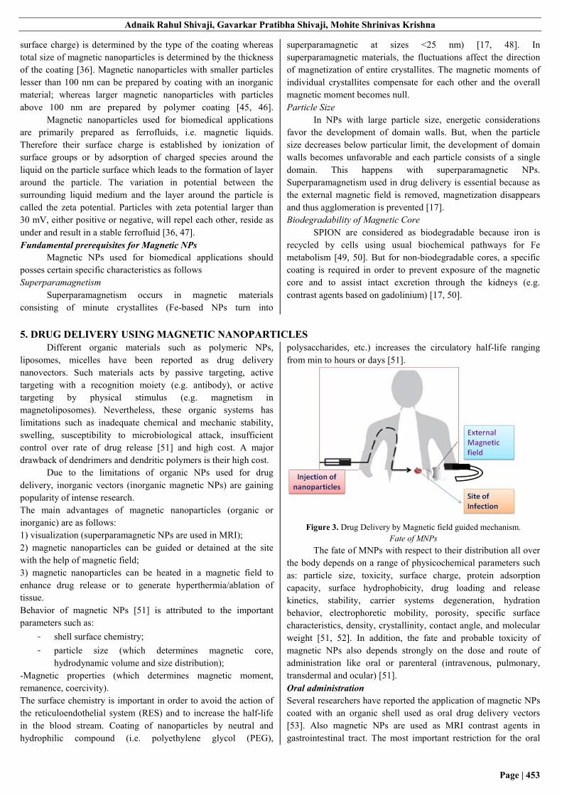
Figure 3. Drug Delivery by Magnetic field guided mechanism.
Fate of MNPs
The fate of MNPs with respect to their distribution all over
the body depends on a range of physicochemical parameters such
as: particle size, toxicity, surface charge, protein adsorption
capacity, surface hydrophobicity, drug loading and release
kinetics, stability, carrier systems degeneration, hydration
behavior, electrophoretic mobility, porosity, specific surface
characteristics, density, crystallinity, contact angle, and molecular
weight [51, 52]. In addition, the fate and probable toxicity of
magnetic NPs also depends strongly on the dose and route of
administration like oral or parenteral (intravenous, pulmonary,
transdermal and ocular) [51].
Oral administration
Several researchers have reported the application of magnetic NPs
coated with an organic shell used as oral drug delivery vectors
[53]. Also magnetic NPs are used as MRI contrast agents in
gastrointestinal tract. The most important restriction for the oral

Magnetite nanostructures: a novel delivery system for enhanced antimicrobial therapy
Page | 454
delivery of peptides and proteins is that they are degraded in
gastric acid, have low absorption, undergo first-pass metabolism
and shows considerable increase in initial drug concentration.
Feng et al. has studied fate of chemotherapeutic nanoparticles in
oral delivery wherein it reports that particles below 5 μm can be
removed via lymphatic drainage, particles upto 500 nm can cross
epithelial cell membrane by endocytosis, and particles less than 50
nm can reach paracellular passage between intestinal epithelial
cells [51, 54].
Intravenous administration
The carrier for magnetic NPs used in parenteral applications
should be nontoxic, nonimmunogenic, and has size such that it
avoids embolization of capillary ducts. On reaching bloodstream
NPs are cleared by moocytes into the liver, spleen, and bone
marrow where NPs are captured by resident cells (e.g. Kupffer
cells in the liver) prior to degradation. Some NPs which are
present in the Kupffer cells may be incorporated into the bile and
removed in the feces depending on biodegradability and their size.
Remaining NPs are filtered by kidneys and secreted in urine.
Generally, smaller NPs are rapidly eliminated by kidney, whereas
larger NPs are uptaken by liver, spleen, and bone marrow [55-61].
Large NPs are removed by cells which are able to undergo
endocytosis (i.e. by B and T lymphocytes). Biodegradable
magnetic NPs are taken up by any cell by means of pinocytosis
[51].
Influence of the Magnetic Field on Human Body
The magnetic fields required to generate magnetic effect in the
body must be very large. All body components are either dia-,
para-, superpara-, ferri-, or ferromagnetic in nature. Even RBC
which contains hemoglobin (Fe containing pigment), shows
comparatively low response to large or steep field gradients.
Although this low value is sufficient to be used in functional MRI
(fMRI). The other natural Fe-containing compounds in the body
are hemosiderin, ferritin, transferrin, and the cytochromes [51].
In 1987 Schenck reported that, the US FDA classified magnets
having field strength lesser than 2 T as nonsignificant risk devices
[62]. Further positive results made FDA to extend this threshold to
4 T in 1996 and again up to 8 T in 2003 for adults. However the
rate of human blood flow has been reported to be reduced by 30%
in in vitro tests in experiments conducted with strong static
magnetic fields (8 T) [51, 63]. Also it has been reported that
magnetic fields more than 3 T may affect conventional behavior of
erythrocytes. Clinically relevant adverse effects in physiological
or neuro-cognitive functions occurring due to exposure to static
magnetic fields effects (up to 8 T) has not been observed in human
subjects studied [51, 64]. Kangarlu and Robitaille has reported
that human imaging obtained at fields more than 10 T can most
likely be achieved and such comes are currently being planned
[51, 65].
Toxicity of MNPs
The toxicity of MNPs, depends on numerous factors includes but
not limited to the dose, chemical composition, method of
administration, size, biodegradability, solubility,
pharmacokinetics, biodistribution, surface chemistry, shape, and
structure. MNPs must be also be assessed with respect to the risk-
benefit ratio in order to justify their risk. The most important
characteristics of MNPs regarding cytotoxicity include their size,
surface area, shape, composition, and coating (Macaroff, 2006;
Arruebo, 2006). The toxicity of NP can be reduced by the
modifying their surface characteristics (Park, 2006). It has been
reported that the big surface-to-volume ratio of all nanosized
particles will probably cause unfavorable biological responses,
once such NP are inhaled and afterward absorbed via the
respiratory organ or engulfed followed by absorption across the
gastrointestinal tract [51, 67]. Apparently, it's conjointly been
reported that, in 20-100 mg/ml concentrations, large magnetic
particles show higher toxicity than smaller ones even when
normalizing for surface area [68]. In any case, toxicity studies
ought to take into account not solely acute toxicity however
additionally that of degradation products, the attainable
stimulation of cells with future release of inflammatory mediators
[69] and long term toxicity .
The magnetic carriers used as potential drug delivery vectors [51]
should be specifically analyzed for:
- Toxicity in cellular and animal models (acute, subacute, and
chronic toxicity, teratogenicity and mutagenicity);
- Hematocompatibility;
- Biodegradation;
- Immunogenicity;
- Pharmacokinetics.
6. DESIGN OF MNPs FOR BIOMEDICAL APPLICATIONS MNPs used for biomedical applications are commonly prepared as
a core/shell structure. Core/shell structure are designed such that
the inorganic magnetic core is enclosed by an outer layer of shell
(coating). Proper selection of magnetic core and surface coating
material is a key for successful design of MNPs [70]. Magnetic
core chiefly determines the abilities (heating, sensing etc) of
MNPs linked with application efficiency. Surface coating
determines the interaction of MNPs with physiological
environment.
Magnetic Core
The magnetic core must be crystalline and smaller than a critical
size so that it shall contain only one magnetic domain which
enables single-domain nanoparticles to display superparamagnetic
behaviour in the absence and a very high magnetization in the
presence of an external field. Due to this particles are dispersed
and concentrated in solution in-vitro or in blood circulation in-
vivo, without forming magnetized clusters. They may also respond
to an instantly applied field with some kind of magnetic on/off
switching behavior. The size distribution of magnetic cores must
be small. The magnetic cores in a particular sample must have a
unique and uniform shape (monodispersed) since; the magnetic
and physico-chemical properties strongly depend on size and
shape of the magnetic cores [70]. The elements used for the
magnetic core are transition metals such as Fe, Ni, Co and Mn
since they present high magnetization values. But if they are not
specially treated they undergo oxidation very quickly in the
synthesis step. Instead transition metal oxide compounds called
ferrites are used since they are stable, have acceptable

Adnaik Rahul Shivaji, Gavarkar Pratibha Shivaji, Mohite Shrinivas Krishna
Page | 455
magnetizations and are generally used for biomedical applications
[70]. Superparamagnetic iron oxide (SPION), belonging to ferrite
family, is the most commonly employed one in biomedical
applications. İron oxide generally exists as two stable forms called
magnetite (Fe3O4) and its γ phase maghemite (γ-Fe2O3).
Magnetic alloy nanoparticles consisting of 2 or 3 different kind of
metals like FeCo, FePt and NiCu. FePt are also used as magnetic
cores and becoming more popular due to its chemical stability and
high magnetic anisotropy [70].
MNPs can be prepared with several techniques including inert gas
condensation [71] mechanical milling [72], spray pyrolysis [73],
sol-gel [74], vapor deposition [75] and wet chemical processes
[76]. However for biomedical applications the most common
method used for synthesis of MNPs is by hydrothermal chemical
decomposition method. Basically, in such type of liquid phase
synthesis method, some organometalic precursors are placed in
reaction in the presence of some organic surfactants or polymers.
An inert gas is continuously fluxed through the mixture during the
decomposition process at high temperature, and desired
nanomaterial is obtained as a precipitate at the end of the reaction
[70].
Another common method used in MNP synthesis is the
co-precipitation method. It has some limitations as compared with
thermal decomposition method like lower crystallinity and lower
monodispersity. However the method is easier and larger quantity
of product can be obtained. The method is based on the
simultaneous nucleation and growth of magnetic cores by
dissolving metal salt precursors in aqueous environment with
changing pH and temperature [77]. Instead this type of reaction
can be carried out in a confined environment by microemulsion
method in which the co-precipitation reaction takes place nano-
reactors called micelles [70].
Figure 4. Schematic presentation of formation of the core-shell structure
of MNPs.
Surface Coating [78, 79, 80]
The coating will contains long-chain organic ligans or
inorganic/organic polymers, wherever these ligands or polymers
are often introduced throughout (in-situ coating) or when (post-
synthetic coating) synthesis. Unaltered coating is employed for co-
precipitation synthesis technique whereas post-synthetic coating is
used with thermal decomposition technique. Throughout unaltered
coating, precursors of magnetic cores and coating materials are
dissolved within the same reaction answer, and therefore the
nucleation of core and also the coating occurs at the same time. In
post-synthetic coating the surface coating materials are introduced
when the magnetic cores are formed. In every procedures either
end-grafting or surface-encapsulation are followed in order to link
the surface molecules to magnetic cores.
The coatings on MNPs often serve manifold purposes such as it
reduces leaching of the cores and it often facilitates the
stabilization of NPs in slightly alkaline pH or significant salt
concentration [51]. The external surface of silica coatings can be
functionalized to allow the binding of biomolecules.
Additionally, coatings play an important role in retarding clearance
by the RES. Uncoated NPs after systemic administration are
rapidly up taken by the mononuclear phagocyte system and are
cleared to the liver, spleen, and bone marrow. Antibodies bind to
the surface of foreign bodies and accelerate phagocytosis of the
particles. Biodegradable (e.g. dextran) and nonbiodegradable
organic and inorganic coatings are used to obstruct detection and
uptake by the macrophages. The most widely used coating for this
reason is PEG which is a linear neutral polyether. Attachment of
NP to PEG surfaces provides a ‘stealth’ shielding effect, delaying
the action of the RES [81]. PEG shows very low toxicity and
immunogenicity, and intact excretion is possible, either via the
kidneys or in the feces [51, 82]. The organic/inorganic surface
coating of MNPs plays a significant role in medicine applications
for [70].
Preventing agglomeration of MNPs due to the higher interparticle
interactions and ultimately providing the mixture stability of
ferrofluids ready with MNPs;
Providing biocompatibility of MNPs by preventing escape of
nephrotoxic particle from magnetic core into biological
surroundings;
Serving as a base for additional anchoring of functional groups
like biomarkers, antibodies, peptides etc. [70].
Table 1. Polymers / organic used as Nanomaterials [70].
Polymers / organic
molecule
Properties Advantages / Disadvantages
Chitosan -natural, cationic, hydrophilic,
linear, biodegredable
-They are used in non-viral gene delivery
Dextran -natural, branched, hydrophilic,
biocompatible
-They permits the anchoring of biovectors and drugs when functionalized
with amino groups
Polyethyleneglycol
(PEG)
-synthetic, neutral, hydrophilic,
linear, biocompatible
-They are stable even at high ionic strengths of solutions with varying pH,
enhances blood circulation time (a few hours) and spermits
functionalization
Polyethyleneimine
(PEI)
-synthetic, cationic, linear or
branched, non-biodegredable,
toxic
-They form strong covalent bonds with MNP’s surface.
They are used for DNA and RNA delivery, but exhibit cytotoxity

Magnetite nanostructures: a novel delivery system for enhanced antimicrobial therapy
Page | 456
Polymers / organic
molecule
Properties Advantages / Disadvantages
Polyvinylealcohol
(PVA)
-synthetic, hydrophilic,
biocompatible
They irreversibly binds on MNP’s surface but can be used in temperature
sensitive heating or drug release applications due to its decomposition
temperatures (40-50 oC),
Polyvinylpyrolidone
(PVP)
-synthetic, branched, hydrophilic They forms covalent bonds with drugs containing nucleophilic functional
groups
Functionalization of MNPs [83]
In order to modify the application of MNPs in biomedical
applications, the MNPs are functionalized by conjugating with
functional teams. The surface coating permits appropriate base to
attach useful groups on MNPs. These groups (antibodies, peptides,
polysaccharides etc.) allow specific recognition of cell varieties
and target the NPs to a particular tissue or cell by binding to cell
surface receptor. Several linker molecules like 1-ethyl-3-(3-
dimethylaminopropyl) carbodimide coordination compound
(EDCI), N-succinimidyl 3-(2-pyridyldithio) propionate (SPDP), N-
hydroxysuccinimide or N, N’-methylene bis amide (MBA) are also
used to attach the initial hydrophilic coated molecules to those
targets [83]. However in practice the fraction of targeted cells
which interact with the protein attached MNP is relatively low even
though the targeted cell population is recognized with high
specificity. In fact the effectiveness of biomedical applications
depend more on cell-nanoparticle interactions rather than particle
targeting. The cell membranes play a vital role in cell-nanoparticle
interactions. In order to explain the character of cell-nanoparticle
interactions, sometimes lipid bilayers mimicking cell membrane are
employed in studies (Umut, 2013).
Figure 5. Representation of magnetic core-shell structure and multi-
functional surface decoration; MNPs consist of a magnetic iron oxide core
coated with a biocompatible material (e.g. polysaccharide, lipid, protein,
small silane linkers, etc.) and Functional groups on the surface of coatings
to link ligand molecules.
7. METHODS FOR PREPARATION OF MAGNETIC PARTICLES
Ramazan Asmatulu et al. method [83]
Magnetic nanoparticles were prepared by the chemical co-
precipitation of Fe2+ and Fe3+ salts in the presence of a strong base.
2 g of FeCl3 and 0.736 g FeCl2 dissolved in 2ml of deoxygenated
water. Dry nitrogen was purged into a 25ml 3-necked round
bottom flask so as to ensure an inert atmosphere. The prepared salt
solutions were added in the flask. This was followed by the
addition of deoxygenated ammonium hydroxide (50/5 aqueous)
until the pH of the solution was 9.5. The solution turns black
immediately upon addition of the base, suggesting that magnetite
(Fe3O4) was formed. The reaction was allowed to continue under a
nitrogen purge for 3min. Then a stabilizer solution (oleic acid and
dichloromethane) was added by a syringe. The reaction was stirred
for an additional 0.5-2 hrs. It was then neutralized with 1M HCl to
a pH 7 and allowed to stir for an additional 3min. The produced
material was decanted into a beaker where it separated into
aqueous and organic layers. The magnetite nanoparticles were
positioned in the organic phase as they were stabilized by oleic
acid. After removing the aqueous layer, the organic layer was
rinsed with fresh deionized water to remove any remaining salt.
Kathryn M. Spiers et al. method [84]
Magnetite particles were synthesized by oxidative
hydrolysis of a ferrous sulphate in an alkaline medium. A solution
of 6.46 g potassium nitrate (KNO3) and 44.9 g potassium
hydroxide (KOH) in 240 ml oxygen-free double distilled water
was added dropwise over 5 min to a solution of 80 g ferrous
sulphate (FeSO4•7H2O) in 560 ml water. The solution was heated
to 900C and flushed with nitrogen. It was stirred continuously
using a magnetic stirrer. About 23 g of magnetite is synthesized by
his reaction. In order to limit the growth of the magnetite particles,
14.4 ml oleic acid was added 15 min after the addition of a
KNO3/KOH solution. The reaction vessel was immediately
removed from the heat, and allowed to cool. Afterwards the
solution was acidified to pH 3 by the addition of dilute nitric acid.
Addition of hexane to this solution leads to the separation of
magnetite into the organic phase, indicative of functionalisation of
the magnetite by the oleic acid. The magnetite powder was
collected and dried [17]
Huiling Bao et al.method [84]
Briefly the hydroxylated copolymer was dissolved in 1ml
tetrahydroflourane (THF), and 10ml Fe2+ solution was added. The
reaction was continued for 4 h with magnetic stirring at 700C.
Thereafter, the reaction mixture was cooled to room temperature
and the latex was separated from the medium by centrifugation.
Then obtained latex was filtered with methanol, and the
precipitation was washed with water and methanol three times in
turn. After vacuum dried at 600oC, the final product was obtained.
A. Pich et al. method [85]
Synthesis of iron oxide particles
Solutions of FeCl2 and FeCl3 were prepared in separate
flasks by keeping molar ratio of FeCl3:FeCl2 constant at2:1. These
solutions are added to stirred dispersion under nitrogen. NH4OH
solution was added drop-wise to start iron oxide formation.
Immediately the solution became dark-brown demonstrating that

Adnaik Rahul Shivaji, Gavarkar Pratibha Shivaji, Mohite Shrinivas Krishna
Page | 457
iron oxide has been produced in the system. After 30 min formed
composite particles were removed from reaction vessel and
cleaned by precipitation to remove all by-products [17]. Magnetic
nanoparticles were washed with 0.01-M HCl solution. Magnetic
dispersion with HCl solution was centrifuged (Universal 16 A) at
a speed of 3000-U/min for 25-min to precipitate the particles. This
procedure was repeated for four times. After that precipitated
magnetic particles were cleaned with distilled water. Then
calculated amount of sodium oleate was added to the required
amount of magnetic dispersion (15-mg Na-oleate in 50-ml of 1.1-
mg/ml magnetic dispersion, keeping the ratio constant at 1:1).
Then the dispersion was heated to 80C for 5-min and finally
sonicated for another 5-min.
A. Ibrahim et al. method [86]
Magnetically responsive polyalkylcyanoacrylate
nanoparticles were prepared by anionic polymerization of the
monomer in the presence of ultrafine magnetite particles of
between 0.01 to 0.05 m. After 1g of glucose and 1g of citric acid
had been dissolved in 100 ml of distilled water, 0.7 g of magnetite
particles were dispersed by ultrasonic treatment for 15 min. The
suspension was passed through a fritted glass filter (pore size 9-15
m) to avoid magnetite agglomerates. Dactinomycin (2 ml) and
isobutylcyanoacrylate monomer (1.5 ml) were added and stirred
ultrasonically (40W). After 3 hrs, nanoparticles were formed and
filtered through a fritted glass filter (suspension A). To separate
magnetized nanoparticles, the suspension was allowed to flow
through a magnetic field at a rate of 1 ml per 3 min, using a
pumping circulation tub system. Four permanent magnets were
attached to the external surface of the circulation tubes. After
removal of the magnets, the nanoparticles attached to the internal
surface are washed with 10ml of an aqueous solution containing
sodium chloride (0.7 %) and calcium chloride (0.2 %). This
magnetically responsive particle suspension was finely
resuspended by ultrasonic treatment for 15 min, at 40W and
filtered through fritted glass.
4. CHARACTERIZAZTION OF MAGNETIC PARTICLES Particle Size and Shape
Magnetic particles are of variable sizes. Conventional light
microscopy (LM) and scanning electron microscopy (SEM) are
most commonly used techniques to visualize microparticles. Both
techniques can be used to determine the shape and outer structure
of the microparticles. Particle size and its distribution are
determined by light microscopy, scanning electron microscopy,
transmission electron microscopy, etc. Confocal laser scanning
microscopy (CLSM) is applied as a nondestructive visualization
technique for microparticles. CLSM allows visualization and
characterization of structures not only on the surface, but also
inside the particles, provided the material is sufficiently
transparent and can be fluorescently labeled. By collecting several
coplanar cross sections, a three-dimensional reconstruction of the
inspected object is possible.
Chemical Analysis
Electron spectroscopy for chemical analysis (ESCA) is used to
determine the surface chemistry of the microspheres. ESCA
determines the atomic composition of the surface. Degradation of
the polymeric matrix carrier system is determined by the Fourier
Transform Infrared Spectroscopy (FTIR). The surface of the
microspheres is investigated measuring total attenuated reflectance
(ATR). The surface carboxylic acid residue is measured by using
radioactive glycine. The radioactive glycine conjugate is prepared
by reaction of 14C-glycine ethyl ester hydrochloride with the
microspheres. The radioactivity of conjugate is measured using
scintillation counter. Surface associated amino acid residue is
determined by the radioactive 14C- acetic acid conjugate. The
carboxylic acid residue is measured through the liquid scintillation
counter and hence the amino acid residue can be determined
indirectly.
Drug Loading
The capture efficiency or the drug loading of the microspheres or
the percent entrapment can be determined by allowing washed
microspheres to lyse the lysate is then subjected to the
determination of active compound by suitable method. The
percent encapsulation efficiency is calculated using following
equation:
% Entrapment = (actual content / theoretical content) x 100
Magnetic Properties
Magnetic properties of nanocomposite particles were
characterized by using vibrating sample magnetometer (VSM).
The magnetic moment of each dried magnetic particles measured
over a range of applied fields between -800and +800Gauss with a
sensitivity of 0.1 emu/g. the prepared samples can be characterized
by weight or volume in VSM. The dry samples are weighed (0.075
g), while the fluids are injected into the sample holder (~ 0.05 ml).
In this system, when a magnetic sample is placed between two
coils of an electromagnet creating a uniform magnetic field
gradient, the applied field induces the magnetic domains to line up
with the field through dipole interactions. As the magnetic field is
increased, number of domains will be also enhanced until the
particles reach saturation levels. During magnetic field alignment,
the particles undergo a sinusoidal motion and produce an electrical
signal in a set of stationary pick-up coils. This signal is
proportional to magnetic moment, vibration amplitude and
vibrational frequency. After the measurements, magnetic
saturation values of the materials are calculated for each sample
by dividing the saturation magnetization by the weight of samples.
Thermo gravimetric Analysis
Differential scanning calorimetry and other gravimetric methods
are used to determine the extent of interaction of polymers with
magnetite and such other magnetic materials. Moreover the
stability of ferrous and ferric ions can be assessed by
thermogravimetric methods.
Stability Measurements
Stability measurements can be performed by using separation
analyser (e.g. LUMiFuge). Measurements are made in glass tubes
at accelerated velocities from 50to 300rpm. The slope of
sedimentation curve can be used to calculate sedimentation
velocity and stability data can be found.
ξ - Potential measurements

Magnetite nanostructures: a novel delivery system for enhanced antimicrobial therapy
Page | 458
ξ - Potential measurements can be made using an instrument like
Zetasizer 2000. The zeta potential is measured at different pH
values and stability of magnetic particles can be predicted
4. ANTIMICROBIAL ACTIVITY OF MAGNETIC NANOPARTICLES Magnetic nanoparticles (MNPs) commonly consist of
magnetic elements such as iron, nickel, cobalt and manganese or
zinc, and most often by ferrite as magnetite (Fe3O4) or maghemite
(γ-Fe2O3). When these nanoparticles when dispersed in colloidal
solutions constituted magnetic fluids (MFs), stable suspensions of
magnetic nanoparticles in inorganic or organic solvent carrier. In
these solutions, the particle-liquid interactions are so strong such
that their magnetic behaviors are transmitted to the liquid as a
whole [87].
Magnetic nanoparticles bind with the drugs, proteins,
enzymes, antibodies, or nucleotides and can be directed to an
organ. Although MNPs are mostly used in cancer research and
treatment, new antibiotic coated magnetic nanoparticles intended
as magnetically controllable agents for the recovery of bacteria
loaded tissues and organs. The conjugation of antimicrobial drugs
with magnetic nanoparticles can merge the best properties of both,
generating an improved antimicrobial nanoparticle, and enhance
therapeutic effectiveness of antimicrobial drugs in the treatment of
infectious diseases. Antimicrobial activity of MNPs was studied
by Grumezescu and collaborators which revealed activity of
different antibiotics for the synergistic effect of the synthesized
water dispersible magnetic nanocomposites against Gram-positive
and Gram-negative bacterial strains [88]. Similarly, Dong and co-
workers have shown that the combination of barbituric acid based
N-halamine with magnetic nanoparticles reveals higher biocidal
activity than the bulk powder barbituric acid-based N-halamine
[89]. Nevertheless, magnetic nanoparticles are recognized by
macrophages of phagocyte system and are eliminated from the
body. In order to improve biocompatibility, to reduce toxicity and
to ensure non-immuno-genicity, particles have been encapsulated
(e.g., with chitosan, dextran, lactic acid). In addition it consists of
iron, which acts to maintain the operation of essential metabolic
pathways present in living organisms [52, 90]. For pathogenic
microorganisms, especially P. brasiliensis, the ability to acquire
iron is critical for the establishment of infection, so that the ability
to capture this element from the host is considered to be a
virulence factor [91]. Moreover, studies show that P. brasiliensis,
both the yeast and mycelial forms requires iron for metabolic
reactions [92]. Cano and colleagues demonstrated that the
restriction of iron was one of the mechanisms for inhibiting the
transformation of yeast in activated macrophages in the form of
conidia subsequently yeast growth within macrophages [93].
Similarly, studies with chloroquine, a drug which affects the
metabolism of iron in macrophages decreases the survival of the
yeast P. brasiliensis in macrophages by interfering with the
acquirement of iron by the fungus [94, 95, 96]. Further studies
also demonstrate antimicrobial effect of zinc oxide (ZnO)
nanoparticles. Vani et al studied that they can be used effectively
for the control of microorganisms and the prevention of infections
caused by Staphylococcus aureus [97]. The antibacterial activity
of ZnO nanoparticles also has been studied by Jones et al [98].
Reports suggest that these nanoparticles have a potential
application as bacteriostatic agent in visible light and might be
useful for controlling the spread as well as infection of a variety of
bacterial strains.
Nanomaterials for Antimicrobial activity
Nanomaterials as antibacterials complementary to
antibiotics are highly promising and are gaining large interest as
they might be proves beneficial to fill the gaps where antibiotics
commonly fail. This includes combating multidrug-resistant
mutants and biofilm Antimicrobial nanoparticles currently used
are classified as follows.
Inorganic Nanoparticles: These include Metals and metal
oxides which are used for their extremely potent antibacterial
effect. Metal oxide nanoparticles demonstrate bactericidal
properties through generation of reactive oxygen species (ROS)
even though some are effective due to their physical structure and
release of metal ion. E.g. Silver (Ag), iron oxide (Fe3O4),
titanium oxide (TiO2), copper oxide (CuO) and zinc oxide (ZnO).
Organic nanoparticles: Polymeric nanoparticles kill
microorganisms either by releasing antibiotics, antimicrobial
peptides, and antimicrobial agents or by contact-killing cationic
surfaces such as quaternary ammonium compounds, alkyl
pyridiniums, or quaternary phosphonium. E.g. Poly-�-lysine,
Quaternary Ammonium Compounds, Cationic Quaternary
Polyelectrolytes, N-Halamine Compounds, Polysiloxanes.Benzoic
Acid, Phenol, and p-Hydroxy Benzoate Esters, Triclosan.
The description, mechanism, properties and Antimicrobial
spectrum of Inorganic Nanoparticles is summarized in the table 2
and table 3.
Table 2. The description, mechanism, properties and antimicrobial spectrum of inorganic nanoparticles. Inorganic
Nanoparticles Nanoparticle description/Mechanism Antimicrobial spectrum
Silver (Ag)
The antimicrobial effectiveness of Ag nanoparticles,
was reported to be size-dependent [99]. Ag
nanoparticles produced “pits” in the cell wall of E. coli,
by increasing the membrane permeability and
inactivating the respiratory chain [100, 101].
In another work it has been reported that Ag ion can
inhibit and disrupt protein structure by binding to thiol
and amino groups since Ag has an affinity for sulfur
Ag nanoparticles used as an effective antimicrobial
agent against bacteria, fungi, and viruses [114].
In medicine, Ag compounds are generally applied to
treat burns, wounds, and a variety of infectious diseases
[115, 116, 117].

Adnaik Rahul Shivaji, Gavarkar Pratibha Shivaji, Mohite Shrinivas Krishna
Page | 459
Inorganic
Nanoparticles Nanoparticle description/Mechanism Antimicrobial spectrum
and nitrogen [101].
In conclusion it was suggested that silver NM are
photocatalytic [102] and can induce ROS [103, 104,
105].
Titanium Oxide
(TiO2)
TiO2 nanoparticles are photocatalytic. Their toxicity is
induced by visible light, near-UV or UV [11] as well as
it stimulates ROS burst. The ROS damage the
membrane, DNA, and many other macromolecules and
functions of the bacterial cell [13].
TiO2 nanoparticles kill both Gram-positive and Gram-
negative bacteria [118].
It is also effective against various viral species and
parasites [81, 82, 119].
TiO2 is effective against many bacteria including spores
of Bacillus [120], which is the most resistant organism
known.
Zinc Oxide.
(ZnO)
ZnO nanoparticles damage bacterial cells by two
pathways [106, 107, 108]
by binding to membranes and disrupting their potential and integrity
by inducing ROS production In addition Zn nanoparticles are weak mutagenics
[109].
ZnO nanoparticles inhibit the growth of methicillin-
sensitive S. aureus (MSSA), methicillin-resistant S.
aureus (MRSA), and methicillin-resistant S.
epidermidis (MRSE) strains. They are effective
bactericidal agents against such bacteria which were
not affected by the drug-resistant mechanisms of
MRSA and MRSE [121, 122].
Zinc oxide (ZnO) NM are of relatively low cost and
effective in size dependency [123] against a wide range
of bacteria [124, 125]
such as Klebsiella pneumonia [126], Listeria
monocytogenes, Salmonella enteritidis [106],
Streptococcus mutans,Lactobacillus [127], and E. coli
[106, 107] with low toxicity to human cells [128].
Iron Oxide and Gold.
Fe3O4 nanoparticles
and gold (Au)
Fe3O4 in its bulk form and Au are normally considered
inert and lack antimicrobial properties. However they
can be tailored to induce antimicrobial properties when
synthesized as nanosize particles. Antibacterial activity
of Au-NM is enhanced by binding to nonantibiotic
molecules such as amino-substituted pyrimidines [110]
and citrate, which along with light energy, induce ROS
production and mutations used in therapy against
cancer cells [111].
Au nanoparticles and nanorods are bactericidal when
photothermally functionalized [129].
Au-NM bound to antibiotics such as ampicillin [130,
131], vancomycin [132], the antibacterial enzyme
lysozyme [133] and other NM [134] were bactericidal
to many multidrug-resistant pathogens, as well as
penicillin and vancomycin resistant bacteria.
Copper oxide (CuO)
Antibacterial efficacy of CuO is fairly weak as
compared to that of Ag or ZnO.
Copper oxide (CuO) NM exert their antibacterial
activity [112, 113] by membrane disruption and ROS
production
Cu NM are effective against B. subtilis and B.
anthracis [135, 136].
Magnesium oxides
(MgO)
Magnesium (Mg) is used in various NM in the form of
MgO or MgX2 (e.g., MgF2) [11, 137]. MgO
nanoparticles induces ROS production and also directly
inhibit essential enzymes of the bacteria [13].
Nano-MgO particles show efficient antimicrobial
activity against bacteria (both Gram-positive and
Gram-negative), spores, and viruses.
Superparamagnetic
iron oxide (SPION)
It is relatively new advancement for using magnetic
particles that cause local hyperthermia in the presence
of a magnetic field [137]. Moreover they can be coated
by other NM such as Ag and Au and their magnetic
effect can be utilized to penetrate and destroy biofilms
Superparamagnetic iron oxide nanoparticles enhances
efficacy of antibiotics against antibiotic-resistant
biofilms

Magnetite nanostructures: a novel delivery system for enhanced antimicrobial therapy
Page | 460
Inorganic
Nanoparticles Nanoparticle description/Mechanism Antimicrobial spectrum
[138, 139, 140, 141].
Aluminium (Al)
Bactericidal effect of aluminum NM is relatively mild
and they work only at high concentrations [11, 142]
unless in combination with other NM such as Ag [143].
The mechanism of action of aluminum NM, as recently
shown for E. coli, is by diffusion and accumulation
inside the cells, causing pit formation, perforation, and
membrane disorganization, leading to cell death [144].
Aluminum nanoparticles are used as antimicrobial
agent against E. coli
Table 3. The description, mechanism, properties and antimicrobial spectrum of
organic nanoparticles [145] Organic Nanoparticles Nanoparticle Description/Mechanism/ Antimicrobial Spectrum
Poly-�-lysine Poly-�-lysine is a cationic homopeptide of L-lysine. It is effective against Gram-positive and Gram-
negative bacteria. It also displays activity against spores of B.coagulans, B. stearothermophilus, and
B. subtilis.
Quaternary Ammonium
Compounds.
benzalkonium chloride, stearalkoniumchloride, and cetrimoniumchloride are well known
disinfectants. Their antimicrobial activity is a attributed to N-alkyl chain length and lipophilicity.
Compounds with alkyl chain length 12–14 of alkyls provide optimum antibacterial activity. They
are effective against Gram-positive bacteria. Alkyls group with 14–16 carbon chains show better
activity against Gram-negative bacteria
Cationic Quaternary
Polyelectrolytes
Cationic Quaternary Polyelectrolytes are acrylic or methacrylic derivatives. Many of these are
synthesized from commercial methacrylic monomers such as 2-(dimethylamino) ethyl methacrylate.
These polymers provide wide structural versatility by the alteration of hydrophobicity, molecular
weight, surface charge, and other parameters.
N-Halamine Compounds. N-halamine compounds contain one or more nitrogen-halogen covalent bonds which are formed
by halogenation of imide, amide, or amine groups, which provide stability and slow release free
active halogen species into the environment. These oxidizing halogens promote the direct transfer of
an active element to the biological target site or through dissociation to free halogen in aqueous
media. These reactive free halogens lead to inhibition or inactivation of a microbial cell.
Polysiloxanes. Polysiloxanes exhibit high antibacterial activity against both Escherichia coli and Staphylococcus
aureus.
Benzoic Acid, Phenol, and p-
Hydroxy Benzoate Esters
The stereo electronic effect of the phenyl group is amajor contributing factor for antimicrobial
activity of p-hydroxyphenyl acrylate derivatives. Benzaldehyde is well known for its bactericidal,
fungicidal, and algaecidal activities. Benzaldehyde containing methyl methacrylate polymers have
been synthesized and tested against Bacillus macroides, Pseudomonas aeruginosa, and Dunaliella
tertiolecta. Polymers show fivefold inhibition of algae growth compared to acid-glass control
surfaces.
Polymeric Nanosized
Antimicrobials.
Polymeric nanosized antimicrobial agents are known to have long-term antimicrobial activity: they
are nonvolatile and chemically stable, can bind to the surface of interest, and hardly permeate
through biological membranes such as the skin.
Table 4. Functionalized magnetite nanoparticles to treat infections. Micro-organism Description of MNPs
MNPs to Inhibit Yeasts Candida albicans The functionalized magnetite nanoparticles with 20 nm maximum diameter were prepared by a precipitation method, using oleic
acid as surfactant, and Rosmarinus officinalis essential oil (NPs-EO) as an antimicrobial agent [147]. The suspended core-shell nanoparticles were used to coat a catheter by applying a magnetic field on the nanofluid. The essential oil was adsorbed in a secondary covering treatment. The fungal adherence ability on the catheter was examined in multiwell plates. The main observed components of R. officinalis were 40.596% eucalyptol, 11.389% camphor, 10.19% caryophyllene, and 18.42% α-pinene. The catheter coated with NPs-EO showed a distinct C. albicans biofilm inhibitory effect in a time dependent manner, for up to 72 h, as the viable cell counts (VCCs) revealed. C. albicans biofilms grown on the nanomodified surfaces diminished with up to approximately 85% as compared to uncoated catheters.
Adnaik Rahul Shivaji, Gavarkar Pratibha Shivaji, Mohite Shrinivas Krishna
Page | 461
Micro-organism Description of MNPs Candida tropicalis
The functionalized magnetite NPs with R. officinalis essential oil coated onto a catheter also showed a distinct inhibition against C. tropicalis [147] compared to uncoated catheter. At 48 and 72 h the number of the VCCs was higher. Such antimicrobial agents of essential oils into MNPs can be used as biofilms of prosthetic devices against pathogenic microorganisms but also as nanocarriers for treating infectious diseases [148].
Candida krusei MNPs have great potential in the treatment of Candida krusei infections. Essential oils have been stabilized with MNPs and used successfully in the prevention of fungal biofilms. In particular, a core/shell/coated-shell hybrid nanobioactive system composed from Anethum graveolens essential oil-magnetic nanoparticles was obtained by a modified Massart method. CLSM on coated and uncoated coverslips colonized with C. krussei Y5 strain revealed a rare adherence of yeast cells on the coated surface, whereas a thin biofilm with an internal canalicular structure was developed on the uncoated surface [149]. Other recent study reports the modulation of C. krusei infections by MNPs [150] has been made where microbial interactions are influenced by interfacial forces, such as the electrostatic field, that plays a significant role on the interaction between MNPs and microbial surfaces [151].
Candida glabrata A. graveolens essential oil-MNPs nanoparticles were used to prevent the growth of C. glabrata. C. glabrata inoculated coupons produce a monostratified biofilm, homogenously distributed on the uncoated as well as on the coated coverslip surface [151].
Saccharomyces cerevisiae
Recently, fatty acid-functionalized magnetite nanostructures were used for the investigation of in vitro microbial biofilms developed on different substrata, using S. cerevisiae strain. The results are very promising, highlighting the importance of magnetic nanoparticles in the inhibition of fungal biofilms. The CLSM technique was used to obtain images of uncovered and nanoparticles-oleic acid covered glass-slips and the growth of S. cerevisiae was monitored after 24, 48 and 72 h. The cover slips coated with oleic acid-MNPs showed higher microbial colonization inhibition compared to the uncoated surfaces [149].
MNPs to Inhibit Bacteria Escherichia coli Magnetite nanoparticles (MNPs) coated with chitosan and grafted with cephalosporins that show great antibacterial drug
properties against E. coli [147]. The magnetic chitosan microspheres were prepared by wet chemical precipitation of Fe2+ and Fe3+ ions in solution with chitosan and hydroxide. The tested antibiotics were cefepime, ceftriaxone, cefuroxime and cefoperazone. The cephalosporins were well encapsulated into the iron ore chitosan microspheres, holding their properties and being advantageous to traditional delivery of the drug since by exploitation of this magnetite/chitosan approach, the minimal inhibitory concentration was reduced from 2 to 7.8 times for the E. coli tested strains. Polyacrylamide doped MNPs (10–20 nm) were reported to show wonderful bactericidal properties, particularly in the elimination of microbes from water [152]. The superoxide and hydroxide radicals made by iron chemical compound appear to be the reason for microorganism harm, since it should lead to oxidative stress, damage of proteins, membranes and deoxyribonucleic acid [152]. Modified MNPs with sodium poly(γ-glutamic acid) (PGA) were found to lower the MIC of the commercial antibiotics linezolid and cefaclor against E. coli, as compared with solutions of the plain antibiotics. For the E. coli ATCC 8739 strain, the PGA-coated MNPs showed a lower MIC worth (<0.5 μg/mL) than linezolid (16 μg/mL) and cefaclor (8 μg/mL), however higher MIC values for the CaPGA-coated MNPs (128 μg/mL). it had been found that the coated PGA-MNPs don't reduce the MIC values for the E. coli O157:H7 strain compared to business antibiotics, however nonetheless they showed a particular inhibition against all tested strains. In another work, MNPs stabilized with thioglycerol showed effective inhibition against E. coli. Such stabilized iron oxide nanoparticles have potential applications within the medical specialty field, chiefly as antimicrobial agents. Dextran and disaccharide coated MNPs (with diameter of 5.8 and 7.3 nm respectively) showed good inhibition against E. coli ATCC 25922 strain, particularly at concentrations between 0.01–0.625 mg/mL [153]. This study conjointly disclosed that dextran is more effective as a coating medium relating to bactericidal activity compared to sucrose. Further the authors have demonstrated that parameters like hydroxyl group compounds, oxygen generated species, reduced size of nanoparticles and usage of specific sugars for the microbic enzymatic conversion play important roles in the germicidal bactericidal and mechanism.
Staphylococcus aureus
Magnetite NPs cross-linked with chitosan made grafted with 2 selected aminoglycoside were conjointly reported [154]. The chitosan-magnetite NPs synthesised by the co-precipitation technique and coated with aminoglycosides antibiotics (kanamycin and neomycin) proved to possess exceptional antibiotic activity against S. aureus strains. The concentration of both antibiotic kanamycin and neomycin utilized in the magnetite NPs was significantly less than the one without NPs that was necessary to stop the growth of S. aureus. Approximately the concentration of kanamycin or neomycin used with chitosan-magnetite NPs needed to stop the growth of S. aureus was half the amount of these antibiotics required without MNPs. The reason of this exceptional antimicrobial activity was the higher surface area to volume ratio of the MNPs and hence the greater available surface of the antibiotic that was in contact with the microorganisms but also the control release ratio. Recently, Grumezescu et al. found that spherical magnetites containing eugenol and prepared by the precipitation technique had excellent anti-adherence activity against S. aureus biofilm formation [155]. To prepare such NPs, 3-hydroxybutyric acid-co-3-hydroxyvaleric acid, polyvinyl alcohol and eugenol were used because the organic phaset to organize the emulsion, that when sonication, water addition, chloroform evaporation and centrifugation gave the magnetites. The MNPs with eugenol were fabricated by matrix assisted pulsed laser evaporation (MAPLE). The size of the MNPs was less than 10 nm and the microbiology resulted demonstrated that such MNPs with eugenol had very good anti-biofilm activity against S. aureus. MNPs coated with chitosan-carboxymethylcellulose were found to have improved antibiotic activity once incorporated with best-known antibiotics [156]. Such Fe3O4/chitosan-carboxymethylcellulose MNPs were found to boost significantly (2%–10%) the effectivity and drug delivery of penicillins, macrolides, aminoglycosides, rifampicines and quinolones categories against S. aureus. Thus, such MNPs may be used as potential carriers of antibiotics by enhancing their effectiveness without being cytotoxic or influencing the HCT8 eukaryotic cell cycle.

Magnetite nanostructures: a novel delivery system for enhanced antimicrobial therapy
Page | 462
Micro-organism Description of MNPs Pseudomonas aeruginosa
Magnetite NPs cross-linked with chitosan and coated with aminoglycoside antibiotics (kanamycinand neomycin) were conjointly accustomed to stop the growth of P. aeruginosa [154]. The incorporation of the NPs with these antibiotics clearly increased the antimicrobial activity of the last because of higher surface area to volume ratio and to the controlled release of the aminoglycosides. MNPs functionalized with eugenol had additionally similar anti-adherence activity against P. aeruginosa strains [155], creating them ideal candidates for developing new anti-bacterial materials. Effective against P. aeruginosa were conjointly the MNPs coated with chitosan-carboxymethylcellulose and incorporated with antibiotics [156]. Nanocomposites consisting of biogenic magnetite, silver NPs and chitosan exhibited MIC values between 14 to 50 mg/L against P. aeruginosa, lower the plain drugs [148]. MNPs stabilised with thioglycerol showed additionally a good inhibition against P. aeruginosa with a MIC worth of 0.047 mg/mL [157].
Enterococcus faecalis
Chifiriuc et al., reported the primary study making an attempt to analyze the ability of magnetite nanoparticles to enhance the anti-bacterial activity of current antibiotics against planktonic and biofilm-growing E. faecalis. The antibiotics tested with MNPs were Vancocin, penicillin and streptomycin. The results prompt that magnetite nanoparticles are often thought of effective aminoglycoside antibiotics carriers, so as to get improved methods for elimination of E. faecalis biofilms on biomedical devices or human tissues. Also, it had been found that MNPs improved the antimicrobial activity of streptomycin, both against planktonic and completely different E. faecalis cells most likely as a result of the binding impact of MNPs to bacterium and also the resulting membrane disruption. The authors seen that since this is often the primary work with the uset of MNPs for the elimination of E. faecalis from prosthetic devices or human tissues, additional work is required with the use of a variety of antibiotics to better know the inhibition mechanisms [158].
Functionalized Magnetite Nanoparticles to Treat
Infections
In recent years, researchers have reported the
potential of MNPs used for microbial growth inhibition,
biofilm formation of many pathogenic species, such as
fungi, yeasts and bacteria and also to treat specific infectious
diseases [146]. The various Functionalized Magnetite
Nanoparticles to Treat Infections are summarized in table 4.
4. PERSPECTIVES AND FUTURE CHALLENGES Actually, we are experiencing the early use of MNPs in
practice as drug delivery vectors and as tools for
hyperthermia/thermal ablation. Magnetic drug delivery is
emerging as a promising tool to treat infections and some products
are already in the market. The limitation with the use of external
magnetic fields will be overcome by use of internal magnets
placed within the proximity of the target by minor invasive
surgery [56, 158, 159, 160, 161]. The paramount therapeutic
potential may be correlated with the applications involving
‘intelligent’ particles with a magnetic core (in order to direct the
particles to the proximity of the target), a recognition layer and a
therapeutic load. The challenges like development of appropriate
recognition layers are difficult. In conclusion, uses of magnetic
NPs by the medical specialty is not limited to drug delivery and
hence new applications for magnetic NPs are possibly in magnetic
resonance imaging, where contrast agents may be labeled with a
recognition moiety, cell sorting/targeting, bioseparation [51].
5. REFERENCES [1] A.Coates, Y.Hu, R.Bax, C.Page, The future challenges facing the
development of new antimicrobial drugs, Nat Rev Drug Discov 1, 895-
910, 2002.
[2] C.B.Walker, Selected antimicrobial agents: mechanisms of action,
side effects and drug interactions, Periodontol 2000 10, 12- 28, 1996.
[3] A.P.Magiorakos, A.Srinivasan, R.B. Carey, et al., Multidrug-resistant,
extensively drug-resistant and pandrug-resistant bacteria: an international
expert proposal for interim standard definitions for acquired resistance,
Clinical Microbiology and Infection. vol. 18, no. 3, 268–281, 2012.
[4] P.Nordberg, D.L.Monnet, O. Cars, Antibacterial Drug Resistance
(Background Document forthe WHO Project: Priority Medicines for
Europe and the World, A Public Health Approach to Innovation); WHO:
Geneva, Switzerland, 2004.
[5] WHO Global Strategy for Containment of Antimicrobial Resistance;
World Health Organization: Geneva, Switzerland, 2001.
[6] Common Side Effects, Allergies and Reactions to Antibiotics.
Available online: http://www.drugs.com/article/antibiotic-sideeffects-
allergies-reactions.html.
[7] L.Zhang, F.X.Gu, J.M.Chan, A.Z.Wang, R.S.Langer, O.C. Farokhzad,
Nanoparticles in medicine: therapeutic applications and developments,
Clin. Pharmacol. Ther 83, 761-9, 2008.
[8] M.E.Davis, Z.G.Chen, D.M.Shin, Nanoparticle therapeutics: an
emerging treatment modality for cancer, Nat Rev Drug Discov 7, 771-82,
2008.
[9] D.Peer, J.M.Karp, S.Hong, O.C.Farokhzad, R. Margalit, R.Langer,
Nanocarriers as an emerging platform for cancer therapy, Nat
Nanotechnol 2, 751-60, 2007.
[10] V.Wagner, A.Dullaart, A.-K. Bock, A.Zweck, The emerging
nanomedicine landscape, Nat. Biotechnol 24, 1211-7, 2006.
[11] R.Y.Pelgrift, A.J.Friedman, Nanotechnology as a therapeutic tool to
combat microbial resistance, Advanced Drug Delivery Reviews 65, 13-14,
1803–1815, 2013.
[12] A.J.Huh, Y.J.Kwon, Nanoantibiotics: a new paradigm for treating
infectious diseases using nanomaterials in the antibiotics resistant era,
Journal of Controlled Release 156, 2, 128–145, 2011.
[13] K.Blecher, A.Nasir, A.Friedman, The growing role of
nanotechnology in combating infectious disease, Virulence 2, 5,395–401,
2011.
[14] V.Mody, R.Siwale, A.Singh, H.Mody, Introduction to metallic
nanoparticles, J Pharm Bioallied Sci. 2(4), 282–289, 2010.
[15] M.A.Willard, L.Kurihara, E.E.Carpenter, S.Calvin, V.G.Harris,
Chemically prepared magnetic nanoparticles, Int Mater Rev. 49, 3–4,
2004.
[16] Gajjar.K.Siddharth, G.U.Sailor, A.K.Seth, B. Purva, Patel, A
Review on Targeted Drug Delivery: Magnetic Drug Delivery System,
JPSBR. 1, 2, 125-133, 2011.
[17] M.Arruebo, R.Fernandez-Pacheco,R. Ibarra, J.Santamaria, Magnetic
nanoparticles for drug delivery, Nanotoday 3 (2), 22-32, 2007.

Adnaik Rahul Shivaji, Gavarkar Pratibha Shivaji, Mohite Shrinivas Krishna
Page | 463
[18] M.W.Freeman, A.Arrott A, J.H.LWatson , Magnetism in Medicine,
Appl. Phys 31, S404–S405, 1960.
[19] U.Zimmermann, G.Pilwat, Organ specific application of drugs by
means of cellular capsule systems J, Biosci. 31, 732-736, 1976.
[20] K.J.Widder, A.E.Senyei, D.G.Scarpelli, Magnetic Microspheres: A
Model System of Site Specific Drug Delivery In vivo, Proc. Soc. Exp.
Biol. Med. 158, 141-146, 1978.
[21] T.Kato, et al., Magnetic Microcapsules for Targeted Delivery of
Anticancer Drugs, Appl. Biochem. Biotechnol. 10, 199-211, 1984.
[22] P.K.Gupta, C-T.Hung, N.S.Rao, Ultrastructural disposition of
adriamycin-associated magnetic albumin microspheres in rats J,
Pharmacol. Sci. 78, 290-294, 1989.
[23] Y.Morimoto et al., Biomedical applications of magnetic fluids. i.
Magnetic guidance of ferro-colloid-entrapped albumin microsphere for
site specific drug delivery in vivo. J, Pharmacobio-Dynamics. 3, 264-267,
1980.
[24] U.O.Hafeli et al., Magnetically directed poly(lactic acid) 90Y-
microspheres: novel agents for targeted intracavitary radiotherapy, J.
Biomed. Mater. Res 28, 901-908, 1994.
[25] U.OHafeli et al., Effective targeting of magnetic radioactive 90Y-
microspheres to tumor cells by an externally applied magnetic field.
Preliminary in vitro and in vivo results, Nucl. Med. Biol. 22, 147-155,
1995.
[26] A.S.Lubbe et al., Clinical experiences with magnetic drug targeting:
A Phase I study with 4'-Epidoxorubicin in 14 patients with advanced solid
tumors, Cancer Res. 56, 4686-4693, 1996.
[27] A.S.Lubbe et al., Preclinical experiences with magnetic drug
targeting: Tolerance and efficacy, Cancer Res. 56, 4694-4701, 1996.
[28] H.J.Hathaway, Detection of breast cancer cells using targeted
magnetic nanoparticles and ultra-sensitive magnetic field sensors, Breast
Cancer Res. 3, 13(5), R108, 2011.
[29] U.K.Dutta, S.K.Ghosal, T.K.Pai, Indian Drugs 32, 484-487, 1995.
[30] Ekapop.Viroonchatapan et al, Release of 5-fluorouracil from
thermosensitive magnetoliposomes induced by an electromagnetic field, J
Controlled Release 46, 263-271, 1997.
[31] T.Neuberger et al., Superparamagnetic nanoparticles for biomedical
applications: Possibilities and limitations of a new drug delivery system,
J. Magn. Magn. Mater. 293, 483, 2005.
[32] Li.Xiaohong, Sun.Zonghua, Synthesis of Magnetic Polymer
Microspheres and Application for Immobilization of Proteinase of
Balillus sublitis. J, App. Pol. Sci. 58, 1991-1997, 1995.
[33] S.E.Leucuta, Drug Development and Industrial Pharmacy 12, 11-13,
2281-2288, 1986.
[34] S.Gubin, Introduction, In: Gubin S, (Eds.), Magnetic nanoparticles,
Wiley-VCH, Weinheim, pp. 1-24, 2009.
[35] J.F.Bondi, K.D.Oyl er,X. Ke, X, P.Schiffer, R.E.Schaak, Chemical
synthesis of air stable manganese nanoparticles, J Am Chem Soc 131,
9144-5, 2009.
[36] S. Prijic, G.Sersa, Magnetic nanoparticles as targeted delivery
systems in oncology Radiol Oncol. 45, 1-16, 2011.
[37] C.Alexiou, R.Jurgons, Magnetic drug targeting, In: Andra W, Nowak
H, (Eds.), Magnetism in Medicine: A Handbook, Wiley-VCH, Berlin, pp.
596-605, 2007.
[38] T.Lowery, Nano materials-based magnetic sensors switch biosensors.
In: Kumar C, editor. Nanomaterials for the life sciences, Weinheim:
Wiley-VCH. 3-54, 2009.
[39] C.Wilhelm, A.Cebers, J.C.Bacri, F.Gazeau , Deformation of
intracellular endosomes under a magnetic field,Eur Biophys J. 32, 655-60,
2003.
[40] J.L.Kirschvink, A.Kobayashi-Kirschvink, Woodford, B.J., Magnetite
biomineralization in the human brain, Proc Natl Acad Sci. U S A. 89,
7683-7, 1992.
[41] P.P.Schultheiss-Gr assi, R.Wessiken, J.Dobson, TEM investigations
of biogenic magnetite extracted from the human hippocampus, Biochim
Biophys Acta. 1426, 212-6, 1999.
[42] S.J.Soenen, M.De Cuyper, Assessing cytotoxicity of (iron oxide-
based) nanoparticles: an overview of different methods exemplified with
cationic magnetoliposomes, Contrast Media Mol Imaging 4, 207-19,
2009.
[43] A.K.Gupta, M.Gupta, Synthesis and Surface Engineering of Iron
Oxide Nanoparticles for Biomedical Applications, Biomaterials 26, 3995-
4021, 2005.
[44] T.Phenrat, N.Saleh, K.Sirk, R.D.Tilton, G.V.Lowry, Aggregation
and sedimentation of aqueous nanoscale zerovalent iron dispersions,
Environ Sci Technol. 41, 284-90, 2007.
[45] S.Prijic, J.Scancar, R.Romih, M.Cemazar, V.B.Bregar, A.Znidarsic
et al., Increased cellular uptake of biocompatible superparamagnetic iron
oxide nanoparticles into malignant cells by an external magnetic field, J
Membr Biol. 236, 167-79, 2010.
[46] B.Chertok, A.E.David, V.C.Yang, Polyethyleneimine-modified iron
oxide nanoparticles for brain tumor drug delivery using magnetic
targeting and intra-carotid administration. Biomaterials 31, 6317-24,
2010.
[47] R.Hunter, Electrokinetics and the zeta potential, In: Hunter R, (Eds.),
Foundations of Colloid Science, Oxford University Press, New York, pp.
373-434, 2001.
[48] J.Lee et al., J. Colloid Interface Sci. 177, 490, 1996.
[49] J.W.M.Bulte, D.L.Kraitchman, NMR Biomed 17, 484, 2004.
[50] K.B.Saebo, Comprehensive summaries of Uppsala Dissertations
form the Faculty of Medicine, Uppsala University, Sweden, 2004.
[51] M.Arruebo et al., Development of magnetic nanostructured silica-
based materials as possible vectors for drug-delivery applications, Chem.
Mater 18, 1911-1919, 2006.
[52] U.E.Schaible, S.H.E. Kaufmann, Iron and microbial infection,
Nature Rev Microbiol 2, 12, 946- 953, 2004.
[53] J.Cheng et al., Magnetically responsive polymeric microparticles for
oral delivery of protein drugs, Pharm Res. Pharm. Res 23, 557-564, 2006.
[54] S.S.Feng, S.Chien, Chemotherapeutic engineering: application and
further development of chemical engineering principles for chemotherapy
of cancer and other diseases, Chem. Eng. Sci. 58, 4087-4114, 2003.
[55] A.Vonarbourg et al., Parameters influencing the stealthiness of
colloidal drug delivery systems, Biomaterials 27, 4356-4373, 2006.
[56] R.Fernandez Pacheco et al., Magnetic nanoparticles for local drug
delivery using magnetic implants, J. Magn. Magn. Mater 311, 318-322,
2006.
[57] S.Mornet et al., Magnetic nanoparticle design for medical diagnosis
and therapy, J. Mater. Chem. 14, 2161-2175, 2004.
[58] P.Gould, Nanomagnetism shows in vivo potential, Nano Today 1, 2,
34-39, 2006.
[59] J.Lu et al., Solid-state synthesis of monocrystalline iron oxide
nanoparticle based ferrofluid suitable for magnetic resonance imaging
contrast application, Nanotechnology 17, 5812-5820, 2006.
[60] B.Bonnemain, Superparamagnetic agents in Magnetic Resonance
Imaging: physicochemical characteristics and clinical applications, A
Review. J. Drug Targeting 6, 167-174, 1998.
[61] R.Weissleder et al., Ultrasmall superparamagnetic iron oxide:
characterization of a new class of contrast agents for MR imaging,
Radiology 175, 489-493, 1990.
[62] J.F.Schenck, Physical interactions of static magnetic fields with
living tissues, Prog. Biophys. Mol. Biol. 87, 185-204, 2005.
[63] Y.Haik et al., Apparent Viscosity of Human blood in a high Static
Magnetic field, J. Magn. Magn. Mater. 225, 180-186, 2001.
[64] D.W.Chakeres, F. De Vocht, Static magnetic field effects on human
subjects related to Magnetic Resonance Imaging systems, Prog. Biophys.
Molec. Biol 87, 255-265, 2005.
[65] A.Kangarlu, P.M.L.Robitaille, Biological effects and health
implications in magnetic resonance imaging, Concepts Magn. Reson. 12,
321-359, 2000.

Magnetite nanostructures: a novel delivery system for enhanced antimicrobial therapy
Page | 464
[66] P.P.Macaroff et al., Studies of cell toxicity and binding of magnetic
nanoparticles with blood stream macromolecules, J. Appl. Phys. 99,
08S102, 2006.
[67] R.Duncan, L.Izzo, Dendrimer biocompatibility and toxicity, Adv.
Drug Delivery Rev 57, 2215-2237, 2005.
[68] H.Yin et al., The effects of particle size and surface coating on the
cytotoxicity of nickel ferrite. Biomaterials, Biomaterials 26, 5818-5826,
2005.
[69] T.Neuberger et al., Superparamagnetic nanoparticles for biomedical
applications: Possibilities and limitations of a new drug delivery system,
J. Magn. Magn. Mater. 293, 483, 2005.
[70] E.Umut, Surface Modification of Nanoparticles Used in Biomedical
Applications, In: Mahmood Aliofkhazraei, (Eds.), Modern Surface
Engineering Treatments, 2013.
[71] N.H.Hai, R.Lemoine, S. Remboldt, M.Strand, J.E. Shield,
D.Schmitter, R.H.Kraus, M. Espy, D.L. Leslie-Pelecky, Iron and Cobalt-
based Magnetic Fluids Produced by Inert Gas, Condensation, Journal of
Magnetism and Magnetic Materials 293 (1), 75-79, 2005.
[72] V.M.Chakka, B.Altuncevahir, Z.Q.Jin, Y. Li, J.P. Liu, Magnetic
Nanoparticles Produced by Surfactant-asisted Ball Milling, Journal of
Applied Physics 99,08E912, 2006.
[73] W.Wang, Y.Itoh, I.W. Lenggoro, K.Okuyama, Nickel and Nickel
Oxide Nanoparticles Prepared from Nickel Nitrate Hexahydrate by a Low
Pressure Spray Pyrolysis, Materials Science and Engineering: B. 111, 1,
69-76, 2004.
[74] D.H.Chen, X.R. He, Synthesis of Nickel Ferrite Nanoparticles by
Sol-gel Method, Materials Research Bulletin 36, 7-8, 1369-1377, 2001.
[75] K.L.Klug, V.P.Dravid, Johnson D.L., Silica-encapsulated Magnetic
Nanoparticles Formed by a Combined Arcevaporation / Chemical Vapor
Deposition Technique, Journal of Material Research Society. 18, 4, 988-
993, 2003.
[76] K.Maaz, A.Mumtaz, S.K.Hasanain, A. Ceylan, Synthesis and
Magnetic Properties of Cobalt Ferrite (CoFe2O4) Nanoparticles Prepared
by Wet Chemical Route, Journal of Magnetism and Magnetic Materials.
308, 2, 289- 295, 2007.
[77] R.Arulmurugan, B.Jeyadevan, G.Vaidyanathan, S.Sendhilnathan,
Effect of Zinc Substitution on C0-Zn and Mn- Zn Ferrite Nanoparticles
Prepared by Co-precipitation, Journal of Magnetism and Magnetic
Materials 288, 470-477, 2005.
[78] R.H.Kodama, Magnetic Nanoparticles, Journal of Magnetism and
Magnetic Materials 200, 1-3, 359-372, 1999.
[79] Y.Labaye, O.Crisan, L.Berger, J.M.Greneche, J.M.D.Coey, Surface
Anisotropy in Ferromagnetic Nanoparticles, Journal of Applies Physics.
91, 10, 8715-8717, 2002.
[80] C.R.Vestal, Z.J.Zhang, Effects of Surface Coordination Chemistry on
the Magnetic Properties of MnFe2O4 Spinel Ferrite Nanoparticles,
Journal of American Chemical Society 125, 9828-9833, 2003.
[81] L.Zan, W.Fa, T.Peng, Z.K.Gong, Photocatalysis effect of nanometer
TiO2 and TiO2-coated ceramic plate on Hepatitis B virus, Journal of
Photochemistry and Photobiology B. Biology 86, 2, 165–169, 2007.
[82] A.M.Allahverdiyev, E.S.Abamor, M.Bagirova et al., Investigation of
antileishmanial activities of Tio2-Ag nanoparticles on biological
properties of L. tropica and L. infantum parasites, in vitro, Experimental
Parasitology 135, 1, 55–63, 2013.
[83] Ramazan Asmatulu et al., Synthesis, characterization and targeting
of biodegradable magnetic nanocomposite particles by external magnetic
fields, J. Magn. Magn. Mater 292, 108-119, 2005.
[84] Huiling Bao et al., Preparation of magnetic nanoparticles modified
by amphiphilic copolymers, Materials Letters 60, 17-18, 2167-2170,
2006.
[85] A.Pich, A et al., Composite magnetic particles: 2. Encapsulation of
iron oxide by surfactant-free emulsion polymerization, Polymer. 46, 13,
4596-4603, 2005.
[86] A.Ibrahim, et al., New magnetic drug carrier, J. Pharm. Pharmacol
35, 59-61, 1983.
[87] R.Rosensweig, Directions in ferrohydrodynamics, J Appl Physi. 57,
8, 4259-4264, 1985.
[88] A.M.Grumezescu, E.Andronescu, A.M.Holban, A.Ficai, D.Ficai,
G.Voicu, V.Grumezescu et al., Water dispersible cross-linked magnetic
chitosan beads for increasing the antimicrobial efficiency of
aminoglycoside antibiotics, Intern J Pharmaceut 454, 233–240, 2013.
[89] A.Dong, Y. Sun, S. Lan, Q.Wang, Q.Cai, X.Qi et al., Barbituric
acid-based magnetic N-Halamine nanoparticles as recyclable antibacterial
agents, ACS Appl. Mater. Interfaces 5, 8125−8133, 2013.
[90] A.Sheftel, O.Stehling, R.Lill, Iron-sulfur proteins in health and
disease, Trend Endocrinol Metab 21, 5, 302-314, 2010.
[91] D.Kornitzer, Fungal mechanisms for host iron acquisition, Curr Opin
Microbiol 12, 4. 377- 383, 2009.
[92] R.Arango, A.Restrepo, Growth and production of iron chelants by
Paracoccidioides brasiliensis mycelia and yeast forms, Med Mycol 26, 2,
113-118, 1998.
[93] L.E.Cano, B.Gomez, E.Brummer, A.Restrepo, D.A.Stevens,
Inhibitory effect of deferoxamine or macrophage activation on
transformation of Paracoccidioides brasiliensis conidia ingested by
macrophages: reversal by holotransferrin, Infec Immun 62, 4, 1494-1496,
1994.
[94] L.A.Dias-Melicio, A.P.Moreira, S.Aparecida Calvi, A.M.V.de
Campos Soares, Chloroquine inhibits Paracoccidioides brasiliensis
survival within human monocytes by limiting the availability of
intracellular iron, Microbiol Immun 50, 4, 307-314, 2006.
[95] L.A.Dias-Melicio, S.A.Calvi, M.T.S.Peraçoli, A.M.V.C.Soares,
Inhibitory effect of deferoxamine on Paracoccidioides brasiliensis
survival in human monocytes: reversal by holotransferrin not by
apotransferrin, Rev Inst Med Trop S Paulo 47, 5, 263-266, 2005.
[96] L.A.Dias-Melicio, S.A.Calvi,A.P.Bordon, M.A.Golim, M.T.S.
Peraçoli, A.M.V.C. Soares, Chloroquine is therapeutic in murine
experimental model of paracoccidioidomycosis, FEMS Immunol Med
Microbiol 50, 1, 133-143, 2007.
[97] R.Vani, S.B.Raja, T.S.Sridevi, K.Savithri, S.N.Devaraj, E.K.Girija et
al., Surfactant free rapid synthesis of hydroxyapatite nanorods by a
microwave irradiation method for the treatment of bone infection,
Nanotechnology 22, 28, 285701, 2011.
[98] N.Jones, B.Ray, K.T.Ranjit, A.C.Manna, Antibacterial activity of
ZnO nanoparticle suspensions on a broad spectrum of microotganisms,
FEMS Microbiol Lett. 279, 1, 71-76, 2008.
[99] S.Poulose, T.Panda, P.P.Nair, T.Th´eodore, Biosynthesis of silver
nanoparticles, Journal of Nanoscience and Nanotechnology (14), 2,
2038–2049, 2014.
[100] N.Beyth, I.Yudovin-Farber, M.Perez-Davidi, A.J.Domb, E.I.Weiss,
Polyethyleneimine nanoparticles incorporated into resin composite cause
cell death and trigger biofilm stress in vivo, Proceedings of the National
Academy of Sciences of the United States of America 107, 51, 22038–
22043, 2010.
[101] O.Choi, K.K.Deng, N.J.Kim, L. Ross Jr, R.Y.Surampalli, Z.Hu, The
inhibitory effects of silver nanoparticles, silver ions, and silver chloride
colloids on microbial growth, Water Research 42, 12, 3066–3074, 2008.
[102] A.Kumar, V.Palanichamy, S.M.Roopan, Photocatalytic action
ofAgCl nanoparticles and its antibacterial activity, Journal of
Photochemistry and Photobiology B. Biology 138, 302–306, 2014.
[103] S.Ninganagouda, V.Rathod, D.Singh et al., Growth kinetics and
mechanistic action of reactive oxygen species released by silver
nanoparticles from Aspergillus niger on Escherichia coli, BioMed
Research International, 2014.
[104] C.Carlson, S.M.Hussein, A.M.Schrand et al., Unique cellular
interaction of silver nanoparticles: size-dependent generation of reactive
oxygen species, Journal of Physical Chemistry B 112, 43, 13608–13619,
2008.
[105] M.J.Piao, K.A.Kang, I.K. Lee et al., Silver nanoparticles induce
oxidative cell damage in human liver cells through inhibition of reduced

Adnaik Rahul Shivaji, Gavarkar Pratibha Shivaji, Mohite Shrinivas Krishna
Page | 465
glutathione and induction of mitochondriainvolved apoptosis, Toxicology
Letters. 201, 1, 92– 100, 2011.
[106] T.Jin, D.Sun, J.Y.Su, J.Y., H.Zhang, H.J.Sue, Antimicrobial
efficacy of zinc oxide quantum dots against Listeria monocytogenes,
Salmonella Enteritidis, and Escherichia coli O157:H7, Journal of Food
Science 74, 1, M46–M52, 2009.
[107] Y.Liu, L.He, A.Mustapha, H.Li, Z.Q.Hu, M.Lin, Antibacterial
activities of zinc oxide nanoparticles against Escherichia coli O157:H7,
Journal of Applied Microbiology 107, 4, 1193–1201, 2009.
[108] R.Pati, R.K.Mehta, S.Mohanty et al., Topical application of zinc
oxide nanoparticles reduces bacterial skin infection in mice and exhibits
antibacterial activity by inducing oxidative stress response and cell
membrane disintegration in macrophages, Nanomedicine:
Nanotechnology, Biology, and Medicine. 10, 6, 1195–1208, 2014.
[109] X.Pan, J.E.Redding, P.A. Wiley, L.Wen, J.S.McConnell, B.Zhang,
Mutagenicity evaluation of metal oxide nanoparticles by the bacterial
reverse mutation assay, Chemosphere 79, 1, 113–116, 2010.
[110] Y.Zhao, Y.Tian, Y.Cui, W. Liu, W.Ma, X.Jiang, X., Small
molecule-capped gold nanoparticles as potent antibacterial agents that
target gram-negative bacteria, Journal of the American Chemical Society
132, 35, 12349–12356, 2010.
[111] V.Raji, J.Kumar, C.S.Rejiya, M.Vibin, V.N.Shenoi, A.Abraham,
Selective photothermal efficiency of citrate capped gold nanoparticles for
destruction of cancer cells, Experimental Cell Research 317, 14, 2052–
2058, 2011.
[112] L.Esteban, F.Malpartida Tejeda, A.Esteban-Cubillo, C.Pecharromn,
J.S.Moya, Antibacterial and antifungal activity of a soda-lime glass
containing copper nanoparticles, Nanotechnology 20, 50, 2009.
[113] M.Hans, A.Erbe, S.Mathews, Y.Chen, M.Solioz, F.Mucklich, Role
of copper oxides in contact killing of bacteria, Langmuir 29, 52, 16160–
16166, 2013.
[114] M.Rai, A.Yadav, A.Gade, Silver nanoparticles as a new generation
of antimicrobials, Biotechnology Advances 27, 1, 76–83, 2009.
[115] A.Avalos, A.I.Haza, D.Mateo, P.Morales, Interactions of
manufactured silver nanoparticles of different sizes with normal human
dermal fibroblasts, International Wound Journal, 2014.
[116] C.Elliott, The effects of silver dressings on hronic and burns wound
healing, British Journal of Nursing 19, 15, S32–S36, 2010.
[117] N.P.Aditya, P.G.Vathsala, V.Vieira, R.S.R.Murthy, E.B. Souto,
Advances in nanomedicines for malaria treatment, Advances in Colloid
and Interface Science 201-202, 1–17, 2013.
[118] C.Wei, W.Y.Lin, Z.Zalnal et al., Bactericidal activity of TiO2
photocatalyst in aqueous media: toward a solar-assisted water disinfection
system, Environmental Science and Technology 28, 5, 934–938, 1984.
[119] A.S.Brady-Est´evez, S.Kang, Elimelech, M., A singlewalled-
carbon-nanotube filter for removal of viral and bacterial pathogens, Small
4, 4, 481–484, 2008.
[120] D.B.Hamal, J.A.Haggstrom, G.L. Marchin, M.A.Ikenberry, K.
Hohn, K.J.Klabunde, A multifunctional biocide/ sporocide and
photocatalyst based on titanium dioxide (TiO2) codoped with silver,
carbon, and sulfur, Langmuir 26, 4, 2805–2810, 2010.
[121] M.A.Ansari, H.M. Khan, A.A.Khan, A.Sultan, A.Azam,
Characterization of clinical strains of MSSA, MRSA and MRSE isolated
from skin and soft tissue infections and them antibacterial activity of ZnO
nanoparticles, World Journal of Microbiology & Biotechnology 28, 4,
1605–1613, 2012.
[122] E.Malka, I.Perelshtein, A.Lipovsky et al., Eradication ofmultidrug
resistant bacteria by a novel Zn-doped CuO nanocomposite, Small. 9, 23,
4069–4076, 2013.
[123] L.Palanikumar, S.N.Ramasamy, C.Balachandran, Sizedependent
antimicrobial response of zinc oxide nanoparticles, IET
Nanobiotechnology 8, 2, 111–117, 2014.
[124] Z.Huang, X.Zheng, D.Yan, et al., Toxicological effect of ZnO
nanoparticles based on bacteria, Langmuir 24, 8, 4140–4144, 2008.
[125] S.Hakraborti, A.K.Mandal, S.Sarwar, P.Singh, R.Chakraborty,
P.Chakrabarti, Bactericidal effect of polyethyleneimine capped ZnO
nanoparticles on multiple antibiotic resistant bacteria harboring genes of
high-pathogenicity island, Colloids and Surfaces B: Biointerfaces 121C,
44–53, 2014.
[126] L.S.Reddy, M.M.Nisha, M.Joice, P.N.Shilpa, Antimicrobial activity
of zinc oxide (ZnO) nanoparticle against Klebsiella pneumoniae,
Pharmaceutical Biology 52, 11, 1388–1397, 2014.
[127] S.Kasraei, L.Sami, S.Hendi, M.Y.AliKhani, L.Rezaei Soufi,
Z.Khamverdi, Antibacterial properties of composite resins incorporating
silver and zinc oxide nanoparticles on Streptococcusmutans and
Lactobacillus, RestorativeDentistry& Endodontics 39, 2, 109–114, 2014.
[128] K.M.Reddy, K.Feris, J.Bell, D.G.Wingett, C.Hanley, A.Punnoose,
Selective toxicity of zinc oxide nanoparticles to prokaryotic and
eukaryotic systems, Applied Physics Letters 90, 21, 2007.
[129] R.S.Norman, J.W.Stone, A.Gole, C.J.Murphy, T.L.Sabo-Attwood,
Targeted photothermal lysis of the pathogenic bacteria, pseudomonas
aeruginosa, with gold nanorods, Nano Letters. 8, 1, 302–306, 2008.
[130] A.N.Brown, K.Smith, T.A.Samuels, J. Lu, S.O.Obare, M.E.Scott,
Nanoparticles functionalized with ampicillin destroy multiple-antibiotic-
resistant isolates of Pseudomonas aeruginosa and Enterobacter aerogenes
and Methicillin-resistant Staphylococcus aureus, Applied and
Environmental Microbiology 78, 8, 2768–2774, 2012.
[131] M.Chamundeeswari, L.S.S.Sobhana, J.P.Jacob et al., Preparation,
characterization and evaluation of a biopolymeric gold nanocomposite
with antimicrobial activity, Biotechnology and Applied Biochemistry 55,
1, 29–35, 2010.
[132] M.Varisco, N.Khanna, P.S.Brunetto, K.M.Fromm, New
antimicrobial and biocompatible implant coating with synergic silver-
vancomycin conjugate action, Chem. Med. Chem 9, 6, 1221–1230, 2014.
[133] W.Y.Chen, J.Y.Lin, W.J.Chen,L.Luo, E.Wei-Guang Diau,
Y.C.Chen, Functional gold nanoclusters as antimicrobial agents for
antibiotic-resistant bacteria, Nanomedicine 5, 5, 755–764, 2010.
[134] H.Y.Chang, J.Cang, P.Roy, H.T.Chang, Y.C.Huang, C.C.Huang,
Synthesis and antimicrobial activity of gold/silvertellurium
nanostructures, ACS Applied Materials & Interfaces 6, 11, 8305–8312,
2014.
[135] P.Pey, M.S.Packiyaraj, H.Nigam, G.S.Agarwal, B.Singh,
M.K.Patra, Antimicrobial properties of CuO nanorods and multi-armed
nanoparticles against B. anthracis vegetative cells and endospores,
Beilstein Journal of Nanotechnology 5, 789–800, 2014.
[136] J.P.Ruparelia, A.K.Chatterjee, S.P.Duttagupta, S.Mukherji, Strain
specificity in antimicrobial activity of silver and copper nanoparticles,
Acta Biomaterialia 4, 3, 707–716, 2008.
[137] H.Park, H.J.Park, J.A.Kim et al., Inactivation of Pseudomonas
aeruginosa PA01 biofilms by hyperthermia using superparamagnetic
nanoparticles, Journal of Microbiological Methods 84, 1, 41–45, 2011.
[138] N.G.Durmus, E.N.Taylor, K.M. Kummer, T.J.Webster, Enhanced
efficacy of superparamagnetic iron oxide nanoparticles against antibiotic-
resistant biofilms in the presence of Metabolites, Advanced Materials
(Deerfield Beach, Fla) 25, 40, 5706–5713, 2013.
[139] E.N.Taylor, K.M.Kummer, N.G.Durmus, K.Leuba, K.M.Tarquinio,
T.J.Webster, Superparamagnetic iron oxide nanoparticles (SPION) for the
treatment of antibiotic-resistant biofilms, Small. 8, 19, 3016–3027, 2012
[140] E.N.Taylor, T.J.Webster, The use of superparamagnetic
nanoparticles for prosthetic biofilm prevention, International Journal of
Nanomedicine 4, 145–152, 2009.
[141] M.J.Hajipour, K.M.Fromm, A.A.Ashkarran et al., Antibacterial
properties of nanoparticles, Trends in Biotechnology 30, 10, 499–511,
2012.
[142] Z.Qiu, Y.Yu, Z.Chen et al., Nanoalumina promotes the horizontal
transfer of multiresistance genes mediated by plasmids across genera,
Proceedings of the National Academy of Sciences of the United States of
America 109, 13, 4944–4949, 2012.

Magnetite nanostructures: a novel delivery system for enhanced antimicrobial therapy
Page | 466
[143] J.J.Buckley, P.L.Gai, A.F.Lee, L.Olivi, K.Wilson, Silver carbonate
nanoparticles stabilised over alumina nanoneedles exhibiting potent
antibacterial properties, Chemical Communications 34, 4013–4015, 2008.
[144] M.A.Ansari, H.M.Khan, A.A.Khan, S.S.Cameotra, Q.Saquib,
J.Musarrat, Interaction of Al2O3 nanoparticles with Escherichia coli and
their cell envelope biomolecules, Journal of Applied Microbiology 116,
772–783, 2014.
[145] N.Beyth et al., Alternative Antimicrobial Approach: Nano-
Antimicrobial Materials, Evidence-Based Complementary and Alternative
Medicine, 1-16, 2015.
[146] I.Liakos, A.M.Grumezescu, A. M. Holban, Magnetite
Nanostructures as Novel Strategies for Anti-Infectious Therapy,
Molecules 19 (8), 12710-12726, 2014.
[147] C.Chifiriuc, V.Grumezescu, A.M.Grumezescu, C.Saviuc, V.Lazăr,
E.Andronescu, Hybrid magnetite nanoparticles/Rosmarinus officinalis
essential oil nanobiosystem with antibiofilm activity, Nanoscale Res. Lett
7, 209, 2012.
[148] Z.Markova, K.Siskova, J.Filip, K.Safarova, R.Prucek, A.Panacek,
M.Kolar, R.Zboril, Chitosan-based synthesis of magnetically-driven
nanocomposites with biogenic magnetite core, controlled silver size, and
high antimicrobial activity, Green Chem. 14, 2550–2558, 2012.
[149] C.Saviuc, A.M.Grumezescu, C.M.Chifiriuc, D.E. Mihaiescu,
R.Hristu, G.Stanciu, E.Oprea, V.Radulescu, V.Lazar, Hybrid
Nanosystem for Stabilizing Essential Oils in Biomedical Applications,
Dig. J. Nanomater. Biostruct 6, 1657–1666, 2011.
[150] X.C.Fan, J.J.Chen, Q.Shen, Docetaxel-nicotinamide complex-
loaded nanostructured lipid carriers for transdermal delivery, Int. J.
Pharm 458, 296–304, 2013.
[151] C.Saviuc, A.M.Grumezescu, M.C.Chifiriuc, C.Bleotu, G.Stanciu,
R.Hristu, D.E.Mihaiescu, V.Lazar, In vitro methods for the study of
microbial biofilms, Biointerface Res. Appl. Chem. 1, 31–40, 2011.
[152] M.Mukherje, In vitro antimicrobial activity of polyacrylamide
doped magnetic iron oxide nanoparticles, Int. J. Mater. Mech. Manuf. 2,
64–66, 2014.
[153] S.L.Iconaru, A.M.Prodan, M.Motelica-Heino, S.Sizaret, D.Predoi,
Synthesis and characterization of polysaccharide-maghemite composite
nanoparticles and their antibacterial properties, Nanoscale Res. Lett. 7,
576, 2012.
[154] A.M.Grumezescu, E.Andronescu, A.M.Holban, A.Ficai, D.Ficai,
G.Voicu, V.Grumezescu et al., Water dispersible cross-linked magnetic
chitosan beads for increasing the antimicrobial efficiency of
aminoglycoside antibiotics, Intern J Pharmaceut 454, 233–240, 2013.
[155] V.Grumezescu, A.M.Holban, F.Iordache, G.Socol, G.D.Mogoşanu,
A.M.Grumezescu, A.Ficai, B.Ş Vasile, R.Truşca, M.C.Chifiriuc et al.,
MAPLE fabricated magnetite@eugenol and (3-hidroxybutyric acid-co-3-
hidroxyvaleric acid)-polyvinyl alcohol microspheres coated surfaces with
anti-microbial properties, Appl. Surf. Sci 306, 16–22, 2014.
[156] A.M.Grumezescu, E.Andronescu, A.Ficai, C.Bleotu,
D.E.Mihaiescu, M.C.Chifiriuc, Synthesis, characterization and in vitro
assessment of the magnetic chitosan – carboxymethylcellulose
biocomposite interactions with the prokaryotic and eukaryotic cells, Int. J.
Pharm 436, 771–777, 2012.
[157] C.Ramteke, B.K.Sarangi, T.Chakrabarti, S.Mudliar, D.Satpute,
R.A.Pandey, Synthesis and broad spectrum antibacterial activity of
magnetite ferrofluid, Curr. Nanosci 6, 587–591, 2010.
[158] M.R.Ibarra et al., Spanish Patent Application 02803, 2006, 2013.
[159] B.B.Yellen et al., Targeted drug delivery to magnetic implants for
therapeutic applications, J. Magn. Magn. Mater 293, 647-654, 2005.
[160] A.J.Rosengart et al., Magnetizable implants and functionlized
magnetic cariers: A novel approch for nonivasive yet targeted drug
delivery, J. Magn. Magn. Mater. 293, 633, 2005.
[161] R.Fernandez-Pacheco et al., Carbon coated nanoparticles for local
drug delivery using magnetic implants. In: Laudon M, Romanowicz B,
(Eds.), Technical Proceedings of the 2005 NSTI Nanotechnology
Conference and Trade Show, Nanotech. Vol. 1. Anaheim, CA: Nano
Science and Technology Institute; pp. 144–147, 2005.
© 2016 by the authors; licensee AMG Transcend, Bucharest, Romania. This article is an open access article distributed
under the terms and conditions of the Creative Commons Attribution license (http://creativecommons.org/licenses/by/4.0/).