void characteristics and tortuosity of calcium silicate
TRANSCRIPT
Chung et al. BMC Oral Health (2021) 21:565 https://doi.org/10.1186/s12903-021-01940-2
RESEARCH
Void characteristics and tortuosity of calcium silicate-based cements for regenerative endodontics: a micro-computed tomography analysisSang‑Yeop Chung1†, Yun Hyeong Kim2†, Yong Kwon Chae3, Su‑Sung Jo1, Sung Chul Choi3 and Ok Hyung Nam3*
Abstract
Background: Internal voids of materials can serve a hub for microorganism and affect the sealing ability. This study aimed to evaluate the sealing performance of calcium silicate‑based cements in immature teeth treated with regen‑erative endodontics.
Methods: Twenty single root canals from immature permanent premolars were prepared using regenerative endo‑dontic protocols. The root canals were randomly divided into two groups and sealed with mineral trioxide aggregate (MTA) and Biodentine (BD). The teeth were kept in humid environment for 7 days and scanned using micro‑computed tomography. The voids within the cements were segmented and visualized using image processing, incorporating the modified Otsu algorithm. The porosity of each sample was also calculated as the ratio between the number of voxels of voids and the volume of the cements. Tortuosity was also calculated using the A‑star algorithm.
Results: Voids larger than 70 μm were predominantly observed in the top and interfacial surface of cements. The others were evenly distributed. MTA and BD showed the same level of porosity and tortuosity at interfacial surfaces. In inner surfaces, MTA showed more less porosity and tortuosity compared to BD (p < 0.05).
Conclusions: There were no differences in sealing performance between MTA and BD.
Keywords: Calcium silicate cement, Dental materials, Regenerative endodontic protocol, Sealing ability, Tortuosity
© The Author(s) 2021. Open Access This article is licensed under a Creative Commons Attribution 4.0 International License, which permits use, sharing, adaptation, distribution and reproduction in any medium or format, as long as you give appropriate credit to the original author(s) and the source, provide a link to the Creative Commons licence, and indicate if changes were made. The images or other third party material in this article are included in the article’s Creative Commons licence, unless indicated otherwise in a credit line to the material. If material is not included in the article’s Creative Commons licence and your intended use is not permitted by statutory regulation or exceeds the permitted use, you will need to obtain permission directly from the copyright holder. To view a copy of this licence, visit http://creativecommons.org/licenses/by/4.0/. The Creative Commons Public Domain Dedication waiver (http://creativecom‑mons.org/publicdomain/zero/1.0/) applies to the data made available in this article, unless otherwise stated in a credit line to the data.
BackgroundAnatomy of immature permanent teeth is characterized by wide root canals with open apex and thin dentinal walls. Thus, endodontic treatment of necrotic immature permanent teeth is challenging because of the unique
anatomy [1]. Recently, regenerative endodontic pro-cedures (REPs) have been suggested as an alternative approach for the treatment of necrotic immature perma-nent teeth. The clinical protocol of REPs includes disin-fection, provocation of bleeding, and a coronal seal [2]. Ideal situation for REPs requires a blood clot below the cementoenamel junction (CEJ) and sealing with a bio-compatible material [3]. The material used should pro-vide a tight seal against microorganism as well as induce mineral formation [3, 4]. Calcium silicate cements, such as mineral trioxide aggregate (MTA) and Biodentine
Open Access
*Correspondence: [email protected]†Sang‑Yeop Chung and Yun Hyeong Kim have contributed equally to this work as first authors3 Department of Pediatric Dentistry, School of Dentistry, Kyung Hee University, Kyungheedae‑Ro 26, Dongdaemoon‑Gu, Seoul 02447, Republic of KoreaFull list of author information is available at the end of the article
Page 2 of 8Chung et al. BMC Oral Health (2021) 21:565
(BD), are recognized as suitable sealing materials for this purpose [5].
Clinically, a void-free seal is difficult to be accom-plished. Residual microorganisms can re-grow within the voids. The voids within the materials are less critical in this topic, as they are trapped in an isolated environment. However, voids at the interface between the material and the dentinal wall may serve as hubs for microorganism, leading to microleakage [6]. Coronal microleakage has been regarded as potential cause of treatment failure after endodontic treatment. A previous in vivo study demon-strated that coronal microleakage could lead to periapi-cal inflammation [7]. And these voids could be a cause of re-infection as they are in contact with potentially infected canal walls [8]. Furthermore, such voids act as structural defects that can serve as potential stress con-centration points, resulting in the reduction of strength [6, 9]. Therefore, the sealing ability of the materials can be influenced by void formation.
To evaluate sealing ability, leakage studies using dye or bacteria, scanning electron microscopy (SEM), and ani-mal models have been employed [10, 11]. Recently, for a more detailed analysis of the sealing ability, micro-com-puted tomography (micro-CT) has been used to effec-tively reveal internal void structure without damaging the samples. Studies using micro-CT have successfully demonstrated the characteristics of materials, particu-larly the pore structures, and their effects on the sealing ability [12–14]. Several studies have evaluated the seal-ing ability of calcium silicate cements in various clinical situations. However, few studies have been conducted on sealing materials used in REPs. Therefore, this study aimed to evaluate void structures of MTA and BD, which are dominant factors in determining the sealing of the cement-based materials. In addition, tortuosity, a param-eter that can represent a percolation characteristic, was examined, and its correlation with the sealing ability was also discussed. The null hypothesis of this study was that sealing performance of MTA was superior to BD.
MethodsSample preparationThis study proposal was reviewed and approved by the Ethics Committee of Kyung Hee University Dental Hos-pital, Kyung Hee University (KH-D20-038). This study followed the CRIS guidelines for in-vitro studies, as dis-cussed in the 2014 concept note [15]. Sample size calcu-lation was performed using G* Power 3.1.9.4 program (Heinrich Heine, University Düsseldorf, Düsseldorf, Ger-many). The size was estimated with α of 0.05 and β power of 0.95. The effective size was calculated based on a pre-vious study [16]. The required sample size was 10 sam-ples per group.
Twenty immature premolars with a single root canal were collected. Teeth with any damage to root surfaces, such as root fractures or cracks, were excluded. The teeth were immersed in 4% sodium hypochlorite (NaOCl) for 24 h and subsequently in 0.1% thymol at 4 °C. The teeth were randomly divided into two groups (n = 10); one group was sealed with ProRoot MTA (Dentsply, Tulsa Dental, JC, USA) and the other with BD (Septodont, Saint-Maur-des-fossés, France). The clinical procedure of REPs was performed in accordance to the guidelines of revitalization procedures by the European Society of Endodontology, except pulp extirpation and induc-ing blood clot because pulps are already removed by 4% NaOCl and 0.1% Thymol during storage [17]. An endo-dontic access opening was prepared. The canals were irrigated with 2% NaOCl (20 mL, 5 min) and sterile physiological saline (5 mL), following which they were dried using paper points. Thereafter CollaCote (Inte-gra LifeSciences Corp, Plainsboro, NJ, USA) was placed approximately 4 mm below the CEJ. The materials were applied 3–4 mm thickness over the CollaCote with gentle vertical pressure via hand plugger (S-Kondenser; Obtura Corporation, Spartan, USA). The materials were mixed according to the manufacturer’s instructions. The cavity was closed using a Caviton (GC, Tokyo, Japan). Finally, the teeth were maintained in Hank’s balanced salt solu-tion in sealed containers at 37 °C for seven days as previ-ously described [6, 12]. All experiment procedures were performed by one pediatric dental specialist.
Micro‑CT imaging processThis study employed micro-CT (Skyscan 1172, Bruker micro-CT, Kontich, Belgium) to analyze the three-dimensional (3D) microstructure of the cements. The scanning parameters were 100 kV, 100 μA, and 1180 ms. With the micro-CT device, the tooth specimen was scanned to obtain sequential 2D images of the internal cross-sections, which were then merged and converted into 3D images. The initially reconstructed images for the study were composed of pixels with a size of 5.9 μm.
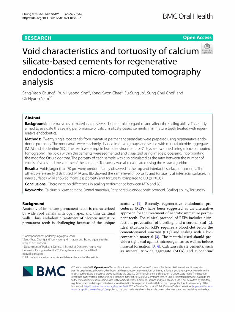
Briefly, a 3D image of the specimen was obtained by subsequent stacking of the binary images, and the pro-duced image was visualized and binarized to analyze the characteristics of the voids (Fig. 1). To characterize the spatial distribution of voids, image processing was conducted to separately analyze the void characteristics at the interface between the canal wall and the cement, as well as the inner cement. First, the cement part was segmented from the teeth using a multi-Otsu threshold algorithm [18], and the cement part was classified into two parts: interfacial and inner cement. For the classifi-cation, the boundary of the cement was outlined by rec-ognizing the internal geometry of the targeted tooth, and
Page 3 of 8Chung et al. BMC Oral Health (2021) 21:565
a region of 40 pixels (approximately 236 μm) from the canal wall was set as the interfacial cement; the cement part excluding the interfacial region was defined as the inner cement. The same procedure was applied to all the samples.
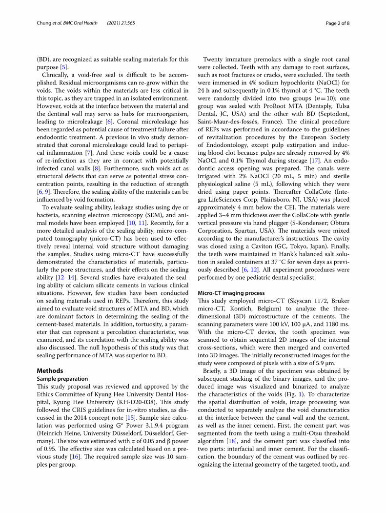
For a more effective analysis, the void characteristics in both the interfacial and inner cements were investi-gated using binarized micro-CT images. Binarization of each sample was conducted based on the histogram of grayscale micro-CT images. For this purpose, a proper threshold value needed to be selected, and pixels with values lower than the threshold were considered as voids (Fig. 2). To select the threshold, the modified Otsu method and a modified watershed algorithm [19] were adopted, and the image processing was carried out using an imaging toolbox in MATLAB R2020b (Mathworks, Inc., Natick, MA, USA).
Void structure analysisThe void size distributions of the specimens were inves-tigated to determine the spatial distribution of the voids according to their sizes. Next, the porosity of the inter-face and the inner parts was computed as the number of
void voxels compared to the volume of the interface/inner cement area. For this study, only voids larger than 5.9 μm were considered for the calculation by considering the res-olution of the image used.
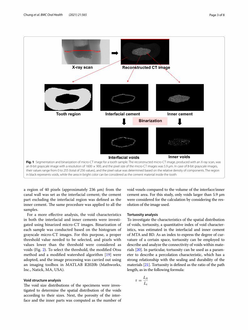
Tortuosity analysisTo investigate the characteristics of the spatial distribution of voids, tortuosity, a quantitative index of void character-istics, was estimated in the interfacial and inner cement of MTA and BD. As an index to express the degree of cur-vature of a certain space, tortuosity can be employed to describe and analyze the connectivity of voids within mate-rials [20]. In particular, tortuosity can be used as a param-eter to describe a percolation characteristic, which has a strong relationship with the sealing and durability of the materials [21]. Tortuosity is defined as the ratio of the path length, as in the following formula:
τ =
LA
Ls
Fig. 1 Segmentation and binarization of micro‑CT image for a tooth sample. The reconstructed micro‑CT image, produced with an X‑ray scan, was an 8‑bit grayscale image with a resolution of 1600 × 900, and the pixel size of the micro‑CT images was 5.9 μm. In case of 8‑bit grayscale images, their values range from 0 to 255 (total of 256 values), and the pixel value was determined based on the relative density of components. The region in black represents voids, while the area in bright color can be considered as the cement material inside the tooth
Page 4 of 8Chung et al. BMC Oral Health (2021) 21:565
Here, τ refers to the tortuosity, LA denotes the actual distance of the void path, and Ls is the shortest distance between the starting and end points on the void path.
In this study, the A-star algorithm was used to calcu-late LA , the actual distance of the pore path (Fig. 3). This algorithm is used to find the shortest void path from an arbitrary starting point to the end point [22]. The short-est path was calculated using two functions: AC(t) and HC(t). First, AC(t), the function to express the actual distance from the selected starting point to a tempo-rary point (t) within voids, was calculated. The heuristic estimate (HC(t)) from the temporary to the end points was then subsequently added with AC(t), and the total sum of the functions, TC(t), was computed. For the cal-culation of AC(t), the vertical, horizontal, and diagonal
distances from the start point to the temporary points were considered, and the temporary point was moved from the start point to the end point by considering the shortest function values at each point. Simultaneously, HC(t) was calculated considering only the horizontal and vertical distances from the temporary point, this approach being known as the Manhattan distance. The sum of the functions, TC(t), was calculated iteratively by moving the temporary point and considering the minimum value of the functions; the path of the tempo-rary point was set as LA . The tortuosity of the interfa-cial and inner cement was computed for all specimens to describe the complexity of the void structure within each material and each section.
Statistical analysisData were analyzed using SPSS version 20.0 (SPSS Inc., Chicago, IL, USA). The normality test was performed using the Shapiro–Wilk test, which revealed that the data were normally distributed. Thus, the results were ana-lyzed using the independent T-test. P-values < 0.05 were considered to indicate statistical significance.
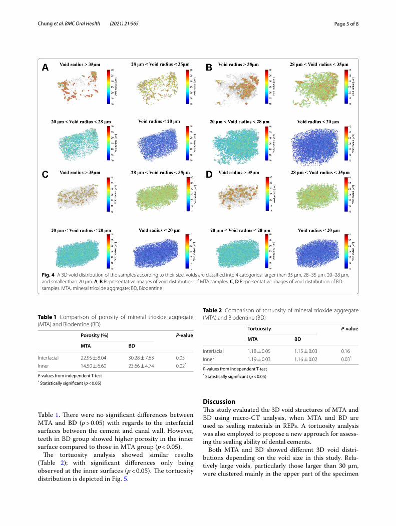
ResultsFigure 4 shows the 3D distribution of the voids in MTA and BD groups. In both the groups, relatively small voids with a radius of < 28 μm were evenly distributed through-out the specimen. However, relatively large voids with a radius > 35 μm were concentrated in the upper part of the specimen or the interface.
The porosities of the interfacial and inner surfaces of each specimen as obtained following a quantitative analysis of voids between MTA and BD are shown in
Fig. 2 Segmentation of pores for interfacial and inner cement regions in 3D volume
Fig. 3 Schematic of the A‑start algorithm. AC(t) denotes the actual distance between the starting point and a temporary point, while HC(t) is the distance between the temporary and end points at iteration process t. The total cost TC(t) is computed by adding the two functions
Page 5 of 8Chung et al. BMC Oral Health (2021) 21:565
Table 1. There were no significant differences between MTA and BD (p > 0.05) with regards to the interfacial surfaces between the cement and canal wall. However, teeth in BD group showed higher porosity in the inner surface compared to those in MTA group (p < 0.05).
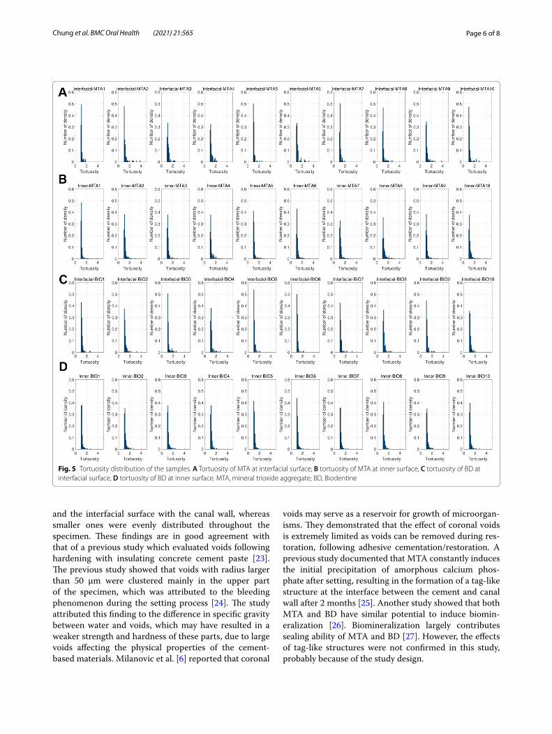
The tortuosity analysis showed similar results (Table 2); with significant differences only being observed at the inner surfaces (p < 0.05). The tortuosity distribution is depicted in Fig. 5.
DiscussionThis study evaluated the 3D void structures of MTA and BD using micro-CT analysis, when MTA and BD are used as sealing materials in REPs. A tortuosity analysis was also employed to propose a new approach for assess-ing the sealing ability of dental cements.
Both MTA and BD showed different 3D void distri-butions depending on the void size in this study. Rela-tively large voids, particularly those larger than 30 μm, were clustered mainly in the upper part of the specimen
Fig. 4 A 3D void distribution of the samples according to their size. Voids are classified into 4 categories: larger than 35 μm, 28–35 μm, 20–28 μm, and smaller than 20 μm. A, B Representative images of void distribution of MTA samples, C, D Representative images of void distribution of BD samples. MTA, mineral trioxide aggregate; BD, Biodentine
Table 1 Comparison of porosity of mineral trioxide aggregate (MTA) and Biodentine (BD)
P-values from independent T-test* Statistically significant (p < 0.05)
Porosity (%) P‑value
MTA BD
Interfacial 22.95 ± 8.04 30.28 ± 7.63 0.05
Inner 14.50 ± 6.60 23.66 ± 4.74 0.02*
Table 2 Comparison of tortuosity of mineral trioxide aggregate (MTA) and Biodentine (BD)
P-values from independent T-test* Statistically significant (p < 0.05)
Tortuosity P‑value
MTA BD
Interfacial 1.18 ± 0.05 1.15 ± 0.03 0.16
Inner 1.19 ± 0.03 1.16 ± 0.02 0.03*
Page 6 of 8Chung et al. BMC Oral Health (2021) 21:565
and the interfacial surface with the canal wall, whereas smaller ones were evenly distributed throughout the specimen. These findings are in good agreement with that of a previous study which evaluated voids following hardening with insulating concrete cement paste [23]. The previous study showed that voids with radius larger than 50 μm were clustered mainly in the upper part of the specimen, which was attributed to the bleeding phenomenon during the setting process [24]. The study attributed this finding to the difference in specific gravity between water and voids, which may have resulted in a weaker strength and hardness of these parts, due to large voids affecting the physical properties of the cement-based materials. Milanovic et al. [6] reported that coronal
voids may serve as a reservoir for growth of microorgan-isms. They demonstrated that the effect of coronal voids is extremely limited as voids can be removed during res-toration, following adhesive cementation/restoration. A previous study documented that MTA constantly induces the initial precipitation of amorphous calcium phos-phate after setting, resulting in the formation of a tag-like structure at the interface between the cement and canal wall after 2 months [25]. Another study showed that both MTA and BD have similar potential to induce biomin-eralization [26]. Biomineralization largely contributes sealing ability of MTA and BD [27]. However, the effects of tag-like structures were not confirmed in this study, probably because of the study design.
Fig. 5 Tortuosity distribution of the samples. A Tortuosity of MTA at interfacial surface, B tortuosity of MTA at inner surface, C tortuosity of BD at interfacial surface, D tortuosity of BD at inner surface. MTA, mineral trioxide aggregate; BD, Biodentine
Page 7 of 8Chung et al. BMC Oral Health (2021) 21:565
In this study, the porosity of MTA and BD was approxi-mately 14.5–30.3%. These values were higher than those reported in other micro-CT studies that compared porosity after placing MTA and BD into silicone molds [28, 29]. The difference may be attributed to the different experimental conditions, since the application and com-paction method employed, and the thickness of cement may affect porosity. BD showed a higher porosity in the inner surface than MTA, which may be due to the differ-ent setting times in the early hydration process. As BD allows a shorter setting time, it can have more voids on its inner surface. This finding indicates that MTA pos-sesses a denser structure than BD as previously reported [30]. Conversely, MTA and BD did not show a signifi-cant difference in porosity at the interfacial surface. This finding is in line with the results of a previous study that evaluated the 3D morphology of MTA and BD following application of root-end filling materials [16].
To quantify the sealing ability of MTA and BD, tortu-osity analysis was conducted. Tortuosity is an indicator of the shortest path in which fluid flows in the specimen, which represents the percolation and sealing ability of materials [20, 31]. The minimum value of the tortuosity is 1, which indicates that the line formed by connecting any point in the void cluster is almost linear without any curvature. In other words, a high tortuosity value would imply that the voids are connected in a complicated pat-tern in the specimen, denoting difficulties in the diffu-sion of moisture and fluid, and indicating a relatively low possibility of being impacted by an external environment [32]. Interestingly, there were no significant differences in tortuosity between MTA and BD at the interfacial sur-face. This reflects that the interfacial surfaces of MTA and BD show similar void connectivity and indicates that both MTA and BD can present similar sealing abilities when used for REPs. A randomized clinical study demon-strated that both MTA and BD were acceptable to create a tight coronal seal [33]. However, there has been contro-versy about sealing ability between MTA and BD in the previous studies. Cechella et al. [34] reported that BD had a lower level of sealing than MTA when they used for apical plugs. A previous leakage study reported that BD showed superior sealing ability as a root-end filling mate-rial compared to MTA by assessing the depth and volume of the penetrated silver nitrate [35]. A recent systematic review regarding sealing ability of root filling materials showed BD has superior sealing ability against MTA [36].
In this study, a 3D analysis of the void structures based on micro-CT images facilitated a more precise and effective evaluation of the spatial characteristics of MTA and BD. However, this study has a few limitations. First, a relatively short-term evaluation was conducted;
thus, the effects of biomineralization on the sealing ability were excluded. Second, the normalization of the root canal space was insufficient. Next, MTA and BD have different radiopacifiers that affect the setting and hydration reactions [37, 38]. Thus, these differences may affect void distribution. Finally, the effect of sev-eral body fluids such as blood, exudate or saliva were not considered because the experiment was conducted extraorally with extracted human teeth.
ConclusionsIn conclusion, this study evaluated the 3D void struc-ture of MTA and BD used in REPs. This study con-firmed that MTA is composed of a denser internal structure than BD and that there are no differences in the sealing abilities of MTA and BD.
AbbreviationsREPs: Regenerative endodontic procedures; CEJ: Cementoenamel junction; MTA: Mineral trioxide aggregate; BD: Biodentine; SEM: Scanning electron microscopy; micro‑CT: Micro‑computed tomography; NaOCl: Sodium hypochlorite; 3D: Three‑dimensional.
AcknowledgementsThe authors declare no conflicts of interest.
Authors’ contributionsS‑YC: Conceptualization, Methodology, Supervision, Funding acquisition; YHK: Data curation, Writing‑Original draft preparation; YKC: Visualization; S‑SJ: Software; SCC: Validation; OHN: Conceptualization, Methodology, Writing‑Original draft preparation, Supervision, Funding acquisition. All authors read and approved the final manuscript.
FundingThis work was supported by the National Research Foundation of Korea (NRF) grant funded by the Korea government (MSIT) (No. 2020R1C1C1006937) and by the Korea Agency for Infrastructure Technology Advancement (KAIA) grant funded by the Ministry of Land, Infrastructure and Transport (Grant 21NANO‑B156177‑02). The funding bodies had no role in design, collection, analysis, interpretation of data, or writing of the manuscript.
Availability of data and materialsAll data generated or analyzed from this study are included in this published article.
Declarations
Ethics approval and consent to participateThis study was performed in accordance with the Declaration of Helsinki. This study protocol was reviewed and approved by the Ethics Committee of Kyung Hee University Dental Hospital, Kyung Hee University (KH‑D20‑038).
Competing interestsThe authors declare that they have no competing interests to declare.
Author details1 Department of Civil and Environmental Engineering, Sejong University, Seoul, Republic of Korea. 2 Department of Dentistry, Graduate School, Kyung Hee Uni‑versity, Seoul, Republic of Korea. 3 Department of Pediatric Dentistry, School of Dentistry, Kyung Hee University, Kyungheedae‑Ro 26, Dongdaemoon‑Gu, Seoul 02447, Republic of Korea.
Page 8 of 8Chung et al. BMC Oral Health (2021) 21:565
Received: 29 August 2021 Accepted: 30 October 2021
References 1. Trope M. Treatment of the immature tooth with a non‑vital pulp and api‑
cal periodontitis. Dent Clin N Am. 2010;54(2):313–24. 2. Galler KM. Clinical procedures for revitalization: current knowledge and
considerations. Int Endod J. 2016;49(10):926–36. 3. Banchs F, Trope M. Revascularization of immature permanent
teeth with apical periodontitis: new treatment protocol? J Endod. 2004;30(4):196–200.
4. Moreno‑Hidalgo MC, Caleza‑Jimenez C, Mendoza‑Mendoza A, Iglesias‑Linares A. Revascularization of immature permanent teeth with apical periodontitis. Int Endod J. 2014;47(4):321–31.
5. Ha WN, Duckmanton P, Kahler B, Walsh LJ. A survey of various endodontic procedures related to mineral trioxide aggregate usage by members of the Australian Society of Endodontology. Aust Endod J. 2016;42(3):132–8.
6. Milanovic I, Milovanovic P, Antonijevic D, Dzeletovic B, Djuric M, Miletic V. Immediate and long‑term porosity of calcium silicate‑based sealers. J Endod. 2020;46(4):515–23.
7. Friedman S, Torneck CD, Komorowski R, Ouzounian Z, Syrtash P, Kaufman A. In vivo model for assessing the functional efficacy of endodontic filling materials and techniques. J Endod. 1997;23(9):557–61.
8. Somma F, Cretella G, Carotenuto M, Pecci R, Bedini R, De Biasi M, Angerame D. Quality of thermoplasticized and single point root fillings assessed by micro‑computed tomography. Int Endod J. 2011;44(4):362–9.
9. Nomoto R, McCabe JF. Effect of mixing methods on the compressive strength of glass ionomer cements. J Dent. 2001;29(3):205–10.
10. Torabinejad M, Parirokh M. Mineral trioxide aggregate: a comprehensive literature review—part II: leakage and biocompatibility investigations. J Endod. 2010;36(2):190–202.
11. Shipper G, Teixeira FB, Arnold RR, Trope M. Periapical inflammation after coronal microbial inoculation of dog roots filled with gutta‑percha or resilon. J Endod. 2005;31(2):91–6.
12. Gandolfi MG, Parrilli AP, Fini M, Prati C, Dummer PM. 3D micro‑CT analysis of the interface voids associated with Thermafil root fillings used with AH Plus or a flowable MTA sealer. Int Endod J. 2013;46(3):253–63.
13. Malkoç MA, Sevimay M, Tatar İ, Çelik HH. Micro‑CT detection and charac‑terization of porosity in luting cements. J Prosthodont. 2015;24(7):553–61.
14. Aragão AC, Pintor AVB, Marceliano‑Alves M, Primo LG, Silva ASS, Lopes RT, Neves AA. Root canal obturation materials and filling techniques for primary teeth: in vitro evaluation in polymer‑based prototyped incisors. Int J Paediatr Dent. 2020;30(3):381–9.
15. Krithikadatta J, Gopikrishna V, Datta M. CRIS Guidelines (Checklist for Reporting In‑vitro Studies): a concept note on the need for standard‑ized guidelines for improving quality and transparency in reporting in‑vitro studies in experimental dental research. J Conserv Dent. 2014;17(4):301–4.
16. Torres FFE, Jacobs R, EzEldeen M, Guerreiro‑Tanomaru JM, Dos Santos BC, Lucas‑Oliveira É, Bonagamba TJ, Tanomaru‑Filho M. Micro‑computed tomography high resolution evaluation of dimensional and morphologi‑cal changes of 3 root‑end filling materials in simulated physiological conditions. J Mater Sci Mater Med. 2020;31(2):14.
17. Galler KM, Krastl G, Simon S, Van Gorp G, Meschi N, Vahedi B, Lambrechts P. European society of endodontology position statement: Revitalization procedures. Int Endod J. 2016;49(8):717–23.
18. Huang D‑Y, Wang C‑H. Optimal multi‑level thresholding using a two‑stage Otsu optimization approach. Pattern Recognit Lett. 2009;30(3):275–84.
19. Chung S‑Y, Abd Elrahman M, Kim J‑S, Han T‑S, Stephan D, Sikora P. Com‑parison of lightweight aggregate and foamed concrete with the same density level using image‑based characterizations. Constr Build Mater. 2019;211:988–99.
20. Shanti NO, Chan VW, Stock SR, De Carlo F, Thornton K, Faber KT. X‑ray micro‑computed tomography and tortuosity calculations of percolating pore networks. Acta Mater. 2014;71:126–35.
21. Cao T, Zhang L, Sun G, Wang C, Zhang Y, Yan N, Xu A. Model for predicting the tortuosity of transport paths in cement‑based materials. Materials (Basel). 2019;12(21):3623.
22. Zeng W, Church RL. Finding shortest paths on real road networks: the case for A. Int J Geogr Inf Sci. 2009;23(4):531–43.
23. Chung S‑Y, Abd Elrahman M, Stephan D, Kamm PH. Investigation of characteristics and responses of insulating cement paste specimens with Aer solids using X‑ray micro‑computed tomography. Constr Build Mater. 2016;118:204–15.
24. Peng Y, Jacobsen S. Influence of water/cement ratio, admixtures and filler on sedimentation and bleeding of cement paste. Cem Concr Res. 2013;54:133–42.
25. Reyes‑Carmona JF, Felippe MS, Felippe WT. Biomineralization ability and interaction of mineral trioxide aggregate and white portland cement with dentin in a phosphate‑containing fluid. J Endod. 2009;35(5):731–6.
26. Cosme‑Silva L, Dal‑Fabbro R, Gonçalves LO, Prado ASD, Plazza FA, Viola NV, Cintra LTA, Gomes Filho JE. Hypertension affects the biocompatibility and biomineralization of MTA, High‑plasticity MTA, and Biodentine®. Braz Oral Res. 2019;33:e060.
27. Gandolfi MG, Taddei P, Tinti A, Prati C. Apatite‑forming ability (bioactivity) of ProRoot MTA. Int Endod J. 2010;43(10):917–29.
28. Basturk FB, Nekoofar MH, Gunday M, Dummer PM. Effect of various mix‑ing and placement techniques on the flexural strength and porosity of mineral trioxide aggregate. J Endod. 2014;40(3):441–5.
29. Guerrero F, Berástegui E. Porosity analysis of MTA and Biodentine cements for use in endodontics by using micro‑computed tomography. J Clin Exp Dent. 2018;10(3):e237–40.
30. Chung S‑Y, Lehmann C, Abd Elrahman M, Stephan D. Pore character‑istics and their effects on the material properties of foamed concrete evaluated using micro‑CT images and numerical approaches. Appl Sci. 2017;7(6):550.
31. Chung S‑Y, Sikora P, Kim DJ, El Madawy ME, Abd Elrahman M. Effect of different expanded aggregates on durability‑related characteristics of lightweight aggregate concrete. Mater Charact. 2021;173:110907.
32. Chung S‑Y, Han T‑S, Kim S‑Y, Lee T‑H. Investigation of the permeability of porous concrete reconstructed using probabilistic description methods. Constr Build Mater. 2014;66:760–70.
33. Aly MM, Taha SEE, El Sayed MA, Youssef R, Omar HM. Clinical and radio‑graphic evaluation of Biodentine and mineral trioxide aggregate in revas‑cularization of non‑vital immature permanent anterior teeth (randomized clinical study). Int J Paediatr Dent. 2019;29(4):464–73.
34. Cechella B, de Almeida J, Kuntze M, Felippe W. Analysis of sealing ability of endodontic cements apical plugs. J Clin Exp Dent. 2018;10(2):e146–50.
35. Tang JJ, Shen ZS, Qin W, Lin Z. A comparison of the sealing abilities between Biodentine and MTA as root‑end filling materials and their effects on bone healing in dogs after periradicular surgery. J Appl Oral Sci. 2019;27:e20180693.
36. Solanki NP, Venkappa KK, Shah NC. Biocompatibility and sealing ability of mineral trioxide aggregate and biodentine as root‑end filling material: A systematic review. J Conserv Dent. 2018;21(1):10–5.
37. Bortoluzzi EA, Broon NJ, Bramante CM, Felippe WT, Tanomaru Filho M, Esberard RM. The influence of calcium chloride on the setting time, solubility, disintegration, and pH of mineral trioxide aggregate and white Portland cement with a radiopacifier. J Endod. 2009;35(4):550–4.
38. Li Q, Coleman NJ. Impact of Bi(2)O(3) and ZrO(2) radiopacifiers on the early hydration and C‑S‑H gel structure of white portland cement. J Funct Biomater. 2019;10(4):46.
Publisher’s NoteSpringer Nature remains neutral with regard to jurisdictional claims in pub‑lished maps and institutional affiliations.