vitamin d plasma binding...
TRANSCRIPT

Vitamin D Plasma Binding Protein
TURNOVERANDFATE IN THE RABBIT
J. G. HADDAD, D. R. FRASER, and D. E. M. LAWSON,Dunn Nutritional Laboratory,University of Cambridge and Medical Research Council, Milton Road,Cambridge CB4 1XJ, England
A B S T RA C T The metabolic disposition of theplasma binding protein (DBP) for vitamin D and itsmetabolites was studied in adult rabbits. Apo-DBP waspurified from rabbit plasma and enzymatically labeledwith radioiodine. The radioiodine-labeled proteinretained its ability to bind vitamin D sterols and itsphysicochemical properties. When 125I-labeled DBPand '3II-labeled rabbit albumin were simultaneouslyinjected intravenously, the 1251 was cleared from plasmaat a faster rate (t1/2 = 1.7 d) than 131I (tl/2 = 5 d) and 125Iwas present in excess of 131I in kidney, liver, skeletalmuscle, heart, lung, intestine, testis, and bone 1 h afterinjection. In contrast to DBP, 25(OH)D3 was clearedmore slowly (t1/2 = 10.7 d). Compared to albumin, DBPradioactivity appeared earlier and in greater quantity inthe urine of catheterized rabbits. Gel filtration analysesof plasma revealed most of the 1251 to elute in the posi-tion of DBP, with only small amounts in the <1,000-dalton region. In contrast, almost all of the urine 1251eluted in this small molecular weight fraction. Themolar ratio of DBP to 25(OH)D3 in normal rabbitplasma was 138/1. The extravascular pool of DBPwas calculated to be 1.5-2.4 times larger than theintravascular DBPpool, and the molar replacement rateof DBPwas 1,350-fold higher than that for 25(OH)D3.The plasma disappearance curves of holo-DBP,prepared either by saturating with 25(OH)D3 or bycovalently linking 3,8-bromoacetoxy-25(OH)D3, werevery similar to that of apo-DBP. Neuraminidasetreatment of DBPdid not alter its plasma survival.
These studies indicate that DBPor DBP-25(OH)D3complex is removed from plasma by a variety of tissues,that the DBPmoiety is degraded during this process,
This work was carried out during Dr. Haddad's tenure as arecipient of a Josiah Macy, Jr. Foundation Faculty ScholarAward (1978-1979). Dr. Haddad is now at the University ofPennsylvania School of Medicine, Department of Medicine,Philadelphia, Pa. 19104.
Received for publication 26 February 1980 and in revisedform 29 December 1981.
1550
and that a significant recirculation of 25(OH)D3 prob-ably occurs. The molar excess of DBPto 25(OH)D3 inplasma, and the relatively rapid turnover of DBPindicate that a high capacity, high affinity, and dynamictransport mechanism for vitamin D sterols exists inrabbit plasma.
INTRODUCTION
Although recognized for 20 yr (1), the human plasmabinding protein (DBP)l for vitamin D and its metabo-lites has only recently been isolated and characterized(2-4). It is an alpha globulin of -58,000 mol wt,containing one sterol binding site per moleculewith the highest binding affinity for the vitamin Dmetabolites, 25-hydroxycholecalciferol [25(OH)D3]and 24,25-dihydroxycholecalciferol [24,25(OH)2D3].This protein is now known to be physicochemically andimmunologically identical to the group specific com-ponent (Gc) human plasma protein (2, 3, 5). There issome evidence to suggest that this protein is synthe-sized in the liver (6, 7) and it occurs in high concentra-tions (6-8 ,uM) in plasma of man (5, 7-9) and rat(10, 11), whether measured by immunoassay or bysaturation analysis of plasma 25(OH)D bindingcapacity. Whencompared with the likely total contentof the vitamin D sterols (0.1-0.2 AM) in plasma in vita-min D-sufficient mammals it is apparent that the apo-DBP is present in remarkable excess of its ligand.
The plasma concentration of DBPis unaffected by awide variety of disorders of mineral homeostasis (5, 7,9) and unlike the retinol-binding protein, which de-clines in concentration during deficiency of vitamin A(12), the plasma level of DBPdoes not decrease duringvitamin D deficiency. Although patients with cirrhosisand nephrotic syndrome (7) have a reduced plasmaconcentration of DBP, it nevertheless remains far in
' Abbreviations used in this paper: 25(OH)D3, 25-hydroxy-vitamin D; 1,25(OH)2D3, 1,25-dihydroxyvitamin D;DBP, binding protein in plasma for 25(OH)D3.
J. Clin. Invest. © The American Society for Clinical Investigation, Inc. - 0021-9738/81/05/1550/11 $1.00Volume 67 May 1981 1550-1560

excess of that required to bind the normal amounts ofvitamin D sterols in the blood. In analyses of over80,000 different samples of human plasma (13) nonewere found where DBP(Gc) was absent, thus suggest-ing a vital role for this plasma protein.
To date, there is no information on the plasma turn-over rate or the metabolic fate of DBP. To help definethese purified rabbit plasma DBP was labeled withradioiodine and its plasma clearance and tissue ac-cumulation were studied in rabbits. These experimentsreveal that DBP, either in the apo or the holo form, iscleared quickly from plasma by a number of tissues andduring this process is degraded to small molecularweight fragments.
METHODS
Materials. Crystalline rabbit plasma albumin, bovineserum albumin, type V, lactoperoxidase (milk, 67 U/mgprotein), neuraminidase (type VIII, Clostridium perfringens,dithiothreitol, iodoacetamide, and blue dextran were pur-chased from Sigma Chemical Co., Dorset, England. 125lodine(carrier-free) and "3'iodine (free from reducing agents) wereobtained from The Radiochemical Centre, Bucks., England.Bromoacetic acid (practical grade) and dicyclohexylcarbo-diimide were obtained from Eastman Organic Chemicals Div.,Eastman Kodak Co., Rochester, N. Y. Calibration proteins fordetermination of molecular weight were obtained as a kit fromBoehringer Corp., East Sussex, England.
Sterol studies. 25-hydroxy [26,27-3H]cholecalciferol(25(OH)[26,27-3H]JD3) (9.6 Ci/mmol, The RadiochemicalCentre) was 99% pure as determined by silica gel GF254 thin-layer chromatography (14). Unlabeled 25(OH)D3 was a giftfrom Hoffmann-La Roche, Basel, Switzerland, and its purityand quantity were assessed by ultraviolet spectrometry. Forstudies in vivo, 1.5 ml rabbit plasma was mixed with 3 i.Ciof 25(OH)[26,27-3H]D3 dissolved in 10 ,lI ethanol and keptat 4°C overnight. Polyacrylamide gel electrophoresis revealed>95% of the 3H to be associated with a protein in the alphaglobulin region. After intravenous injection of the 25(OH)-[27,27-3H]D3 complex into rabbits, blood samples weretaken at intervals for plasma analysis. These samples (1 ml)were extracted with chloroform-methanol (15). Radioac-tivity was measured in the 25(OH)D3 fraction obtained bysilicic acid chromatography (16).
Purification of rabbit DBP. DBP, labeled with tracerquantities of 25(OH)D[26,27-3H]D3, was isolated in pure formfrom 100 ml of normal pooled rabbit plasma by dialysis, DEAEion-exchange column chromatography, gel filtration, on an AcA44 column, and preparative 7% polyacrylamide gel electro-phoresis. The final product was purified 175-fold relative toplasma and was judged to be homogeneous by analytical poly-acrylamide gel electrophoresis (Fig. 1). In addition, the finalpreparation induced a monospecific antiserum in sheepwhich recognized only DBP in rabbit serum on agar gel dif-fusion or electrophoresis. The sheep anti-rabbit DBP anti-serum did not react with DBP in human, monkey, rat, orchicken plasma. The concentration of DBP in rabbit serumwas measured using the sheep anti-rabbit preparation in radialimmunodiffuision assay (17), on gels made with 1% agarosein 0.025 M sodium barbitone buffer, pH 8.6.
Labelinig of proteins vith "25I antd 131I. Purified rabbitplasma DBP and rabbit plasma albumin were labeled with1251 and '31I, respectively by the lactoperoxidase method ofMiyachi et al. (18). After labeling with radioiodine, 0.5 ml of
0.05 M sodium phosphate buffer, pH 7.5, containing 1.0 mgpotassium iodide was added. The entire incubation volumewas then layered onto a 0.5 x 17-cm column of SephadexG-50 (Pharmacia Fine Chemicals Ltd., London, England)equilibrated in 0.02 M sodium phosphate containing 0.3 Msodium chloride, pH 7.4. The column had been precoated byrunning through it 1 ml 0.5% bovine serum albumin in thecolumn buffer and the void volume had been determined withblue dextran. The latter phase of the void volume was col-lected. Radioiodine was removed by this step, and additionalanalyses by polyacrylamide gel electrophoresis or gel filtra-tion on 1 x 40-cm columns of Ultragel AcA54 (LKB, Bromma,Sweden) were carried out. Specific activities of 25-50 ,Ci/,ugprotein were routinely obtained with this procedure. De-natured DBPwas produced by treating 125I-labeled DBPin 1%sodium dodecyl sulphate and 2 mMdithiothreitol overnight atroom temperature, followed by the addition of 0.02 Miodoacetamide for a further hour. More than 99% of thedialyzed product's radioactivity precipitated in the presenceof cold trichloroacetic acid.
Preparation of holo-25(OH)D3-DBP. Three different ap-proaches were taken in the studies with holo-25(OH)D3-DBP.(a) Radioiodinated apo-DBP was incubated in vitro with atwofold molar excess of unlabeled 25(OH)D3. The radio-labeled DBP, in 1.5 ml cold 0.02 M sodium phosphate, 0.15M sodium chloride, pH 7.5, was added to the 25(OH)D3 in10,ul ethanol and the preparation was mixed and incubatedat 0°C for 2 h before injection into rabbits. Preliminary ex-periments with 25(OH)[26,27-3H]D3 were conducted toassess the abilities of unlabeled and 125I-labeled apo-DBPto bind this sterol. After incubation the unbound sterol wasremoved with dextran-coated charcoal (16). The supernatewas counted in a gamma spectrometer and extracted withchloroform-methanol (2:1, vol/vol). The chloroform phase waswashed three times with water and assayed for 3H. Calcula-tions based on the known specific activities of '251-DBP and25(OH)[26,27-3H]D3 revealed that both preparations werecapable of binding nearly equimolar amounts of the sterol.
(b) In one experiment an attempt was made to saturateplasma DBP in vivo by injecting an amount of unlabeled25(OH)D3 equivalent to 1.5 times the molar amount of DBPin the plasma pool. The sterol was injected intravenously inpropylene glycol, 10 min before the injection of holo-DBPlabeled with radioiodine.
(c) A covalent holo 25(OH)D3-DBP preparation was madeby a modification of an affinity labeling procedure pre-viously described (19). Monobromoacetic acid was distilledand the fraction of boiling point 202-204°C was collected.Tetrahydrofuran was passed over a column of neutral aluminaand dried over potassium hydroxide pellets. To a stirred solu-tion of 1.2 mg (3 ,umol) 25(OH)D3 and 2 x 166 dpm 25(OH)-[26,27-3H]D3 in 1.1 ml dry tetrahydrofusion at 22°C wereadded 0.5 mg (3.6 ,umol) bromoacetic acid in 0.5 ml of tetra-hydrofuran. After 5 min 30 ,lI (0.36 ,umol) dry pyridine wasadded and the mixture was stirred for 2 h at 22°C. The solu-tion was filtered through a fritted funnel and dried undernitrogen. The material was dissolved in chloroform/acetone(19:1, vol/vol) and applied to 0.4 mmsilica gel thin-layerplates. Ascending chromatography was carried out in the samesolvent mixture to a 15 cm height. The products, 3,8-bromo-acetoxy-25(OH)D3 (retardation factor; RF= (0.60) and 25-(OH)D3 (RF = 0.36) were located under uiltraviolet (UV) light.The esterified sterol band was scraped from the plate and thesilica gel was extracted with three 10-ml vol of diethyl ether.After removing the solvent under nitrogen the product had aUV spectrum in ethanol identical to that of 25(OH)D3 and ittransformed to 25(OH)D3 during saponification in 0.1 N potas-sium hydroxide in ethanol. Specific activity and UVextinction
Vitamin D Plasma Binding Protein 1551

measurements indicated a 61%yield of ester. In equilibrationcompetition experiments, the ester was 8- to 10-fold lesspotent than equimolar amounts of 25(OH)D3 in displacing25(OH)[26,27-3H]D3 from DBP. However, when the ester wasincubated with equimolar amounts of DBP for 4 h at 22°C,more than 85% of subsequently added 25(OH)[3H]D3 was notbound by the DBP. The ester was covalently bound to theprotein, as indicated by the presence of 3H in slices of sodiumdodecyl sulfate-polyacrylamide gels loaded with the ester-pro-tein mixture previously heated at 700C in 1%sodium dodecylsulphate for 1 h. On the basis of these observations, 30 ,ug125I-DBP was incubated with 321 ng 3,8-bromoacetoxy-25-25(OH)D3 for 4 h at 22°C, and this preparation was used invivo for assessment of the disposition of holo-DBP with co-valently bound ligand.
Animal experiments. New Zealand White and DutchBelted rabbits, 1.8-3.6 kg, were supplied with standard rabbitchow (Crane Meal Mills, Herts., England) ad lib., and given0.005% potassium iodide drinking water starting at least 48 hbefore the experiment. Male rabbits were sedated with asingle intravenous injection of pentobarbital (25 mg/kg bodywt) for the purpose of bladder catheterization with a number8 paediatric bladder catheter (Warne Surgical Products Ltd.,Hants., England). Radioactive DBP (1.9-5.5 ,g; 106-275,uCi 125I) and/or radioactive albumin (1.5-4.9 ,ug; 75-243 ,uCi131I) was injected in 0.5 to 1.5 ml 0.02 M sodium phosphate,0.15 M sodium chloride, pH 7.5 into a marginal ear vein.Blood samples from the marginal vein of the opposite earwere collected into heparinized tubes. Plasma was ob-tained by centrifugation at 500 g for 15 min at 4°C. Plasma orcytosol protein, precipitated with cold trichloroacetic acid(7% wt/vol), was counted in a gamma spectrometer. Plasmaand urine were also analyzed by gel filtration and gelelectrophoresis. Urine was obtained by gravity flow, syringeaspirations of the bladder catheter and a bladder rinsing with15 ml 0.9% (wt/vol) sodium chloride at intervals during eachexperiment. Rabbits were killed by the rapid intravenous in-jection of 240 mg pentobarbital. Organs and tissues were re-moved within 20 min of death. They were rinsed in 0.9%sodium chloride and weighed before analyzing for radioac-tivity. Abdominal wall adipose tissue was excised and the en-tire right tibia was pulverized for a representative bonesample. Skeletal muscle was obtained from the right thigh andduodenum was used as the sample of small intestine. Gutlumenal contents were washed out before weighing andanalysis of the intestinal tissue. Cytosols were prepared byhomogenizing tissue (1:1, vol/wt) in cold 0.05 MTris/HCl buf-fer pH 7.4 containing 0.15 Msodium chloride, 2 mMdithio-threitol and 2 mMEDTA. After centrifugation at 500 g for 10min at 4°C the supernates were centrifuged at 100,000 g for60 min at 40C. The particle-free supernates were stored at-200C before analysis.
In bred hooded rats (200 g) of the Dunn Nutritional Labora-tory strain, given adequate diet and potassium iodide drinkingwater, were used in preliminary experiments to study the fateof labeled rabbit DBP.
Measurement of radioactivity. Tritium was assayed in aPackard Tri-Carb model 3375 scintillation spectrometer(Packard Instrument Co. Inc., Downers Grove, Ill.) us-ing toluene or toluene-Triton X-100 scintillation solu-tions. Quenching was corrected by automatic externalstandardization.
Radioiodine in all the samples was analyzed in the sametype of plastic counting vials and corrections were made forvariation in counting efficiency, isotope decay and isotopespillover in a well-type, double-channel gammaspectrometer(ICN Instruments Division, Antwerp, Belgium). Variation inthe counting efficiency for 1251 was minimal in samples fromdifferent tissues. 30 min after intravenous injection of 1251_
labeled and '3II-labeled rabbit serum albumin or 125I-labeledand 131I-labeled rabbit DBPinto rabbits, the ratio of 125I to 1311in the plasma was within 1%of the original value, but the ratiowas reduced by factors of 0.95 in liver and 0.91 in bone. Sincein all other tissues the ratio was within 2% of the preinjec-tion value, corrections for the presence of 125I in tissue weremade only for liver and bone.
Other methods. Gel filtration analyses on columns ofUltragel AcA54 and AcA44 were performed under constantpump pressure at 40C. Polyacrylamide gel-electrophoresis(20), sodium dodecyl sulphate-polyacrylamide-gel electro-phoresis (21), serum creatinine (22) and 25(OH)D binding as-says (16) were performed as previously described.
Statistical analysis. The turnover data was calculated by anoncompartmental, mathematical analysis of the plasmaspecific radioactivity curves (23). In this method, replacementrate was obtained by dividing the injected dose by the areaunder the plasma curve from zero time to om. Minimal transittime and minimal body pool are the transit time and poolsize if it is assumed that catabolism is solely from thesampling pool. Maximal transit time and maximal body poolare the transit time and pool size if it is assumed that catabo-lism all occurred at a site removed as far as possible from thesampling pool. In the calculation of the maximal transit time,a sum of the reciprocals of the exponents was not employed, asthe early exponential slopes were not comparable in magni-tude to the terminal one; (a fit of a 2 exponential model tothe data indicated the earlier exponentials to be 10 times theterminal one). Since the semilogarithmic plot of the plasmacurve attained a terminal constant slope, maximal transit timewas calculated from the reciprocal of the terminal exponential,and calculation of maximal body pool assumed that the frac-tional rate of catabolism was uniform throughout the sys-tem. Minimal transit time was calculated graphically. Syn-thesis was equated to the calculated replacement rate,assuming a steady state to have prevailed throughout thestudy. This was actually the case as judged by rabbit weights,hematocrit, and serum protein values.
The significance of the differences between the ratio of125I/1311 in the tissue and the ratio of the injected material (seeTable II) was analyzed by the Student's t test.
RESULTS
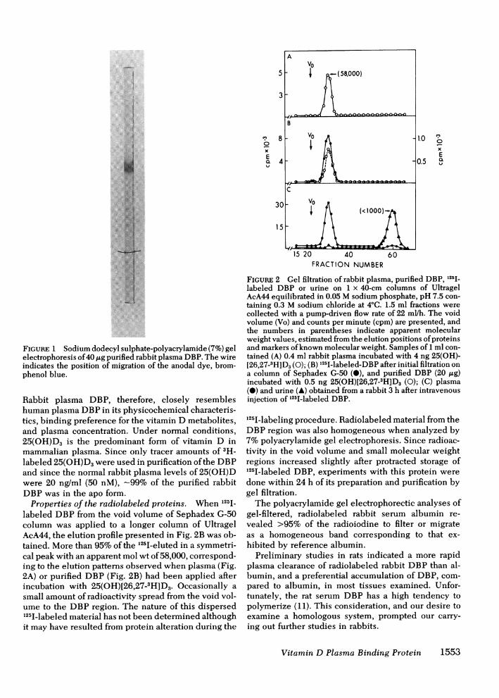
Properties of rabbit plasma DBP. The finalpreparation of purified DBPwas homogeneous whenanalyzed by polyacrylamide gel electrophoresis (Fig.1). Gel filtration analyses revealed that the 25(OH)-[26,27-3H]D3-DBP complex had eluted in a single peakwith an apparent mol wt of 58,000 (Fig. 2B). This wasidentical to the elution of radioactivity when thecolumn was charged with normal rabbit plasma incu-bated with tracer quantities of 25(OH)[26,27-3H]D3(Fig. 2A). The purified DBP from rabbit plasma hadalpha mobility on electrophoretic analyses in 7%poly-acrylamide gels. The absorption spectrum of purifiedrabbit plasma DBP revealed maximum absorbance at275 to 280 nm with a 280/260-nm absorption ratio of1.20. As revealed by saturation analyses, rabbit DBPbound 25(OH)D3 with higher affinity than cholecalcif-erol or 1,25-dihydroxycholecalciferol (1,25(OH)2D3).Radial immunodiffusion quantitation of DBPin rabbitplasma, utilizing purified DBPas reference standard,indicated the concentration to be 400 ng/ml (6.9 ,uM).
1552 J. G. Haddad, D. R. Fraser, and D. E. M. Lawson
T0xEXlQ
FIGURE 1 Sodium dodecyl suiphate-polyacrylamide (7%) gelelectrophoresis of 40 ug purified rabbit plasma DBP. The wireindicates the position of migration of the anodal dye, brom-phenol blue.
Rabbit plasma DBP, therefore, closely resembleshuman plasma DBPin its physicochemical characteris-tics, binding preference for the vitamin D metabolites,and plasma concentration. Under normal conditions,25(OH)D3 is the predominant form of vitamin D inmammalian plasma. Since only tracer amounts of 3H-labeled 25(OH)D3 were used in purification of the DBPand since the normal rabbit plasma levels of 25(OH)Dwere 20 nglml (50 nM), -99% of the purified rabbitDBPwas in the apo form.
Properties of the radiolabeled proteins. When i251_labeled DBP from the void volume of Sephadex G-50column was applied to a longer column of UltragelAcA44, the elution profile presented in Fig. 2B was ob-tained. More than 95%of the i2iilIeluted in a symmetri-cal peak with an apparent mol wt of 58,000, correspond-ing to the elution patterns observed when plasma (Fig.2A) or purified DBP (Fig. 2B) had been applied afterincubation with 25(OH)[26,27_3 H]D3. Occasionally asmall amount of radioactivity spread from the void vol-ume to the DBP region. The nature of this dispersed1251I-labeled material has not been determined althoughit may have resulted from protein alteration during the
0xE
FRACTION NUMBER
FIGuRE 2 Gel filtration of rabbit plasma, purified DBP, 125I_labeled DBP or urine on 1 x 40-cm columns of UltragelAcA44 equilibrated in 0.05 Msodium phosphate, pH 7.5 con-taining 0.3 M sodium chloride at 4°C. 1.5 ml fractions werecollected with a pump-driven flow rate of 22 ml/h. The voidvolume (Vo) and counts per minute (cpm) are presented, andthe numbers in parentheses indicate apparent molecularweight values, estimated from the elution positions of proteinsand markers of known molecular weight. Samples of 1 ml con-tained (A) 0.4 ml rabbit plasma incubated with 4 ng 25(OH)-[26,27-3H]D3 (0); (B) 125IIlabeled-DBP after initial filtration ona column of Sephadex G-50 (0), and purified DBP (20 ,ug)incubated with 0.5 ng 25(OH)[26,27-3H]D3 (0); (C) plasma(0) and urine (A) obtained from a rabbit 3 h after intravenousinjection of i25I-labeled DBP.
i25I-labeling procedure. Radiolabeled material from theDBPregion was also homogeneous when analyzed by7%polyacrylamide gel electrophoresis. Since radioac-tivity in the void volume and small molecular weightregions increased slightly after protracted storage ofi25I1labeled DBP, experiments with this protein weredone within 24 h of its preparation and purification bygel filtration.
The polyacrylamide gel electrophorectic analyses ofgel-filtered, radiolabeled rabbit serum albumin re-vealed >95% of the radioiodine to filter or migrateas a homogeneous band corresponding to that ex-hibited by reference albumin.
Preliminary studies in rats indicated a more rapidplasma clearance of radiolabeled rabbit DBP than al-bumin, and a preferential accumulation of DBP, com-pared to albumin, in most tissues examined. Unfor-tunately, the rat serum DBP has a high tendency topolymerize (11). This consideration, and our desire toexamine a homologous system, prompted our carry-ing out further studies in rabbits.
Vitamin D Plasma Binding Protein 1553

Plasma survival of radiolabeled proteins and25(OH)D3. Fig. 3 presents the early disappearance of125I-labeled DBPand 131I-labeled rabbit albumin fromthe plasma of a normal rabbit after the simultaneousintravenous injection of these proteins. During the firstminutes, both proteins disappeared from the plasma atsimilar rates but a more rapid egress of DBP wasapparent by 1 h after injection. At this time the mean125I_/131 1 ratio found in four rabbits was 0.95, whichwas significantly less (P < 0.02) than the ratio of theinjected material. The curves describing the disappear-ance of both proteins were multiexponential within thefirst day. After a brief delay, both isotopes appeared inthe urine and a greater rate of urinary excretion of 125Iwas observed. At 24 h, the plasma disappearance rate ofDBPwas clearly more rapid and only 16% of the doseremained in the vascular compartment. Over 24 h-22% of the dose of 1251 and 10% of the dose of 131I ap-peared in urine. The divergent rates of plasma clearanceof these proteins remained distinct after 48 h when only9%of the injected DBPwas present in plasma. In con-trast to the behavior of these proteins, denatured DBPwas cleared more rapidly with <10% of the dose re-.maining in plasma 8 h after injection (Fig. 3).
The later phases of the plasma disappearance of thetwo proteins and 25(OH)[26,27-3H]D3 are depicted in
100
3;
U
I.-
u
>
07-'
D
50
X 20
c~10
Fo24
wpat
-o00 4
12
Plosmo
Urine
Fig. 4. The apparent plasma removal rate of the sterolwas similar to those of the proteins only during the firstfew hours after injection (Fig. 3). The later rate of steroldisappearance described a slope indicating a plasmat,/2 of - 11 days. The terminal slope of 125I-labeled DBPplasma disappearance indicated a tl/2 of 1.7 d and thatfor 131I-labeled albumin was 5 d. A total of five rabbitswas studied and these were found to exhibit plasmasurvival curves of DBPand albumin similar to thoseshown in Figs. 3 and 4. An additional rabbit, injectedwith 25(OH)[26,27-3H]D3 bound to normal rabbitplasma, displayed a terminal plasma disappearance rateof this sterol closely similar (t12 = 10.5 d) to that indi-cated in Fig. 4. Base-line plasma concentrations of DBPor 25(OH)D were almost identical to those measuredduring and after the experiments. Table I summarizesthe analyses of the plasma survival curves.
Plasma survival of neuraminidase-treated DBPandholo-25(OH)D3-DBP. Factors which might affect theplasma clearance rate of DBPhave been considered.The role of sialic acid residues in the uptake by liverof plasma proteins was shown by Ashwell and Morell(24) but we found that the presence of this residue onDBP is not essential for its normal turnover rate. Pre-sumptive evidence for heterogeneity in purified rabbitDBPwas revealed by neuraminidase treatment (3 U/0.1 ml in 0.1 M sodium acetate, pH 5.3, for 15 h at37°C) of 3 ,g 1251-labeled DBP. When this preparationand 131I-labeled DBP incubated without enzyme wereanalyzed by polyacrylamide gel electrophoresis, ahigher 1251/1311 ratio was observed in cathodal slices ofthe DBPband. These findings suggest that sialic acidwas present in anodal portions of the DBPpreparationas had been reported for human DBP (25-27).
100125i
50
20
4 8 12 16 20 24
HOURS AFTER INJECTION
FIGuRE 3 Early plasma survival and cumulative urinaryexcretion of radioactivity during the initial 24 h after the simul-taneous injection of 125I-labeled DBP(0) and 1311-labeled rab-bit serum albumin (0) into a rabbit. Urinary excretion of 125I(0) and 1311 (0) was also measured in these animals. In anotherexperiment, 133 ng 25(OH)[26,27-3H]D3 (A), previously incu-bated with 1.5 ml normal rabbit plasma, as described underMethods was injected intravenously and plasma25(OH)[26,27-3H]D3 was isolated by lipid chromatography.In the third experiment shown here, 4 ,tg of denatured 1251_labeled DBP(A) was intravenously injected into a third rabbit.
U-0Tv
0- 4- .0_
E
O'-U 04
_) OC: -
< I_
oc0
101
5
0-5W. _ ,
2 4 6 8 10 12 14 21DAYS AFTER INJECTION
FIGuRE 4 Later plasma survival curves of 25(OH)[26,27-3H]-D3 (A) injected intravenously into one rabbit, and 125I-labeledDBP(0) and 131I-labeled albumin (0) simultaneously injectedintravenously into another rabbit.
1554 J. G. Haddad, D. R. Fraser, and D. E. M. Lawson
l

TABLE IParameters of DBPand 25(OH)D3 Turnover in the Rabbit
Molar ratioParameter DBP 25(OH)D3* DBP/25(OH)D3
Plasma concentration 0.401±0.013 mg/ml 20 ng/ml 138:1Replacement rate 0.925+0.018 mg/h/kg 4.7 ng/h/kg 1,356:1Minimal mean transit time 43.52+ 1.00 h 363.5 hMinimal total body pool 40.25±0.95 mg/kg 1708 ng/kg 162:1Maximal mean transit time 58.82±0.72 h 371.9 hMaximal total body pool 54.43±1.13 mg/kg 1748 ng/kg 215:1Plasma pool, percent total body pool 29.4-39.8% 45.7-46.8%-Fractional catabolic rate, fraction of
intravascular pool per day 0.408±0.005 0.0645Plasma tl2 40.6±0.46 h 257.8 h
Calculations for DBPrepresent the mean±SEMor range of means obtained in five separate experiments,and those for 25(OH)D3 are the mean or range of values in two separate experiments.* These calculations for 25(OH)D3 assume, probably incorrectly, an irreversible removal of the25(OH)D3 from the sampling (plasma) pool.
The plasma disappearance of trichloroacetic acid-precipitable 125I and 1311 was studied in a rabbit injectedsimultaneously with 125I-labeled DBPwhich had beentreated with neuraminidase and 131I-labeled DBPwhich had not been exposed to this enzyme in vitro.The plasma survival curves of these two preparationsare shown in Fig. 5. The removal of sialic acid from the"fast" electrophoretic moiety of DBPdid not cause anyapparent alteration in its plasma survival.
It is also possible that the turnover rate of DBP isaffected by the presence or absence of 25-(OH)D3.Saturation of 1251-labeled DBPwith 25(OH)D3 in vitrodid not alter its rate of egress from plasma (data not
100
50
n 20a'
-o0-
5
u 0
<I.-
0*4
Il
DAYS AFTER INJECTION
FIGURE 5 Plasma survival curves of neuraminidase-treated125I-labeled DBP(0) and untreated, 131I-labeled DBP(-) aftertheir simultaneous intravenous injection into a rabbit.
presented). A similar preparation was injected 10 minafter the slow intravenous injection of 480 ,ug (1.2 mmol)of 25(OH)D3, which was a 1.5-molar excess of ligand forthe calculated intravascular pool of DBP(-50 mgor 0.8,umol for a 3-kg rabbit). As indicated in Fig. 6 the plasmadisappearance of the 125I-labeled DBP was not sub-stantially changed by these procedures. Plasma creati-nine was not increased after this 25(OH)D3 injectionbut plasma 25(OH)D3 concentrations were only nearDBPsaturation levels (2.7 ,ug/ml) at 5 min after injec-tion and they fell rapidly over 24 h to -0.9 ,ug/ml,reaching a level of 0.2 Ag/ml 7 d later.
The plasma survival of 125I-labeled DBPbound to theaffinity labeling reagent, 3,8-bromoacetoxy-25(OH)D3,is also shown in Fig. 6. With this preparation of ligandcovalently bonded to its carrier protein, the plasma sur-vival of 125I-labeled DBPwas unchanged compared toapo-DBP. Total urinary excretion of 125I was similar tothat found after injection of apo-DBP as was the pres-ence of only low-molecular weight 125I in urine and thepredominantly DBP-like 125I in gel-filtration analysesof plasma.
Properties of radiolabeled materials in plasma andurine. Analyses of plasma obtained 5 min after injec-tion of 125I-labeled DBPrevealed a gel filtration profilesimilar to the one shown in Fig. 2B. Samples taken at9 d after injection revealed no less than 92% of theradioactivity to elute in a single peak with an ap-parent mol wt of 58,000 (Fig. 3C). The remainder of theradioactivity was found in the region of apparent mol wt< 1,000. Polyacrylamide gel electrophoresis revealedthe large-molecular weight material to be DBPand thesmall-molecular weight material to have a net negativecharge, running near the bromphenol blue front. Thenature of this material has not been precisely deter-mined but iodotyrosine, free iodine, or iodine attached
Vitamin D Plasma Binding Protein 1555

100
501
X 20
- c
10
a 2 5 ~<
o<I-0
o.s2 4 6 8 10
DAYS AFTER INJECTION
FIGURE 6 Plasma survival curves of holo-DBP preparationsin two rabbits. One rabbit (0) received holo-25(OH)D3, 125I1labeled DBP intravenously after injection of 1.2 ,umol of25(OH)D3 intravenously 10 min earlier and oral cholecalcif-erol (2 mg in arachis oil) 24 h earlier. The other rabbit (0)was injected intravenously with 125I-labeled DBPcovalentlybonded to 3,8-bromoacetoxy-25(OH)D3.
to small peptides are likely possibilities. This small-sized material comprises the major portion of the radio-activity in urine collected at any time within 48 h ofinjection of 125I-labeled DBP (Fig. 2C). Frequently asmall amount of apparently intact 1251-labeled DBPap-peared in urine samples but this never accounted for>1% of the urine radioactivity. Gel filtration pro-files of urine from rabbits injected with 131I-labeledalbumin revealed almost all of the 1311 to be in a smallmol wt form (data not presented). Tissue, rather thanplasma alone, appeared to be required for the formationof low-molecular weight 1251-material since more than99%of 1251-labeled DBPincubated with plasma at 37°Cfor 1 h remained intact. In a single experiment, thelow-molecular weight 125I-material was found inplasma 2 h after the injection of 1251-labeled DBPintoa bilaterally nephrectomized rabbit.
After injection of 25(OH)[26,27-3H]D3, the major dis-tribution of 3H in plasma was observed to be associatedwith DBP. More than 90%of the radioactivity in plasmaco-migrated with the binding protein on gel filtration orelectrophoresis. Chromatography of lipid extracts ofplasma indicated that >88% of plasma-3H ran withstandard 25(OH)D3.
Tissue distribution of radiolabeled proteins. Inpreliminary experiments with rats, the simultaneousintravascular injection of 125I-labeled rabbit DBPand131I-labeled rabbit albumin gave results suggesting therelatively greater accumulation of DBP in a variety of
tissues at all times from 1 to 24 h after injection. Studiesin one rabbit at each of several times after the simul-taneous intravenous injection of 125I-labeled DBPand131I-labeled rabbit albumin revealed an excess of 125Iin many tissues shortly after injection. The significanceof these early results was confirmed by statistical analy-sis of data obtained from four rabbits sacrificed 60 minafter injection (Table II). When compared to the doseratio and the values in the 5 min plasma, the plasmaDBP/albumin ratio was significantly reduced in plasmaobtained 60 min after injection. The previously demon-strated difference between the plasma survival of DBPand albumin, which are proteins of similar size, sug-gested the possibility of a tissue uptake of DBPthat wasnot solely attributable to diffusion into the extracellularfluid. When tissue ratios were calculated on the basisof the total amount of each isotope administered, a sig-nificant excess of 125I was found in all tissues examinedexcept adipose tissue. Highest ratios were found in kid-ney, intestine, and muscle.
Whencorrected for extracellular fluid contamination,the calculated organ distribution of 125I-labeled DBPstill revealed highest accumulations in kidney, liver,and skeletal muscle (Table III). Four rabbits that hadreceived the radiolabeled proteins 24 h earlier werealso sacrificed. At this time the tissue data revealed 125Iaccumulation to be increased in bone and adipose tis-sue compared to the 1-h data. Whenconsidered as theamount of 1251 accumulated per gram wet weight of tissue,kidney was the most efficient extractor and high con-centrations were also noted in lung, liver, and heart.
Properties of radiolabeled material in tissuecytosols. Cytosols from tissues of one rabbit sacrificed
TABLE IIRelative Tissue Distribution of 125J and 131I at 1 h after the
Simultaneous Intravenous Injection of 1251-labeledDBPand 131I-labeled Rabbit Serum Albumin
Organ or tissue 1251/1311
Plasma 0.95±0.01*Liver 1.20±0.02tKidney 1.83±0.09tSkeletal muscle 1.49±0.04tHeart 1.19±0.04*Lung 1.24±0.05*Small intestine 1.43±0.06$Large intestine 1.62±0.06tTestis 1.22±0.02tBone 1. 12±0.02tAdipose tissue 1.04±0.02
Four rabbits were injected. The ratios were calculated on thebasis of the percentage of the total amount of each isotopeadministered. Values are mean±SEM.* The value differs from 1.00 at P < 0.02.t The value differs from 1.00 at P < 0.01.
1556 J. G. Haddad, D. R. Fraser, and D. E. M. Lawson
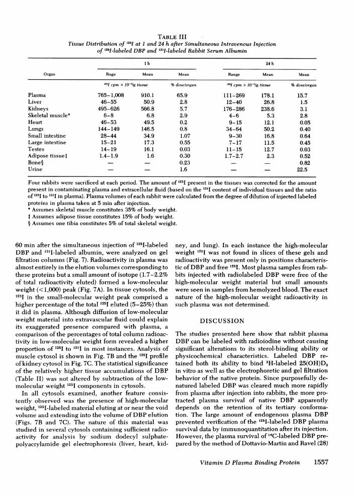
TABLE IIITissue Distribution of 125I at 1 and 24 h after Simultaneous Intravenous Injection
of 1251-labeled DBPand 1311-labeled Rabbit Serum Albumin
lh 24 h
Organ Rnge Mean Mean Range Mean Mean
'251 cpm x 10-3g tissue %doselorgan 1231 Cpm x 10-3/g tissue %doselorgan
Plasma 765-1,008 910.1 65.9 111-269 178.1 15.7Liver 46-55 50.9 2.8 12-40 26.8 1.5Kidneys 495-626 566.8 5.7 176-286 238.6 3.1Skeletal muscle* 6-8 6.8 2.9 4-6 5.3 2.8Heart 46-53 49.5 0.2 9-15 12.1 0.05Lungs 144-149 146.5 0.8 34-64 50.2 0.40Small intestine 28-44 34.9 1.07 9-30 16.8 0.64Large intestine 15-21 17.3 0.55 7-17 11.5 0.45Testes 14-19 16.1 0.03 11-15 12.7 0.03Adipose tissuet 1.4-1.9 1.6 0.30 1.7-2.7 2.3 0.52Bone§ 0.23 0.82Urine - 1.6 22.5
Four rabbits were sacrificed at each period. The amount of 125I present in the tissues was corrected for the amountpresent in contaminating plasma and extracellular fluid (based on the 131I content of individual tissues and the ratioof '25I to 131I in plasma). Plasma volumes of each rabbit were calculated from the degree of dilution of injected labeledproteins in plasma taken at 5 min after injection.* Assumes skeletal muscle constitutes 35% of body weight.t Assumes adipose tissue constitutes 15% of body weight.§ Assumes one tibia constitutes 5%of total skeletal weight.
60 min after the simultaneous injection of 1251-labeledDBPand 135I-labeled albumin, were analyzed on gelfiltration columns (Fig. 7). Radioactivity in plasma wasalmost entirely in the elution volumes corresponding tothese proteins but a small amount of isotope (1.7-2.2%of total radioactivity eluted) formed a low-molecularweight (<1,000) peak (Fig. 7A). In tissue cytosols, the125I in the small-molecular weight peak comprised ahigher percentage of the total 125I eluted (5-25%) thanit did in plasma. Although diffusion of low-molecularweight material into extravascular fluid could explainits exaggerated presence compared with plasma, acomparison of the percentages of total column radioac-tivity in low-molecular weight form revealed a higherproportion of 1251 to 131I in most instances. Analysis ofmuscle cytosol is shown in Fig. 7B and the 125I profileof kidney cytosol in Fig. 7C. The statistical significanceof the relatively higher tissue accumulations of DBP(Table II) was not altered by subtraction of the low-molecular weight 1251 components in cytosols.
In all cytosols examined, another feature consis-tently observed was the presence of high-molecularweight, 125I-labeled material eluting at or near the voidvolume and extending into the volume of DBPelution(Figs. 7B and 7C). The nature of this material wasstudied in several cytosols containing sufficient radio-activity for analysis by sodium dodecyl sulphate-polyacrylamide gel electrophoresis (liver, heart, kid-
ney, and lung). In each instance the high-molecularweight 1251 was not found in slices of these gels andradioactivity was present only in positions characteris-tic of DBPand free 1251. Most plasma samples from rab-bits injected with radiolabeled DBPwere free of thehigh-molecular weight material but small amountswere seen in samples from hemolyzed blood. The exactnature of the high-molecular weight radioactivity insuch plasma was not determined.
DISCUSSION
The studies presented here show that rabbit plasmaDBPcan be labeled with radioiodine without causingsignificant alterations to its sterol-binding ability orphysicochemical characteristics. Labeled DBP re-tained both its ability to bind 3H-labeled 25(OH)D3in vitro as well as the electrophoretic and gel filtrationbehavior of the native protein. Since purposefully de-natured labeled DBPwas cleared much more rapidlyfrom plasma after injection into rabbits, the more pro-tracted plasma survival of native DBP apparentlydepends on the retention of its tertiary conforma-tion. The large amount of endogenous plasma DBPprevented verification of the 125I-labeled DBPplasmasurvival data by immunoquantitation after its injection.However, the plasma survival of 14C-labeled DBPpre-pared by the method of Dottavio-Martin and Ravel (28)
Vitamin D Plasma Binding Protein 1557

3.0 - 1.2s x
E EcL1.5 --0.6
10
5-
15 20 40 60FRACTION NUMBER
FIGURE 7 Gel filtration of plasma and tissue cytosols ob-tained 1 h after the simultaneous intravenous injection of125I-labeled DBPand 131I-labeled rabbit serum albumin (RSA).Analyses were carried out as described for Fig. 2 except thatfractions of 1.7 ml were collected. The arrows depict the elu-tion positions of blue dextran (Vo), rabbit serum albumin(RSA) and pancreatic chymotrypsinogen (PC) during columncalibration. Samples analyzed were: (A) 1 h plasma containing40,000 cpm of 1251-labeled DBPand 31,000 cpm of 131I-labeledRSA; (B) muscle cytosol containing 19,300 cpm of 1251 and7,200 cpm of RSA; and (C) kidney cytosol containing 54,000cpm of 1251.
(data not presented) was observed to be very similar4tl2= 1.65 d) to that of the 125I-labeled protein (t12= 1.69 d, Table I). It is therefore concluded that thefate of the 125-labeled preparation closely approxi-mated that of native plasma DBP.
Experiments employing 1251-labeled DBP and 1311_labeled albumin revealed the relatively rapid clearanceof DBP from rabbit plasma. The fractional catabolicrate for DBPwas three times faster than that of albumin.Assuming no recirculation of DBP nor of 25(OH)D3in a 1-kg rabbit the molar replacement rate of DBP(383 nmol/d) would be - 1,350 times that for 25(OH)D3(283 pmol/d) (Table I). Since the normal rabbit plasmamolar ratio of DBP to 25(OH)D3 is 138:1, the meta-bolic turnover of plasma DBPis almost 10 times that of25(OH)D3. The recognition of the faster turnover ofplasma proteins in smaller animals (29) does not sub-stantially detract from the conclusion that the transportcapacity (concentration x turnover rate) of DBP inmammals is remarkably high.
Experiments with 3H-labeled 25(OH)D3 indicatedthat as in man, this sterol has an apparently long plasmasurvival (t12 = 10.7 d) (Fig. 4). Since its carrier proteinhas a considerably faster turnover rate, the metabolic
disposition of this sterol and the carrier clearlydiverge at some point. The experiments with 125I-la-beled rabbit DBP indicate that this protein is clearedfrom plasma by a large number of tissues. The proteinappears to be degraded during this process sinceshortly after the intravenous injection of 125I-labeledDBP, a form of 125I with a mol wt < 1,000 appearsin the plasma and is rapidly excreted in the urine. Fromthe assumption that the difference between theplasma survival of 125I-labeled DBPand 131I-labeled al-bumin represents the tissue uptake of 125I that is notsolely attributable to diffusion into the extracellularfluid, an association of DBPwith tissue elements mustbe considered. The preferential accumulation of 1251_labeled DBPcompared to 131I-labeled albumin is sup-portive in this regard. However, the precise nature ofthe tissue accumulation and degradation of DBPis notrevealed by our studies. After injection of 25(OH)-[26,27-3H]D3 and 125I-labeled DBP, similar rates ofdisappearance from plasma were seen for only a verybrief period (Fig. 3) with subsequent increases in theplasma 3H to 125I ratio. A dissociation of the sterol-protein complex must occur at some site yet to beidentified.
25(OH)D3 has been found distributed to many humantissues (30) and can exert a prolonged biological actionafter a single dose (31). Kinetic analyses of 25(OH)D3distribution suggest its tissue entry and subsequentegress into plasma (32). The apparently protractedplasma survival of 25(OH)D3 (33, 34) may therefore re-flect the difference between unmetabolized 25(OH)D3that recirculates between plasma and tissue and thatwhich is metabolically transformed (Figs. 3 and 4). It isalso possible that the dissociation of 25(OH)D3 fromDBPmight occur in the extracellular compartment. Theexperiments with apo and holo-25(OH)D3 DBPpreparations (Figs. 4 and 6) indicated a very similarplasma clearance of these materials and therefore donot suggest a different clearing mechanism for theseforms of DBP. Since we did not examine the relativetissue accumulations of apo-DBP and holo-DBP, asubtle difference in their tissue uptake remains a pos-sibility. Our experiments with 125I1-labeled DBPcovalently bonded to 3,8-bromoacetoxy-25(OH)D3(Fig. 6) support the data obtained with in vivo and invitro saturation of 1251-labeled DBP, but in vivo hydroly-sis of the sterol ester may have occurred.
A substantial metabolism of plasma proteins is recog-nized to occur in liver, kidney, and intestine (35). Ex-periments with neuraminidase-treated DBP(Fig. 5) re-vealed that this plasma protein is not likely cleared bybinding to hepatic receptors that recognize galactoseresidues after terminal sialic residues are removed (24).Since bilateral nephrectomy did not affect the initialplasma turnover rate of DBP, it seems that renal tissueis not the main site of DBPmetabolism. Our failure toidentify intermediate size fragments of DBP in the
1558 J. G. Haddad, D. R. Fraser, and D. E. M. Lawson

plasma or urine, and the appearance of small molecularweight fragments in these fluids and tissue cytosolsshortly after intravenous injection of DBP, suggest theaction of multiple proteases in the degradation of thisprotein. This suggests the possibility of lysosomal ac-tivity which has been linked to receptor-mediatedendocytosis of several proteins (36-38). Our experi-ments in vivo do not provide direct evidence for cel-lular association or uptake of DBP, and further studiesare required to examine this possibility. It is of interest,however, that a cellular protein is recognized to bindDBP(39-41) and that DBPcomplexed to this proteinhas been observed in cytosols from well-washed, cul-tured fibroblasts (41), well-perfused kidney (42), inwell-washed kidney subcellular fractions (42, 43) andin well-washed, cultured bone cells (44). Others, how-ever, have not observed the complexing of DBP byextracts from well-washed cells cultured without serum(39). Recent evidence has been presented that thecellular DBP binding protein is actin. This provoca-tive finding has yet to be explored, but does not guaran-tee an intracellular disposition of DBPbecause DBPmight function to scavenge actin released into the cir-culation (45).
The metabolic pathway for vitamin D implicatesmany tissues in its formation, transformation, and bio-logical action. Vitamin D itself and its potent metabo-lite, 1,25(OH)2D3, for example, are recognized to haveplasma clearance rates and tissue distributions whichare distinct from those of 25(OH)D3 (30, 46-48). It istherefore difficult to present a unified hypothesiswhereby DBP, the only specific binding protein forcholecalciferol sterols in mammalian plasma, plays adelivery function for all these sterols. Cholecalciferoland 1,25(OH)2D3 are bound by DBPwith lower affinitythan 25(OH)D3 (2) and probably have additionalmechanisms for their tissue delivery. The hepatic andadipose tissue accumulation of vitamin D may derivefrom the relatively greater association of cholecalciferolto plasma lipoproteins (49). The solubility of the morepolar metabolite, 1,25(OH)2D3 may be such that at itsusual plasma concentration (30 pg/ml) it is more avail-able in the free form for uptake by target tissues forassociation with their high-affinity, soluble intra-cellular receptors (50-53).
Since the major sterol that is most avidly bound tomammalian plasma DBPis 25(OH)D3, it is appropriateto consider the biological significance of it having aplasma transport protein of high capacity which turnsover rapidly. For the transformation of 25(OH)D3 toother metabolic forms, such a transport system wouldguarantee a continuing supply of substrate, even whencirculating levels of 25(OH)D3 are low (54), duringwhich time DBPlevels are unchanged (7). This inter-pretation, however, would only be attractive if carrier-mediated delivery of 25(OH)D3 occurs. The rapid re-placement of DBPwith newly synthesized apo-DBP
might also protect cells against excessive accumulationof 25(OH)D3 by ensuring the cellular egress ofunutilized 25(OH)D3, away from 1,25-(OH)2D3 recep-tor sites. A precise definition of the biological role ofDBP, however, shall require further studies of the tis-sue disposition of this protein.
ACKNOWLEDGMENTWeare grateful to Dr. J. G. Dann for the gift of partly puri-fied rabbit DBPand DBPantiserum, and to Mr. D. Brown forhis advice and assistance with data analysis.
REFERENCES
1. Thomas, W. C., H. G. Morgan, T. B. Connor, L. Haddock,C. E. Bills, and J. E. Howard. 1959. Studies of antiricketicactivity in serum from patients with disorders of calciummetabolism and preliminary observations on the modeof transport of vitamin D in human serum.J. Clin. Invest.38: 1078-1085.
2. Haddad, J. G., and J. Walgate. 1976. 25-hydroxyvitaminD transport in human plasma. Isolation and partial charac-terisation of calcifidiol-binding protein. J. Biol. Chem.251: 4803-4809.
3. Bouillon, R., H. Van Baelen, W. Rombauts, and P. DeMoor. 1976. The purification and characterisation of thehuman-serum binding protein for the 25-hydroxychole-calciferol. Eur. J. Biochem. 66: 285-291.
4. Imawari, M., K. Kida, and DeW. S. Goodman. 1976. Thetransport of vitamin D and its 25-hydroxymetabolite inhuman plasma. Isolation and partial characterisation ofvitamin D and 25-hydroxyvitamin D binding protein. J.Clin. Invest. 58: 514-523.
5. Imawari, M., and DeW. S. Goodman. 1977. Immunologi-cal and immunoassay studies of the binding protein forvitamin D and its metabolites in human serum. J. Clin.Invest. 59: 432-442.
6. Prunier, J. H., A. G. Beam, and H. Cleve. 1964. Site offormation of the group-specific protein and certain otherserum proteins. Proc. Soc. Exp. Biol. Med. 115: 1005-1007.
7. Haddad, J. G., and J. Walgate. 1976. Radioimmunoassayof the binding protein for vitamin Dand its metabolites inhuman serum. Concentrations in normal subjects and pa-tients with disorders of mineral homeostasis. J. Clin. In-vest. 58: 1217-1222.
8. Haddad, J. G., L. Hillman, and S. Rojanasathit. 1976. Hu-man serum binding capacity and affinity for 25-hydroxy-ergocalciferol and 25-hydroxycholecalciferol. J. Clin.Endocrinol. Metab. 43: 86-91.
9. Bouillon, R., H. Van Baelen, and P. De Moor. 1977. Themeasurement of the vitamin D-binding protein in humanserum. J. Clin. Endocrinol. Metab. 45: 225-231.
10. Rojanasathit, S., and J. G. Haddad. 1977. Ontogeny and ef-fect of vitamin D deprivation on rat serum 25-hydroxy-vitamin D binding protein. Endocrinology. 100:642-647.
11. Bouillon, R., H. Van Baelen, W. Rombauts, and P. DeMoor. 1978. The isolation and characterisation of the vita-min D-binding protein from rat serum.J. Biol. Chem. 253:4426-4431.
12. Muto, Y., J. E. Smith, P. O. Milch, and DeW. S. Goodman.1972. Regulation of retinol-binding protein metabolismby vitamin A status in the rat. J. Biol. Chem. 247: 2542-2550.
13. Cleve, H. 1973. The variants of the group specific com-ponent. Isr. J. Med. Sci. 9: 1133-1146.
14. Fraser, D. R., and E. Kodicek. 1970. Unique biosynthe-
Vitamin D Plasma Binding Protein 1559

sis by kidney of a biologically active vitamin Dmetabolite. Nature (Lond.). 228: 764-766.
15. Bligh, E. G., and W. J. Dyer. 1959. A rapid method oftotal lipid extraction and purification. Can. J. Biochem.Physiol. 37: 911-917.
16. Haddad, J. G., and K. J. Chyu. 1971. Competitive proteinbinding radioassay for 25-hydroxycholecalciferol. J.Clin. Endocrinol. Metab. 33: 992-995.
17. Ouchterlony, 0. 1962. Diffusion in gel methods for im-munological analysis. Prog. Allergy. 6: 30-154.
18. Miyachi, Y., J. L. Vaitukaitis, E. Nieschlag, and M. B.Lipsett. 1972. Enzymatic radioiodination of gonado-tropin. J. Clin. Endocrinol. Metab. 34: 23-28.
19. Warren, J. C., and J. R. Mueller. 1977. Labeling ofsteroid systems. Methods Enzymol. 46: 447-460.
20. Davis, B. J. 1964. Disc electrophoresis: method and appli-cation to human serum proteins. Ann. N. Y. Acad. Sci.121: 404-427.
21. Weber, K., and M. Osborn. 1969. The reliability of molecu-lar weight determinations by sodium dodecyl sulphatepolyacrylamide gel electrophoresis. J. Biol. Chem. 244:4406-4412.
22. Bonsnes, R. W., and H. H. Taussky. 1945. On the colori-metric determination of creatinine by the Jaffe reaction.
J. Biol. Chem. 158: 581-591.23. Katz, J., H. Rostami, and A. Dunn. 1974. Evaluation of
glucose turnover, body mass, and recycling with re-versible and irreversible tracers. Biochem. J. 142:161- 170.
24. Ashwell, G., and A. G. Morell. 1974. The role of surfacecarbohydrates in the hepatic recognition and transport ofcirculating glycoproteins. Adv. Enzymol. 41: 99-128.
25. Svasti, J., and B. H. Bowman. 1978. Human group spe-cific component. J. Biol. Chem. 253: 4188-4194.
26. Van Baelen, H., R. Bouillon, and P. De Moor. 1978. Theheterogeneity of human G, globulin. J. Biol. Chem. 252:6344-6345.
27. Cleve, H., and W. Patutschnick, 1979. Neuraminidasetreatment reveals sialic acid differences in certaingenetic variants of the G, system. Hum. Genet. 47:193-198.
28. Dottavio-Martin, D., and J. M. Ravel. 1978. Relationshipof liver composition to intensity of protein metabolismin different mammals. Anal. Biochem. 87: 562-565.
29. Munro, H. N., and E. D. Downie. 1964. Relationship ofliver composition to intensity of protein metabolism indifferent mammals. Nature (Lond.). 203: 603-604.
30. Mawer, E. B., J. Backhouse, C. A. Holman, G. A. Lumb,and S. W. Stanbury. 1972. The distribution and storage ofvitamin D and its metabolites in human tissues. Clin.Sci. 43: 413-431.
31. Balsan, S., and M. Garabedian. 1972. 25-Hydroxy-cholecalciferol, a comparative study in deficiency ricketsand different types of resistant rickets.J. Clin. Invest. 51:749-759.
32. Omdahl, J. L., G. Jelinek, and R. P. Eaton. 1977. Kineticanalysis of 2S-hydroxyvitamin D3 metabolism in strontiuminduced rickets in the chick. J. Clin. Invest. 60: 1202-1210.
33. Smith, J. E., and DeW. S. Goodman. 1971. The turnoverand transport of vitamin D and of a polar metabolite withthe properties of 25-hydroxycholecalciferol in humanplasma.J. Clin. Invest. 50: 2159-2167.
34. Bec, P., F. Bayard, and J. P. Louvet. 1972. 25-Hydroxy-cholecalciferol dynamics in human plasma. Rev.Europ. Etud. Clin. Biol. 17: 793-796.
35. Schultze, H. E., and J. F. Heremans. 1966. Molecular
Biology of Human Proteins. Elsevier, Amsterdam491-492.
36. Goldstein, J. L., R. G. W. Anderson, and M. S. Brown.1979. Coated pits, coated vesicles, and receptor-mediatedendocytosis. Nature (Lond.). 279: 679-685.
37. Goldstein, J. L., and M. S. Brown. 1977. The low densitylipoprotein pathway and its relation to atherosclerosis.Ann. Rev. Biochem. 46: 897-930.
38. Youngdahl-Turner, P., L. E. Rosenberg, and R. H. Allen.1978. Binding and uptake of transcobalamin 11 by humanfibroblasts.J. Clin. Invest. 61: 133-141.
39. Van Baelen, H., R. Bouillon, and P. De Moor. 1977. Bind-ing of 25-hydroxycholecalciferol in tissues.J. Biol. Chem.252: 2515-2518.
40. Cooke, N. E., J. Walgate, and J. G. Haddad. 1979. Humanserum binding protein for vitamin D and its metabolites.1. Physicochemical and immunological identification inhuman tissues.J. Biol. Chem. 254: 5958-5964.
41. Cooke, N. E., J. Walgate, and J. G. Haddad. 1979. Humanserum binding protein for vitamin D and its metabolites.II. Specific, high affinity association with a protein in nu-cleated tissue.J. Biol. Chem. 254: 5965-5971.
42. Botham, K. M., Y. Tanaka, and H. F. DeLuca. 1974. 25-Hydroxy vitamin D3-1-hydroxylase. Inhibition in vitro byrat and pig tissues. Biochemistry. 13: 4961-4966.
43. Ghazarian, J. G., B. Kream, K. M. Botham, M. W.Nickells, and H. F. DeLuca. 1978. Rat plasma 25-hydroxy-vitamin D3 binding protein: an inhibitor of the 25-hydroxyvitamin D3-la-hydroxylase. Arch. Biochem. Biophys. 189:212-220.
44. Chen, T. L., M. A. Hirst, and D. Feldman. 1979. A recep-tor-like binding macromolecule for la, 25-dihydroxy-cholecalciferol in cultured mouse bone cells. J. Biol.Chem. 254: 7491-7494.
45. Van Baelen, H., R. Bouillon, and P. DeMoor. 1980. Vita-min D-binding protein (Gc-globulin) binds actin.J. Biol.Chem. 255: 2270-2272.
46. Mawer, E. B., M. Davies, J. Backhouse, L. F. Hill, and C.M. Taylor. 1976. Metabolic fate of administered 1,25-dihy-droxycholecalciferol in controls and in patients withhypoparathyroidism. Lancet. 1: 1203-1205.
47. Gray, R. W., A. E. Caldas, D. R. Wilz, J. Lemann, G. A.Smith, and H. F. DeLuca. 1978. Metabolism and excretionof 1,25-(OH)2-vitamin D in healthy adults. J. Clin. Endo-crinol. Metab. 46: 756-765.
48. Frolik, C. A., and H. F. DeLuca. 1972. Metabolism of1,25-dihydroxycholecalciferol in the rat. J. Clin. Invest.51: 2900-2906.
49. Fraser, D. R. 1979. The physiological economy of vitaminD. In Pediatric Disease Related to Calcium. C. Anast, andH. F. DeLuca, editors. Elsevier-North HollandInc., New York.
50. Liao, S. 1975. Cellular receptors and mechanisms of actionof steroid hormones. Inter. Rev. Cytol. 41: 87-172.
51. Tsai, H. C., and A. W. Norman. 1973. Evidence for a cyto-plasmic factor for 1,25-dihydroxyvitamin D in the in-testinal tissue.J. Biol. Chem. 248: 5967-5975.
52. Lawson, D. E. M., and P. W. Wilson. 1974. Intranuclearlocalization and receptor proteins for 1,25-dihydroxy-cholecalciferol in chick intestine. Biochem. J. 144:573-583.
53. Hughes, M. R., and M. R. Haussler. 1978.1,25-Dihydroxy-vitamin D3 receptors in parathyroid glands.J. Biol. Chem.253: 1065-1073.
54. Eastwood, J. B., H. E. de Wardener, R. W. Gray, and J. L.Lemann. 1979. Normal plasma 1,25(OH)2D3 concen-trations in nutritional osteomalacia. Lancet. I: 1377- 1378.
1560 J. G. Haddad, D. R. Fraser, and D. E. M. Lawson