visualization of neutrophil extracellular traps and fibrin
TRANSCRIPT

Visualization of Neutrophil Extracellular Traps and Fibrin Meshwork in HumanFibrinopurulent Inflammatory Lesions: III. Correlative Light and ElectronMicroscopic Study
Takanori Onouchi1, Kazuya Shiogama1, Yasuyoshi Mizutani1, Takashi Takaki2,3 andYutaka Tsutsumi1
1Department of Pathology, Fujita Health University School of Medicine, Toyoake, Japan, 2Techinical Support Center, JEOLLtd., Akishima, Japan and 3Department of Pathology, Tohoku University Graduate School of Medicine, Sendai, Japan
Received August 24, 2016; accepted September 16, 2016; published online October 26, 2016
Neutrophil extracellular traps (NETs) released from dead neutrophils at the site ofinflammation represent webs of neutrophilic DNA stretches dotted with granule-derivedantimicrobial proteins, including lactoferrin, and play important roles in innate immunityagainst microbial infection. We have shown the coexistence of NETs and fibrin meshwork invaried fibrinopurulent inflammatory lesions at both light and electron microscopic levels. Inthe present study, correlative light and electron microscopy (CLEM) employing confocal laserscanning microscopy and scanning electron microscopy was performed to bridge light andelectron microscopic images of NETs and fibrin fibrils in formalin-fixed, paraffin-embedded,autopsied lung sections of legionnaire’s pneumonia. Lactoferrin immunoreactivity and 4'-6-diamidino-2-phenylindole (DAPI) reactivity were used as markers of NETs, and fibrin wasprobed by fibrinogen gamma chain. Of note is that NETs light microscopically represented aslactoferrin and DAPI-colocalized dots, 2.5 μm in diameter. CLEM gave super-resolutionimages of NETs and fibrin fibrils: “Dotted” NETs were ultrastructurally composed of finefilaments and masses of 58 nm-sized globular materials. A fibrin fibril consisted of clusters ofsmooth-surfaced filaments. NETs filaments (26 nm in diameter) were significantly thinnerthan fibrin filaments (295 nm in diameter). Of note is that CLEM was applicable to formalin-fixed, paraffin-embedded sections of autopsy material.
Key words: neutrophil extracellular traps, fibrin, correlative light and electron microscopy,scanning electron microscopy, confocal laser scanning microscopy
I. Introduction
Neutrophils are one of the effector cells in the innateimmune system involved in host defense against microbialinfections [14, 17, 21]. In 2004, Brinkmann et al. reported anew protective function of neutrophils, called neutrophilextracellular traps (NETs) [3]. NETs consist of extracellularspider’s web-like structures, resulting from cell death ofactivated neutrophils [3, 30]. The framework is formed by
* Correspondence to: Yutaka Tsutsumi, M.D., Department of Pathology,Fujita Health University School of Medicine, Toyoake, Aichi 470–1192,Japan. E-mail: [email protected]
neutrophilic DNA stretches, 15 to 17 nm in diameter [3,30]. The DNA-based filaments are dotted with globularmaterials, around 50 nm in diameter, composed of variedneutrophilic granule-derived antimicrobial proteins such asneutrophil elastase, myeloperoxidase, gelatinase and lacto-ferrin (LF) [3, 17, 30]. NETs capture and kill bacteria andfungi [3, 26, 30], to prevent them from spreading and colo-nizing host cell surfaces [28]. NETs are commonly formedin fibrinopurulent inflammatory lesions such as pneumoniaand abscess, and often coexist with fibrin meshwork [20,23].
Fluorescence microscopy and confocal laser scanningmicroscopy (CLSM) visualize the localization of target
Acta Histochem. Cytochem. 49 (5): 141–147, 2016doi: 10.1267/ahc.16028
© 2016 The Japan Society of Histochemistry and Cytochemistry

proteins in cells and tissues at the light microscopic level[8, 18], but the background tissue structure is often unclearand the optical resolution is not high enough [7, 10, 18].Scanning electron microscopy (SEM) and transmissionelectron microscopy (TEM) map the cellular fine structurewith a nanometer scale resolution [18], but the cellularultrastructure is displayed as a black and white image [7].Correlative light and electron microscopy (CLEM) is aunique microscopic method that can combine the advan-tages of light and electron microscopy by examining thesame area of the specimen simultaneously using bothmethods [2, 18]. CLEM has received much attentionamong micromorphologists in recent years [6, 11, 18, 22,24, 32]. The cellular ultrastructure-function associationanalysis by CLEM is investigated firstly by observing withfluorescence microscopy and CLSM, followed by imagingwith SEM or TEM. A super-resolution fluorescent imagecan be obtained finally by overlaying the light and electronmicroscopic pictures [11, 32].
In our previous reports [20, 23], we successfullyobserved NETs and fibrin meshwork in formalin-fixed,paraffin-embedded sections at both the light and electronmicroscopic levels. LF served as a reliable marker of NETs,and LF positivity often coexisted with fibrin fibrils, demon-strated as fibrinogen gamma chain (FGG) immunoreactiv-ity [20, 23]. In the present study, an autopsied lung tissue oflegionnaire’s pneumonia, the same sample used in our pre-vious study [20], was analyzed by CLEM to co-visualizelight and electron microscopic features of NETs and fibrinfibrils. This is the first report describing the fine structuralfeatures of NETs and fibrin fibrils with CLEM.
II. Materials and MethodsSample
A lung tissue of legionnaire’s pneumonia was obtainedat autopsy in Fujita Health University Hospital, Toyoake,Japan. The fresh lung tissue was cut to confirm the compli-cation of lobar pneumonia, and then routinely fixed in 10%formalin and embedded in paraffin wax. We have focusedto analyze the area of fibrinopurulent inflammation accom-panying deposition of fibrillar meshwork structures, whichwere confirmed with hematoxylin-eosin (HE) staining, asdescribed in our previous studies [20, 23].
Immunoperoxidase stainingA paraffin section at 3 μm thickness was mounted on a
coated glass slide New Silane II (Muto Pure Chemicals,Tokyo, Japan), deparaffinized with xylene, and rehydratedthrough graded ethanol. For amino acid polymer immuno-histochemical staining, endogenous peroxidase activity wasquenched with 0.3% hydrogen peroxide in methanol for 30min at room temperature. After a brief dip in tap water, theantigenicity was retrieved by heating with a pressure pancooker (Delicio 6L, T-FAL, Rumily, France) in 1 mM eth-ylenediamine tetraacetic acid solution, pH 8.0, for 10 min,
and the section was left for 30 min at room temperaturefor cooling. A phosphate-buffered saline (PBS, pH 7.2)rinse was interposed between every step. Anti-Legionellapneumophila serogroup 1 rabbit polyclonal antibody (di-luted at 1:500, Denka Seiken, Tokyo, Japan) was incubatedovernight at room temperature. As the second layer re-agent, Simple Stain MAX-PO (Nichirei Bioscience, Tokyo,Japan) was incubated for 30 min at room temperature. Thesite of antigen localization was visualized in 50 mM Tris-HCl buffer, pH 7.6, containing 20 mg/dl diaminobenzidinetetrahydrochloride and 0.006% hydrogen peroxide. Final-ly, the nuclei were lightly counterstained with Mayer’shematoxylin.
Correlative light and electron microscopy (CLEM) usingconfocal laser scanning microscopy (CLSM) and scanningelectron microscopy (SEM)
A paraffin section at 3 μm thickness was mounted on acoated glass slide New Silane II, deparaffinized withxylene, rehydrated through graded ethanol, heat-treatedwith a pressure pan cooker in 10 mM citrate buffer, pH 6.0,for 10 min, and left for 30 min at room temperature forcooling. A PBS rinse was interposed between every step.The section was incubated with a mixture of anti-LF rabbitantiserum (diluted at 1:300, GenWay Biotech, San Diego,CA, USA) and anti-FGG mouse monoclonal antibody(clone: 1F2, diluted at 1:300, Abnova, Taipei, Taiwan)overnight at room temperature, followed by incubation witha mixture of Alexa Fluor 488 (green)-labeled goat anti-rabbit IgG antibody (diluted 1:300, Molecular Probes,Tokyo, Japan), Alexa Fluor 568 (red)-labeled goat anti-mouse IgG antibody (diluted 1:300; Molecular Probes) and4'-6-diamidino-2-phenylindole (DAPI) solution (diluted at1:1,000, Thermo Fisher Scientific, Yokohama, Japan) for1 hr at room temperature. The section was dehydrated ingraded ethanol and tertial-butyl alcohol, dried in a freeze-drying apparatus (JFD-310, JEOL, Tokyo, Japan), andobserved without using a cover slip on a confocal laserscanning microscope (LSM710, Carl Zeiss, Oberkochen,Germany). The section was then sputter-coated with goldpalladium by JFC-1500 (JEOL), and observed on a scan-ning electron microscope (S-4000, Hitachi, Tokyo, Japan).Confocal laser scanning microscopic and scanning electronmicroscopic images were merged by using an imaging soft-ware (Adobe Photoshop CS5.1, Adobe Systems, San Jose,USA). The fluorescent signal strength of Alexa Fluor 488(green), Alexa Fluor 568 (red) and DAPI (blue) was ad-justed using the same imaging software (Adobe PhotoshopCS5.1).
Measurement of the size of fibrils and globular structuresOn light microscopic CLSM images, the diameter of
randomly selected 30 LF and DAPI-colocalized dots(representing NETs-related structures) and 30 FGG-positivefibrils (representing fibrin fibrils) was measured. On ultra-structural CLEM images, the diameter of randomly selected
142 Onouchi et al.

30 DAPI-positive filaments (representing NETs filaments),30 FGG-positive filaments (representing fibrin filaments)and 30 LF-positive NETs-related globular materials wasevaluated. Values were presented as the mean±standarderror of the mean. The diameters of DAPI-positive fila-ments and FGG-positive filaments on the CLEM imageswere statistically compared with two-tailed Student’s t-test.Values of p<0.05 were considered to indicate statistical sig-nificance.
Ethical issueThe use of human material was approved by the ethi-
cal review board for clinical and epidemiological inves-tigations at Fujita Health University, Toyoake (approvalnumber: HM15-583).
III. ResultsLight microscopic observation
HE and immunohistochemical stains were performedfor evaluating the localization of the microbe, Legionellapneumophila serotype 1, LF and FGG in consecutive paraf-fin sections sampled from legionnaire’s pneumonia (Fig. 1).DNA was detected as DAPI fluorescence. Inflammatorycells (neutrophils) phagocytizing Legionella pneumophilawere dispersed among eosinophilic fibrin deposits (Fig. 1a,b). The cytoplasm and nuclei of neutrophils were denselylabeled for LF (green in color) and DAPI (blue in color),respectively. In addition, LF and DAPI co-existed extra-cellularly as a dotted structure (Fig. 1c–f). It is knownthat DNA is a major structural component of NETs [3, 29],and LF forms a complex with DNA on NETs [13, 29]. Atthe site we analyzed, LF and DAPI reactivities did not forma fibrillar network at light microscopic level, while FGG(red in color) formed a meshwork structure of thick fibrils(Fig. 1d, e, f).
Next, high-magnification CLSM observation was per-formed to examine structural correlations among LF, FGGand DAPI. As shown by white arrows in Figure 2 (a–d),green-colored LF and blue-stained DNA were colocalizedon the dotted structure, representing NETs. Red-coloredfibrin fibrils (FGG immunoreactivity) forming thick fibril-lar structures are indicated by black arrows in Figure 2 (e–h). As illustrated by arrowheads in Figure 2 (i–l), LF andDAPI (DNA) double-positive dotted signals were also seenon the FGG-positive fibrin fibrils. Bacillary entrapmentwas not observed at the site of evaluation.
Correlative light and electron microscopic observationHigh magnification CLSM observation clearly distin-
guished NETs-related structures from fibrin fibrils. SEMobservation expectedly visualizes the ultrastructure of thefibrils. To evaluate the fine structure of NETs and fibrinfibrils, the very same area of the section was observed withboth methods. Figure 3 (a, d, g) demonstrates high magnifi-cation CLSM features of NETs (Fig. 3a), fibrin (Fig. 3d)
and the colocalization of NETs and fibrin (Fig. 3g). Thecorresponding SEM features are shown in Figure 3 (b, e,h). Figure 3 (c, f, i) illustrates merged images of CLSM andSEM. It is evident that NETs appeared as clusters of globu-lar materials (Fig. 3a–c), and that fibrin fibrils seen byCLSM are composed of a cluster of filamentous compo-nents (Fig. 3d–f). At the site of colocalization of NETs andfibrin, fibrin filaments were covered with NETs-relatedglobular materials (Fig. 3g–i).
The ultrastructure of NETs and fibrin fibrils wasfurther analyzed by using the merged (CLEM) images of
Localization of LF, FGG and DAPI (DNA) in a paraffin-embedded lung tissue of legionnaire’s pneumonia. Consecutive sectionsdemonstrate HE-stained microscopic features (a), immunoperoxidasestaining for Legionella pneumophila, serotype 1 (b), and CLSM images(c–f) of immunofluorescence staining for LF (green) and FGG (red)and DAPI-induced DNA fluorescence (blue). Dual fluorescence illus-trates LF and DAPI (c), FGG and DAPI (d) and LF and FGG (e). Triplefluorescence is shown in (f). The brown-stained pathogens are observedin the cytoplasm of neutrophils. Fibrin meshwork is shown not only inHE staining (eosinophilic fibrils) but also with FGG red fluorescence.The cytoplasm and nuclei of neutrophils stain green and blue, respec-tively. The “ dotted ” (fine granular) light microscopic localizationpattern of LF and DAPI shown here is different from the commonNETs-related fibrillary structure. Bars=50 μm.
Fig. 1.
CLEM Analysis of Neutrophil Extracellular Traps and Fibrin 143

High-powered fluorescent CLSM images of LF, FGG and DAPI (DNA) in NETs and fibrin fibrils. High-powered CLSM images of NETs (a–d),fibrin fibrils (e–h) and the colocalization of NETs and fibrin fibrils (i–l) are shown. NETs are stained with both LF (green) and DAPI (blue), and fibrinfibrils are stained with FGG (red). Two-color merged images are illustrated in (a–c, e–g, i–k). Three-color merged images are seen in (d, h, l). Whitearrows indicate dotted NETs. Black arrows demonstrate fibrin fibrils. White arrowheads indicate the site of colocalization of NETs and fibrin fibrils:fine NETs-related dots are observed on the fibrin fibrils. Bars=5 μm.
Fig. 2.
Correlation between CLSM and SEM images of NETs and fibrin fibrils. CLSM images (a, d, g) and SEM images (b, e, h) of NETs (a, b), fibrinfibrils (d, e) and the colocalization of NETs and fibrin fibrils (g, h) are shown. NETs are dually stained for LF (green) and DAPI (blue), while fibrinfibrils are stained for FGG (red). Merged features of the CLSM and SEM images are illustrated in (c, f, i). NETs are observed here as a cluster ofglobular materials (a–c). Note that thick fibrin fibrils are composed of a cluster of smooth-surfaced filaments (d–f). At the site of colocalization, fibrinfilaments are attached with globular materials (g–i). Bars=5 μm.
Fig. 3.
144 Onouchi et al.
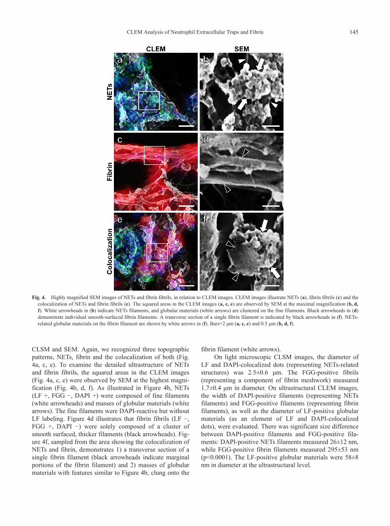
CLSM and SEM. Again, we recognized three topographicpatterns, NETs, fibrin and the colocalization of both (Fig.4a, c, e). To examine the detailed ultrastructure of NETsand fibrin fibrils, the squared areas in the CLEM images(Fig. 4a, c, e) were observed by SEM at the highest magni-fication (Fig. 4b, d, f). As illustrated in Figure 4b, NETs(LF +, FGG −, DAPI +) were composed of fine filaments(white arrowheads) and masses of globular materials (whitearrows). The fine filaments were DAPI-reactive but withoutLF labeling. Figure 4d illustrates that fibrin fibrils (LF −,FGG +, DAPI −) were solely composed of a cluster ofsmooth surfaced, thicker filaments (black arrowheads). Fig-ure 4f, sampled from the area showing the colocalization ofNETs and fibrin, demonstrates 1) a transverse section of asingle fibrin filament (black arrowheads indicate marginalportions of the fibrin filament) and 2) masses of globularmaterials with features similar to Figure 4b, clung onto the
fibrin filament (white arrows).On light microscopic CLSM images, the diameter of
LF and DAPI-colocalized dots (representing NETs-relatedstructures) was 2.5±0.6 μm. The FGG-positive fibrils(representing a component of fibrin meshwork) measured1.7±0.4 μm in diameter. On ultrastructural CLEM images,the width of DAPI-positive filaments (representing NETsfilaments) and FGG-positive filaments (representing fibrinfilaments), as well as the diameter of LF-positive globularmaterials (as an element of LF and DAPI-colocalizeddots), were evaluated. There was significant size differencebetween DAPI-positive filaments and FGG-positive fila-ments: DAPI-positive NETs filaments measured 26±12 nm,while FGG-positive fibrin filaments measured 295±53 nm(p<0.0001). The LF-positive globular materials were 58±8nm in diameter at the ultrastructural level.
Highly magnified SEM images of NETs and fibrin fibrils, in relation to CLEM images. CLEM images illustrate NETs (a), fibrin fibrils (c) and thecolocalization of NETs and fibrin fibrils (e). The squared areas in the CLEM images (a, c, e) are observed by SEM at the maximal magnification (b, d,f). White arrowheads in (b) indicate NETs filaments, and globular materials (white arrows) are clustered on the fine filaments. Black arrowheads in (d)demonstrate individual smooth-surfaced fibrin filaments. A transverse section of a single fibrin filament is indicated by black arrowheads in (f). NETs-related globular materials on the fibrin filament are shown by white arrows in (f). Bars=2 μm (a, c, e) and 0.5 μm (b, d, f).
Fig. 4.
CLEM Analysis of Neutrophil Extracellular Traps and Fibrin 145

IV. Discussion
NETs represent an extracellular spider’s web-like struc-ture of neutrophilic DNA stretches dotted with granule-derived antimicrobial proteins, including lactoferrin (LF),functionally preventing microbial invasions [3, 26, 28].The pathophysiological significance of NETs has beenreported in a variety of diseased conditions such as acuteappendicitis, dysentery, preeclampsia, necrotizing fasciitisand pneumonia [1, 3, 4, 9]. In these acute inflammatorylesions, fibrin meshwork commonly deposited togetherwith neutrophilic infiltration [5, 31], and NETs forming aweb-like network were also demonstrated by SEM [20, 27].
We previously evaluated differences in the fibrillarstructures of NETs and fibrin in formalin-fixed, paraffin-embedded sections of inflammatory lesions [20, 23]. LFfunctioned a good immunohistochemical marker of NETs,while fibrin was demonstrated by FGG immunostaining. Inthe light microscopic analysis, fibrils were categorized intothree types: thin, thick and clustered thick [23]. Thin fibrilsbelonged to NETs and thick fibrils were composed of eithermixed NETs and fibrin or fibrin alone, while clustered thickfibrils were solely composed of fibrin [23]. In the electronmicroscopic analysis using an autopsied lung tissue oflegionnaire’s pneumonia, NETs were composed of finefilaments and globular materials attached onto the fibrinfibrils [20]. The smooth-surfaced fibrin filaments weremuch thicker than the NETs filaments [20]. A discrepancyto be solved is that the diameter of the fibrils at the lightmicroscopic level appeared much larger than that seen atthe electron microscopic level [20].
CLEM has successfully been utilized in a variety ofmorphologic studies. For example, Sindbis virus exit path-way [19] and macrophage uptake of cylindrical microparti-cles [25] were investigated by CLEM. These studies wereperformed without causing damages to the ultrastructure ofthe cell and tissue. In the present CLEM analysis using aroutinely prepared autopsy lung tissue (legionnaire’s pneu-monia), the same area of the inflammatory lesion wassimultaneously observed with both CLSM and SEM [18]. Itis of note that CLSM did not damage the SEM features.Both types of microscopy should be used at their full capa-bilities [15, 18, 32]. The results were consistent with thoseof our previous studies [20, 23]. NETs (LF +, FGG −,DAPI +) were composed of fine filaments, 26±12 nm indiameter, and masses of globular materials. Fibrin filaments(LF −, FGG +, DAPI −), 295±53 nm in diameter, weremuch thicker than the NETs filaments.
The CLEM analysis explained the discrepancybetween the light and electron microscopic observationsregarding the diameter of the fibrils and the shape of themeshwork structure. Fibrin, seen as a thick (1.7 μm-sized)fibril at the light microscopic level, consisted ultrastructur-ally of a cluster of fine filamentous skeletons. NETs-relateddots, measuring 2.5±0.6 μm, seen at the light microscopiclevel were ultrastructurally composed of clustered globular
materials, 58±8 nm in diameter, on the fine filamentousstructure. These sizes of the filaments and globules wereconsistent with those of the previous reports [3, 16, 17, 30].It should be of note that light microscopically, NETs canpresent as a form of clustered dots, but not in the form ofspider’s web-like framework. The importance of therecognition of this variant of NETs should be emphasized,particularly when NETs are evaluated under pathologicconditions. Poor development of spider’s web-like NETsmay be related to the intracellular growth features of thepathogen, Legionella pneumophila [12]. In fact, bacteria,the target of NETs functions, were not entrapped in theextracellular space of the lung where we evaluated. Asdescribed in our previous study using immunoperoxidasevisualization of LF and FGG [20], LF and FGG double-positive meshwork structures were clearly demonstrated inthe same pneumonia sample, where accumulation of LF-positive globules on the FGG-positive fibrin filament wasaccelerated. The appearance of the “dotted” NETs may bedependent upon the balance of neutrophilic LF release andfibrin meshwork formation.
The present study is the first report demonstrating, bymeans of LF, FGG and DAPI as probes, the ultrastructuralfeatures of NETs and fibrin fibrils in an inflammatorylesion by CLEM, combining CLSM and SEM. It should beemphasized that the CLEM analysis was applicable to rou-tinely prepared formalin-fixed, paraffin-embedded sectionsof autopsy materials and that the ultrastructural localizationof specific proteins was successfully demonstrated evenafter heating treatment. We sincerely hope that the CLEManalysis can be expanded to morpho-functional studies ofNETs and fibrin fibrils in archival pathology materialsunder varied inflammatory conditions.
V. Competing Interest StatementWe have no conflict of interest to be claimed.
VI. AcknowledgmentsWe are grateful to Senior Assistant Prof. Gen Niimi,
Ph.D. and Assistant Prof. Tomihiko Ide, Ph.D., Division ofElectron Microscopy, Institute of Joint Research, FujitaHealth University, Toyoake, for their technical advice. Weare also grateful to Prof. Ken-ichi Inada, M.D., Ph.D. andAssistant Prof. Kouhei Sakurai, Ph.D., Department of Diag-nostic Pathology, Banbuntane-Houtokukai Hospital, FujitaHealth University School of Medicine, Nagoya, for theirhelpful discussion. Ms. Yukika Hasegawa, Ms. SayakaTakeuchi, Ms. Mika Maeshima, and Ms. Chikayo Yashiro,Department of Pathology, Fujita Health University Schoolof Medicine, Toyoake, are cordially acknowledged for theirpositive cooperation in our research activity. This work wassupported by a Research Grant from Fujita Health Univer-sity, 2015–2016 (no specific grant number given).
146 Onouchi et al.

VII. References
1. Beiter, K., Wartha, F., Albiger, B., Normark, S., Zychlinsky, A.and Henriques-Normark, B. (2006) An endonuclease allowsStreptococcus pneumoniae to escape from neutrophil extra-cellular traps. Curr. Biol. 16; 401–407.
2. Benedetti, L., Sogne, E., Rodighiero, S., Marchesi, D., Milani, P.and Francolini, M. (2014) Customized patterned substrates forhighly versatile correlative light-scanning electron microscopy.Sci. Rep. 4; 7033.
3. Brinkmann, V., Reichard, U., Goosmann, C., Fauler, B.,Uhlemann, Y., Weiss, D. S., Weinrauch, Y. and Zychlinsky, A.(2004) Neutrophil extracellular traps kill bacteria. Science 303;1532–1535.
4. Buchanan, J. T., Simpson, A. J., Aziz, R. K., Liu, G. Y., Kristian,S. A., Kotb, M., Feramisco, J. and Nizet, V. (2006) DNaseexpression allows the pathogen group A Streptococcus to escapekilling in neutrophil extracellular traps. Curr. Biol. 16; 396–400.
5. Chung, C. L., Chen, Y. C. and Chang, S. C. (2003) Effect ofrepeated thoracenteses on fluid characteristics, cytokines, andfibrinolytic activity in malignant pleural effusion. Chest 123;1188–1195.
6. Cortese, K., Diaspro, A. and Tacchetti, C. (2009) Advancedcorrelative light/electron microscopy: current methods and newdevelopments using Tokuyasu cryosections. J. Histochem.Cytochem. 57; 1103–1112.
7. de Boer, P., Hoogenboom, J. P. and Giepmans, B. N. (2015)Correlated light and electron microscopy: ultrastructure lightsup! Nat. Methods 12; 503–513.
8. Giepmans, B. N., Adams, S. R., Ellisman, M. H. and Tsien, R. Y.(2006) The fluorescent toolbox for assessing protein location andfunction. Science 312; 217–224.
9. Gupta, A. K., Hasler, P., Holzgreve, W., Gebhardt, S. and Hahn,S. (2005) Induction of neutrophil extracellular DNA latticesby placental microparticles and IL-8 and their presence inpreeclampsia. Hum. Immunol. 66; 1146–1154.
10. Hell, S. W. (2007) Far-field optical nanoscopy. Science 316;1153–1158.
11. Hellstrom, K., Vihinen, H., Kallio, K., Jokitalo, E. and Ahola, T.(2015) Correlative light and electron microscopy enables viralreplication studies at the ultrastructural level. Methods 90; 49–56.
12. Jules, M. and Buchrieser, C. (2007) Legionella pneumophilaadaptation to intracellular life and the host response: clues fromgenomics and transcriptomics. FEBS Lett. 581; 2829–2838.
13. Kanyshkova, T. G., Semenov, D. V., Buneva, V. N. andNevinsky, G. A. (1999) Human milk lactoferrin binds two DNAmolecules with different affinities. FEBS Lett. 451; 235–237.
14. Kinnula, V. L., Soini, Y., Kvist-Makela, K., Savolainen, E. R.and Koistinen, P. (2002) Antioxidant defense mechanisms inhuman neutrophils. Antioxid. Redox Signal. 4; 27–34.
15. Kopek, B. G., Shtengel, G., Xu, C. S., Clayton, D. A. and Hess,H. F. (2012) Correlative 3D superresolution fluorescence andelectron microscopy reveal the relationship of mitochondrialnucleoids to membranes. Proc. Natl. Acad. Sci. U S A 109;6136–6141.
16. Krautgartner, W. D., Klappacher, M., Hannig, M., Obermayer,A., Hartl, D., Marcos, V. and Vitkov, L. (2010) Fibrin mimicsneutrophil extracellular traps in SEM. Ultrastruct. Pathol. 34;
226–231.17. Liu, F. C., Chuang, Y. H., Tsai, Y. F. and Yu, H. P. (2014) Role of
neutrophil extracellular traps following injury. Shock 41; 491–498.
18. Liv, N., Zonnevylle, A. C., Narvaez, A. C., Effting, A. P.,Voorneveld, P. W., Lucas, M. S., Hardwick, J. C., Wepf, R. A.,Kruit, P. and Hoogenboom, J. P. (2013) Simultaneous correlativescanning electron and high-NA fluorescence microscopy. PLoSOne 8; e55707.
19. Martinez, M. G., Snapp, E. L., Perumal, G. S., Macaluso, F. P.and Kielian, M. (2014) Imaging the alphavirus exit pathway. J.Virol. 88; 6922–6933.
20. Onouchi, T., Shiogama, K., Matsui, T., Mizutani, Y., Sakurai, K.,Inada, K. and Tsutsumi, Y. (2016) Visualization of neutrophilextracellular traps and fibrin meshwork in human fibrinopurulentinflammatory lesions: II. Ultrastructural study. Acta Histochem.Cytochem. 49; 117–123.
21. Rigby, K. M. and DeLeo, F. R. (2012) Neutrophils in innatehost defense against Staphylococcus aureus infections. Semin.Immunopathol. 34; 237–259.
22. Rilla, K. and Koistinen, A. (2015) Correlative light and electronmicroscopy reveals the HAS3-induced dorsal plasma membraneruffles. Int. J. Cell Biol. 2015; 769163.
23. Shiogama, K., Onouchi, T., Mizutani, Y., Sakurai, K., Inada, K.and Tsutsumi, Y. (2016) Visualization of neutrophil extracellulartraps and fibrin meshwork in human fibrinopurulent inflam-matory lesions: I. Light microscopic study. Acta Histochem.Cytochem. 49; 109–116.
24. Sjollema, K. A., Schnell, U., Kuipers, J., Kalicharan, R. andGiepmans, B. N. (2012) Correlated light microscopy andelectron microscopy. Methods Cell Biol. 111; 157–173.
25. Tscheka, C., Hittinger, M., Lehr, C. M., Schneider-Daum, N.and Schneider, M. (2015) Macrophage uptake of cylindricalmicroparticles investigated with correlative microscopy. Eur. J.Pharm. Biopharm. 95; 151–155.
26. Urban, C. F., Reichard, U., Brinkmann, V. and Zychlinsky, A.(2006) Neutrophil extracellular traps capture and kill Candidaalbicans yeast and hyphal forms. Cell. Microbiol. 8; 668–676.
27. Veklich, Y., Francis, C. W., White, J. and Weisel, J. W. (1998)Structural studies of fibrinolysis by electron microscopy. Blood92; 4721–4729.
28. Vitkov, L., Klappacher, M., Hannig, M. and Krautgartner,W. D. (2009) Extracellular neutrophil traps in periodontitis. J.Periodont. Res. 44; 664–672.
29. Vogel, H. J. (2012) Lactoferrin, a bird’s eye view. Biochem. CellBiol. 90; 233–244.
30. Vorobjeva, N. V. and Pinegin, B. V. (2014) Neutrophil extra-cellular traps: mechanisms of formation and role in health anddisease. Biochemistry 79; 1286–1296.
31. Wait, M. A., Sharma, S., Hohn, J. and Dal Nogare, A. (1997) Arandomized trial of empyema therapy. Chest 111; 1548–1551.
32. Watanabe, S., Punge, A., Hollopeter, G., Willig, K. I., Hobson,R. J., Davis, M. W., Hell, S. W. and Jorgensen, E. M. (2011)Protein localization in electron micrographs using fluorescencenanoscopy. Nat. Methods 8; 80–84.
This is an open access article distributed under the Creative CommonsAttribution License, which permits unrestricted use, distribution, andreproduction in any medium, provided the original work is properly cited.
CLEM Analysis of Neutrophil Extracellular Traps and Fibrin 147