visual, gustatory, & olfactory microanatomy audrone r. biknevicius, phd ohio university college...
TRANSCRIPT

Visual, Gustatory, & Olfactory
MicroanatomyAudrone R. Biknevicius, PhD
Ohio UniversityCollege of Osteopathic Medicine
CPC 2 – Fall quarter 2005

Sclera(with episclera anteriorly)
Cornea
Fibrous LayerUveal Layer
Choroid
Iris
Ciliary Body
Retinal Layer
Retina
Modified from Netter, 1989
Inside/Inner
Outside/Outer
Limbus
Sclera(with episclera anteriorly)
Cornea
Fibrous LayerDura mater

Modified from Netter, 1989
Light
Cornea
Anterior Compartment (Aqueous humor):•Anterior chamber•Pupil•Posterior chamber
Lens
Posterior Compartment (Vitreous humor)
Retina

Gartner & Hiatt, 2001. Figure 22-7
Sclera
Choroid
Retina
Light

Cell Types in the Retina
Support Cells - Müller cells(neuroglial cells)
Retinal Pigment Epithelium
Modified from Gartner & Hiatt, 2001. Figure 22-8

Cell Types in the Retina
Photoreceptors: Rods & Cones
Ganglion cells
Integrating neurons
Neurons:
Horizontal
Bipolar
Amacrine
Modified from Gartner & Hiatt, 2001. Figure 22-8

Vitreous body
Choroid
Modified from Young & Heath, 2000. Figure 21.8
--- Pigment Epithelium
--- Outer Nuclear Layer
--- Inner Nuclear Layer
--- Ganglion Cell Layer
--- Outer Plexiform Layer
--- Inner Plexiform Layer
--- Photoreceptors (rods & cones proper)
--- Optic Nerve Fiber Layer
--- Inner Limiting Membrane
--- Outer Limiting Membrane
Mu
ller
Cel
l
10 Histological LayersO
ute
r
I
nne
r

Modified from Gartner & Hiatt, 2001. Figure 22-7
--- Pigment Epithelium
--- Outer Nuclear Layer
--- Inner Nuclear Layer
--- Ganglion Cell Layer
--- Outer Plexiform Layer
--- Inner Plexiform Layer
--- Photoreceptors (rods & cones proper)
--- Optic Nerve Fiber Layer--- Inner Limiting Membrane
--- Outer Limiting Membrane
Light
Neu
ral
laye
r
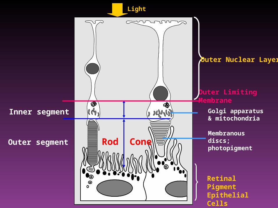
Outer segment
Inner segment
Outer Limiting Membrane
Rod Cone
Light
Retinal Pigment Epithelial Cells
Golgi apparatus & mitochondria
Membranous discs;photopigment
Outer Nuclear Layer

Rods Cones
Rhodopsin IodopsinIodopsinIodopsin
Individual cones have only one type of
iodopsin
All over the retina, more numerous
toward outer edge
All over the retina, but concentrated
in the maculae (fovea)
Low light Bright light
Light
Scotopic vision Peripheral vision
Phototopic visionCentral vision

Eye development
www.img.cas.cz/resrep/ zk/mouse-eye.html

Clinical question: Between which layers of the retina does retinal detachment occur?
www.emedicine.com/ oph/topic411.htm
Ph
oto
rece
pto
rP
igm
ent
Ep
ith
eliu
m
Young & Heath, 2000

Information Path
Young & Heath, 2000. Figure 21.8
Action potential!
Light Path
choroid

medlib.med.utah.edu
Visual Acuity
Macula lutea:•Bounded by temporal retinal vascular arcades•Used primarily for central and color vision (phototopic vision)
Fovea centralis:•Depression at center of macula; thinned outer nucleus layer; high density of cones
Foveola:•Only cone receptors•Almost 1 cone : 1 optic tract neuron•Retinal avascular zone
•central vision; maximum visual discrimination•movements of eyes bring light to focus on macula

Netter, 1989
Optic Disc
Optic disc (optic papilla):•Ganglion cell axons exit eye (through lamina cribrosa) to form the optic nerve; lacks photoreceptors (blind spot) •Retinal vessels enter/exit eye
superior
lateral

Netter, 1989
UVEAL (VASCULAR) LAYER
•Choroid•Choroidal vessels (choriocapillaris)supply photoreceptors
•Iris (melanocytes) •Dilator pupillae m.•Sphincter pupillae m.
•Ciliary body•Ciliary m.•Suspensory ligament•Lens
Innervation of intrinsic eye muscles:
Sympathetics: dilator pupillae m.
Parasympathetics (CN III): sphincter pupillae m. ciliary mm.

Arterial Supply to Retina
webvision.med.utah.edu
Central Retinal Artery•Supplies inner 2/3 of retina (including ganglion cells)
Choriocapillaris (capillary lamina of the choroid)•Supplies outer 1/3 of retina (retinal pigment cells & photoreceptors)

Netter, 1989
AVASCULAR TISSUES
•LENS•CORNEA
Metabolites are exchanged via:
•Aqueous humor flow-Produced by ciliary processes in posterior chamber of the anterior compartment-Reabsorbed by canal of Schlemm (venous) at the angle of the anterior chamber
•Oxygen diffuses from corneal surface

Wilson-Pauwels et al., 1988
Visual PathwayPat O’Connor

ChemosensationTaste
Smell
Dysgeusia = Alteration in normal taste sensationAgeusia = Loss of taste sensation
Parosmia = Distortion in the perception of odor Anosmia = Loss of sense of smell
•Flavor = combination of taste + smell
•Chemoreceptor cells regenerate(~2 weeks gustatory; 6-8 weeks olfactory)
•Both senses require that chemical signal molecules be dissolved or suspended in a watery medium

Modified from Netter, 1989
Sweet
Sour
Salty
Bitter
TASTE MODALITIES

Filiform Papilla(“filaments”)
Fungiform Papilla (“mushroom-like”)
Circumvallate Papilla(“around an elevation”)
Von Ebner’s Glands(serous secretion)Modified from Netter, 1989
Lingual Papillae

Vallate(Circumvallate)~50% taste buds
Foliate ~30% taste buds
Fungiform~20% taste buds
Modified from Kandel, et al., 1991
= Taste chemoreceptor(Taste bud)Trough

Basal Cell Taste Receptor Cell
Support Cell
Kandel, et al., 1991
Mo
difi
ed
fro
m Y
ou
ng
& H
ea
th,
20
00
. F
igu
re 2
1.1
TastePore
Trough
Primary Sensory Neuron

w3.ouhsc.edu/histology
Von Ebner’s Glands

Ant. 2/3 Post. 1/3 Epiglottis(oral) (pharyngeal) (laryngeal)
Taste Facial n. Glossopharyngeal n. Vagus n.(Chorda tympani) (Sup.laryneal n.)
Touch Trigeminal n. Glossopharyngeal n. Vagus n.(Lingual n.) (Sup.laryneal n.)
Agur (1991) Grant’s Atlas of Anatomy
Anterior 2/3 of tongue(oral tongue)
Posterior 1/3 of tongue(pharyngeal tongue includingvallate papillae)
(also some extrapapillary taste buds scattered in the palate & pharynx)

Kandel, et al., 1991
Project 1st to nucleus of the solitary tract (nucleus solitarius)

CN VII CN IX [CN X]
Nucleus of the Solitary Tract – rostral part (brainstem medulla) (gustatory nucleus)
Nucleusambiguus(CN IX &X)
Dorsal motor nucleus of
CN X
Medullar reticular
formation
Ventropostero- medial nucleus
(thalamus)
Hypothalmus Amygdaloid complex
Parasymp.to viscera
Somatic motor: larynx, pharynx & palate (part)
Reflexes:swallowing,
vomiting
Feeding reflexes, emotions
Gustatory cortex (conscious
perception of taste)
Perception of sensory
stimuli; fear conditioning
(Kinsley Concise Text of Neuroscience)
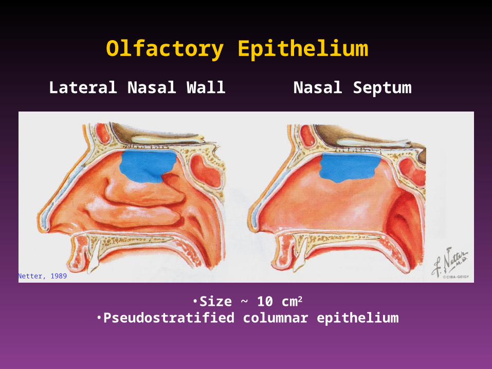
Lateral Nasal Wall Nasal Septum
Olfactory Epithelium
Netter, 1989
•Size ~ 10 cm2
•Pseudostratified columnar epithelium

Kinsley Concise Text of Neuroscience
Basal cell
Olfactory epithelium:•Olfactory cells•Sustentacular (support) cells•Basal cells
Olfactory Mucosa
Lamina propria:•Bowman’s gland (serous)•Highly vascularized

Heimer The Human Brain and Spinal Cord
Olfactory TractOlfactory Bulb`
Olfactory Nerve (CN I)= Axons of olfactory receptor cells (1st order sensory neurons)- Cells replaced

Heimer The Human Brain and Spinal Cord
•Olfactory receptor cells synapse with mitral cells & tufted cells in the olfactory bulb (glomeruli)
•Numerous interneurons enable complex interactions (recall integrating neurons in retina!)
•Olfactory tract (axons of mitral and tufted cells)
Projections to (without thalamic relay):•Piriform & entorhinal cortex•Amygdala

Basal cell
Trigeminal Nerve & Chemosensation
•Provides most of the sensory innervation of the face: mechanical, proprioception, nociception
•Also sensitive to chemical stimuli in the nose, oral cavity and eye
•Protective mechanism against potentially dangerous chemicals