visian icl (implantable collamer lens) for … glossary 3 2.0 introduction ... 1.0 glossary this...
TRANSCRIPT

Visian ICL™ (Implantable Collamer Lens)
For Nearsightedness Facts You Need To Know About STAAR Surgical’s Visian ICL SURGERY
PATIENT INFORMATION BOOKLET
For Nearsightedness (Myopia) between –3 to –20 Diopters
with 2.5 Diopters or less of Astigmatism
Please read this entire booklet. Discuss its contents with your eye doctor so that you have all of your questions answered to your satisfaction. Ask any questions you may have before
you agree to this surgery.
Distributed and Manufactured by: STAAR Surgical Company
1911 Walker Avenue Monrovia, CA 91016 USA
Tel: (800) 352-7842 FAX: (800) 952-4923
Copyright 2017 by STAAR Surgical Company
This booklet may be reproduced only by a treating physician, for use with patients considering Visian ICL Surgery. All other rights are reserved.

1
This page left blank intentionally.
2
TABLE OF CONTENTS
1.0 Glossary ........................................................................................................................ 3
2.0 Introduction ................................................................................................................... 8
3.0 What Is Nearsightedness? ............................................................................................. 8
4.0 How Does Visian ICL Correct Nearsightedness? ......................................................... 9
5.0 Other Treatments to Correct Nearsightedness? .......................................................... 12
6.0 Benefits And Risks Of The Visian ICL for Nearsightedness ..................................... 12
7.0 Contraindications ........................................................................................................ 16
8.0 Warnings ..................................................................................................................... 16
9.0 Precautions .................................................................................................................. 17
10.0 Are You A Good Candidate For Visian ICL Surgery? ............................................. 19
11.0 What Should You Expect During Visian ICL Surgery? ........................................... 20
12.0 Questions To Ask Your Doctor ................................................................................ 22
13.0 Self-Test .................................................................................................................... 23
14.0 Clinical Studies ......................................................................................................... 24
15.0 Adverse Events Observed In The Visian ICL Clinical Studies ................................ 27
16.0 Summary of Important Information .......................................................................... 30
17.0 Patient Assistance Information ................................................................................. 32

3
1.0 Glossary
This section summarizes important terms used in this information booklet or that your doctor may discuss with you. Please discuss any related questions with your doctor.
Acute: Of sudden, rapid onset, usually with notable symptoms.
Acute Corneal Swelling: A sudden swelling of the usually clear front surface of the eye (cornea).
Anisocoria: Unequal pupil size.
Anterior Chamber: Front chamber of the eye; anterior chamber depth is the space between the back of the cornea to the front part of the crystalline lens. Anterior chamber angle is the location where the cornea and iris meet.
Antibiotic Medication: A drug used to treat or prevent infection. Your doctor may prescribe this medication after Visian ICL surgery.
Anti-inflammatory Medication: A drug that reduces inflammation or the body’s reaction to injury or disease. Any eye surgery can cause inflammation. Your doctor may prescribe the medication after Visian ICL surgery.
Astigmatism: A focusing error that results in blurred distant and/or near vision. The cornea is more curved in some directions than others, and causes light rays to focus at different points inside the eye. Parts of objects appear clearer than other parts.
Cataract: Opacity, or clouding, of the crystalline lens inside the eye that can blur vision.
Collagen: A gel-like supporting substance found in the cornea, skin and other connective tissue of the body.
Collamer: Hydroxyethyl methacrylate (HEMA)/porcine-collagen based polymer material (STAAR proprietary product).
Conjunctival Irritation: An irritation of the white portion of the eyeball and inner eyelid.
Contraindications: Any special conditions that result in the treatment not being recommended.
Cornea: The clear front layer of the eye that lets light enter. Surgery such as PRK, LASIK and RK reshapes the cornea to improve distant vision.
Corneal Edema: Abnormal fluid build-up/swelling in the cornea. The condition is usually temporary after surgery with no significant effect on vision. Persistent corneal swelling may cause a loss of vision.
4
Corneal Endothelium: A thin, single layer of cells on the innermost surface of the cornea, responsible for keeping the cornea clear. These cells do not reproduce and decrease in number with age.
Corneal Flap: A thin slice of tissue on the surface of the cornea made at the beginning of a LASIK procedure. This flap is folded back before the laser shapes the inner layer of the cornea.
Corneal Transplant: Removal and replacement of cornea.
Crystalline Lens: A structure inside the eye that helps to focus light onto the back surface (retina) of the eye.
Cystoid Macular Edema: Swelling of the macula, located in the center of the retina.
Diabetic Retinopathy: Damage to the back surface of the eye responsible for sensing light due to diabetes.
Diopter: A unit of focusing power, used to describe the amount of nearsightedness and astigmatism of an eye. Abbreviated as “D”.
Double Vision: Seeing multiple images of the object being looked at.
Endophthalmitis: Severe infection or inflammation of the entire eyeball.
Endothelial Cell Loss: A thin, single layer of cells (endothelial cells) on the innermost surface of the cornea keeps the cornea clear by pumping water out of it. Normally, these cells slowly decrease in number as you age. Additional loss of these cells beyond the normal amount can occur following many kinds of eye surgery. If too many cells are lost, the cornea can become cloudy, which can decrease vision.
Endothelium: See Corneal Endothelium.
Glare: A harsh or uncomfortable bright light. Glare symptoms are usually caused by a distortion of light that would otherwise be tolerable without the distortion.
Glaucoma: An eye disease usually associated with high eye pressure. Glaucoma damages the optic nerve of the eye and usually causes a progressive loss of vision.
Halos: Circular flares or rings of light that may appear around a headlight or other lighted object. This symptom may occur after surgery.
Hyphema: Blood in the front (anterior) chamber of the eye.
Hypopyon: Discharge in the front chamber of the eye.
Implantable Collamer Lens (ICL): A lens made of collagen based polymer which is implanted in the eye behind the iris and in front of the crystalline lens in order to correct or reduce nearsightedness. The Visian ICL can be removed.

5
Inflammation of the Eye: The eye’s response to injury, infection or irritation which can cause redness of the eye, pain, blurred vision and/or light sensitivity.
Intraocular Lenses: An artificial lens that is placed in the eye to correct refractive errors such as nearsightedness.
Intraocular Pressure (IOP): The amount of pressure of the fluid inside your eye.
Iris: Colored part of the eye.
Iris Prolapse: A movement of the colored portion of the eye through a surgical wound to a position outside the eye.
Iritis: Inflammation in the front (anterior) chamber of the eye.
Laser Assisted In-Situ Keratomileusis (LASIK): A type of eye surgery that uses a device to create a thin, hinged flap of tissue on the cornea which is then folded back. A laser then reshapes the tissue under the flap and the flap is put back on the eye so the tissue heals.
Lens: Natural crystalline lens in the eye which helps focus light properly into the back of the eye.
Lens Opacities: A cloudiness of the crystalline lens.
Macular Degeneration: A reduction in your central vision due to the thinning of a part of your retina responsible for fine (reading) vision.
Macular Edema: Swelling in the area responsible for fine (reading) vision on the back surface of the eye (retina).
Macular Hemorrhage: Bleeding in the area responsible for fine (reading) vision on the back surface of the eye (retina).
Myopia: A focusing error that results in blurrier vision at distance than near. Myopia is also called nearsightedness.
Narrow Anterior Chamber Angle: A decrease in the size of the front chamber of the eye which could block the flow of fluid from inside of the eye to the outside resulting in a raised eye pressure (glaucoma).
Nearsighted/Nearsightedness: A focusing error that results in blurrier vision at distance than near. Nearsightedness is also called myopia.
Non-reactive Pupil: A condition where the colored portion of the eye does not get larger or smaller when light is shined in the eye.
Ocular Hypertension: Increased eye pressure.
Peripheral Anterior Synechiae: Scar tissue at the outer edges of the front chamber of the eye.

6
Peripheral Iridotomy: A small hole placed at the outer edge of the colored portion of the eye, usually using an Yttrium Aluminum Garnet (YAG) laser beam.
Persistent: Lasts for a period of time during the study follow-up usually at least until the end of the study.
Phakic Intraocular Lens: Placement of a man-made lens in an eye that still has its natural crystalline lens.
Photorefractive Keratectomy (PRK): A type of eye surgery that uses a laser to reshape the front surface of the eye to improve vision. After the epithelium (outermost layer) of the cornea is first scraped away, the laser removes tissue from the exposed surface. After the surgery, the epithelium grows back.
Pigment Dispersion: An abnormal release of pigment particles from cells in the eye that could block the drainage of fluid from the inside to the outside of the eye.
Pseudoexfoliation: A condition where flakes of material can come off the surface of the crystalline lens and block the drainage of fluid from the inside to the outside of the eye.
Pupil: The black part of the eye; fluctuates in size allowing varying degrees of light into the eye.
Pupillary Block Glaucoma: The inability of fluid to flow from the back chamber of the eye to the front chamber frequently blocking drainage of fluid out of the eye and raising the pressure in the eye (glaucoma).
Radial Keratotomy (RK): A type of eye surgery that changes the shape of the front surface of the eye by making a special pattern of cuts in the cornea to correct nearsightedness.
Retina: The layer of nerve tissue at the back of the eye that captures images, similar to film in a camera, and sends information about these images to the brain. Light must be focused correctly on the retina to form clear images.
Retinal Detachment: Separation of the retina from its natural position on the back surface of the eyeball.
Subretinal Hemorrhage: Bleeding under the retina.
Uveitis: Inflammation of the middle layer of tissue in the eye.
Viscoelastic: gel-like fluid placed inside the eye during eye surgery to help maintain the shape of the eye
Visian ICL: see Implantable Collamer Lens

7
Visual Acuity: A measure of the sharpness of vision using a letter chart. Best Corrected Visual Acuity (BCVA) is the best vision with eyeglasses. Uncorrected Visual Acuity (UCVA) is the best vision without eyeglasses or contact lenses.
Vitreous Loss: The loss of a clear gel like material from the farthest back chamber of the eye during a surgical procedure.
YAG Laser: Yttrium Aluminum Garnet laser beam used in ophthalmology to produce a small
hole at the outer edge of the colored portion of the eye (peripheral iridotomy).
8
2.0 Introduction
The purpose of this booklet is to help you decide if you want to have the Visian Implantable
Collamer Lens (ICL) placed in one or both of your eyes to treat your nearsightedness (or
myopia). It is important for you to understand both the benefits and risks of this surgery before
you make a decision. The “Glossary” in this booklet explains the meaning of all words printed in
italics. Please read this entire booklet carefully and discuss your questions with a doctor who is
trained in Visian ICL surgery.
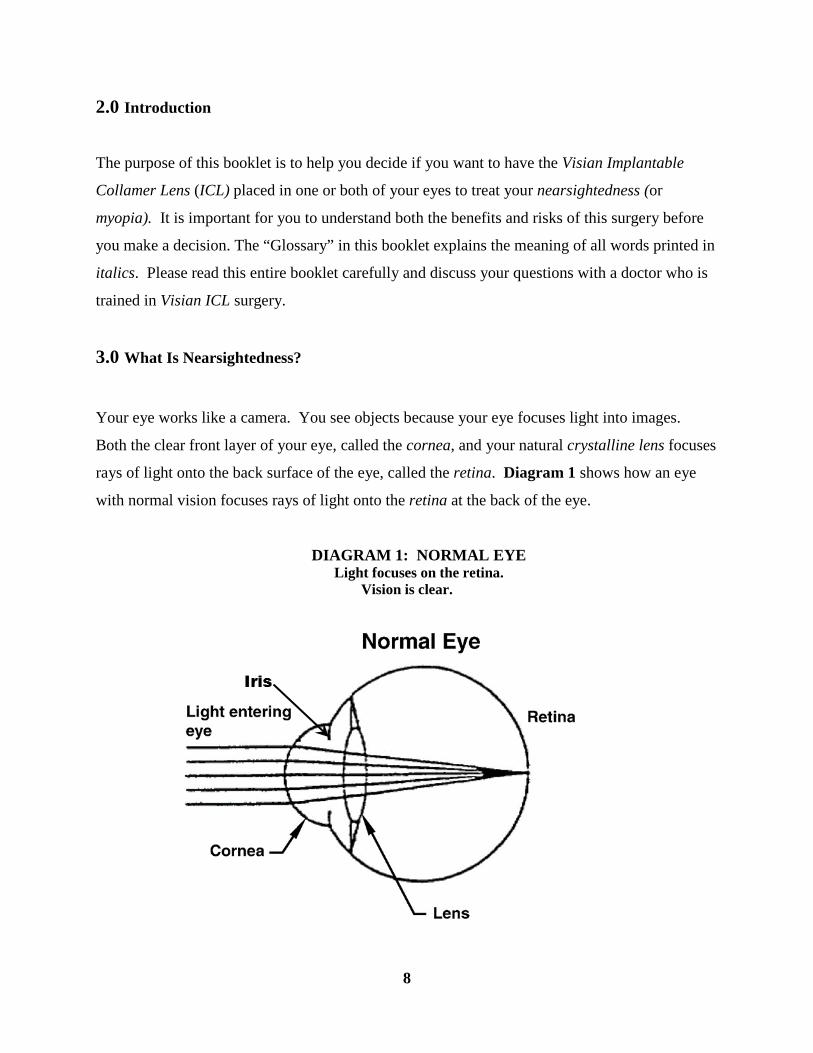
3.0 What Is Nearsightedness?
Your eye works like a camera. You see objects because your eye focuses light into images.
Both the clear front layer of your eye, called the cornea, and your natural crystalline lens focuses
rays of light onto the back surface of the eye, called the retina. Diagram 1 shows how an eye
with normal vision focuses rays of light onto the retina at the back of the eye.
DIAGRAM 1: NORMAL EYE Light focuses on the retina.
Vision is clear.
9
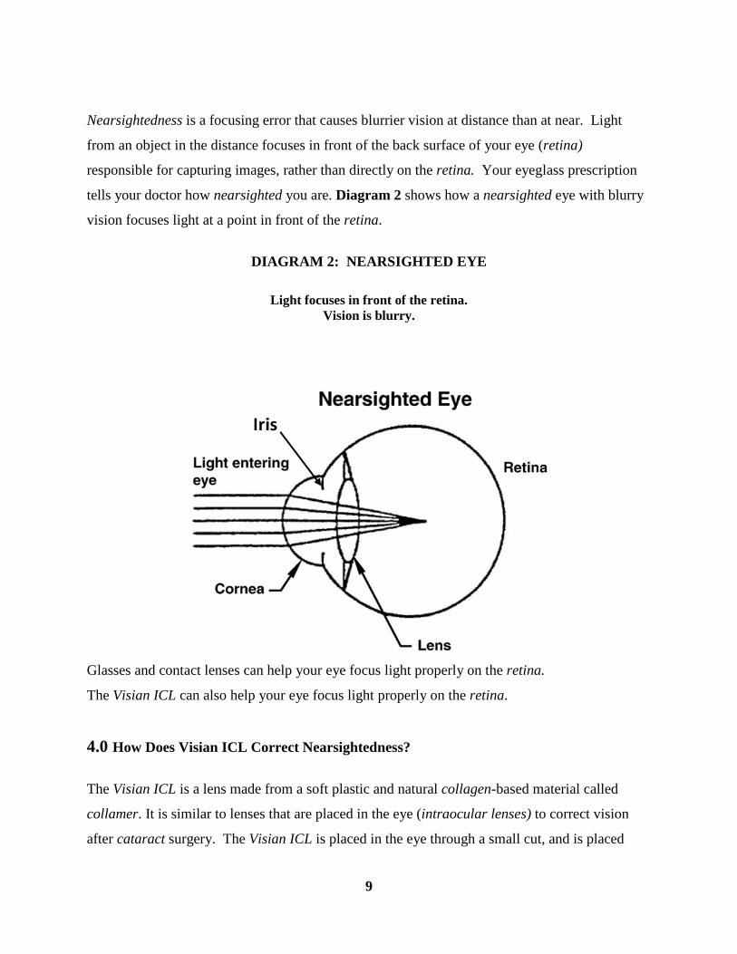
Nearsightedness is a focusing error that causes blurrier vision at distance than at near. Light
from an object in the distance focuses in front of the back surface of your eye (retina)
responsible for capturing images, rather than directly on the retina. Your eyeglass prescription
tells your doctor how nearsighted you are. Diagram 2 shows how a nearsighted eye with blurry
vision focuses light at a point in front of the retina.
DIAGRAM 2: NEARSIGHTED EYE
Light focuses in front of the retina. Vision is blurry.
Glasses and contact lenses can help your eye focus light properly on the retina.
The Visian ICL can also help your eye focus light properly on the retina.
4.0 How Does Visian ICL Correct Nearsightedness?
The Visian ICL is a lens made from a soft plastic and natural collagen-based material called
collamer. It is similar to lenses that are placed in the eye (intraocular lenses) to correct vision
after cataract surgery. The Visian ICL is placed in the eye through a small cut, and is placed
10
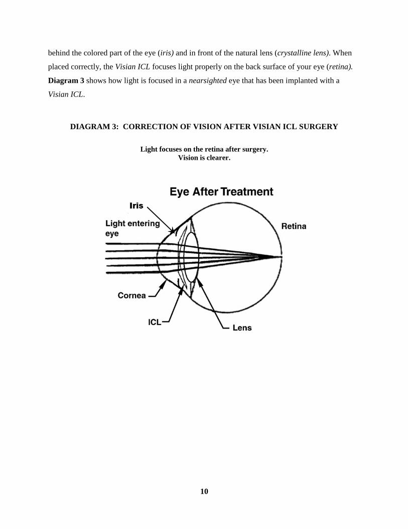
behind the colored part of the eye (iris) and in front of the natural lens (crystalline lens). When
placed correctly, the Visian ICL focuses light properly on the back surface of your eye (retina).
Diagram 3 shows how light is focused in a nearsighted eye that has been implanted with a
Visian ICL.
DIAGRAM 3: CORRECTION OF VISION AFTER VISIAN ICL SURGERY
Light focuses on the retina after surgery.
Vision is clearer.

11
A diopter (D) is a unit of focusing power used to describe the amount of nearsightedness or
focusing error (astigmatism) in the eye. Visian ICL surgery is designed to treat nearsightedness
between –3D to –15D, and reduce nearsightedness up to -20D in eyes with up to 2.5D of
astigmatism. If you have nearsightedness within these ranges, Visian ICL surgery may improve
your distance vision without eyeglasses or contact lenses.
The Visian ICL will not correct any astigmatism you may have. Astigmatism is a focusing error
of the eye that results in blurred vision. In eyes with astigmatism, the clear front layer of the eye
that lets light enter (cornea) is more curved in some directions than others. This causes light rays
to focus at different points inside the eye and some parts of objects will appear clearer than other
parts. Diagram 4 shows how an eye with astigmatism may focus light.
DIAGRAM 4: NEARSIGHTED and ASTIGMATIC EYE
Visian ICL surgery is permanent as long as the Visian ICL stays in your eye. The Visian ICL can
be removed at a future date. However, the residual effect of the Visian ICL on your eye after it is
removed is not known. If your physician removes the Visian ICL, you will lose the benefit of
your nearsightedness correction. This means that your vision may not return to what it was like
before the Visian ICL surgery.
12
5.0 Other Treatments to Correct Nearsightedness?
Other treatments for nearsightedness include eyeglasses, contact lenses or other eye surgeries.
Eye surgeries available to correct nearsightedness include Radial Keratotomy (RK),
Photorefractive Keratectomy (PRK), Laser Assisted in situ Keratomileusis (LASIK) and Phakic
Intraocular Lens implantation (the Visian ICL is a phakic intraocular lens). These surgeries may
not meet the vision requirements for some careers, such as military service. Eye surgeries can
either change the shape of the front surface of the clear layer at the front of your eye (cornea),
including RK, PRK and LASIK, or require the insertion of a lens into the eye. RK uses a surgical
instrument to make fine cuts in the cornea. PRK and LASIK use a laser to reshape the cornea.
For LASIK, an instrument cuts a thin flap of tissue from the front of your cornea. This corneal
flap is folded back and a laser removes tissue under the flap to change the shape of the cornea.
The flap is then put back in place for the eye to heal.
6.0 Benefits And Risks Of The Visian ICL for Nearsightedness
Benefits
Visian ICL surgery can safely correct nearsightedness between –3D to –15D, and partially
correct nearsightedness up to -20D in eyes with up to 2.5D of astigmatism.
If your eyeglass prescription is in these ranges, the Visian ICL may make your distance vision
without eyeglasses or contact lens correction better. Placing the Visian ICL into the eye requires
surgery, and all eye surgery carries potentially serious risks. Please review this booklet and
discuss the risks with your doctor.
Risks
This part of the booklet explains the risks of Visian ICL surgery. Please see 14.0 Clinical Studies
of the Visian ICL for more information about the FDA Clinical Study.
In a clinical study of the Visian ICL, patients were examined by the study doctor until 36 months
(3 years) after surgery in the first part of the study. In the second part of the study, patients
returned for visits until 60 months (5 years) or longer after Visian ICL surgery. The study
doctors collected information on the below items:
• Additional surgeries;
13
• Cataract formation;
• Loss of best corrected vision (BCVA)
• Raised pressure inside the eye (intraocular pressure) and damage to the optic nerve
caused by increased pressure in the eye (glaucoma);
• Loss of cells on the innermost surface of the cornea (endothelial cells);
• Other complications.
Additional (Secondary) Surgery
Another surgery to take out, replace or adjust the position of the Visian ICL may be necessary.
In the FDA clinical study, the Visian ICL was removed most often because of cataract surgery.
Additional (secondary) surgery may also be necessary to lower eye pressure. Please see the
section on “Raised intraocular pressure (IOP) and Glaucoma Development” below.
Cataract Formation
After Visian ICL surgery, patients have increased risk of developing cloudiness of the natural
lens (cataract), including risk of a cataract that may need surgery. The risk of a cataract
continues to rise with each year that the Visian ICL is in the eye. Because of this, you should see
your doctor regularly for an eye exam to check you for cataracts. The long term risk of a
cataract and additional surgery may be higher in older patients and those with higher levels of
nearsightedness (myopia). The long-term risk of cataract beyond 7 years is unknown. If your
doctor recommends cataract surgery, both the Visian ICL and the cataract is removed and
another intraocular lens is implanted, just as is done in any routine cataract surgery.
Loss of best corrected vision
Eighteen eyes in the FDA study lost vision of two or more lines as measured on an eye chart.
The most common reason was clouding of the natural lens (cataract). Vision got better in these

14
eyes after cataract surgery. In other eyes, vision improved without treatment. In 7 of the 18
eyes, however, vision did not get better after 5 or more years.
Raised intraocular pressure (IOP) and glaucoma development
Normal eye pressure (intraocular pressure or IOP) can vary, but is often considered to be from
10-21 millimeters of mercury (mmHg). An IOP higher than normal is called ocular hypertension
and if left untreated, can cause damage to the optic nerve (glaucoma) and cause permanent vision
loss. Patients with high levels of nearsightedness are also at increased risk of developing
glaucoma. In the clinical study, some patients had an increase in IOP to values greater than
10mmHg higher than before Visian ICL surgery or to higher than 25mmHg. Other patients
experienced an increase in eye pressure requiring treatment beyond just the use of medicine,
most often at 1 to 2 days after surgery. In most of these eyes, another hole was placed in the
extreme outer edge of the colored portion of the eye (peripheral iridotomy) to reduce the
pressure.
A few patients in the study developed damage to the optic nerve (glaucoma). The first case of
glaucoma was diagnosed at 5 months after Visian ICL surgery and the last case happened at over
6 years (73 months) after surgery.
Endothelial Cell Loss
A thin, single layer of cells (endothelial cells) on the surface of the cornea closest to the inside of
your eye, keeps the cornea clear by pumping water out of it. Normally, these cells slowly
decrease in number as you age. Additional loss of these cells beyond the normal amount can
happen after many kinds of eye surgery. If too many cells are lost, the cornea can become
cloudy, which can decrease vision.
Loss of endothelial cells can happen after Visian ICL surgery. Before your surgery, you will
have an eye exam that will help your doctor decide if you are a candidate for Visian ICL surgery.
Patients implanted with the Visian ICL experience some loss of endothelial cells and a
continuing loss of endothelial cells over time that is greater than that expected from aging.
Amount of loss varies, but 11% of those checked at 5 -7 years from surgery had more than 30%
endothelial cell loss. If loss reaches a critical level, there could be a build-up of fluid or swelling
15
of the cornea (corneal edema). Corneal edema may require that your cornea be removed and
replaced (corneal transplantation).
Other Complications
Other risks associated with Visian ICL surgery may include:
• movement of the colored portion of the eye (iris) through a surgical wound to a position
outside the eye (iris prolapse),
• bleeding in the area on the retina responsible for reading vision (macular hemorrhage),
• bleeding under the retina (subretinal hemorrhage),
• increase in focusing error (astigmatism),
• lifting or pulling of the retina from its natural position (retinal detachment),
• unequal pupil size (anisocoria).
Potential complications are not limited to those reported during the clinical study. The following
represent potential complications/adverse events reported with refractive surgery in general:
• irritation of the white portion of the eyeball and inner eyelid (conjunctival irritation),
• temporary severe abnormal fluid build-up/swelling in the cornea (acute corneal swelling)
after surgery that does not cause a loss of vision,
• continuing abnormal fluid build-up/swelling in the cornea (persistent corneal swelling)
that may cause a loss of vision,
• partial or total eye infection (endophthalmitis),
• significant harsh or uncomfortable bright light (glare) or circular flares or rings of light
that may appear around a headlight or other lighted object (halos),
• blood in the eye (hyphema),
• discharge in the eye (hypopyon),
• Visian ICL dislocation,
• cyst-like swelling with of the center of the retina with fluid (cystoid macular edema),
• condition where the colored portion of the eye does not get larger or smaller when light is
shined in the eye (non-reactive pupil),
16
• the inability of fluid to flow from the back chamber of the eye to the front chamber,
frequently blocking drainage of fluid out of the eye and raising the pressure in the eye
(pupillary block glaucoma),
• severe inflammation of the eye,
• inflammation in the front part of the eye (iritis),
• inflammation in the middle layer of tissue in the eye (uveitis),
• loss of clear gel-like material from the farthest back chamber of the eye during surgery
(vitreous loss) and
• removal and replacement of the cornea (corneal transplant).
7.0 Contraindications
You should NOT have Visian ICL surgery if you:
• are less than 21 years of age;
• have a narrow front (anterior) chamber as measured by a special eye test by your doctor,
or if your doctor finds that the shape of your eye is not adequate to fit the Visian ICL
(anterior chamber depth less than 3.0 millimeters);
• are pregnant or nursing
• do not meet the minimum endothelial cell density for your age at the time of
implantation as determined by your eye doctor
8.0 Warnings
• Two holes in the extreme outer edge of the colored portion of the eye (peripheral
iridotomies) must be performed 90º apart using a laser at between 2 to 3 weeks before
implantation of the Visian ICL.
• The long-term effects of the Visian ICL on the thin, single layer of cells on the surface of
the cornea closest to the inside of your eye, that keep the cornea clear (corneal
endothelium) are not known. In the FDA clinical study with the Visian ICL, some patients
had 30% or greater loss of corneal endothelial cells. You should be aware that a greater
than normal build-up of fluid or swelling of the cornea (corneal edema) can happen. The
corneal edema may even require that your cornea be removed and replaced (corneal

17
transplantation). You should see your doctor regularly for an exam to check your
endothelium as long as you have the Visian ICL in your eye(s). This will help your doctor
monitor the long-term health of your cornea.
• After Visian ICL surgery, patients have increased risk of developing cloudiness of the
natural lens (cataract), including risk of a cataract that may need surgery. The risk of
cataract continues to rise with each year that the Visian ICL is in the eye. Because of this,
you should see your doctor regularly for an eye exam to check you for cataracts. The
long term risk of a cataract and additional surgery may be higher in older patients and
those with higher degrees of nearsightedness (myopia). The long-term risk of a cataract
beyond 7 years is unknown.
• The potential of the Visian ICL to raise the pressure inside your eye (intraocular
pressure) and the long-term risks of the following are unknown:
damage to the optic nerve caused by increased pressure (glaucoma),
scar tissue at the outer edges of the front chamber of the eye (peripheral anterior
synechiae), and
abnormal release of pigment particles from cells in the eye that could block the
drainage of fluid from the inside to the outside of the eye (pigment dispersion).
9.0 Precautions
1. Patients with higher amounts of nearsightedness had worse results. The Visian ICL was
less effective in correcting nearsightedness and there was a higher risk of complications
in these patients.
2. The effect of pupil size on visual symptoms is not known.
3. The relationship between the Visian ICL and future cloudiness of the lens (cataract) and
lifting or pulling of the retina from its natural position (retinal detachment) is not known.
4. The ability of ultraviolet absorbing lenses to reduce the incidence of retinal disorders has
not been established. Examples of retinal disorders include damage to your eye caused by

18
sun gazing or reduction in your central vision due to the thinning of a part of your retina
(macular degeneration).
5. The safety of and ability of the Visian ICL to correct moderate to high nearsightedness
has NOT been established in patients with:
• unstable or worsening nearsightedness;
• history or clinical signs of inflammation inside the eye (iritis/uveitis);
• damage to the layer of the nerve tissue at the back of the eye that captures images
(retina) caused by diabetes (diabetic retinopathy);
• damage to the optic nerve caused by increased pressure in the eye (glaucoma);
• with history of previous eye surgery such as removal and replacement of the cornea
(corneal transplant) or surgery to repair the layer of the nerve tissue at the back of the
eye that captures images (retina) after it has separated from its natural position on the
back surface of the eyeball (retinal detachment);
• life-threatening non-ocular disease (e.g., end-stage heart failure or kidney disease);
• progressive sight-threatening disease other than nearsightedness;
• a diagnosis of high pressure inside the eye (ocular hypertension);
• insulin-dependent diabetes;
• flakes of material blocking normal fluid drainage from the eye (pseudoexfoliation);
• abnormal release of pigment inside the eye (pigment dispersion);
• greater than -20D of nearsightedness; greater than 2.5D of astigmatism.

19
10.0 Are You A Good Candidate For Visian ICL Surgery?
Your doctor will conduct a thorough eye examination to determine if you are a candidate for
Visian ICL surgery. In addition, if you are considering Visian ICL surgery for nearsightedness
you must:
• be between the ages of 21 and 45;
• have between –3D and –20D of nearsightedness and no more than 2.5D of astigmatism;
• understand that the Visian ICL is indicated for the correction of nearsightedness between
–3D and ≤ -15D and the reduction of nearsightedness between
> -15D and –20D;
• have the shape of your eye able to fit the Visian ICL (have an anterior chamber depth of
3.0 millimeters or greater);
• have a minimally acceptable density of the thin, single layer of cells (endothelial cells) on
the innermost surface of the cornea, responsible for keeping the cornea clear. If your
doctor determines that your endothelial cell density is below the minimum level, you will
be at greater risk of swelling of your cornea (corneal edema), possibly requiring removal
and replacement of your cornea (corneal transplantation);
• have written evidence that your nearsightedness has been stable for at least 1 year;
• understand the risks and benefits of Visian ICL for nearsightedness surgery compared to
other available treatments for nearsightedness;
• be able to lie flat on your back;
• have no known allergies to any of the medications that your doctor may discuss will be
used before, during and after your surgery;
• not be pregnant or nursing;
• understand that at between 2 to 3 weeks before Visian ICL surgery you will need to have
holes made in the extreme outer edge of the colored portion of the eye (peripheral
iridotomy) using a laser;
• be willing to sign an Informed Consent Form provided by your doctor.
You and your doctor will determine if you are a suitable candidate for the Visian ICL and the
frequency of follow-up required to monitor the health of your eye.
20
11.0 What Should You Expect During Visian ICL Surgery?
Before the Surgery
Before surgery, your doctor needs to determine your complete medical and eye history and check
the health of both your eyes. This exam will determine if your eyes are healthy and if you are a
good candidate for Visian ICL surgery. This examination will include a measurement of the
inner layer of your cornea (endothelium).
If you wear contact lenses, it is very important that you stop wearing them 2 to 4 weeks before
your eye examination and surgery for the doctor to obtain a stable eye measurement. Failure to
do this may lead to suboptimal results of your surgery.
Tell your doctor if you take any medications, have any eye conditions, have undergone previous
eye surgery, have any medical conditions or have any allergies. Ask your doctor if you should
eat or drink right before the surgery. You should also arrange for transportation since you
must not drive immediately after surgery. Your doctor will let you know when your vision is
good enough to drive again.
Two to Three Weeks before Surgery
Two to three weeks before your Visian ICL surgery, your doctor will make two holes in the
extreme outer edge of the colored portion of the eye (peripheral iridotomies) to prepare your eye
for implantation of the Visian ICL. This is necessary to make sure that the fluid flows properly
from the back chamber to the front chamber of the eye to prevent a buildup of pressure within
the eye after Visian ICL surgery. The doctor will usually apply numbing drops to the eye and
make tiny openings in the colored portion of the eye (iris) with a laser beam. Usually this
doesn’t affect your ability to drive home after this procedure but check with your doctor.

21
After the peripheral iridotomy procedure, you will be prescribed eye drops for you to use. It is
important that you follow all medication instructions. Your doctor will instruct you to
discontinue the use of these medications before the day of surgery.
The Day of Surgery
On the day of surgery, eye drops will be placed in your eye to enlarge (dilate) the black part of
your eye (pupil).
Once your pupil is fully dilated, your doctor will put numbing eye drops in your eye and/or inject
a needle with numbing medication into your eye and ask you to lie on your back on the treatment
table/chair in the treatment room. Your doctor may discuss alternative anesthetic/sedation
options with you before surgery.
A small incision is made into the clear front layer of the eye that lets light enter (cornea) and the
Visian ICL is inserted and positioned in its proper position in the eye as illustrated in Diagram 3
at the beginning of this booklet. . The entire procedure will usually take approximately 20 to 30
minutes or less.
After the surgery is complete, your doctor will place some eye drops/ointment in your eye. For
your eye protection and comfort, your doctor may apply a patch or shield over your eye. The
procedure is painless because of the numbing medication. It is important that you do not
drive yourself home and make arrangements before the day of surgery for transportation
home.
The First Days after Surgery
Your doctor will need to see you the day after surgery to conduct an eye exam which will include
monitoring the pressure in your eye.
You may be sensitive to light and have a feeling that something is in your eye. Sunglasses may
make you more comfortable. Also, your eye may hurt. Your doctor can prescribe pain
medication to make you more comfortable during the first few days after the surgery. If you
22
experience severe pain in the eye, please contact your doctor immediately. You will need to use
eye drop/ointment drugs in the first week to treat or prevent infection (antibiotic) and reduce
inflammation in the eye (anti-inflammatory) in the first week.
IMPORTANT: Use the eye medications as directed by your eye doctor. (Your results may
depend upon your following your doctor’s instructions).
DO NOT rub your eyes, especially for the first 3 to 5 days after surgery. If you notice any
sudden decrease in your vision, you should contact your doctor immediately.
Long Term Care: In a small number of cases, Visian ICL replacement and/or removal may
become necessary. Visian ICL replacement may be performed if your doctor believes a different
lens may either fit your eye better or provide you better vision. Visian ICL removal may be
necessary if you develop a cataract and your doctor recommends surgery. If you need to have
cataract surgery, the intraocular lens used to replace your natural crystalline lens can often
correct your nearsightedness.
If your doctor removes the Visian ICL, you will lose the benefit of your nearsightedness
correction. This means that your vision may not return to what it was like before the Visian ICL
surgery. After Visian ICL surgery it is important that you follow your doctor’s recommendations
for eye care and follow-up visits.
12.0 Questions To Ask Your Doctor
You may want to ask the following questions to help you decide if Visian ICL surgery for
nearsightedness is right for you:
• What are my other options to correct my nearsightedness?
• Will I have to limit my activities after surgery and for how long?
• What are the benefits of Visian ICL surgery for my amount of nearsightedness?
• What quality of vision can I expect in the first few months after surgery?
23
• If Visian ICL surgery does not correct my vision, what is the possibility that my
eyeglasses would need to be stronger than before? Could my need for eyeglasses
increase over time? Could I undergo a different type of eye surgery for the
correction of my vision?
• How is Visian ICL surgery likely to affect my need to wear eyeglasses or contact
lenses as I get older?
• Will my eye heal differently, if injured after implantation of the Visian ICL?
• Should I have Visian ICL surgery in my other eye?
• How long will I have to wait before I can have surgery in my other eye?
• What vision problems might I experience if I have n Visian ICL only in 1 eye?
Discuss the cost of surgery and follow-up care needs with your doctor. Most health
insurance policies do not cover eye surgery for the correction of nearsightedness.
13.0 Self-Test
Are You An Informed And Educated Patient?
Take the test below to see if you can answer the following questions after reading this
booklet.
True False
1. Visian ICL surgery for nearsightedness is the same as laser surgery. □ □
2. Visian ICL surgery is risk-free. □ □
3. It does not matter if I wear my contact lenses before Visian ICL surgery when my doctor told me not to wear them. □ □
4. After the surgery, there is a good chance that I will depend less on eyeglasses or contact lenses to see distant objects.
□ □
5. There is a risk I may lose some best corrected vision after Visian ICL surgery. □ □
6. It does not matter if I am pregnant or nursing. □ □

24
7.
If my doctor finds that I have decreased size of the front chamber of the eye (narrow anterior chamber angles) which could block the flow of fluid from the inside to the outside of the eye, I am still a good candidate for Visian ICL surgery.
□ □
8. The Visian ICL will correct my astigmatism. □ □
9. It is important I follow my doctor’s specific instructions concerning medications. □ □
10. My doctor does not need to know about my full medical history (conditions not dealing with the eye). □ □
You can find the answers to Self-Test at the end of Section 16- Summary of Important
Information.
14.0 Clinical Studies
A clinical study was conducted to evaluate the benefits and risks of Visian ICL surgery. The
study was conducted in two phases: the first phase lasted three years after surgery to collect
effectiveness and safety information. The second phase involved collection of more safety data
to at least five years after Visian ICL Surgery.
• Description of Study Patient Group:
− 526 eyes of 294 patients were implanted with a Visian ICL
− Most patients were white (Caucasian) and over half of the patients were female
− Patients ranged from 21 to 45 years of age at time of surgery
− Nearsightedness before surgery ranged between –3D and –20D. The average was –
10.06D.
• Visual Acuity without Glasses after Surgery:
− Visual acuity measures the sharpness of vision using a letter chart. In the United States, a
visual acuity of 20/40 or better measured on an eye chart is required in most states to
drive a car without glasses or contact lenses. Three years after insertion of the Visian
ICL, 94.7% of eyes in the Visian ICL study saw 20/40 or better without glasses or contact
lenses.
25
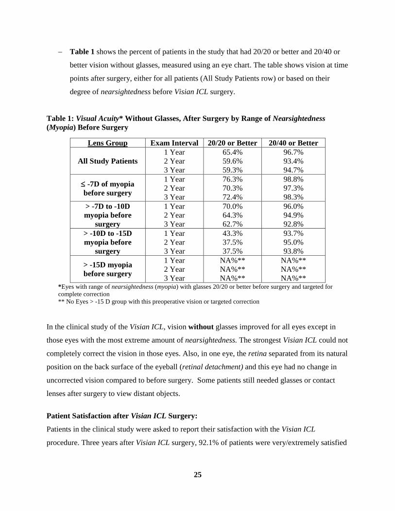
− Table 1 shows the percent of patients in the study that had 20/20 or better and 20/40 or
better vision without glasses, measured using an eye chart. The table shows vision at time
points after surgery, either for all patients (All Study Patients row) or based on their
degree of nearsightedness before Visian ICL surgery.
Table 1: Visual Acuity* Without Glasses, After Surgery by Range of Nearsightedness (Myopia) Before Surgery
Lens Group Exam Interval 20/20 or Better 20/40 or Better
All Study Patients 1 Year 2 Year 3 Year
65.4% 59.6% 59.3%
96.7% 93.4% 94.7%
≤ -7D of myopia before surgery
1 Year 2 Year 3 Year
76.3% 70.3% 72.4%
98.8% 97.3% 98.3%
> -7D to -10D myopia before
surgery
1 Year 2 Year 3 Year
70.0% 64.3% 62.7%
96.0% 94.9% 92.8%
> -10D to -15D myopia before
surgery
1 Year 2 Year 3 Year
43.3% 37.5% 37.5%
93.7% 95.0% 93.8%
> -15D myopia before surgery
1 Year 2 Year 3 Year
NA%** NA%** NA%**
NA%** NA%** NA%**
*Eyes with range of nearsightedness (myopia) with glasses 20/20 or better before surgery and targeted for complete correction ** No Eyes > -15 D group with this preoperative vision or targeted correction
In the clinical study of the Visian ICL, vision without glasses improved for all eyes except in
those eyes with the most extreme amount of nearsightedness. The strongest Visian ICL could not
completely correct the vision in those eyes. Also, in one eye, the retina separated from its natural
position on the back surface of the eyeball (retinal detachment) and this eye had no change in
uncorrected vision compared to before surgery. Some patients still needed glasses or contact
lenses after surgery to view distant objects.
Patient Satisfaction after Visian ICL Surgery:
Patients in the clinical study were asked to report their satisfaction with the Visian ICL
procedure. Three years after Visian ICL surgery, 92.1% of patients were very/extremely satisfied
26
and 7.3% were moderately/fairly satisfied with their vision. Only 0.6% of patients were
unsatisfied.
Quality of Vision after Visian ICL Surgery:
Quality of vision reported by patients as very good/excellent improved from 55% before the
Visian ICL to 77% at 3 years after the Visian ICL procedure. Patients reporting poor/very poor
vision dropped in half at 3 years (5.8%) compared to before the Visian ICL (11.6%).
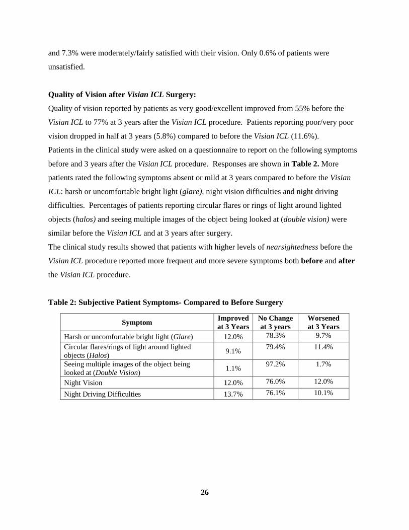
Patients in the clinical study were asked on a questionnaire to report on the following symptoms
before and 3 years after the Visian ICL procedure. Responses are shown in Table 2. More
patients rated the following symptoms absent or mild at 3 years compared to before the Visian
ICL: harsh or uncomfortable bright light (glare), night vision difficulties and night driving
difficulties. Percentages of patients reporting circular flares or rings of light around lighted
objects (halos) and seeing multiple images of the object being looked at (double vision) were
similar before the Visian ICL and at 3 years after surgery.
The clinical study results showed that patients with higher levels of nearsightedness before the
Visian ICL procedure reported more frequent and more severe symptoms both before and after
the Visian ICL procedure.
Table 2: Subjective Patient Symptoms- Compared to Before Surgery
Symptom Improved at 3 Years
No Change at 3 years
Worsened at 3 Years
Harsh or uncomfortable bright light (Glare) 12.0% 78.3% 9.7% Circular flares/rings of light around lighted objects (Halos) 9.1% 79.4% 11.4%
Seeing multiple images of the object being looked at (Double Vision) 1.1% 97.2% 1.7%
Night Vision 12.0% 76.0% 12.0%
Night Driving Difficulties 13.7% 76.1% 10.1%
27
15.0 Adverse Events Observed In The Visian ICL Clinical Studies Patients in the Visian ICL clinical study were followed for 36 months (3 years) after surgery in
the first phase of the study and up to 60 months (5 years) or longer in the second phase of the
study. The safety follow-up of study patients included the following events:
• Additional surgeries;
• Cataract formation;
• Loss of best corrected vision with eyeglasses (BCVA)
• Raised pressure inside the eye (intraocular pressure) and damage to the optic nerve
caused by increased pressure in the eye (glaucoma);
• Loss of cells on the innermost surface of the cornea (endothelial cells);
• Other complications.
Additional (Secondary) Surgery
A total of 8.2% of eyes in the FDA clinical study had a secondary surgery to change the position
of, remove or replace the Visian ICL, or to treat raised pressure inside the eye (intraocular
pressure or IOP).
A second surgery to change the position of the Visian ICL was done in 0.8% of eyes
while 1.5% of eyes had a second surgery to replace the Visian ICL, and 1.9% of eyes had
a second surgery to remove the Visian ICL. One eye (0.2%), had both a replacement and
removal of the Visian ICL. In all cases, the reason for Visian ICL removal was associated
with cataract surgery.
A second surgery to treat raised pressure inside the eye was done in (3.8%) of eyes in the
clinical study. Of these, 3.2% of eyes underwent an additional YAG laser treatment. In
the other 0.6% of eyes, the surgeon had to perform another surgery to remove the
remaining gel-like fluid used during eye surgery (viscoelastic fluid).
Cataract Formation
A cataract is a clouding of the natural lens inside the eye that can decrease vision. Because the
Visian ICL is placed inside the eye near the natural lens, there is a risk of developing a cataract.
Long-term follow up of patients in the FDA clinical study suggests that older age and higher

28
levels of nearsightedness increase the risk for cataract after Visian ICL surgery. Cataracts that
affect vision may require surgery to remove the cataract. In this case, both the Visian ICL and
the cataract is removed and another intraocular lens is implanted, just as is done in any routine
cataract surgical procedure. In the FDA clinical study, 45 eyes developed some form of
cataract, out of 526 eyes implanted with the Visian ICL, with 334 eyes followed for 5 – 7 years.
Loss of best corrected vision with eyeglasses (BCVA)
Eighteen eyes of 16 patients in the study lost vision of two or more lines as measured on an eye
chart. The most common reason was clouding of the natural lens (cataract). Vision got better in
these eyes after cataract surgery. In other eyes, vision improved without treatment. In 7 of the
18 eyes, however, vision did not get better after 5 or more years.
Raised intraocular pressure (IOP) and glaucoma development
Normal eye pressure (intraocular pressure or IOP) can vary, but is often considered to be from
10-21 millimeters of mercury (mmHg). An IOP higher than normal is called ocular hypertension
and if left untreated, can cause damage to the optic nerve (glaucoma) and cause permanent vision
loss. Patients with high levels of nearsightedness are also at increased risk of developing
glaucoma.
In the early days after surgery, an increase in eye pressure requiring treatment beyond just the
use of medicine was reported for 3.8% of eyes in the FDA clinical study of the Visian ICL, most
often at 1 to 2 days after surgery. In most of these eyes, another hole was placed in the extreme
outer edge of the colored portion of the eye (peripheral iridotomy) to reduce the pressure.
An increase in IOP to values greater than 10mmHg higher than before Visian ICL surgery or to
higher than 25mmHg was reported in 2.7% of eyes through 5 years or more after surgery. A total
of 7 eyes in 4 patients developed damage to the optic nerve (glaucoma) through 5 or more years
after ICL surgery. The first case of glaucoma was diagnosed at 5 months after Visian ICL
surgery and the last case happened at over 6 years (73 months) after surgery.
Endothelial Cell Loss
A thin, single layer of cells on the innermost surface of the cornea (endothelial cells) keep the
cornea clear by pumping water out of the cornea. Normally, these cells slowly decrease in
29
number as you age. Additional loss of these cells beyond the normal amount can occur following
many kinds of eye surgery. If too many cells are lost, the cornea can become cloudy, which can
cause decreased vision.
Loss of endothelial cells has been reported after surgery to implant the Visian ICL. Before your
surgery, your doctor will evaluate the health of your corneal endothelium to aid in determining
whether you are a candidate for Visian ICL surgery. It is important to note that while most
patients in the study reported no significant endothelial cell loss, 13 eyes of 10 patients reported
more than 30% endothelial cell loss (11.3% of those available for evaluation 5 years or more
after surgery). Three of the 13 eyes reported this loss within the first year after surgery. The
timing of the loss for these 3 eyes suggests that the loss may have been caused by the surgical
procedure used to insert the Visian ICL into the eye. The remaining 10 eyes had endothelial cell
loss 5 years or more after surgery.
Other Complications
One case each of the following complications were reported in the Visian ICL FDA study:
• movement of the colored portion of the eye (iris) through a surgical wound to a position
outside the eye (iris prolapse) at 1 day after surgery,
• bleeding in the area on the retina responsible for reading vision (macular hemorrhage) at
1 week after surgery and,
• bleeding under the retina (subretinal hemorrhage) at 3 months after surgery.
A >2 diopter increase in focusing error (astigmatism) caused by a change in the curvature of the
cornea was seen in 2 eyes at 3 years after Visian ICL surgery. Lifting or pulling of the retina
from its natural position (retinal detachment) was reported in 3 eyes at 4, 22 and 31 months after
Visian ICL implantation.
A case of unequal pupil size (anisocoria) was reported for a patient implanted with an ICL in
another clinical study.
Potential complications are not limited to those reported during the clinical study. The following
represent potential complications/adverse events reported with refractive surgery in general:
• An irritation of the white portion of the eyeball and inner eyelid (conjunctival irritation)
30
• Severe abnormal fluid build-up/swelling in the cornea (acute corneal swelling). The
condition is usually temporary after surgery with no significant effect on vision.
• Continuing abnormal fluid build-up/swelling in the cornea (persistent corneal swelling).
Persistent corneal swelling may cause a loss of vision.
• Total eye infection (endophthalmitis)
• Significant harsh or uncomfortable bright light (glare) or circular flares or rings of light
that may appear around a headlight or other lighted object (halos). These are usually
caused by a distortion of light that would normally be tolerable without the distortion.
• Blood in the eye (hyphema),
• Discharge in the eye (hypopyon),
• Eye infection,
• Visian ICL dislocation,
• Cyst-like swelling with of the center of the retina with fluid (cystoid macular edema),
• A condition where the colored portion of the eye does not get larger or smaller when light
is shined in the eye (non-reactive pupil),
• The inability of fluid to flow from the back chamber of the eye to the front chamber
frequently blocking drainage of fluid out of the eye and raising the pressure in the eye
(pupillary block glaucoma),
• Severe inflammation of the eye,
• Inflammation in the front part of the eye (iritis),
• Inflammation in the middle layer of tissue in the eye (uveitis),
• Loss of clear gel-like material from the farthest back chamber of the eye during surgery
(vitreous loss) and,
• Removal and replacement of the cornea (corneal transplant).
16.0 Summary of Important Information
• Visian ICL Surgery provides a permanent correction of your nearsightedness as long as
the Visian ICL remains in the eye. The Visian ICL may be removed. If your physician
removes the Visian ICL, you will lose the benefit of your nearsightedness correction. This
means that your vision may not return to what it was like before the Visian ICL surgery.
31
• Visian ICL surgery does not eliminate the need for reading glasses, even if you have
never worn them before.
• Your vision must be stable before Visian ICL surgery. You must provide written
evidence that your nearsightedness has changed no more than 0.50 D each year for at
least 1 year.
• Pregnant and nursing women should wait until they are not pregnant and not nursing to
have the Visian ICL surgery.
• Visian ICL surgery has some risks. Please read and understand this entire booklet before
you agree to the surgery. The sections on Risks (Section 6.0), Warnings (Section 8.0)
and Precautions (Section 9.0) are especially important to read carefully.
• Some other options to correct nearsightedness include glasses, contact lenses, RK, PRK
and LASIK.
• Before considering Visian ICL surgery you should:
a. have a complete eye examination.
b. talk with at least one eye care professional about Visian ICL surgery, especially
the potential benefits, risks, and complications. You should discuss the time
needed for healing after surgery.
• Certain eye diseases, eye conditions, previous eye surgery, systemic medical conditions
may have an impact on the results after Visian ICL surgery. It is important that you
provide your doctor with your complete medical history so your doctor may determine if
you are a good candidate for the Visian ICL for correction of nearsightedness..
32
• The Visian ICL is intended to improve your vision. However, because you are
nearsighted, you should consult with your eye doctor on a regular basis (i.e., once a year)
to verify the overall health of your eye.
Answers to Self-Test Questions: 1. F 6. F 2. F 7. F 3. F 8. F 4. T 9. T 5. T 10. F
17.0 Patient Assistance Information
To be completed by you or your Primary Eye Care Professional as a reference.
Primary Eye Care Professional Name: Address: Phone: Visian ICL Doctor Name: Address: Phone: Treatment Location Name: Address: Phone:

33
Visian ICL Manufacturer and Distributor:
STAAR Surgical Company
1911 Walker Avenue Monrovia, CA 91016 USA
Tel: (800) 352-7842 FAX: (800) 952-4923
MKT-0125 Rev 2