walghazzawi.kau.edu.sawalghazzawi.kau.edu.sa/getfile.aspx?id=159313&lng=ar&fn=... · web...
TRANSCRIPT

King Abdulaziz UniversityFaculty of Science
Department of Biochemistry Girls Section
General Biochemistry Lab
BIOC 201
Organized by: Sharifa Al-Ghamdi& Huda Al-ShaibiPage 1

Table of Contents
Lab # Experiment name Page #
1 Laboratory Wares 3
2 TERMS USED IN BIOCHEMISTRY & CALCULATIONS 12
3 Definition of pH and buffer 21
4 COLORIMETRY & SPECTROPHOTOMETR 26
5 ProteinsQualitative Tests for Proteins
3439
8 Qualitative Tests for Amino Acids 42
9 CARBOHYDRATESQUALITATIVE TESTS FOR CARBOHYDRATES
48
58
11Lipids 70
Determination of acid value 72
13 Paper chromatography 75
14 SEPARATION OF AMINO ACIDS BY PAPER CHROMATOGRAPHY 80
Organized by: Sharifa Al-Ghamdi& Huda Al-ShaibiPage 2

Laboratory Wares
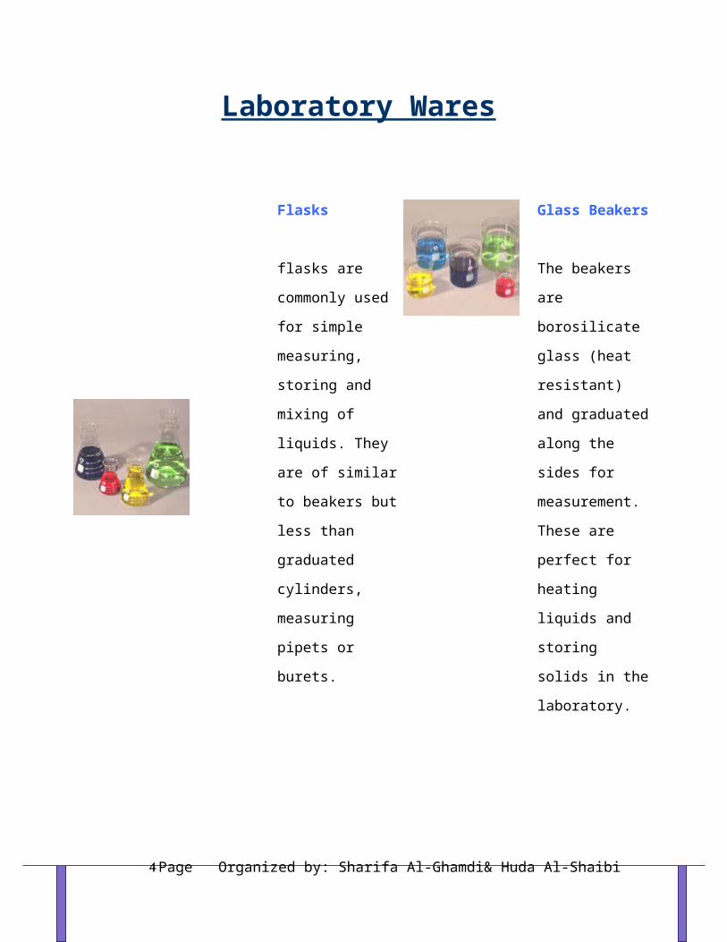
Flasks
flasks are commonly
used for simple
measuring, storing
and mixing of liquids.
They are of similar to
beakers but less than
graduated cylinders,
measuring pipets or
burets.
Glass Beakers
The beakers are
borosilicate glass
(heat resistant) and
graduated along the
sides for
measurement. These
are perfect for
heating liquids and
storing solids in the
laboratory.
Dropping Pipet
Used to transfer
liquids in qualitative
test. It does not allow
a accurately
measurement
Burets
Ground and finished
stopcocks for leak-
free operation. They
feature durable,
permanent
markings; fine,
sharp lines and
large, easy-to-read
numbers. Our
Burets meet ASTM
specifications.
Organized by: Sharifa Al-Ghamdi& Huda Al-ShaibiPage 3
Pipets
Pipets are used to
measure and transfer
small volumes (10
mL or less) of
liquids. Pipets are
long graduated tubes
that allow one to
accurately measure
and transfer small
volumes.
Pippet Bulbs
The orange pipette
bulb can be used
with the 10ml pipet.
The thumbwheel
pipetter is designed
for use with the 2ml
pipettes but will
also work with
small sizes. Neither
work with corrosive
liquids.
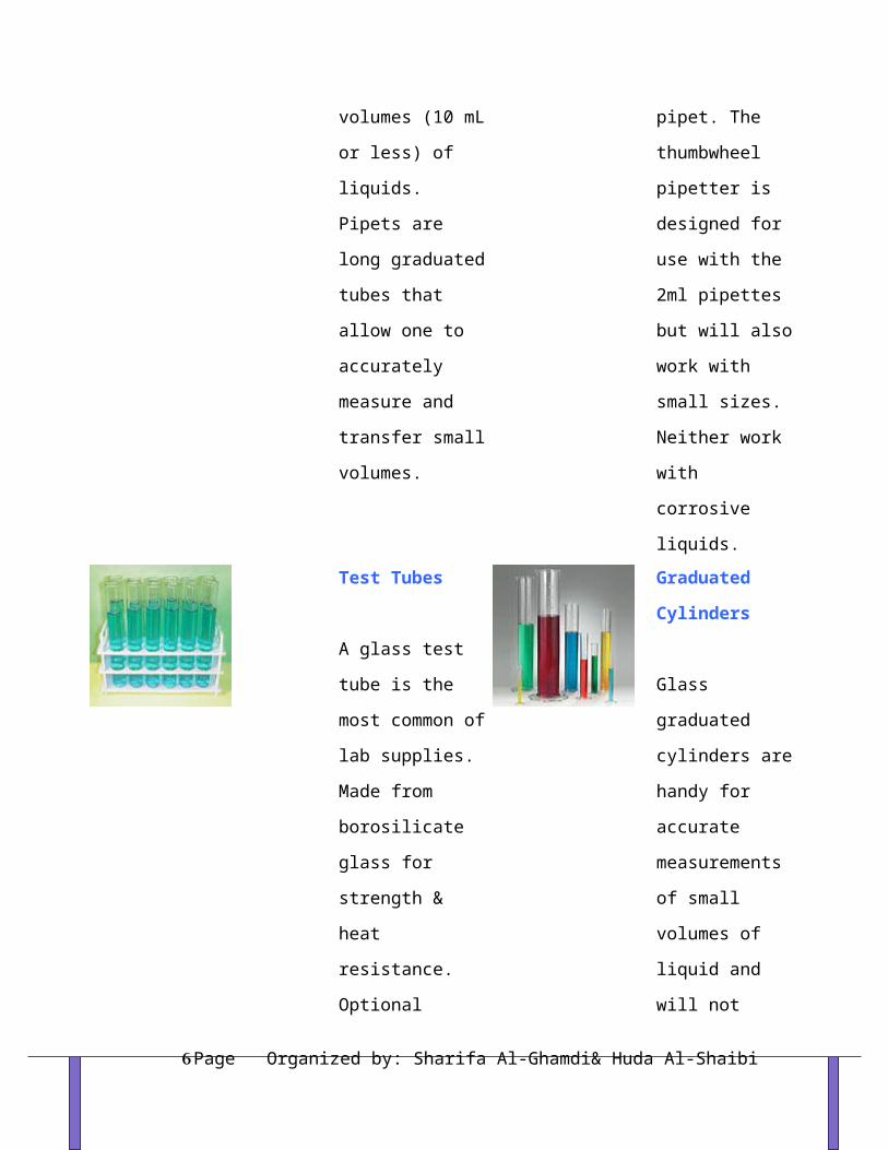
Test Tubes
A glass test tube is
the most common of
lab supplies. Made
from borosilicate
glass for strength &
heat resistance.
Optional marking
spot allows for pencil
notations.
Graduated
Cylinders
Glass graduated
cylinders are handy
for accurate
measurements of
small volumes of
liquid and will not
cloud if exposed to
materials such as
concentrated NaOH,
or any hydrocarbon
Organized by: Sharifa Al-Ghamdi& Huda Al-ShaibiPage 4

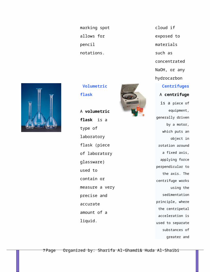
Volumetric flask
A volumetric flask
is a type of laboratory
flask (piece of
laboratory glassware)
used to contain or
measure a very
precise and accurate
amount of a liquid.
Centrifuges
A centrifuge is a piece of equipment,
generally driven by a
motor, which puts an
object in rotation around
a fixed axis, applying
force perpendicular to
the axis. The centrifuge
works using the
sedimentation principle,
where the centripetal
acceleration is used to
separate substances of
greater and lesser
density.
Spectrophotometer
A spectrophotometer
is an instrument
designed to detect the
amount of radiant
light energy absorbed
by molecules.
Hot Plates
hot plate provides
an efficient source
of infrared heat.
Most have solid
state push button
auto ignition and
heat output is fully
adjustable.
Organized by: Sharifa Al-Ghamdi& Huda Al-ShaibiPage 5
pH Meter
A pH meter a device
used for
potentiometric pH
measurements, which
measures essentially
the electro-chemical
potential between a
known liquid inside
the glass electrode
(membrane) and an
unknown liquid
outside.
Scale
an instrument or
machine for weighing.
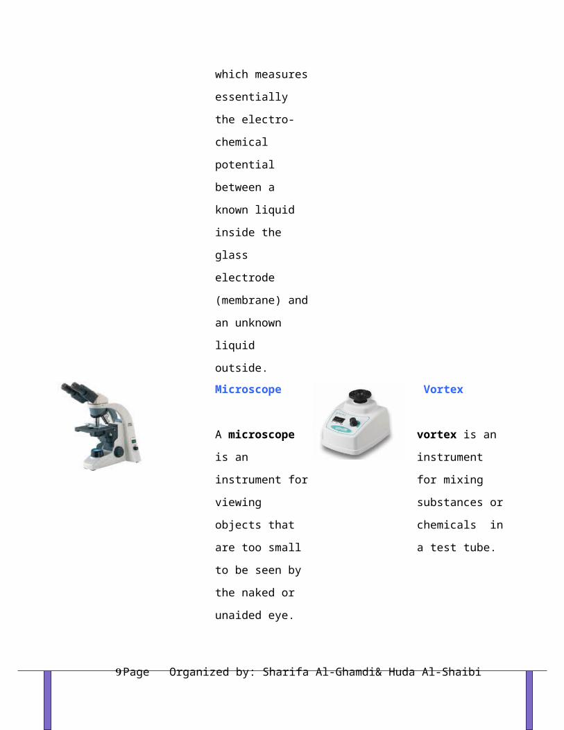
Microscope
A microscope is an
instrument for
viewing objects that
are too small to be
seen by the naked or
unaided eye.
Vortex
vortex is an
instrument for
mixing substances
or chemicals in a
test tube.
Pipets
There are several different types and sizes of pipets, which are used for slightly different
purposes. Be sure that you know how to identify the different types of pipets and that you
can determine the total volume and the gradations on each.
Organized by: Sharifa Al-Ghamdi& Huda Al-ShaibiPage 6
Types of Pipets
1- Volumetric or transfer pipettes are designed to deliver a single volume precisely (the
volume will be indicated near the top of the pipet (i.e., 2 mL).
2- Mohr or measuring pipets are graduated but stop at a baseline before the pipet begins
to narrow.
3- Serological pipets are graduated to deliver (there is no base mark).
The appropriate amount of fluid is drawn into the pipet (with the meniscus precisely on
the correct mark) and the entire amount is transferred.
4- Mechanical
Most mechanical pipets can be set to draw and dispense different volumes. They are
usually set by turn the knurled nob near the top. The volume is read in the window.
Mechanical pipets are operated by depressing the plunger. On the downward stroke of the
plunger there are two stops. The first offers firm resistance, and the second is a hard stop.
To take up a volume in the pipet, place a tip on the end of the pipet. Depress the plunger
to the first stop and insert into the sample to be transferred. Draw the liquid into the pipet
by slowly releasing the plunger. To dispense the liquid from the pipet, place the tip of the
pipet into the opening of the well and slowly depress the plunger all the way to the
second stop. When the liquid has been dispensed withdraw the pipet tip from the well
before releasing the plunger.
Reference :www.biology.lsu.edu/.../1208_pipet.php
Organized by: Sharifa Al-Ghamdi& Huda Al-ShaibiPage 7

CALIBRATION OF VOLUMETRIC EQUIPMENT:
In any experiment, you must ascertain the limits and range of your
measuring instruments.
Buret
1. Calibrating the Buret. Use de-ionized water for all operations. Check your buret for
leakage and drainage time.
Pipet
1. Calibrating the Pipet. Just as in the case of the buret, check your pipet for proper
drainage time. Fill it to the mark with de-ionized water and observe the time for it
to empty while it is held vertically.
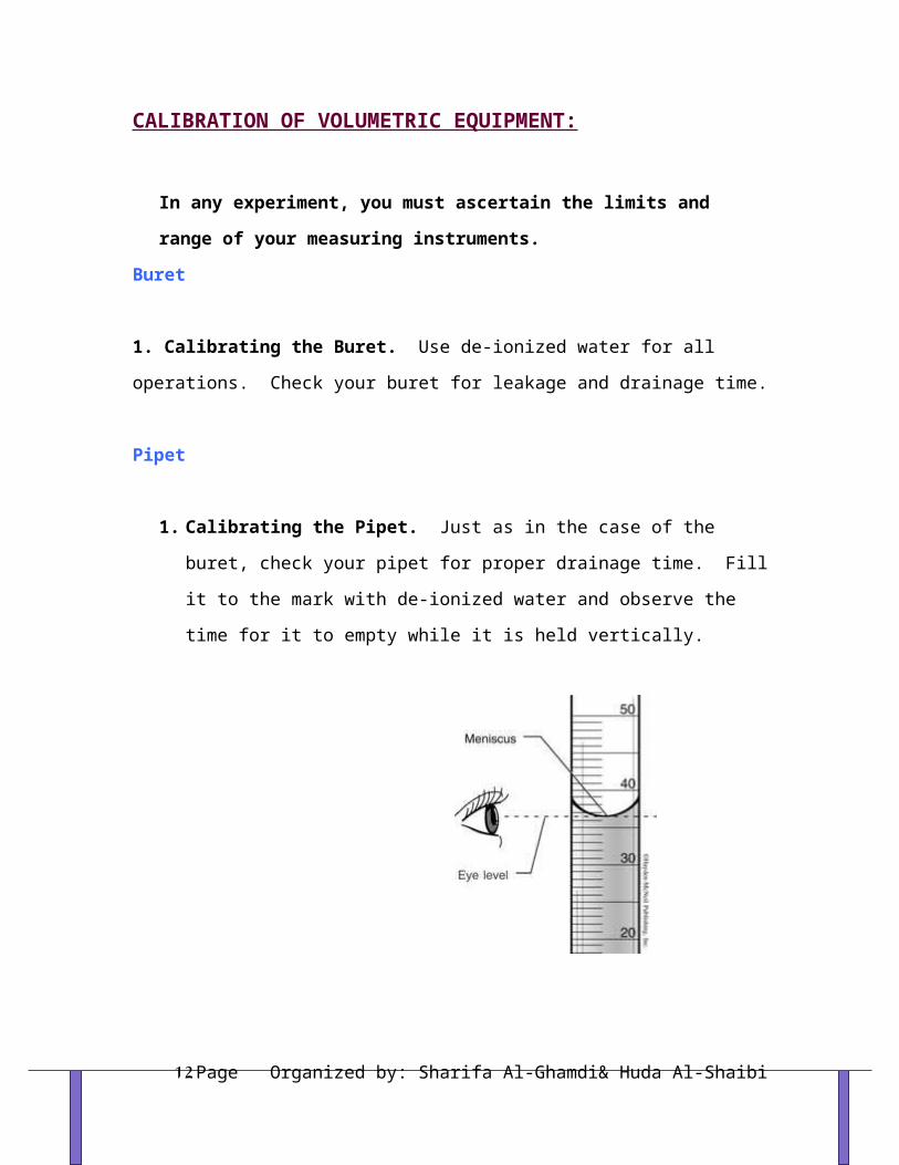
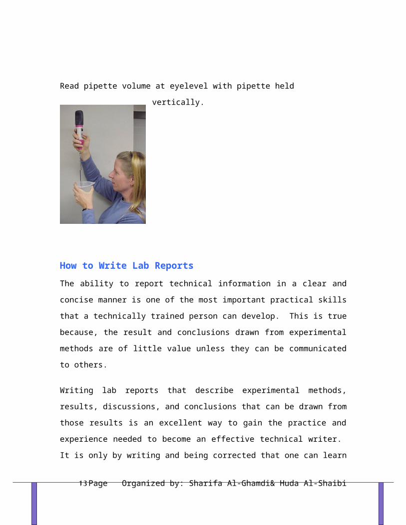
Read pipette volume at eyelevel with pipette held vertically.
Organized by: Sharifa Al-Ghamdi& Huda Al-ShaibiPage 8
How to Write Lab ReportsThe ability to report technical information in a clear and concise manner is one of the
most important practical skills that a technically trained person can develop. This is true
because, the result and conclusions drawn from experimental methods are of little value
unless they can be communicated to others.
Writing lab reports that describe experimental methods, results, discussions, and
conclusions that can be drawn from those results is an excellent way to gain the practice
and experience needed to become an effective technical writer. It is only by writing and
being corrected that one can learn to write. A beginner will find it helpful to follow a
certain format for his or her reports. This will help ensure that the report is complete and
well organized. Written lab reports should consist of the following parts:
Title Page
This should be the first (cover) page of the report. When writing the title page of a lab
report, the following should be included:
1. The title of the experiment.
2. The students name in full.
3. The instructor or person for whom the lab report is being compiled.
4. The date on which the experiment was performed or the date the lab report was
written.
Introduction Page
Organized by: Sharifa Al-Ghamdi& Huda Al-ShaibiPage 9

Under this heading should be an overview of what the experiment was about. A sound
definition of what was learned about the process being carried out during the experiment
should be included.
Materials and Methods
This section should contain a description, in the students own words, of the experimental
procedure that was followed in the performance of the experiment. The materials and
methods section should be complete enough so that another student with the same
background, but unfamiliar with the experiment, could perform the same experiment
without additional instructions. Procedures and equipment used should be written in a
sentence form. Do not list!
Results
The result section should contain raw data. Raw data consist of actual measured values
recorded during the experiment. Use tables to present this information. All tables should
have descriptive titles, and they should show the units of data entries clearly. The data
section should also contain any graphs that are required. This is an effective method for
communicating experimental results. The following steps should be taken into
consideration while plotting a graph:
1. Do not use tiny dots, use symbols like X or O.
2. Do not draw a series of straight line segments between experimental data points
plotted on
a graph. The purpose of many of the experiments is to verify theoretical relationships
between variables.
3. All graphs should have descriptive titles. These titles should tell what the graph is
intended
to show. Each axis of a graph should be labeled with the variable and unit it
represents.
Always use graph paper and always label graph coordinate lines so that it is easy to
see how
Organized by: Sharifa Al-Ghamdi& Huda Al-ShaibiPage 10

many units each division represents.
Discussions and Conclusions
This is the interpretation-and-conclusion of your report. This section should include the
following:
1. How the conduct of the experiment met the objectives.
2. What took place during the process.
3. All questions should be answered within this section in a very logical and clear
manner.
The questions should be put into statement form.
4. The conclusions should be relevant to the experiment that was performed and should
be
based on facts learned as a result of the experiment.
5. You should also include any recommendations that you feel would improve the
experimental
procedure. If you have any further investigations that might be suggested by the data,
you should also include them here.
References:
http://water.me.vccs.edu/reports.htm
Organized by: Sharifa Al-Ghamdi& Huda Al-ShaibiPage 11
TERMS USED IN BIOCHEMISTRY & CALCULATIONS
Tips to Success in Biochemistry Lab:
1. Write down a clear plan before you start working.
2. Keep very careful notes so you can recheck what you did if you get unexpected results.
3. Label all tubes and samples so they don't get mixed up.
4. Work slowly and carefully. Accurate, reproducible results in a biochemical test like this
requires care on your part. If you are sloppy or careless your results will suffer.
Terms used in biochemistry lab:
Liter-------volume unit.
1L = 1000ml = 106 μl (micro liter).
Gram--------mass unit (weight).
1g = 1000mg = 106 μg
Units of Concentrations:
I- Mass unit / volume unit:- Example: mg/ml, mg/l, g/l, g/dl, mg%, etc.
- To convert from:
g>>>>>mg >>>>X 1000
mg>>>>g >>>> / 1000
g >>>> μg >>>> X 1000000
Organized by: Sharifa Al-Ghamdi& Huda Al-ShaibiPage 12
mg>>>> μg >>>> X1000
L>>>>>ml >>> X 1000
L>>>> µl >>>> X 1000000
ml>>>> µl >>>> X 1000
dl >>> ml >>>>> X 100
Problem:Convert 0.2 mg/ml to
- g/l
- g/dl
- mg%
- mg/ µl
- g/ µl
II- Molarity (M):
- Another way of expressing concentration is called molarity. Molarity is the number of
moles of solute dissolved in one liter of solution. The units, therefore are moles per liter,
specifically it's moles of solute per liter of solution.
Molarity = moles of solute
liter of solution
- Rather than writing out moles per liter, these units are abbreviated as M or M.
M = wt X 1000
MW X Vml
- To convert from mg% to mmol/l
mmol/l = mg X 10
Organized by: Sharifa Al-Ghamdi& Huda Al-ShaibiPage 13

MWsubstance
III- Normality (N):
- When you need to compare solutions on the basis of concentration of specific ions or the
amount of charge that the ions have, a different measure of concentration can be very useful.
It is called normality.
- No. of equivalent weight per liter of solution.
Eq.wt = molecular weight (MW)
Valance
- N = wt X 1000
Eq.wt X Vml
Problem:
1- How many grams of glucose are needed to make 100 ml of a 0.6 mol/l solution? (MW glucose
= 180).
2- How can you prepare 0.1 M NaOH solution?
3- Describe the preparation of 5 L of 0.1 M Na2CO3 (MW = 105.99) from the primary
standard solid.
Balance Rules and Instructions:The Figure below illustrates one type of top loading electronic balance. Refer to this
figure when following the steps and precautions for using the balance listed below:
1. Never pour or transfer chemicals over the balance. Spilled chemicals can damage the
balances, which are very expensive to repair or replace. Never weigh warm or hot
objects; if you can feel any heat, the weighing will not be accurate. Always use a container
such as a vial, beaker, flask, or watch glass to weigh a solid or liquid chemical on the
balance to protect the balance pan.
Organized by: Sharifa Al-Ghamdi& Huda Al-ShaibiPage 14

2. Make sure your hands are clean and dry before you handle containers or objects that
are to be weighed. The outside of these containers or objects must also be clean and dry.
Clean up any spills on the balance pan or lab bench around the balance immediately.
3. First open or remove the draft lid or cover (if there is one) and check to make sure
that the balance pan is clean. If the pan is dirty, have your TA show you how to clean it
and gently place it back on the balance.
4. Close or put the draft lid back on the balance and zero the balance by pressing the
"T" or "on/tare" button (re-zero bar on Mettler balances). Wait 5-10 seconds for the
weight display to stabilize. (If the object to be weighed is so large that the draft lid can't
be used, do this step without the draft lid in place.)
5. Open or remove the draft lid and place the object to be weighed on the balance pan.
Then close or place the draft lid back on the balance. (As long as it does not touch the
object to be weighed, leave the lid off if it does touch the object.) After 5-10 seconds the
weight display will stabilize and you can record the mass to ±0.001 g.
6. Never unplug the balance.
Top loading electronic balance
Organized by: Sharifa Al-Ghamdi& Huda Al-ShaibiPage 15

IV- Percent Concentration (%):
1-Volume percent is usually used when the solution is made by mixing two liquids.
- The use of percentages is a common way of expressing the concentration of a solution.
- The percentages can be calculated using volumes as well as weights, or even both
together.
- One way of expressing concentrations is by volume percent. Another is by weight
percent. Still another is a hybrid called weight/volume percent.
Volume percent (v/v) = volume of solute X 100
volume of solution
Example:
Rubbing alcohol is generally 70% by volume isopropyl alcohol. That means that 100 ml
of solution contains 70 ml of isopropyl alcohol. That also means that a liter (or 1000 ml)
of this solution has 700 ml of isopropyl alcohol plus enough water to bring it up a total
volume of 1 liter, or 1000 ml.
2- Weight percent: is expressing the concentration of a solution in weight percent (or
mass percent).
Organized by: Sharifa Al-Ghamdi& Huda Al-ShaibiPage 16

Weight percent (w/w) = weight of solute X 100
weight of solution
Question:
What is the weight percent of glucose in a solution made by dissolving 4.6 g of glucose
in 145.2 g of water?
Analysis:
To get weight percent we need the weight of the solute and the total weight of the
solution.
Determine total weight of solution: 4.6 g+ 145.2 g = 149.8 g
Calculate percent:
Weight % glucose = 4.6 g glucose x 100 = 3.1% glucose
149.8 g solution
3- Weight- volume percent: Another variation on percentage concentration is
weight/volume percent or mass/volume percent. This variation measures the amount of
solute in grams but measures the amount of solution in milliliters. An example would be
a 5 %( w/v) NaCl solution. It contains 5 g of NaCl for every 100 mL of solution.
Weight-Volume percent (w/v) = weight of solute X 100
volume of solution
Dilution:
- Diluted solutions can be prepared from concentrated solutions
M conc X V conc = M dil X V dil
Organized by: Sharifa Al-Ghamdi& Huda Al-ShaibiPage 17

Moles taken from concentrated solution = Moles placed in diluted solution
Problem:How can we prepare 100 ml of 0.04M K2Cr2O7 from 0.2M K2Cr2O7?
Serial Dilution:
- This technique involves the removal of a small amount of an original solution to another
container which is then brought up to the original volume using the required buffer or water.
In the example below, if you have 1 mL of your original solution and you remove 10 µL and
place it in a tube containing 990 µL of water or media you have made a 1:100 dilution.
Here is an example of how to do a series of serial dilutions:
1 ml extract + 4 ml water = 1/5 dilution
1 ml 1/5 dilution + 4 ml water = 1/25 dilution
1 ml 1/25 dilution + 4 ml water = 1/125 dilution
Organized by: Sharifa Al-Ghamdi& Huda Al-ShaibiPage 18

Dilution factor = Final volume
Initial volume
Standard Curve:
- A standard curve is a quantitative research tool, a method of plotting assay data that
is used to determine the concentration of a substance.
- The assay is first performed with various known concentrations of a substance similar
to that being measured. For example a standard curve for protein concentration is
often created using known concentrations of bovine serum albumin.
- The assay procedure may measure absorbance, optical density, luminescence,
fluorescence, radioactivity, or something else.
- This data is used to make the standard curve, plotting concentration on the X axis,
and assay measurement on the Y axis. The same assay is then performed with samples
of unknown concentration.
- To analyze the data, one locates the measurement on the Y-axis that corresponds to
the assay measurement of the unknown substance and follows a line to intersect the
standard curve.
- The corresponding value on the X-axis is the concentration of substance in the
unknown sample.
- Here is an example of how construct a standard curve.
Organized by: Sharifa Al-Ghamdi& Huda Al-ShaibiPage 19

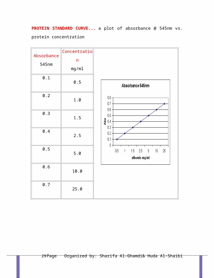
PROTEIN STANDARD CURVE... a plot of absorbance @ 545nm vs. protein concentration
Absorbance 545nm
Concentrationmg/ml
0.10.5
0.21.0
0.31.5
0.42.5
0.55.0
0.610.0
0.725.0
Organized by: Sharifa Al-Ghamdi& Huda Al-ShaibiPage 20
Definition of pH and buffer
pH:- is a measure of acidity and the alkalinity of a solution in terms of hydrogen ion H+ or (hydronium ion concentration) pH= -log[H3O+]
Thus, it is evident that the pH is inversely proportional to the acidity. Lower the pH, higher the acidity or hydrogen ion concentration while higher the pH, the acidity is lower.Just as pH is convenient way to represent the concentration of H3O+ pOH is convenient way to express the concentration of OH-.
pOH= -log[OH-]A solution is acidic if its pH is less than 7 A solution is basic if its pH is grater than 7 (base is any substance that accept H+) A solution is neutral if its pH is equal to 7pH value of some common materials
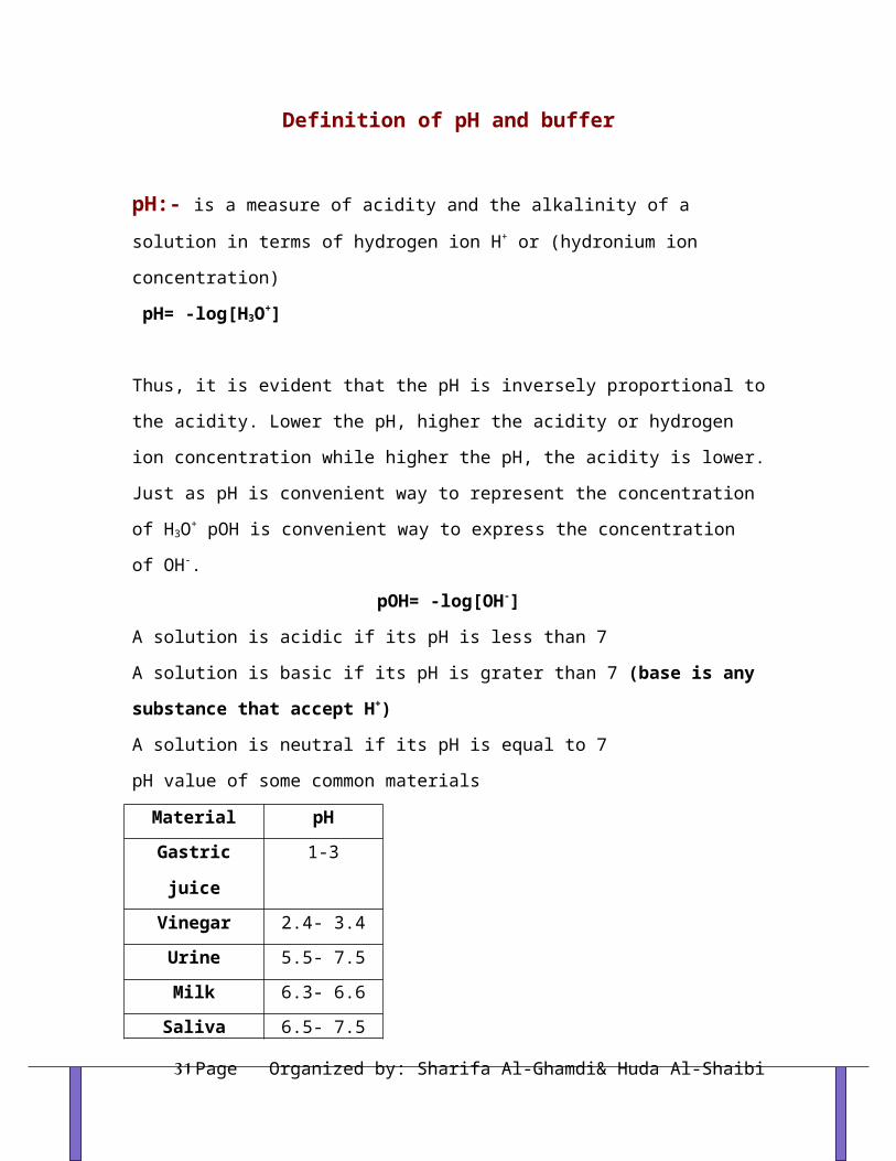
Material pHGastric juice 1-3
Vinegar 2.4- 3.4Urine 5.5- 7.5Milk 6.3- 6.6
Saliva 6.5- 7.5Pure water 7
Blood 7.35- 7.45Sea water 8- 9ammonia 11.7
Organized by: Sharifa Al-Ghamdi& Huda Al-ShaibiPage 21

There are tow ways to measure the pH:1- By using a pH paper which is a plane paper soaked with mixture of
pH indicator.
Some acid-base indicators.
2- By using pH meter this method is more accurate and more precise.
Organized by: Sharifa Al-Ghamdi& Huda Al-ShaibiPage 22

Definition of buffer: Is a solution that resists change in pH when limited amounts of an acid or a base are added to it; buffers consist of weak acids + corresponding salts Or weak base + their salt.ApplicationsTheir resistance to changes in pH makes buffer solutions very useful for chemical manufacturing and essential for many biochemical processes. Buffer solutions are necessary to keep the right pH for enzymes in many organisms to work. Many enzymes work only under very precise conditions. Industrially, buffer solutions are used in fermentation processes and in setting the correct conditions for dyes used in coloring fabrics.The acid-base balance or pH of the body fluids is maintained by a closely regulated mechanism. This involves the body buffers, the respiratory system and the kidney.Selection of the buffer:The selection of a particular buffer for a given application is based on two consideration:
1- the desired pH2- chemical compatibility of the buffer components with the sample
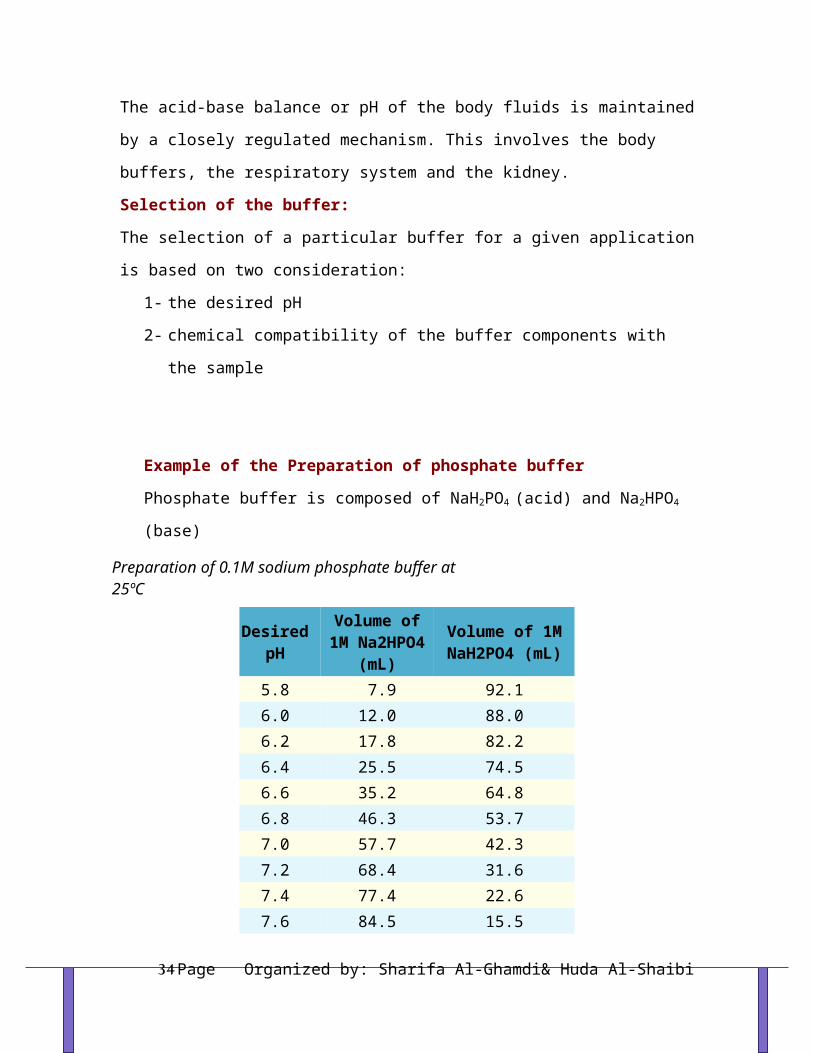
Example of the Preparation of phosphate buffer Phosphate buffer is composed of NaH2PO4 (acid) and Na2HPO4 (base)
Preparation of 0.1M sodium phosphate buffer at 25ºC
Desired pH
Volume of 1M
Na2HPO4 (mL)
Volume of 1M NaH2PO4 (mL)
Organized by: Sharifa Al-Ghamdi& Huda Al-ShaibiPage 23

5.8 7.9 92.1 6.0 12.0 88.0 6.2 17.8 82.26.4 25.5 74.5 6.6 35.2 64.8 6.8 46.3 53.77.0 57.7 42.3 7.2 68.4 31.67.4 77.4 22.67.6 84.5 15.57.8 89.6 10.48.0 93.2 6.8
References:MN Chatterjea, Text book of biochemistry. Jaypee.2004Bettelheim, FA, Introduction to general, organic and biochemistry.2007
Organized by: Sharifa Al-Ghamdi& Huda Al-ShaibiPage 24

RESULTS & LAB REPORT
Organized by: Sharifa Al-Ghamdi& Huda Al-ShaibiPage 25

COLORIMETRY & SPECTROPHOTOMETR
- Many biochemical experiments involve the measurements of compound or group of
compounds present in a complex mixture.
- The most widely used method for determining the concentration of biochemical
compounds is colorimetry, which makes use of the property that when white light passes
through a colored solution, some wavelength are absorbed more than others.
- Many compounds are not themselves colored but can be made to absorb light in visible
region by reaction with suitable reagents.
- These reactions are fairly specific and in most cases very sensitive, so that quantities of
material in the region of mM / L concentrations can be measured.
- The big advantage of is that complete isolation of compound is not necessary and the
constituents of a complex mixture such as blood can be determined after little treatment.
- The depth of the color is proportional to the concentration of the compound being
measured, while the amount of light is proportional to the intensity of the color and hence
the concentration.
Organized by: Sharifa Al-Ghamdi& Huda Al-ShaibiPage 26

Measurement of Extinction:- The earliest colorimeters relied on the human eye to match the color of a solution with
that of one of a series of colored discs. The results obtained were too subjective and not
particularly accurate.
The Colorimeter:
- Colorimeter is generally any tool that characterizes colour samples to provide an
objective measure of colour characteristics. In chemistry, the colorimeter is an apparatus
that allows the absorbance of a solution at a particular frequency (colour) of visual light to
be determined. Colorimeters hence make it possible to determine the concentration of a
known solute, since it is proportional to the absorbance.
- Different chemical substances absorb varying frequencies of the visible spectrum.
Colorimeters rely on the principle that the absorbance of a substance is proportional to its
concentration i.e., a more concentrated solution gives a higher absorbance reading.
- Filter in the colorimeter is used to select the color of light which the solute absorbs the
most, in order to maximize the accuracy of the experiment. Note that the colour of the
absorbed light is the 'opposite' of the colour of the specimen, so a blue filter would be
appropriate for an orange substance. Sensors measure the amount of light which has
Organized by: Sharifa Al-Ghamdi& Huda Al-ShaibiPage 27

passed through the solution, compared to the amount entering, and a display reads the
amount absorbed.
- A quantitative reading for the concentration of a substance can be found by making up a
series of solutions of known concentration of the chemical under study, and plotting a
graph of absorbance against concentration. By reading off the absorbance of the specimen
substance on the graph, a value for its concentration is found.
How colorimeter works?
1- White light from a tungsten lamp passes through a slit, then a condenser lens, to give a
parallel beam which falls on the solution under investigation contained in an absorption
cell or cuvette. The cell is made of glass with the sides facing the beam cut parallel to each
other.
2- Beyond the absorption cell is the filter, which is selected to allow maximum transmission
of the color absorbed. If a blue solution is under examination, then red is absorbed and a
red filter is selected.
NOTE: The color of the filter is complementary to the solution.
3- The light then falls on to a photocell which generates an electrical current in direct
proportion to the intensity of light falling on it.
4- This small electrical signal is increased by the amplifier which passes to a galvanometer
of digital readout to give absorbance reading directly.
Organized by: Sharifa Al-Ghamdi& Huda Al-ShaibiPage 28
-Among the simplest and most common colorimeters are the Spectronic 20 and Spectronic
21. They are commonly called the Spec 20 and Spec 21.
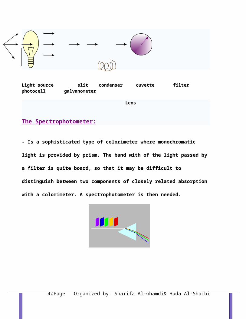
Light source slit condenser cuvette filter photocell galvanometer
Lens
The Spectrophotometer:
- Is a sophisticated type of colorimeter where monochromatic light is provided by prism.
The band with of the light passed by a filter is quite board, so that it may be difficult to
distinguish between two components of closely related absorption with a colorimeter. A
spectrophotometer is then needed.
Organized by: Sharifa Al-Ghamdi& Huda Al-ShaibiPage 29

- All types require a Blank: which is a solution that contains the entire reagents except the
substance to be measured. It is used to adjust the device to zero.
Here is a summary of the steps of operation of a Spec 20 & spectrophotometer:
1- Power >>>>>>>>Turn on power.
2-Warmup>>>>>>Allow about 5 minutes when first turned on.
3-Wavelength>>>>>Select appropriate wavelength.
4- Zero>>>>>With sample holder empty and closed, adjust meter needle to 0%T (or
infinite A) using zero control knob.
5- Blank >>>>Fill tube half full with water. Place in sample holder and close cover. Adjust
meter needle to 100%T (or 0 A) using light control knob.
6- Standard>>>>Measure absorbance (or %T) of known solution. Fill tube half full with
sample of known concentration. Place in sample holder and close cover. Read absorbance
value (or %T) from meter. Repeat this step if making a calibration curve or verifying
proportionality (Beer's Law).
7- Sample>>>>>Measure absorbance (or %T) of solution with unknown concentration as
in previous step.
Organized by: Sharifa Al-Ghamdi& Huda Al-ShaibiPage 30
Types of spectrophotometer:1- Visible spectrophotometer.
2- Ultraviolet (UV) spectrophotometer.
References:
www.wikipedia.org
Organized by: Sharifa Al-Ghamdi& Huda Al-ShaibiPage 31

COLORIMETRIC DETERMINATION OF VITAMIN B-12
- Vitamin B12 is water soluble vitamin.
- RDA= 2.4 µg /day for adult.
- It is found only in animal sources.
Functions:
1- Aids folic acid in synthesis of heme.
2- It prevents anemia.
3- Required for protein digestion and absorption.
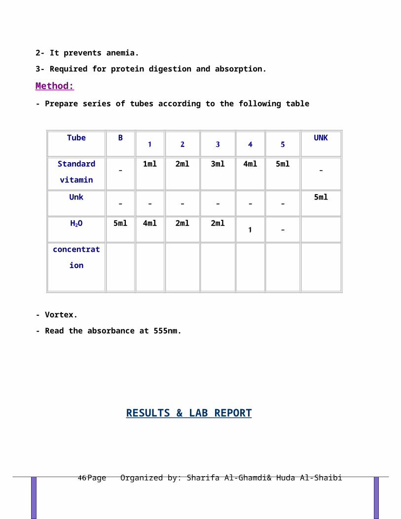
Method:- Prepare series of tubes according to the following table
Tube B 1 2 3 4 5 UNK
Standard
vitamin
- 1ml 2ml 3ml 4ml 5ml -
Unk - - - - - - 5ml
H2O 5ml 4ml 2ml 2ml 1 -
concentration
- Vortex.
- Read the absorbance at 555nm.
Organized by: Sharifa Al-Ghamdi& Huda Al-ShaibiPage 32

RESULTS & LAB REPORT
- You are supplied with the sample of unk vitamin.
- Plot the standard curve to calculate the concentration of unk.
- Calculate the concentration by using the equation>>>> Cunk = Aunk X C std
A std
Organized by: Sharifa Al-Ghamdi& Huda Al-ShaibiPage 33
PROTEINSIntroduction:
- Protein (from the Greek protas meaning "of primary importance") is a complex, high-
molecular-weight organic compound that consists of amino acids joined by peptide bonds.
- Proteins are natural polymer molecules consisting of amino acid units. The number of
amino acids in proteins may range from two to several thousand.
- Proteins are probably the most important class of biochemical molecules, although of
course lipids and carbohydrates are also essential for life. Proteins are the basis for the
major structural components of animal and human tissue.
- Proteins are essential to the structure and function of all living cells and viruses. Many
proteins are enzymes or subunits of enzymes, catalyzing chemical reactions. Other proteins
play structural or mechanical roles, such as those that form the struts and joints of the
cytoskeleton, serving as biological scaffolds for the mechanical integrity and tissue
signaling functions.
- Proteins can be hydrolyzed by acids, bases or specific enzymes.
Primary Protein Structure:
- Proteins are biopolymers built from 20 different L-alpha-amino acids.
- The two ends of the amino acid chain are referred to as the carboxy terminus (C-
terminus) and the amino terminus (N-terminus) based on the nature of the free group on
each extremity.
- Biochemists refer to four distinct aspects of a protein's structure:
Organized by: Sharifa Al-Ghamdi& Huda Al-ShaibiPage 34

1- Primary structure: the amino acid sequence. In order to function properly, peptides and
proteins must have the correct sequence of amino acids.
2- Secondary structure: is the specific geometric shape caused by intramolecular and
intermolecular hydrogen bonding of amide groups. Some combinations of amino acids will
tend to form:
Alpha Helix : In the alpha helix, the polypeptide chain is coiled tightly in the fashion of a
spring. The "backbone" of the peptide forms the inner part of the coil while the side chains
extend outward from the coil. The helix is stabilized by hydrogen bonds between the >N-H
of one amino acid and the >C=O on the 4th amino acid away from it.
Beta Pleated Sheet : In this structure, individual protein chains are aligned side-by-side
with every other protein chain aligned in an opposite direction. The protein chains are held
together by intermolecular hydrogen bonding, that is hydrogen bonding between amide
groups of two separate chains. This intermolecular hydrogen bonding in the beta-pleated
sheet is in contrast to the intramolecular hydrogen bonding in the alpha-helix.
Organized by: Sharifa Al-Ghamdi& Huda Al-ShaibiPage 35

3- Tertiary structure: is the entire three-dimensional shape of the protein. This shape is
determined by the sequence of amino acids. The overall shape of a single protein molecule
primarily formed by hydrophobic interactions, but hydrogen bonds, ionic interactions, and
disulfide bonds are usually involved too.
4- Quaternary structure: the shape or structure that results from the union of more than
one protein molecule, usually called protein subunits in this context, which function as part
of the larger assembly or protein complex.
Organized by: Sharifa Al-Ghamdi& Huda Al-ShaibiPage 36

Denaturation of Proteins:
- Denaturation is the disruption of secondary, tertiary and quaternary structure of proteins
leading to loss of their biological activity.
- Proteins denature when they lose their three-dimensional structure - their chemical
conformation and thus their characteristic folded structure. Proteins may be denatured at
the secondary, tertiary and quaternary structural levels, but not at the primary structural
level.
- Denaturation may be caused by:
1- Physical factors such as heating.
2- Chemical factors such as strong acid or base.
Denaturated proteins are characterized by:
1-Loss of function: Most biological proteins lose their biological function when denatured,
for example, enzymes lose their catalytic activity.
2- They become less soluble. As a result, they are easily precipitated.
Organized by: Sharifa Al-Ghamdi& Huda Al-ShaibiPage 37
3- Reversibility and irreversibility: In many proteins (unlike egg whites), denaturation is
reversible (the proteins can regain their native state when the denaturing influence is
removed).
Organized by: Sharifa Al-Ghamdi& Huda Al-ShaibiPage 38
Qualitative Tests for Proteins
1- Biuret Test:- It is the general test for all proteins.
- Biuret reagent is dilute CuSO4 in strong alkaline medium.
- Alkaline CuSO4 reacts with all compounds containing 2 or more peptide bonds to give a
blue-violet color.
Method:
1 ml of biuret reagent + 1 ml of protein ……mix well>>>> blue-violet color.
2- Denaturation by heat and extreme pH:
- Extreme heating and pH (conc. acids) denature proteins leading to precipitation of
proteins.
Method:
3ml Protein >>>>>>BWB-10min >>>>>> ppt of protein.
3ml Protein >>>>>> drops conc.HCL >>>>>> ppt of protein.
3- Precipitation of proteins by heavy metals:
Organized by: Sharifa Al-Ghamdi& Huda Al-ShaibiPage 39

- Proteins are precipitated in alkaline medium with heavy metals due to the direct union of
cation (Cu++, Ag+, Ba++, Pb++) with anionic groups of proteins, which are formed in basic
medium.
- At alkaline pH 7 and above, proteins are usually negatively charged so the addition of
positively charged ions will neutralize this charge and the proteins come out of solution (i.e.
heavy metals combine with proteins forming insoluble metalloproteine).
Method:
Few drops of heavy metals + 2ml protein + few drops 10% NaOH>>>>ppt
4- Precipitation of proteins by acidic reagent: - Proteins are precipitated in acidic medium with some reagents such as TCA, picric acid
and tannic acid due to the direct union of the anionic group with the cationic groups of the
proteins, which are formed in acidic medium.
- These compounds carry large negative charges which neutralize the positively charged
protein to form insoluble salt complex with protein.
- The acidic reagents are therefore most effective at acidic medium where proteins are
positively charged.
Method:
Few drops of acidic reagent + 2ml protein >>>slowly add dilute NaOH and observe the
result as the pH increase.
3- Detection of Amino acids contents of Protein:Carry on all the experiments you have done in amino acids lab on proteins to detect the
amino acid content of each protein.
References:www.chemtopics.com
www.wikipedia.org
RESULTS & LAB REPORT
Organized by: Sharifa Al-Ghamdi& Huda Al-ShaibiPage 40

- Present your results in a good and full lab report.
Organized by: Sharifa Al-Ghamdi& Huda Al-ShaibiPage 41
Qualitative Tests for Amino Acids
There are a number of qualitative tests to detect the presence of amino acids and these are
largely dependent on the nature of R-group.
Experiment-1:Ninhydrin Reaction:-A color reaction given by amino acids and peptides on heating with the chemical ninhydrin. The
technique is widely used for the detection and quantitation (measurement) of amino acids and
peptides.
-Ninhydrin is a powerful oxidizing agent which reacts with all amino acids between pH 4-8 to
produce a purple colored-compound.
-The reaction is also given by primary amines and ammonia but without the liberation of Co2
-The amino acids proline and hydroxyproline also reacts but produce a yellow color.
Method:
1 ml AA + 1 ml NH---- heat in boiling WB for 5min-----Purple color.
Alpha-amino acid + 2 ninhydrin ---> CO2 + aldehyde + final complex (purple) + 3H2O
In summary, ninhydrin, which is originally yellow, reacts with amino acid and turns deep
purple. It is this purple color that is detected in this method.
Experiment-2: Xanthoproteic Reaction:- This reaction involves the nitration of benzene nucleus in alkaline medium. As a result AAs
that contain aromatic nucleus undergo this reaction.
Organized by: Sharifa Al-Ghamdi& Huda Al-ShaibiPage 42

- Aromatic AAs form yellow nitro derivative on heating with conc. nitric acid, the salts of these
derivatives are orange.
Phenylalanine Tryptophan Tyrosine
Method:
1 ml AA + 1 ml conc. HNO3----- heat the mixture in WB for 30s--cool--add drop-wise
40% NaOH to render the solution alkaline--- Yellow to orange color.
Organized by: Sharifa Al-Ghamdi& Huda Al-ShaibiPage 43

Nitrated tyrosine (a) and tryptophan (b)
Experiment-3: Millon Reaction:- This reaction is used to detect the presence of phenol (hydroxybenzene) which reacts with
Millon's reagent to form red complexes.
- The only phenolic AA is tyrosine.
Tyrosine
Method:
1 ml AA + 5 drops of Millon reagent ----- heat the mixture in BWB for 10min--cool too
room temp--add 5 drops of NaNO2---Brick red color.
Experiment-4: Hopkin-cole Reaction:- This reaction is used to detect the presence of indol group
- The indol group of tryptophan reacts with glyoxalic acid in the presence of conc. H2SO4 to
give purple color.
Organized by: Sharifa Al-Ghamdi& Huda Al-ShaibiPage 44

- The reagent is glyoxalic acid in conc. H2SO4 (Glacial acetic acid which has been exposed to
light contains glyoxalic acid).
Tryptophan
Method:
1 ml AA + 1 ml Hopkin-cole reagent -----mix well--Carefully pour conc. H2SO4 down the
side of the tube so as to form two layers --Purple ring at the interface.
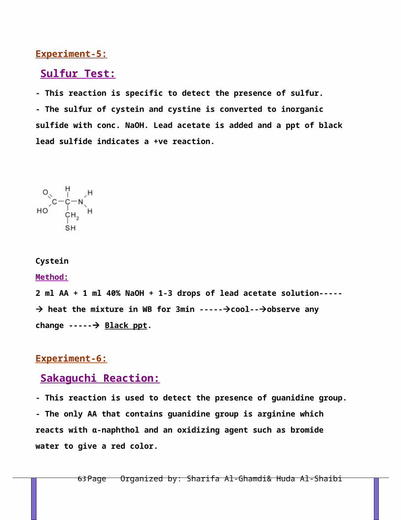
Experiment-5: Sulfur Test:- This reaction is specific to detect the presence of sulfur.
- The sulfur of cystein and cystine is converted to inorganic sulfide with conc. NaOH. Lead
acetate is added and a ppt of black lead sulfide indicates a +ve reaction.
Cystein
Method:
Organized by: Sharifa Al-Ghamdi& Huda Al-ShaibiPage 45
2 ml AA + 1 ml 40% NaOH + 1-3 drops of lead acetate solution----- heat the mixture in
WB for 3min -----cool--observe any change ----- Black ppt.
Experiment-6: Sakaguchi Reaction:- This reaction is used to detect the presence of guanidine group.
- The only AA that contains guanidine group is arginine which reacts with α-naphthol and an
oxidizing agent such as bromide water to give a red color.
Arginine
Method:
2 ml AA + 1 ml 2M NaOH + 1 ml ethanolic 0.02% α-naphthol ----- mix wellcool in
ice-----add 1 ml of alkaline hypochlorite solution---- Red color
References:www.wikipedia.org
www.chemtopics.com
RESULTS & LAB REPORT
Organized by: Sharifa Al-Ghamdi& Huda Al-ShaibiPage 46

- You are supplied with samples of different amino acids, identify them.
-Present your results in a good and full lab report.
Organized by: Sharifa Al-Ghamdi& Huda Al-ShaibiPage 47
CARBOHYDRATES
General Information:
Carbohydrates are the most abundant class of organic compounds found in living organisms.
They originate as products of photosynthesis, an endothermic reductive condensation of
carbon dioxide requiring light energy and the pigment chlorophyll.
+ n H2O + energy CnH2nOn + n O2
As noted here, the formulas of many carbohydrates can be written as carbon hydrates,
Cn(H2O)n, hence their name. The carbohydrates are a major source of metabolic energy, both
for plants and for animals that depend on plants for food. Aside from the sugars and
starches that meet this vital nutritional role, carbohydrates also serve as a structural
material (cellulose), a component of the energy transport compound ATP, recognition sites
on cell surfaces, and one of three essential components of DNA and RNA.
Carbohydrates are called saccharides or, if they are relatively small, sugars.
A- Simple Sugars
1- Contain the elements carbon, hydrogen, and oxygen.
- The name carbohydrate literally means water compounds of carbon.
- The general formula for simple sugars is Cn(H2O)n.
Organized by: Sharifa Al-Ghamdi& Huda Al-ShaibiPage 48

- This class of compounds is better described as Polyhydroxy aldehydes and ketones.
- The simplest carbohydrates are glyceraldehyde and dihydroxyacetone.
A - Methods of Classification:
- Several methods are used to classify carbohydrates.
1-One method of classification is based on whether the carbohydrate can be broken down into smaller units.
Monosaccharides - cannot be broken down into smaller units by hydrolysis. Sometimes
called simple sugars.
Disaccharides - can be broken down (hydrolyzed) into two monosaccharide units.
Oligosaccharides - can be broken into three to six monosaccharide units.
Polysaccharides - composed of 7 or more mono-saccharide units.
2-Another method is based on the number of carbons found in a simple sugar.
- If it has three carbons it is called a triose.
- If it has four carbons it is called a tetrose.
- If it has five carbons it is called a pentose.
- If it has six carbons it is called a hexose.
3-Another method uses the kind of carbonyl group.
A- Aldose - a monosaccharide with an aldehyde group.
Organized by: Sharifa Al-Ghamdi& Huda Al-ShaibiPage 49

B- Ketose - a monosaccharide with a ketone group.
CCC
CH 2OH
CCH 2OH
OH
HH
HOOHOH
esotcurf
- Usually combine the carbonyl classification and the number classification together.
B- Stereoconfigurations of simple sugars.
- Carbohydrates contain many stereocenters.
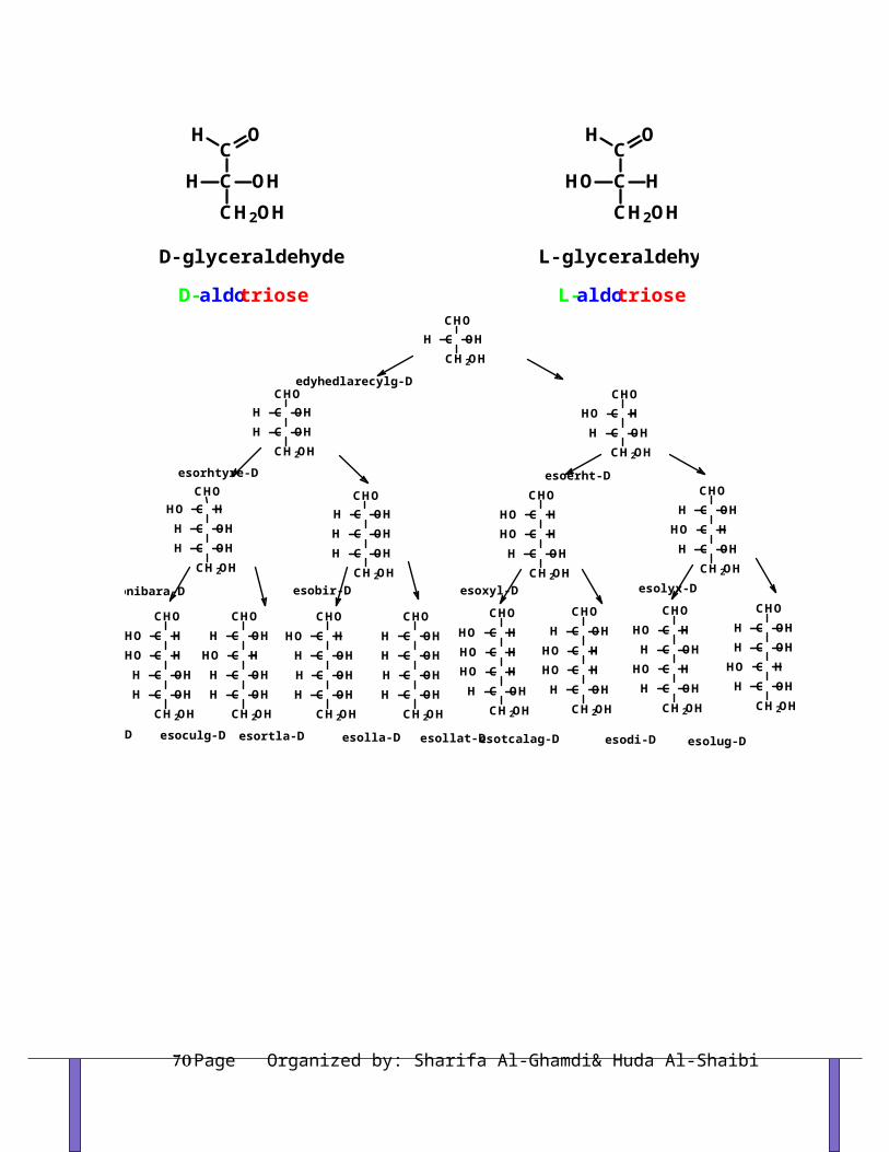
1- If the OH group is found on the right side of the carbon chain, the sugar is designated as
a D sugar.
Organized by: Sharifa Al-Ghamdi& Huda Al-ShaibiPage 50

2- If the OH group is found on the left side of the chain of carbons, the sugar is designated
as an L sugar.
CCCC
CHOHH
HOH
OHOH
HOH
CH 2OH
CCCC
CHOH
HHOH
OHHOHOH
CH 2OH
CCCC
CHOOH
HOHOH
HOH
HH
CH 2OH
CCCC
CHOOH
OHOH
HOH
HHH
CH 2OH
CCCC
CHOH
HHOH
HOOH
HOH
CH 2OH
C OHHCH 2OH
CHO
CC
HOH
HOH
CH 2OH
CHO
CCCC
CHOH
OHOH
HOHO HHH
CH 2OH
CC
OHOH
HH
CH 2OH
CHO
CCC
HHOH
OHHOH
CH 2OH
CHO
CCC
HHOH
HOHOH
CH 2OH
CHOCCC
OHOH
HO HHH
CH 2OH
CHO
CCC
OHOH
H OHHH
CH 2OH
CHO
CCCC
CHOHO
HHOH
HHOHOH
CH 2OH
CCCC
CHOH
HOHOH
HOOH
HH
CH 2OH
edyhedlarecylg-D
esorhtyre-D esoerht-D
esonibara-D esobir-D esolyx-Desoxyl-D
esoculg-Desonnam-D esolla-Desortla-D esollat-D esotcalag-D esodi-D esolug-D
Organized by: Sharifa Al-Ghamdi& Huda Al-ShaibiPage 51
CC OHHCH2OH
OH
D-glyceraldehyde
D-aldotriose
CC HHOCH2OH
OH
L-glyceraldehyde
L-aldotriose

Cyclic Structures:
- Five membered sugar rings are known as furanose rings.
- Six membered sugar rings are known as pyranose rings.
esonarufobir-D- esonarufobir-D-
O
OHH
H
H
OH
CH 2HO
OHH
O
OHH
H
OH
CH 2HO
HH
OH
+
CCCC
CH 2OH
HH
OH
HOHOH
OH
esobir-D
Organized by: Sharifa Al-Ghamdi& Huda Al-ShaibiPage 52

esonarypoculg-D-esonarypoculg-D-
+O
H
OH
OHH
OHHCH 2HO
HO
HO
OH
H
OHH
OHHCH 2HO
HO
H
esoculg-D
CCCC
COH
OHOH
HOH
HHH
CH 2OH
OH
Carbohydrate Anomers:- Formation of either of the cyclic form has created a new stereocenter.
- These stereoisomeric ring forms of carbohydrates are called Anomers.
- Anomers are carbohydrates that differ by the stereo-configuration of the carbon involved
in ring formation.
- The greek letters α and β are used to describe the configuration about the ring forming
carbon.
- The α anomer always has the OH group oriented in a downward fashion on the anomeric
carbon of a D-sugar.
- The β anomer always has the OH group oriented in an upward fashion on the anomeric
carbon of a D-sugar.
Important Carbohydrates:
Monosaccharides
- composed of three to seven carbon atoms. 1- Glucose - most abundant hexose in our diet.
- The building block of complex carbohydrates.- Component of the disaccharides: sucrose, maltose and lactose.- Found in the polysaccharides: starch, cellulose and glycogen.
Organized by: Sharifa Al-Ghamdi& Huda Al-ShaibiPage 53

CCCC
CHOOH
OHOH
HOH
HHH
CH 2OH
O
H
H
H
OHOH
CH 2OH
HOH HO,H
2- Galactose
- Found in the disaccharide, lactose.
- Found in the cellular membranes of the brain and nervous system.
- Galactose is the C-4 epimer of glucose.
CCCC
CHOOH
HOH
HOH
HHOH
CH 2OH
HO,H
O
H
H
OH
CH 2OH
HOH
HO
H
3- Fructose
- Sweetest of the carbohydrates.
- Component of the disaccharide sucrose.
- Fructose is a keto sugar.
Disaccharides
- composed of two monosaccharide units.
1- Maltose - malt sugar.
Used in cereals, candies and the brewing of beverages.
Composed of two D-glucose sugars joined by an α-1,4 linkage.
Organized by: Sharifa Al-Ghamdi& Huda Al-ShaibiPage 54

O OHH
H
HH
H
O
HOHOH H H
OH OHOH
OH
CH 2OH CH 2OH
H
2-Lactose - milk sugar.
- Found in milk and milk products.
- Composed of one galactose and one glucose unit joined by a β-1,4 linkage.
OH
H
H
OH H
OH
OH
CH 2OH
HO
CH 2OH
OH
HOHH
H
OH
H
O
3- Sucrose - table sugar.
- Product of sugar cane and sugar beets.
- Composed of one glucose and one fructose unit.
- Linkage is at both anomeric carbons.
O O
OH
H
OH
H
H
O
HH
H
OH H
HOHCH 2OHOH
CH 2OHCH 2OH
Polysaccharides - composed of many (more than 10) monosaccharide units.
1- Cellulose:
Major structural material of plant cells.
Consists of many glucose units joined by β-1,4 linkages.
2- Starch:
Storage form of glucose found in rice wheat, potatoes, grains and cereals.
Consists of many glucose units joined by α-1,4 linkages.
Organized by: Sharifa Al-Ghamdi& Huda Al-ShaibiPage 55

Maltose is the disaccharide starting material.
3- Glycogen:
Animal starch. Storage form of glucose found in the liver and muscle of animals.
Contains many highly branched glucose units.
Joined by α-1,4 linkages and branched by α-1,6 linkages.
4- Dextrin:
- Mixture of branched and un-branched soluble polysaccharides produced by partial
hydrolysis of starch by acids or amylases.
Reducing sugars:
Any sugar that contains either:
1-A free aldehyde group.
2- An α-hydroxy ketone group.
Organized by: Sharifa Al-Ghamdi& Huda Al-ShaibiPage 56

3-A hemiacetal linkage
-The presence of any of these groups allows the carbohydrate to undergo easy oxidation.
- If the sugar gets oxidized it causes reduction.
- Thus the name “reducing sugar”.
Organized by: Sharifa Al-Ghamdi& Huda Al-ShaibiPage 57

QUALITATIVE TESTS FOR CARBOHYDRATES
Experiment-1:Molisch Test:- It is the general test for all carbohydrates.
- All carbohydrates. Monosaccharides give a rapid positive test. Disaccharides and
polysaccharides react slower.
- The Molisch reagent dehydrates pentoses to form furfural (top reaction) and dehydrates
hexoses to form 5-hydroxymethyl furfural (bottom reaction). The furfurals further react
with -naphthol present in the test reagent to produce a purple product.
→ Condensation with α-naphthol >>>> Purple ring.
Organized by: Sharifa Al-Ghamdi& Huda Al-ShaibiPage 58

Method:
1ml test solution + 2 drops of α-naphthol >> mix well >> add conc. H2SO4 down the side of
the tube to form the ring at the interface of the two layers.
-ve +ve
Experiment-2:Fehling's Test:
- This test is used to differentiate between reducing and non reducing sugars.
- A reducing sugar reacts with Fehling's reagent in alkaline medium to form an orange to red
precipitate. Fehling's reagent is commonly used for reducing sugars but is known to be not
specific for aldehydes.
- Positive result is detected by reduction of the deep blue solution of cupric (II) to a red
precipitate of insoluble cuprous oxide (Cu2O).
- The sucrose does not react with Fehling's reagent. Sucrose is a disaccharide of glucose and
fructose. Most disaccharides are reducing sugars (e.g. lactose and maltose)
Organized by: Sharifa Al-Ghamdi& Huda Al-ShaibiPage 59

- Sucrose is non-reducing sugar because the anomeric carbon of glucose is involved in the
glucose- fructose bond and hence is not free to form the aldehyde in solution.
Fehling's Reagent: Two solutions are required:
Fehling's "A" uses 7 g CuSO4.5H2O dissolved in distilled water containing 2 drops of dilute
sulfuric acid.
Fehling's "B" uses 35g of potassium tartrate and 12g of NaOH in 100 ml of distilled water.
Method:
1ml test solution + 1ml Fehling's reagent > heat the mixture in BWB (5min)>>Reddish
brown ppt
Experiment-3:Benedict's Test:
- This test is used also to differentiate between reducing and non reducing sugars.
- It works on the same principle but Benedict is more stable than Fehling's reagent.
Organized by: Sharifa Al-Ghamdi& Huda Al-ShaibiPage 60

- Benedict's reagent contains blue copper(II) sulfate (CuSO4) · 5H2O which is reduced to red
copper(I) oxide by aldehydes, thus oxidizing the aldehydes to carboxylic acids.
- The copper oxide is insoluble in water and so precipitates. The colour of the final solution
ranges from green to brick red depending on how many of the copper (II) ions are present.
Method:
1ml test solution + 1ml Benedict's reagent > heat the mixture in BWB (5min)>>Reddish
brown ppt
Experiment-4:Barfoid Test:
- It is a test used to differentiate between monosaccharides and disaccharides. This reaction
will detect reducing monosaccharides in the presence of disaccharides. This reagent uses
copper ions to detect reducing sugars in an acidic solution. Barfoed's reagent is copper
acetate in dilute acetic acid (pH 4.6)
- Reducing monosaccharides are oxidized by the copper ions in a weak acidic medium to
form a carboxylic acid and a reddish ppt of Cu2O (cuprous oxide).
- Reducing disaccharides (lactose but not sucrose) undergo the same reaction but at slower
rate.
Method:
- 1 ml of the solution to be tested + 2 ml of freshly prepared Barfoed's reagent. Place test
tubes into a boiling water bath and heat for 2 minutes. Remove the tubes from the bath and
allow to cool.
- Formation of a green, red, or yellow precipitate is a positive test for reducing
monosaccharides. Do not heat the tubes longer than 3 minutes, as a positive test can be
obtained with disaccharides if they are heated long enough.
Organized by: Sharifa Al-Ghamdi& Huda Al-ShaibiPage 61

Experiment-5: Seliwanoff Test:- The test reagent dehydrates ketohexoses to form 5-hydroxymethylfurfural. 5-
hydroxymethylfurfural further reacts with resorcinol present in the test reagent to produce a
red product within two minutes (reaction not shown). Aldohexoses react to form the same
product, but do so more slowly.
Method:
1/2 ml of a sample + 2ml of Seliwanoff's reagent (a solution of resorcinol and HCl) is added.
The solution is then heated in a boiling water bath for two minutes.
- A positive test is indicated by the formation of a red product.
- In case of sucrose, avoid over-boiling because sucrose may be hydrolyzed to its component
(glucose and fructose) and gives false positive result.
-ve +ve
Experiment-5: Hydrolysis test:
Organized by: Sharifa Al-Ghamdi& Huda Al-ShaibiPage 62
- Sucrose is the only non-reducing sugar so it does not reduce the alkaline Cu solutions (Fehling
and Benedict). Sucrose must first be hydrolyzed to its component and then test for.
Method:
6 ml of 1% sucrose in a test tube + 2 drops of concentrated hydrochloric acid (HCl). Heat the tube
in a boiling water bath for 15 minutes.
- Then test for Fehling, Benedict and all the previous tests.
Experiment-6: Iodine test : Test for Polysaccharides
1- Starch:
- 1/2 mL of the fresh starch solution + 1 drop of the iodine solution.
- A dark blue color indicates a positive test for starch. If the yellow color of the iodine
reagent simply becomes diluted, no starch is present. Record the observation as positive
(blue) or negative (yellow).
2- Dextrin:
- 1/2 mL of the fresh dextrin solution + 1 drop of the iodine solution.
- A violet color indicates a positive test for dextrin. If the yellow color of the iodine reagent
simply becomes diluted, no dextrin is present. Record the observation as positive (violet) or
negative (yellow).
Experiment-6: The preparation of osazone :
Organized by: Sharifa Al-Ghamdi& Huda Al-ShaibiPage 63

- Phenyl hydrazine reacts with normal carbonyls to produce phenyl hydrazones.
1- Sugars undergo a variation of this reaction in which 3 molecules of phenylhydrazine
react with the sugar to produce a 1,2-diphenylhydrazone.
2- These 1,2-diphenylhydrazones are known as osazones.
- Because both carbons 1 and 2 are involved in the reaction C-2 epimers produce the same
osazone.
Organized by: Sharifa Al-Ghamdi& Huda Al-ShaibiPage 64

Ketoses with configurations identical to aldoses below C-2 give the same osazones e.g.
glucose and fructose.
Explain glucose and fructose form the same osazone?
Characteristics of osazones: 1- Have a characteristic shape.
2- Have characteristic melting points.
3- Specific time and whether the osazone is formed from hot solutions or only on cooling.
- Glucose + Phenyl hydrazine>>>>>>>>>>> Glucosazone (broomed shape)
- Fructose + Phenyl hydrazine>>>>>>>>>>> Fructosazone (broomed shape)
- Maltose + Phenyl hydrazine>>>>>>>>>>> Maltosazone (spherical shape)
- Lactose + Phenyl hydrazine>>>>>>>>>>> Lactosazone
- Sucrose + Phenyl hydrazine>>>>>>>>>>> -ve (WHY?)
Method:
1/2 g of phenyl hydrazine + 1 spoon sodium acetate + 2ml of glucose solution >>>>>BWB (45 min)
until yellow crystals appear >>> cold and examine a sample of crystals under microscope. Compare
the crystal shapes with the supplied photographs.
References:www.wikipedia.orgwww.chemtopics.com
Organized by: Sharifa Al-Ghamdi& Huda Al-ShaibiPage 65

RESULTS & LAB REPORT
- You are supplied with samples of different sugars carry on all the previous experiments.
- Record your methods, observations and inferences in the three-column format
- Draw the form of crystals.
Organized by: Sharifa Al-Ghamdi& Huda Al-ShaibiPage 66

SCHEME FOR UNKNOWN SUGARSolution of carbohydrate
Benedict’s test
(2ml sugar+2ml Benedicts reagent….boiling 5 min.)
-ve for non reducing sugars +ve for reducing sugars
Iodine test Iodine test
(1ml of iodine + 5 drops of sugar) (1ml of iodine + 5 drops of sugar)
+ve -ve -ve +ve
Starch Non-reducing Reducing
Reducing
Disaccharide monosaccharides
Dextrin
(sucrose) and disaccharides
*Hydrolysis of sucrose
(5ml of sugar + o.5 ml conc. Hcl
heat in boiling bath for 5 min.
Then perform Fehling, Benedict,
Seliwanoff and Barfoid tests)
Barfoed`s Test
(2ml Barfoed reagent+ 1ml sugar >>boiling 5min)
-ve +ve
Reducing disaccharides monosaccharides
(Lactose)
Organized by: Sharifa Al-Ghamdi& Huda Al-ShaibiPage 67

Seliwanoff`s test
(1ml sugar+1ml Seliwanoff reagent>>>boiling 2 min.)
+ve -ve
Keto-sugar Aldo-sugar
RESULTS & LAB REPORTOrganized by: Sharifa Al-Ghamdi& Huda Al-ShaibiPage 68

- You are supplied with samples of unknown samples, identify them.
- Record your methods, observations and inferences in the three-column format
Organized by: Sharifa Al-Ghamdi& Huda Al-ShaibiPage 69
LIPIDS
-The lipids are a large and diverse group of naturally occurring organic compounds that
are related by their solubility in non-polar organic solvents (e.g. ether, chloroform, acetone
& benzene) and general insolubility in water.
- Lipids are defined by their physical behavior rather than by their chemical structures. As
a result there is great structural variety among the lipid class.
- Fats and oils are made from two kinds of molecules: glycerol (a type of alcohol with a
hydroxyl group on each of its three carbons) and three fatty acids joined by dehydration
synthesis. Since there are three fatty acids attached, these are known as triglycerides.
- The main distinction between fats and oils is whether they’re solid or liquid at room
temperature, and this is based on differences in the structures of the fatty acids they
contain.
- The main functions of lipids include:
1- Source of energy.
2- Some fat soluble vitamins have regulatory or coenzyme functions.
3- Structural functions (cell membrane).
1. Fatty Acids (FA):
- The “tail” of a fatty acid is a long hydrocarbon chain, making it hydrophobic. The “head”
of the molecule is a carboxyl group which is hydrophilic. These long-chain carboxylic acids
are generally referred to by their common names, which in most cases reflect their sources.
Natural fatty acids may be saturated or unsaturated.
Organized by: Sharifa Al-Ghamdi& Huda Al-ShaibiPage 70

- The terms saturated, mono-unsaturated, and poly-unsaturated refer to the number of
hydrogen atoms attached to the hydrocarbon tails of the fatty acids as compared to the
number of double bonds between carbon atoms in the tail.
- Fats, which are mostly from animal sources, have all single bonds between the carbons in
their fatty acid tails, thus all the carbons are also bonded to the maximum number of
hydrogen atoms possible. Since the fatty acids in these triglycerides contain the maximum
possible amount of hydrogen atoms, these would be called saturated fats. The hydrocarbon
chains in these fatty acids are, thus, fairly straight and can pack closely together, making
these fats solid at room temperature.
- Oils, mostly from plant sources, have some double bonds between some of the carbons in
the hydrocarbon tail, causing bends or “kinks” in the shape of the molecules. Because some
of the carbons share double bonds, they’re not bonded to as many hydrogen atoms as they
could if they weren’t double bonded to each other. Therefore these oils are called
unsaturated fats. Because of the kinks in the hydrocarbon tails, unsaturated fats can’t pack
as closely together, making them liquid at room temperature.
Essential Fatty Acids:
- Are those that the body can not synthesize them and therefore must be supplied in the
diet.
- Two of FAs are essential in humans>>>Linoleic acid & Linolenic acid.
DETERMINATION OF ACID VALUE Organized by: Sharifa Al-Ghamdi& Huda Al-ShaibiPage 71

Sensitivity of Fats to Oxidation (Rancidity):
- Saturated FAs are relatively resistant to oxidation outside the body.
- Unsaturated FAs are slowly but spontaneously oxidize in the presence of air.
Rancidity:
- Oxidative cleavage of the double bonds in unsaturated FA and peroxide formation which
results in unpleasant taste and smell.
- It produces aldehyde and carboxylic acids of shorter length.
Rancidity is caused by:
1- Atmospheric air.
2- Hydrolysis by microorganisms.
- The amount of free fatty acids present therefore gives an indication of the:
1- Age.
2- Quality of oil.
Acid Value:
- Is the number of mg KOH required to neutralize the free fatty acids present in 1g of fat.
Method:
1- Weigh out 2g of the test compound (fresh and rancid).
2- Suspend the melted fat in about 10ml of fat solvent.
3- Add 2 drops of ph.ph. (Indicator).
Organized by: Sharifa Al-Ghamdi& Huda Al-ShaibiPage 72

4- Mix well.
5- Titer with 0.1M KOH.
6- End point>>>> faint pink color persists 20-30s
7- Record the volume of KOH required.
References:www.wikipedia.org
RESULTS & LAB REPORT
Organized by: Sharifa Al-Ghamdi& Huda Al-ShaibiPage 73

- You are provided with the sample of fresh and rancid oil.
- Calculate the acid value for fresh and rancid oil.
Calculations:
1M KOH 1L contains 56 g/ L of fat
0.1M KOH 1L contains 5.6 mg/ ml of fat
So: 5.6 mg >>>>>>>>>>> 1ml
? mg >>>>>>>>>>>> titer No.
5.6 X titer No. = Y (No. of mg of KOH)
Y >>>>>>>>> 2g of fat
? >>>>>>>>> 1g of fat
Y X 1 / 2 = No of mg of KOH that required to neutralize 1g of fat (i.e. acid value).
Organized by: Sharifa Al-Ghamdi& Huda Al-ShaibiPage 74

PAPER CHROMATOGRAPHY
- Chromatography is a family of analytical chemistry techniques for the separation of
mixtures.
- It was the Russian botanist Mikhail Tsvet (Mikhail Semyonovich Tsvet) who invented the
first chromatography technique in 1901.
- The separation of molecules depends on differences of 1- size 2- shape 3- mass 4- charges
5- solubility and 6- adsorption.
Types of Chromatography:1- Adsorption chromatography.
2- Partition chromatography e.g. paper chromatography
3- Gel-filtration chromatography.
Uses of chromatography:
- Government laboratories used to check
for approved dyes in food
that vegetables contained tiny amounts of pesticides and herbicides
Advantages of using chromatography:
1. Require very minute amount for identification.
2. Can be used to identify substances that cannot be easily melted or distilled.
Organized by: Sharifa Al-Ghamdi& Huda Al-ShaibiPage 75

All types of chromatography involve interaction between:
1- The mixture to be separated.
2- The stationary phase.
3- The mobile phase.
Principle of Paper Chromatography:
- Method of separating and identifying both colored and colorless mixtures.
- Mixtures can be solids, liquids or gases but their components must be able to dissolve in
the same solvent to different extents.
- Generally involves 2 phases:
stationary phase solid support e.g. water on paper
mobile phase solvent or a gas
- Test mixture is applied onto the chromatography paper and a solvent is then allowed to
pass over the paper. As the solvent does so, the components of the mixture travel along with
it.
- The stationary phase retards the passage of the components of the sample. When
components pass through the system at different rates they become separated in time.
The solvent used depends on the substance to be separated
The components will travel at different rates over paper depending on:
their solubility in the solvent
how well the dyes adsorb on the chromatography paper
Generally, the more soluble the component is in the solvent and the less it adsorb onto the
chromatography paper, the faster it would move with the solvent on the paper and hence
the spot appears further up the paper
Organized by: Sharifa Al-Ghamdi& Huda Al-ShaibiPage 76

Result of chromatography is known as the >>>> Chromatogram
Types of Paper Chromatography:
- There are three types of paper chromatography:
I- Ascending Paper Chromatography:
- Solvent running up the paper by capillary action.
II- Descending Paper Chromatography:
- Solvent running down the paper by both capillary action and gravity.
Advantage of the descending method over the ascending method:
- Good for long pieces of paper thus better separation.
- Aided by gravity thus faster.
III- Two-Dimensional Paper Chromatography:
- The mixture is separated in the first solvent which should be volatile then after drying the
paper is turned through 90 and separation is carried out in the second solvent. After
location, a map is obtained and compounds can be identified by comparing their position
with a map of known compounds developed under the same conditions.
Stationary Phase:
- In paper chromatography, cellulose in the form of paper sheets makes an identical
support medium. >>>> WHY?
- Because it has the ability to adsorb water molecules between cellulose fibers and forms a
stationary hydrophilic phase.
Organized by: Sharifa Al-Ghamdi& Huda Al-ShaibiPage 77

- Paper: Watman No. 1 of high quality is the paper most frequently used for analytical
purposes.
Mobile Phase:
- In paper chromatography, mobile phase is a mixture of solvents.
- The choice of solvent depends on the mixture investigated:
1- If the compounds move close to solvent (A) front >>>>> these compounds are highly
soluble in solvent A
2- If the compounds are crowded around the origin >>>>> these compounds are not
sufficiently soluble in solvent B.
Therefore, a suitable solvent for separation would be an appropriate mixture of both
solvent A & B. As a result R f values of the components of the mixture are spread across
the length of the paper.
Retention Factor( R f ):
- The retention is measured as the retention factor Rf, the run length of the compound
divided by the run length of the solvent front:
- Unknown substances could be identified by the Rf values
Rf = dist. Moved by the substance dist. Moved by the solvent
- The Rf of a compound often differs considerably between experiments and
laboratories due to variations of the solvent, the stationary phase, temperature, and the
Organized by: Sharifa Al-Ghamdi& Huda Al-ShaibiPage 78

setup. It is therefore important to compare the retention of the test compound to that of
one or more standard compounds under absolutely identical conditions.
Detection of Spots:
- After development, the spots corresponding to different compounds may be located by
their color
- However, most compounds are colorless and are visualized by:
1- Spraying the paper with specific reagents.
2- Dipping the paper in a solution of the reagent in a volatile solvent.
3- Fluorescent substances can be visualized by ultraviolet (UV) light.
Organized by: Sharifa Al-Ghamdi& Huda Al-ShaibiPage 79

SEPARATION OF AMINO ACIDS BY PAPER CHROMATOGRAPHY
- Separation and identification of amino acids are operations that must be performed
frequently by biochemists. The 20 amino acids present in proteins have similar structures.
However, each amino acid is unique in polarity and ionic characteristics. In this
experiment, we will use paper chromatography to separate and identify the components of
an unknown amino acid mixture.
- The solvent mixture contains several components, one of which is usually water and
another of which is a more non-polar solvent. As the solvent mixture moves up the paper
by capillary action, the water in the mixture binds to the hydrophilic paper (cellulose) and
creates a liquid stationary phase of many small water droplets. The non-polar solvent
continues to move up the paper forming a liquid mobile phase. Since amino acids have
different R-groups, they also have different degrees of solubility in water vs. the non-polar
solvent. An amino acid with a polar R-group will be more soluble in water than in the non-
polar solvent, so it will dissolve more in the stationary water phase and will move up the
paper only slightly. An amino acid with a hydrophobic R-group will be more soluble in the
mobile non-polar solvent than in water, so it will continue to move up the paper. Different
amino acids will move different distances up the paper depending upon their relative
solubilities in the two solvents, allowing for separation of amino acid mixtures.
- The movement of amino acids can be defined by a quantity known as Rf value, which
measures the movement of an amino acid compared to the movement of the solvent. At the
start of the chromatography, the amino acid is spotted at what is called the origin. The
chromatography is then performed, and the procedure is stopped before the solvent runs
all the way up the paper. The level to which the solvent has risen is called the solvent front.
Organized by: Sharifa Al-Ghamdi& Huda Al-ShaibiPage 80

The Rf value of an amino acid is the ratio of the distance traveled by the amino acid from
the origin to the distance traveled by the solvent from the origin.
- Since Rf value for an amino acid is constant for a given chromatography system, an
unknown amino acid can be identified by comparing its Rf value to those of known amino
acids.
Application:Materials:1- Filter paper: Watman No.1.
2- Solvent system: Butanol: glacial acetic acid: water.
3- Ninhydrine reagent.
4- Standard amino acids and mixture of unknown.
Procedure:1- Draw a light pencil line 1-2cm from the bottom of the paper.
2- Place a single drop of compound at intervals 2cm.
3- Dry with hair dryer.
4- Dip the paper in the jar with one of the edges of the paper to which the sample of the
spot is adjacent into the solvent.
5- Allow to run.
6- Remove the paper.
7- Determine the solvent front.
8- Dry.
9- Spray the paper with ninhydrin.
10- Dry the paper.
Organized by: Sharifa Al-Ghamdi& Huda Al-ShaibiPage 81

After some time
References:
www.wikipedia.org
Organized by: Sharifa Al-Ghamdi& Huda Al-ShaibiPage 82

RESULTS & LAB REPORT
- Calculate the R f and then identify the unknown amino acids in the mixture.
- Present your results in a good and full lab report.
Organized by: Sharifa Al-Ghamdi& Huda Al-ShaibiPage 83