version 11.1 2012 final -...
TRANSCRIPT

BSAC Methods for Antimicrobial Susceptibility Testing Version 11.1 May 2012 All enquiries to: Jenny Andrews at: + 44 (0) 121 507 5693 Email: [email protected]

Version 11.1 May 2012 2
Contents Page
Working Party members 5 Abstract 6 Preface 11
Disc Diffusion Method for Antimicrobial Susceptibility Testing 1. Preparation of plates 13 2. Selection of control organisms 14 Table 2 a Control strains to monitor test performance of antimicrobial susceptibility
testing 15
2b Control strains used to confirm that the method will detect resistance 15 3. Preparation of inoculum 15 3.1 Comparison with 0.5 McFarland standard 16 3.1.1 Preparation of the McFarland standard 16 3.1.2 Inoculum preparation by the growth method 16 3.1.3 Inoculum preparation by the direct colony suspension method 16 3.1.4 Adjustment of the organism suspension to the density of the 0.5
McFarland standard 16
3.1.5 Dilution of suspension equivalent to 0.5 McFarland standard in distilled water before inoculation
16
3.2 Photometric standardisation of turbidity of suspension 17 3.3 Direct susceptibility testing of urines and blood cultures 18 4. Inoculation of agar plates 19 5. Antimicrobial discs 19 5.1 Storage and handling of discs 19 5.2 Application of discs 19 6. Incubation 19 6.1 Conditions of incubation 20 7. Measuring zones and interpretation of susceptibility 21 7.1 Acceptable inoculum density 21 7.2 Measuring zones 21 7.3 Use of templates for interpreting susceptibility 21 8. Oxacillin/cefoxitin testing of staphylococci 22 8.1 Detection of oxacillin resistance in Staphylococcus aureus and
coagulase negative staphylococci 22
8.2 Detection of methicillin/oxacillin/cefoxitin resistance in staphylococci by use of cefoxitin as test agent
23
Interpretative tables
Table MIC and zone breakpoints for: 6 Enterobacteriaceae 25 7 Acinetobacter species 30 8 Pseudomonas 31 9 Stenotrophomonas maltophilia 33
Version 11.1 May 2012 3
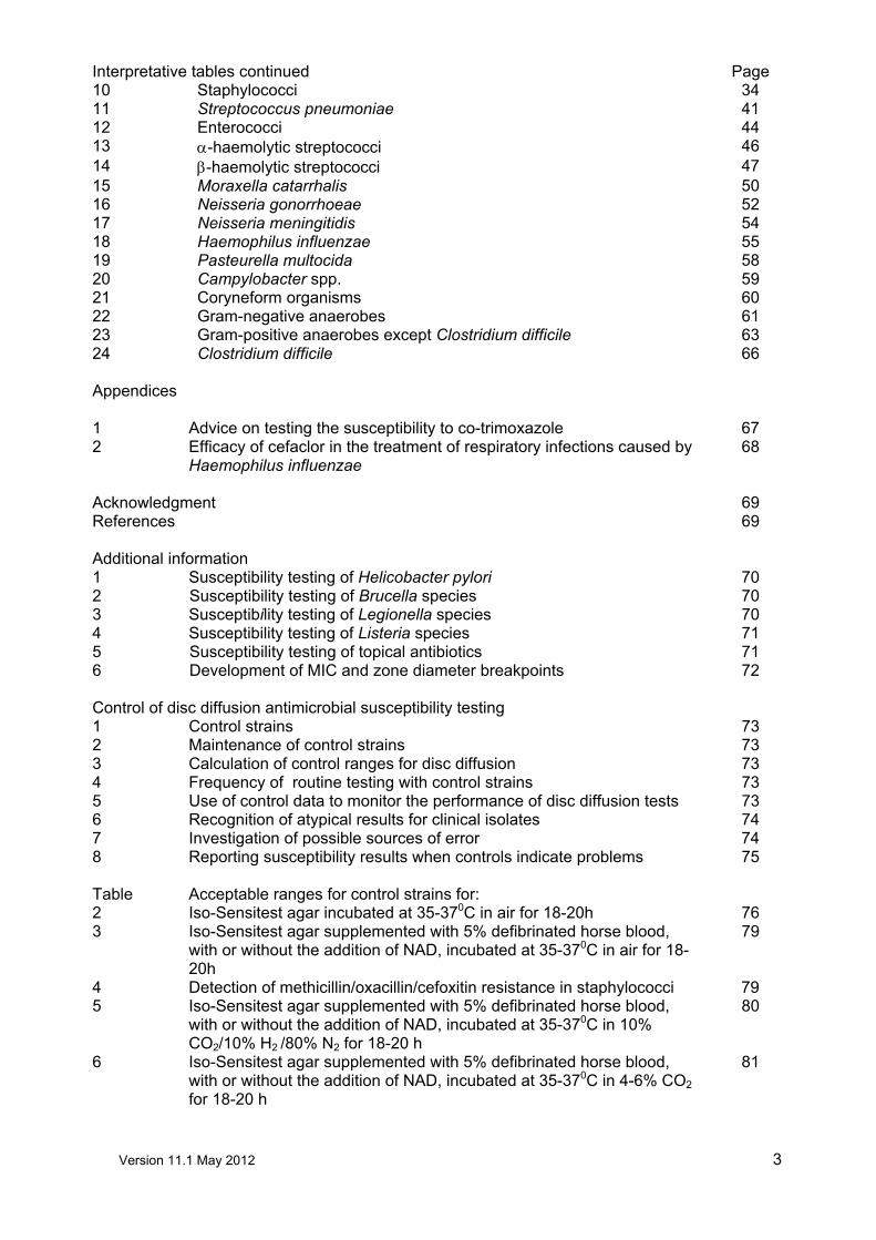
Interpretative tables continued Page 10 Staphylococci 34 11 Streptococcus pneumoniae 41 12 Enterococci 44 13 α-haemolytic streptococci 46 14 β-haemolytic streptococci 47 15 Moraxella catarrhalis 50 16 Neisseria gonorrhoeae 52 17 Neisseria meningitidis 54 18 Haemophilus influenzae 55 19 Pasteurella multocida 58 20 Campylobacter spp. 59 21 Coryneform organisms 60 22 Gram-negative anaerobes 61 23 Gram-positive anaerobes except Clostridium difficile 63 24 Clostridium difficile 66 Appendices 1 Advice on testing the susceptibility to co-trimoxazole 67 2 Efficacy of cefaclor in the treatment of respiratory infections caused by
Haemophilus influenzae 68
Acknowledgment 69 References 69 Additional information 1 Susceptibility testing of Helicobacter pylori 70 2 Susceptibility testing of Brucella species 70 3 Susceptibility testing of Legionella species 70 4 Susceptibility testing of Listeria species 71 5 Susceptibility testing of topical antibiotics 71 6 Development of MIC and zone diameter breakpoints 72 Control of disc diffusion antimicrobial susceptibility testing 1 Control strains 73 2 Maintenance of control strains 73 3 Calculation of control ranges for disc diffusion 73 4 Frequency of routine testing with control strains 73 5 Use of control data to monitor the performance of disc diffusion tests 73 6 Recognition of atypical results for clinical isolates 74 7 Investigation of possible sources of error 74 8 Reporting susceptibility results when controls indicate problems 75 Table Acceptable ranges for control strains for: 2 Iso-Sensitest agar incubated at 35-370C in air for 18-20h 76 3 Iso-Sensitest agar supplemented with 5% defibrinated horse blood,
with or without the addition of NAD, incubated at 35-370C in air for 18-20h
79
4 Detection of methicillin/oxacillin/cefoxitin resistance in staphylococci 79 5 Iso-Sensitest agar supplemented with 5% defibrinated horse blood,
with or without the addition of NAD, incubated at 35-370C in 10% CO2/10% H2 /80% N2 for 18-20 h
80
6 Iso-Sensitest agar supplemented with 5% defibrinated horse blood, with or without the addition of NAD, incubated at 35-370C in 4-6% CO2
for 18-20 h
81

Version 11.1 May 2012 4
Page 9. Control of MIC determinations Table Target MICs for: 7 Haemophilus influenzae, Enterococcus faecalis, Streptococcus
pneumoniae, Bacteroides fragilis and Neisseria gonorrhoeae 83
8 Escherichia coli, Pseudomonas aeruginosa and Staphylococcus aureus
85
9 Pasteurella multocida 87 10 Bacteroides fragilis, Bacteroides thetaiotaomicron and Clostridium
perfringens 87
11 Group A streptococci 87 References 88 Suppliers 89 Useful web sites 90
Version 11.1 May 2012 5
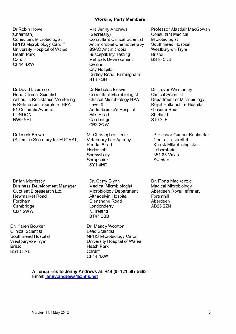
Working Party Members:
Dr Robin Howe (Chairman)
Consultant Microbiologist NPHS Microbiology Cardiff University Hospital of Wales Heath Park Cardiff CF14 4XW
Mrs Jenny Andrews (Secretary) Consultant Clinical Scientist Antimicrobial ChemotherapyBSAC Antimicrobial Susceptibility Testing Methods Development Centre City Hospital Dudley Road, Birmingham B18 7QH
Professor Alasdair MacGowan Consultant Medical Microbiologist Southmead Hospital Westbury-on-Trym Bristol BS10 5NB
Dr David Livermore Head Clinical Scientist Antibiotic Resistance Monitoring & Reference Laboratory, HPA 61 Colindale Avenue LONDON NW9 5HT
Dr Nicholas Brown Consultant Microbiologist Clinical Microbiology HPA Level 6 Addenbrooke's Hospital Hills Road Cambridge CB2 2QW
Dr Trevor Winstanley Clinical Scientist Department of Microbiology Royal Hallamshire Hospital Glossop Road Sheffield S10 2JF
Dr Derek Brown (Scientific Secretary for EUCAST)
Mr Christopher Teale Veterinary Lab Agency Kendal Road Harlescott Shrewsbury Shropshire SY1 4HD
Professor Gunnar Kahlmeter Central Lasarettet Klinisk Mikrobiologiska Laboratoriet 351 85 Vaxjo Sweden
Dr Ian Morrissey Business Development Manager Quotient Bioresearch Ltd. Newmarket Road Fordham Cambridge CB7 5WW
Dr. Gerry Glynn Medical Microbiologist Microbiology Department Altnagelvin Hospital Glenshane Road Londonderry N. Ireland BT47 6SB
Dr. Fiona MacKenzie Medical Microbiology Aberdeen Royal Infirmary Foresthill Aberdeen AB25 2ZN
Dr. Karen Bowker Clinical Scientist Southmead Hospital Westbury-on-Trym Bristol BS10 5NB
Dr. Mandy Wootton Lead Scientist NPHS Microbiology Cardiff University Hospital of Wales Heath Park Cardiff CF14 4XW
All enquiries to Jenny Andrews at: +44 (0) 121 507 5693 Email: [email protected]

Version 11.1 May 2012 6
Abstract
Summary of changes in version 11
Table 5: Incubation conditions for antimicrobial susceptibility tests on various organisms
Change to the incubation temperature and duration of incubation for Campylobacter spp.
8.2 Detection of methicillin/oxacillin/cefoxitin resistance in staphylococci by use of cefoxitin as the test agent
• Amendment stating that testing is for all staphylococci not just Staphylococcus aureus. • Reading cefoxitin zones of inhibition is for all staphylococci (Figure 3)
8.2.7 Interpretation: • For S. saprophyticus
• For coagulase staphylococci other than S. saprophyticus
Table 6. MIC and zone diameter breakpoints for Enterobacteriaceae (including Salmonella and Shigella spp.)
Change in MIC or zone diameter breakpoints
• Co-amoxiclav (UTI) • Azithromycin (S. typhi only)
Change to comments • Cefixime • Cefotaxime • Ceftazidime • Ceftriaxone
Table 8. MIC and zone diameter breakpoints for Pseudomonas spp.
Change in MIC or zone diameter breakpoints • Amikacin
Table 10. MIC and zone diameter breakpoints for staphylococci
Removal of recommendations which can now be found in the “Legacy” section • Mupirocin (5µg disc)
Change in MIC or zone diameter breakpoints • Cefoxitin (S. saprophyticus) • Vancomycin (coagulase negative staphylococci)
Change to comments • Cefoxitin (S. saprophyticus) • Vancomycin (coagulase negative staphylococci)

Version 11.1 May 2012 7
• Clindamycin • Erythromycin
Table 11. MIC and zone diameter breakpoints for Streptococcus pneumoniae
Change in MIC or zone diameter breakpoints • Meropenem (meningitis) • Linezolid
Change to comments • Linezolid
Table 13. MIC and zone diameter breakpoints for α-haemolytic streptococci
Change to comments • Clindamycin • Erythromycin
Table 14. MIC and zone diameter breakpoints for β-haemolytic streptococci
Change in MIC or zone diameter breakpoints • Trimethoprim (Group B streptococci) Change to comments • Clindamycin
Table 15. MIC and zone diameter breakpoints for Moraxella catarrhalis
Change in MIC or zone diameter breakpoints • Cefaclor • Cefuroxime • Cefuroxime axetil • Nalidixic acid • Chloramphenicol
Change to comments • Cefaclor • Chloramphenicol
Table 16. MIC and zone diameter breakpoints for Neisseria gonorrhoeae
Change to comments • Tetracycline

Version 11.1 May 2012 8
Table 18. MIC and zone diameter breakpoints for Haemophilus influenzae
Change in MIC or zone diameter breakpoints • Amoxicillin • Co-amoxiclav • Meropenem (meningitis) • Chloramphenicol
Change to comments • Amoxicillin • Chloramphenicol
Table 20. MIC and zone diameter breakpoints for Campylobacter spp.
Change in MIC or zone diameter breakpoints • Ciprofloxacin • Nalidixic acid • Erythromycin
Change to comments • Erythromycin
Table 24. MIC and zone diameter breakpoints for Clostridium difficile
Change in MIC or zone diameter breakpoints • Daptomycin • Fusidic acid • Metronidazole • Moxifloxacin • Tigecycline • Rifampicin • Vancomycin
Change to comments • Daptomycin • Fusidic acid • Metronidazole • Moxifloxacin • Tigecycline • Rifampicin • Vancomycin
Additional information
Table 1: MIC breakpoints for Helicobacter pylori based on epidemiological “cut-off” values (ECOFFs), which distinguish “wild-type” isolates from those with reduced susceptibility
Change in MIC or zone diameter breakpoints
• Amoxicillin
Version 11.1 May 2012 9
• Clarithromycin • Levofloxacin • Tetracycline • Metronidazole • Rifampicin
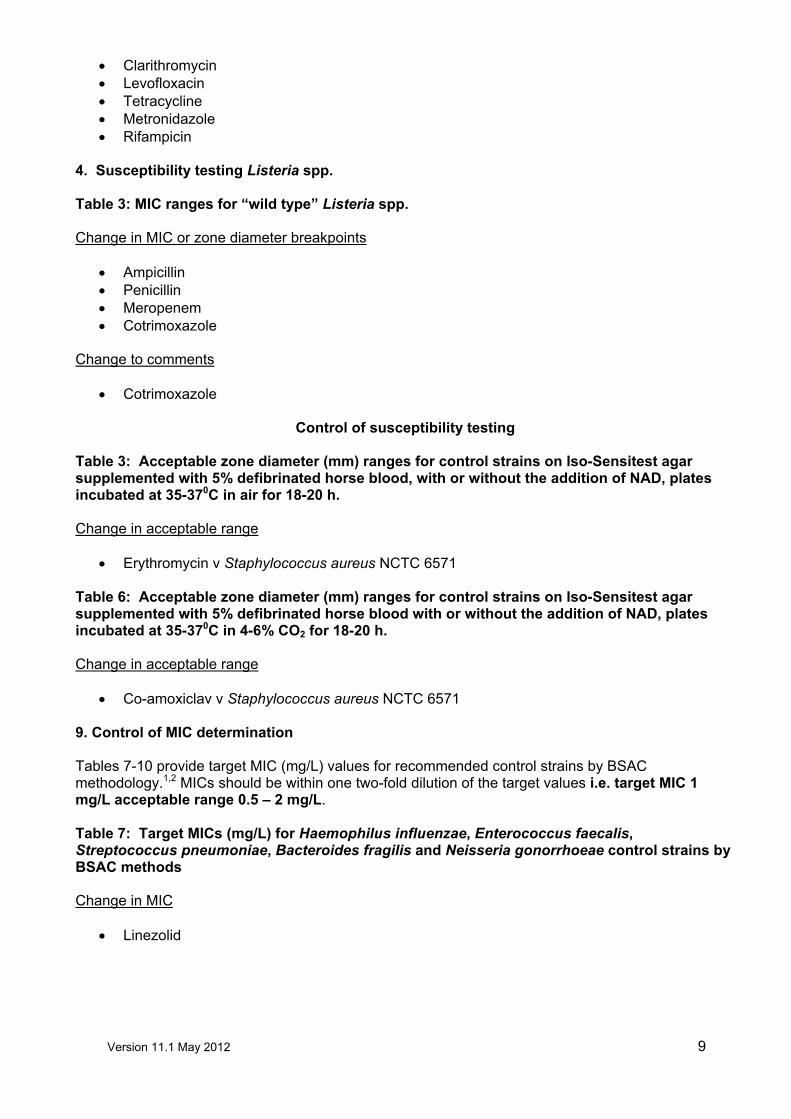
4. Susceptibility testing Listeria spp.
Table 3: MIC ranges for “wild type” Listeria spp.
Change in MIC or zone diameter breakpoints • Ampicillin • Penicillin • Meropenem • Cotrimoxazole
Change to comments • Cotrimoxazole
Control of susceptibility testing
Table 3: Acceptable zone diameter (mm) ranges for control strains on Iso-Sensitest agar supplemented with 5% defibrinated horse blood, with or without the addition of NAD, plates incubated at 35-370C in air for 18-20 h.
Change in acceptable range
• Erythromycin v Staphylococcus aureus NCTC 6571
Table 6: Acceptable zone diameter (mm) ranges for control strains on Iso-Sensitest agar supplemented with 5% defibrinated horse blood with or without the addition of NAD, plates incubated at 35-370C in 4-6% CO2 for 18-20 h.
Change in acceptable range
• Co-amoxiclav v Staphylococcus aureus NCTC 6571
9. Control of MIC determination Tables 7-10 provide target MIC (mg/L) values for recommended control strains by BSAC methodology.1,2 MICs should be within one two-fold dilution of the target values i.e. target MIC 1 mg/L acceptable range 0.5 – 2 mg/L.
Table 7: Target MICs (mg/L) for Haemophilus influenzae, Enterococcus faecalis, Streptococcus pneumoniae, Bacteroides fragilis and Neisseria gonorrhoeae control strains by BSAC methods
Change in MIC
• Linezolid

Version 11.1 May 2012 10
Table 8: Target MICs (mg/L) for Escherichia coli, Pseudomonas aeruginosa and Staphylococcus aureus control strains by BSAC methods
Change in MIC
• Cefoxitin • Colistin • Daptomycin • Doripenem • Piperacillin/tazobactam • Sulphamethoxazole • Tetracycline
NB. All changes to the tables are shown in bold text.
Erratum
In Table 14 (MIC and zone diameter breakpoints for β-haemolytic streptococci) the MIC breakpoints
for tetracycline should be R > 2 mg/L, I = 2 mg/L and S ≤ 1 mg/L. In Table 16 (MIC and zone diameter
breakpoints for Neisseria gonorrhoeae) the MIC breakpoints for penicillin should be R > 1 mg/L, I =
0.12-1mg/L and S ≤ 0.06 mg/L. In Table 18 (MIC and zone diameter breakpoints for Haemophilus
influenzae) the MIC breakpoints for cefuroxime should be R > 2 mg/L, I = 2mg/L and S ≤ 1 mg/L.
In Table 22 (MIC and zone diameter breakpoints for Gram-negative anaerobes) the MIC breakpoints
for penicillin should be R > 0.5 mg/L, I = 0.5mg/L and S ≤ 0.25 mg/L.
For the control of susceptibility testing the acceptable ranges for Staphylococcus aureus NCTC 6571
(Table 3: Acceptable zone diameter (mm) ranges for control strains on Iso-Sensitest agar
supplemented with 5% defibrinated horse blood, with or without the addition of NAD, plates incubated
at 35-370 C in air for 18-20 h) have been expanded to include cefuroxime, amoxicillin, co-amoxiclav,
chloramphenicol and nalidixic acid.
Version 11.1 May 2012 11
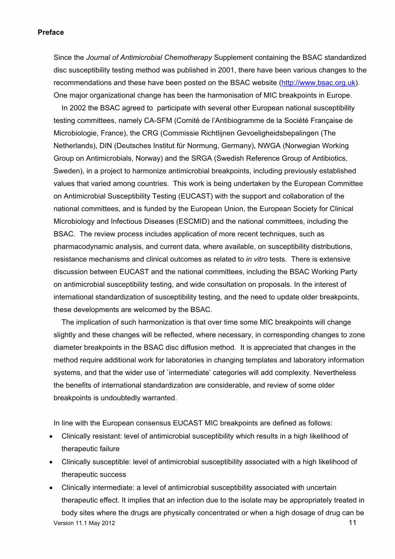
Preface
Since the Journal of Antimicrobial Chemotherapy Supplement containing the BSAC standardized
disc susceptibility testing method was published in 2001, there have been various changes to the
recommendations and these have been posted on the BSAC website (http://www.bsac.org.uk).
One major organizational change has been the harmonisation of MIC breakpoints in Europe.
In 2002 the BSAC agreed to participate with several other European national susceptibility
testing committees, namely CA-SFM (Comité de l’Antibiogramme de la Société Française de
Microbiologie, France), the CRG (Commissie Richtlijnen Gevoeligheidsbepalingen (The
Netherlands), DIN (Deutsches Institut für Normung, Germany), NWGA (Norwegian Working
Group on Antimicrobials, Norway) and the SRGA (Swedish Reference Group of Antibiotics,
Sweden), in a project to harmonize antimicrobial breakpoints, including previously established
values that varied among countries. This work is being undertaken by the European Committee
on Antimicrobial Susceptibility Testing (EUCAST) with the support and collaboration of the
national committees, and is funded by the European Union, the European Society for Clinical
Microbiology and Infectious Diseases (ESCMID) and the national committees, including the
BSAC. The review process includes application of more recent techniques, such as
pharmacodynamic analysis, and current data, where available, on susceptibility distributions,
resistance mechanisms and clinical outcomes as related to in vitro tests. There is extensive
discussion between EUCAST and the national committees, including the BSAC Working Party
on antimicrobial susceptibility testing, and wide consultation on proposals. In the interest of
international standardization of susceptibility testing, and the need to update older breakpoints,
these developments are welcomed by the BSAC.
The implication of such harmonization is that over time some MIC breakpoints will change
slightly and these changes will be reflected, where necessary, in corresponding changes to zone
diameter breakpoints in the BSAC disc diffusion method. It is appreciated that changes in the
method require additional work for laboratories in changing templates and laboratory information
systems, and that the wider use of `intermediate’ categories will add complexity. Nevertheless
the benefits of international standardization are considerable, and review of some older
breakpoints is undoubtedly warranted.
In line with the European consensus EUCAST MIC breakpoints are defined as follows:
• Clinically resistant: level of antimicrobial susceptibility which results in a high likelihood of
therapeutic failure
• Clinically susceptible: level of antimicrobial susceptibility associated with a high likelihood of
therapeutic success
• Clinically intermediate: a level of antimicrobial susceptibility associated with uncertain
therapeutic effect. It implies that an infection due to the isolate may be appropriately treated in
body sites where the drugs are physically concentrated or when a high dosage of drug can be

Version 11.1 May 2012 12
used; it also indicates a buffer zone that should prevent small, uncontrolled, technical factors
from causing major discrepancies in interpretation.
The presentation of MIC breakpoints (mg/L) has also been amended to avoid the theoretical
‘gap’ inherent in the previous system as follows:
MIC ≤ (as previously) MIC breakpoint concentration = organism is susceptible
MIC > (previously≥) MIC breakpoint concentration = organism is resistant
In practice, this does result in changes to breakpoint systems based on two-fold dilutions.
However, the appearance of the tables will change, e.g. R ≥16, S ≤8 will change to R>8, S ≤8.
Version 11.1 May 2012 13
Disc Diffusion Method for Antimicrobial Susceptibility Testing
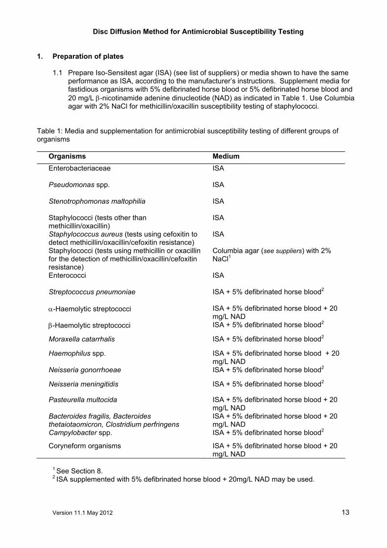
1. Preparation of plates 1.1 Prepare Iso-Sensitest agar (ISA) (see list of suppliers) or media shown to have the same
performance as ISA, according to the manufacturer’s instructions. Supplement media for fastidious organisms with 5% defibrinated horse blood or 5% defibrinated horse blood and 20 mg/L β-nicotinamide adenine dinucleotide (NAD) as indicated in Table 1. Use Columbia agar with 2% NaCl for methicillin/oxacillin susceptibility testing of staphylococci.
Table 1: Media and supplementation for antimicrobial susceptibility testing of different groups of organisms
Organisms Medium Enterobacteriaceae
ISA
Pseudomonas spp. ISA
Stenotrophomonas maltophilia
ISA
Staphylococci (tests other than methicillin/oxacillin)
ISA
Staphylococcus aureus (tests using cefoxitin to detect methicillin/oxacillin/cefoxitin resistance)
ISA
Staphylococci (tests using methicillin or oxacillin for the detection of methicillin/oxacillin/cefoxitin resistance)
Columbia agar (see suppliers) with 2% NaCl1
Enterococci
ISA
Streptococcus pneumoniae
ISA + 5% defibrinated horse blood2
α-Haemolytic streptococci ISA + 5% defibrinated horse blood + 20 mg/L NAD
β-Haemolytic streptococci ISA + 5% defibrinated horse blood2
Moraxella catarrhalis ISA + 5% defibrinated horse blood2
Haemophilus spp. ISA + 5% defibrinated horse blood + 20 mg/L NAD
Neisseria gonorrhoeae ISA + 5% defibrinated horse blood2
Neisseria meningitidis ISA + 5% defibrinated horse blood2
Pasteurella multocida ISA + 5% defibrinated horse blood + 20 mg/L NAD
Bacteroides fragilis, Bacteroides thetaiotaomicron, Clostridium perfringens
ISA + 5% defibrinated horse blood + 20 mg/L NAD
Campylobacter spp. ISA + 5% defibrinated horse blood2
Coryneform organisms ISA + 5% defibrinated horse blood + 20 mg/L NAD
1 See Section 8. 2 ISA supplemented with 5% defibrinated horse blood + 20mg/L NAD may be used.

Version 11.1 May 2012 14
1.2 Pour sufficient molten agar into sterile Petri dishes to give a depth of 4 mm ± 0.5 mm (25 mL in 90 mm diameter Petri dishes).
1.3 Dry the surface of the agar to remove excess moisture before use. The length of time
needed to dry the surface of the agar depends on the drying conditions, e.g. whether a fan-assisted drying cabinet or ‘still air’ incubator is used, whether plates are dried before storage and storage conditions. It is important that plates are not over dried.
1.4 Store the plates in vented plastic boxes at 8-10°C prior to use. Alternatively the plates may
be stored at 4-8°C in sealed plastic bags. Plate drying, method of storage and storage time should be determined by individual laboratories as part of their quality assurance programme. In particular, quality control tests should confirm that excess surface moisture is not produced and that plates are not over-dried.
2. Selection of control organisms
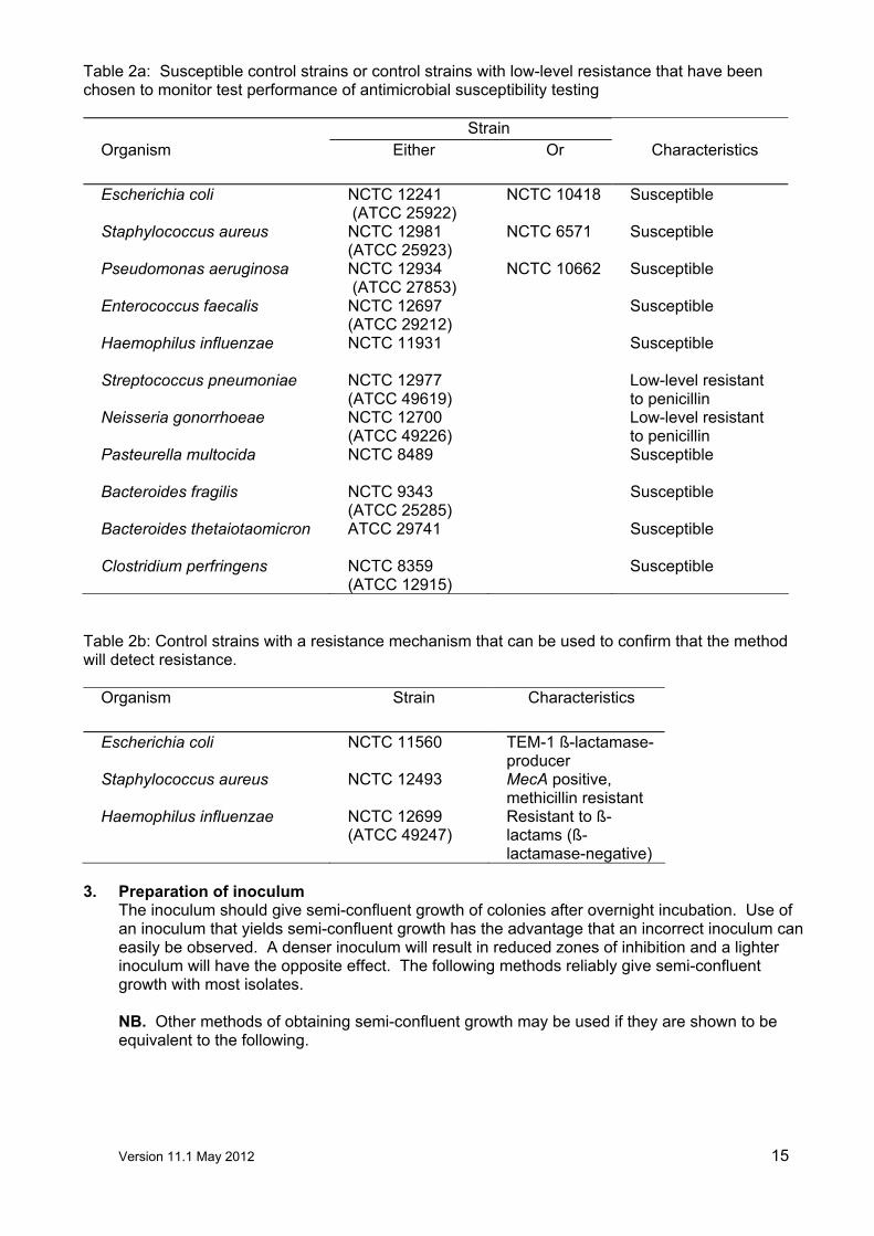
2.1 The performance of the tests should be monitored by the use of appropriate control strains
(see section on control of antimicrobial susceptibility testing). The control strains listed (Tables 2a, 2b) include susceptible strains that have been chosen to monitor test performance and resistant strains that can be used to confirm that the method will detect a mechanism of resistance.
2.2 Store control strains at –70°C on beads in glycerol broth. Non-fastidious organisms may be
stored at –20°C. Two vials of each control strain should be stored, one for an ‘in-use’ supply, the other for archiving.
2.3 Every week subculture a bead from the ‘in-use’ vial on to appropriate non-selective media
and check for purity. From this pure culture, prepare one subculture on each of the following 5 days. For fastidious organisms that will not survive on plates for 5/6 days, subculture the strain daily for no more than 6 days.
Version 11.1 May 2012 15
Table 2a: Susceptible control strains or control strains with low-level resistance that have been chosen to monitor test performance of antimicrobial susceptibility testing
Strain
Organism Either Or Characteristics
Escherichia coli NCTC 12241 (ATCC 25922)
NCTC 10418 Susceptible
Staphylococcus aureus NCTC 12981 (ATCC 25923)
NCTC 6571 Susceptible
Pseudomonas aeruginosa NCTC 12934 (ATCC 27853)
NCTC 10662 Susceptible
Enterococcus faecalis NCTC 12697 (ATCC 29212)
Susceptible
Haemophilus influenzae
NCTC 11931 Susceptible
Streptococcus pneumoniae NCTC 12977 (ATCC 49619)
Low-level resistant to penicillin
Neisseria gonorrhoeae NCTC 12700 (ATCC 49226)
Low-level resistant to penicillin
Pasteurella multocida
NCTC 8489 Susceptible
Bacteroides fragilis NCTC 9343 (ATCC 25285)
Susceptible
Bacteroides thetaiotaomicron
ATCC 29741 Susceptible
Clostridium perfringens NCTC 8359 (ATCC 12915)
Susceptible
Table 2b: Control strains with a resistance mechanism that can be used to confirm that the method will detect resistance.
Organism Strain Characteristics
Escherichia coli NCTC 11560 TEM-1 ß-lactamase-producer
Staphylococcus aureus NCTC 12493 MecA positive, methicillin resistant
Haemophilus influenzae NCTC 12699 (ATCC 49247)
Resistant to ß-lactams (ß-lactamase-negative)
3. Preparation of inoculum
The inoculum should give semi-confluent growth of colonies after overnight incubation. Use of an inoculum that yields semi-confluent growth has the advantage that an incorrect inoculum can easily be observed. A denser inoculum will result in reduced zones of inhibition and a lighter inoculum will have the opposite effect. The following methods reliably give semi-confluent growth with most isolates. NB. Other methods of obtaining semi-confluent growth may be used if they are shown to be equivalent to the following.

Version 11.1 May 2012 16
3.1 Comparison with a 0.5 McFarland standard
3.1.1 Preparation of the 0.5 McFarland standard Add 0.5 mL of 0.048 M BaCl2 (1.17% w/v BaCl2. 2H2O) to 99.5 mL of 0.18 M H2SO4 (1%
w/v) with constant stirring. Thoroughly mix the suspension to ensure that it is even. Using matched cuvettes with a 1 cm light path and water as a blank standard, measure the absorbance in a spectrophotometer at a wavelength of 625 nm. The acceptable absorbance range for the standard is 0.08-0.13. Distribute the standard into screw-cap tubes of the same size and volume as those used in growing the broth cultures. Seal the tubes tightly to prevent loss by evaporation. Store protected from light at room temperature. Vigorously agitate the turbidity standard on a vortex mixer before use. Standards may be stored for up to six months, after which time they should be discarded. Prepared standards can be purchased (See list of suppliers), but commercial standards should be checked to ensure that absorbance is within the acceptable range as indicated above.
3.1.2 Inoculum preparation by the growth method (for non-fastidious organisms, e.g.
Enterobacteriaceae, Pseudomonas spp. and staphylococci) Touch at least four morphologically similar colonies (when possible) with a sterile loop. Transfer the growth into Iso-Sensitest broth or an equivalent that has been shown not to interfere with the test. Incubate the broth, with shaking at 35-37°C, until the visible turbidity is equal to or greater than that of a 0.5 McFarland standard.
3.1.3 Inoculum preparation by the direct colony suspension method (the method of choice for fastidious organisms, i.e. Haemophilus spp., Neisseria gonorrhoeae, Neisseria meningitidis, Moraxella catarrhalis, Streptococcus pneumoniae, α and β-haemolytic streptococci, Clostridium perfringens, Bacteroides fragilis, Bacteroides thetaiotaomicron, Campylobacter spp., Pasteurella multocida and Coryneform organisms). Colonies are taken directly from the plate into Iso-Sensitest broth (or equivalent) or sterile distilled water. The density of the suspension should match or exceed that of a 0.5 McFarland standard. NB. With some organisms production of an even suspension of the required turbidity is difficult and growth in broth, if possible, is a more satisfactory option.
3.1.4 Adjustment of the organism suspension to the density of a 0.5 McFarland standard Adjust the density of the organism suspension to equal that of a 0.5 McFarland standard
by adding sterile distilled water. To aid comparison, compare the test and standard suspensions against a white background with a contrasting black line. NB. Suspension should be used within 15 min.
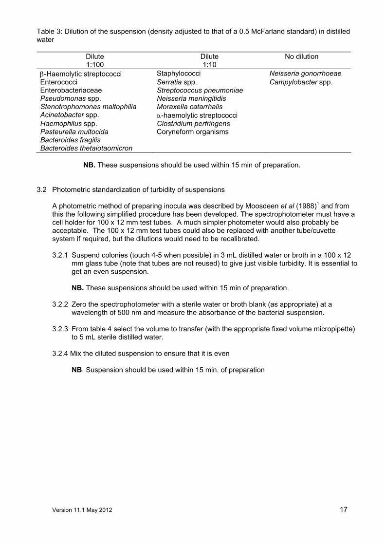
3.1.5 Dilution of suspension in distilled water before inoculation
Dilute the suspension (density adjusted to that of a 0.5 McFarland standard) in distilled water as indicated in Table 3.
Version 11.1 May 2012 17
Table 3: Dilution of the suspension (density adjusted to that of a 0.5 McFarland standard) in distilled water
Dilute 1:100
Dilute 1:10
No dilution
β-Haemolytic streptococci Staphylococci Neisseria gonorrhoeae Enterococci Serratia spp. Campylobacter spp. Enterobacteriaceae Streptococcus pneumoniae Pseudomonas spp. Neisseria meningitidis Stenotrophomonas maltophilia Moraxella catarrhalis Acinetobacter spp. α-haemolytic streptococci Haemophilus spp. Clostridium perfringens Pasteurella multocida Coryneform organisms Bacteroides fragilis Bacteroides thetaiotaomicron
NB. These suspensions should be used within 15 min of preparation.
3.2 Photometric standardization of turbidity of suspensions
A photometric method of preparing inocula was described by Moosdeen et al (1988)1 and from this the following simplified procedure has been developed. The spectrophotometer must have a cell holder for 100 x 12 mm test tubes. A much simpler photometer would also probably be acceptable. The 100 x 12 mm test tubes could also be replaced with another tube/cuvette system if required, but the dilutions would need to be recalibrated. 3.2.1 Suspend colonies (touch 4-5 when possible) in 3 mL distilled water or broth in a 100 x 12
mm glass tube (note that tubes are not reused) to give just visible turbidity. It is essential to get an even suspension.
NB. These suspensions should be used within 15 min of preparation.
3.2.2 Zero the spectrophotometer with a sterile water or broth blank (as appropriate) at a
wavelength of 500 nm and measure the absorbance of the bacterial suspension. 3.2.3 From table 4 select the volume to transfer (with the appropriate fixed volume micropipette)
to 5 mL sterile distilled water. 3.2.4 Mix the diluted suspension to ensure that it is even
NB. Suspension should be used within 15 min. of preparation

Version 11.1 May 2012 18
Table 4: Dilution of suspensions of test organisms according to absorbance reading
Organisms
Absorbance reading at 500 nm
Volume (µL) to transfer to 5 mL sterile distilled water
0.01 - 0.05 250 >0.05 - 0.1 125 >0.1 - 0.3 40 >0.3 - 0.6 20
Enterobacteriaceae Enterococci Pseudomonas spp. Staphylococci
>0.6 - 1.0 10 0.01 - 0.05 500 >0.05 - 0.1 250 >0.1 - 0.3 125 >0.3 - 0.6 80
Haemophilus spp. Streptococci Miscellaneous fastidious Organisms
>0.6 - 1.0 40 NB. As spectrophotometers may differ, it may be necessary to adjust the dilutions slightly to
achieve semi-confluent growth with any individual set of laboratory conditions.
3.3 Direct antimicrobial susceptibility testing of urine specimens and blood cultures Direct susceptibility testing is not advocated as the control of inoculum is very difficult. Direct
testing is, however, undertaken in many laboratories in order to provide more rapid test results. The following methods have been recommended by laboratories that use the BSAC method and. will achieve the correct inoculum size for a reasonable proportion of infected urines and blood cultures If the inoculum is not correct (i.e. growth is not semi-confluent) or the culture is mixed, the test must be repeated. 3.3.1 Urine specimens
3.3.1.1 Method 1 Thoroughly mix the urine specimen, then place a 10 µL loop of urine in the centre of the susceptibility plate and spread evenly with a dry swab.
3.3.1.2 Method 2 Thoroughly mix the urine specimen, then dip a sterile cotton-wool swab in the urine and remove excess by turning the swab against the inside of the container. Use the swab to make a cross in the centre of the susceptibility plate and spread evenly with another sterile dry swab. If only small numbers of organisms are seen in microscopy, the initial cotton-wool swab may be used to inoculate and spread the susceptibility plate.
3.3.2 Positive blood cultures The method depends on the Gram reaction of the infecting organism.
3.3.2.1 Gram-negative bacilli.
Using a venting needle, place one drop of the blood culture in 5 mL of sterile water, then dip a sterile cotton-wool swab in the suspension and remove excess by turning the swab against the inside of the container. Use the swab to spread the inoculum evenly over the surface of the susceptibility plate.
3.3.2.2 Gram-positive organisms.
It is not always possible accurately to predict the genera of Gram-positive organisms from the Gram’s stain. However, careful observation of the morphology, coupled with clinical information, should make an “educated guess” correct most of the time.

Version 11.1 May 2012 19
Staphylococci and enterococci. Using a venting needle, place three drops of the blood culture in 5 mL of sterile water, then dip a sterile cotton-wool swab in the suspension and remove excess by turning the swab against the inside of the container. Use the swab to spread the inoculum evenly over the surface of the susceptibility plate.
Pneumococci, “viridans” streptococci and diptheroids. Using a venting needle, place one drop of the blood culture in the centre of a susceptibility plate, and spread the inoculum evenly over the surface of the plate.
4. Inoculation of agar plate Use the adjusted suspension within 15 min to inoculate plates by dipping a sterile cotton-wool swab into the suspension and remove the excess liquid by turning the swab against the side of the container. Spread the inoculum evenly over the entire surface of the plate by swabbing in three directions. Allow the plate to dry before applying discs.
NB. If inoculated plates are left at room temperature for extended times before the discs are applied, the organism may begin to grow, resulting in reduced zones of inhibition. Discs should therefore be applied to the surface of the agar within 15 min of inoculation.
5. Antimicrobial discs Refer to interpretation tables 6-23 for the appropriate disc contents for the organisms tested.
5.1 Storage and handling of discs.
Loss of potency of agents in discs will result in reduced zones of inhibition. To avoid loss of potency due to inadequate handling of discs the following are recommended: 5.1.1 Store discs in sealed containers with a desiccant and protected from light (this
is particularly important for some light-susceptible agents such as metronidazole, chloramphenicol and the quinolones).
5.1.2 Store stocks at -20°C except for drugs known to be unstable at this temperature. If this is not possible, store discs at <8°C.
5.1.3 Store working supplies of discs at <8°C. 5.1.4 To prevent condensation, allow discs to warm to room temperature before
opening containers. 5.1.5 Store disc dispensers in sealed containers with an indicating desiccant. 5.1.6 Discard discs on the expiry date shown on the side of the container.
5.2 Application of discs
Discs should be firmly applied to the dry surface of the inoculated susceptibility plate. The contact with the agar should be even. A 90 mm plate will accommodate six discs without unacceptable overlapping of zones.
6. Incubation
If the plates are left for extended times at room temperature after discs are applied, larger zones of inhibition may be obtained compared with zones produced when plates are incubated immediately. Plates should therefore be incubated within 15 min of disc application.
6.1 Conditions of incubation Incubate plates under conditions listed in table 5.

Version 11.1 May 2012 20
Table 5: Incubation conditions for antimicrobial susceptibility tests on various organisms Organisms Incubation conditions
Enterobacteriaceae 35-37°C in air for 18-20 h Acinetobacter spp. 35-37°C in air for 18-20 h Pseudomonas spp. 35-37°C in air for 18-20 h Stenotrophomonas maltophilia 30°C in air for 18-20 h Staphylococci (other than methicillin/oxacillin/cefoxitin)
35-37°C in air for 18-20 h
Staphylococcus aureus using cefoxitin for the detection of methicillin/oxacillin/cefoxitin resistance
35°C in air for 18-20 h
Staphylococci using methicillin or oxacillin to detect resistance
30°C in air for 24 h
Moraxella catarrhalis 35-37°C in air for 18-20 h α-Haemolytic streptococci 35-37°C in 4-6% CO2 in air for 18-20 h β-Haemolytic streptococci 35-37°C in air for 18-20 h Enterococci 35-37°C in air for 24 h1
Neisseria meningitidis 35-37°C in 4-6 % CO2 in air for 18-20 h Streptococcus pneumoniae 35-37°C in 4-6 % CO2 in air for 18-20 h Haemophilus spp. 35-37°C in 4-6 % CO2 in air for 18-20 h Neisseria gonorrhoeae 35-37°C in 4-6 % CO2 in air for 18-20 h Pasteurella multocida 35-37°C in 4- 6% CO2 in air for 18-20 h Coryneform organisms 35-37°C in 4-6% CO2 in air for 18-20 h Campylobacter spp. 42°C in microaerophilic conditions for 24 h Bacteroides fragilis, Bacteroides thetaiotaomicron, Clostridium perfringens
35-37°C in 10% CO2/10% H2/80% N2 for 18-20 h (anaerobic cabinet or jar)
1It is essential that plates are incubated for at least 24 h before reporting a strain as susceptible
to vancomycin or teicoplanin.
NB. Stacking plates too high in the incubator may affect results owing to uneven heating of plates. The efficiency of heating of plates depends on the incubator and the racking system used. Control of incubation, including height of plate stacking, should therefore be part of the laboratory’s Quality Assurance programme.

Version 11.1 May 2012 21
7. Measuring zones and interpretation of susceptibility
7.1 Acceptable inoculum density
The inoculum should give semi-confluent growth of colonies on the susceptibility plate, within the range illustrated in Figure 1.
Figure 1: Acceptable inoculum density range for a Gram-negative rod
7.2 Measuring zones
7.2.1 Measure the diameters of zones of inhibition to the nearest millimetre (zone edge should be
taken as the point of inhibition as judged by the naked eye) with a ruler, callipers or an automated zone reader.
7.2.2 Tiny colonies at the edge of the zone, films of growth as a result of the swarming of Proteus spp. and slight growth within sulphonamide or trimethoprim zones should be ignored.
7.2.3 Colonies growing within the zone of inhibition should be subcultured and identified and the test repeated if necessary.
7.2.4 When using cefoxitin for the detection of methicillin/oxacillin/cefoxitin resistance in S. aureus, measure the obvious zone, taking care to examine zones carefully in good light to detect minute colonies that may be present within the zone of inhibition (see Figure 3)
7.2.5 Confirm that the zone of inhibition for the control strain falls within the acceptable ranges in Tables 20-23 before interpreting the test (see section on control of the disc diffusion method).
7.3 Use of templates for interpreting zone diameters
A template may be used for interpreting zone diameters (see Figure 2). A program for preparing templates is available from the BSAC (http://www.bsac.org.uk).
The test plate is placed over the template and the zones of inhibition are examined in relationship to the template zones. If the zone of inhibition of the test strain is within the area marked with an ‘R’, the organism is resistant. If the zone of inhibition is equal to or larger than the marked area, the organism is susceptible.
Lightest acceptable Ideal Heaviest acceptable

Version 11.1 May 2012 22
Figure 2: Template for interpreting zone diameters
R
R
R
R
R
RCZ
CT
PN
CI
G
IM
8. Oxacillin/cefoxitin testing of staphylococci
Methicillin susceptibility testing is difficult with some strains. Expression of resistance is affected by test conditions and resistance is often heterogeneous, with only a proportion of cells showing resistance. Adding NaCl or lowering incubation temperatures increases the proportion of cells showing resistance. Methicillin susceptibility testing of coagulase-negative staphylococci is further complicated as some strains do not grow well on media containing NaCl and are often slower-growing than Staphylococcus aureus. Detection of methicillin resistance in coagulase-negative staphylococci may require incubation for 48 h.
8.1 Method for detection of oxacillin resistance in S. aureus and coagulase-negative staphylococci
8.1.1 Medium
Prepare Columbia (See list of suppliers) or Mueller-Hinton agar (See list of suppliers) following the manufacturer’s instructions and add 2% NaCl. After autoclaving, mix well to distribute the sodium chloride. Pour plates to give a depth of 4 mm (± 0.5 mm) in a 90 mm sterile Petri dish (25 ml). Dry and store plates as previously described (section 1).
8.1.2 Inoculum
Prepare inoculum as previously described (section 3).
8.1.3 Control Susceptible control strains (Staphylococcus aureus ATCC 25923 or NCTC 6571) test the reliability of disc content. Staphylococcus aureus NCTC 12493 is a methicillin resistant strain and is used to check that the test will detect resistant organisms (although no strain can be representative of all the MRSA types in terms of their response to changes in test conditions).
8.1.4 Discs Place a oxacillin 1 µg disc on to the surface of inoculated agar. Discs should be stored and handled as previously described (section 5).
Version 11.1 May 2012 23
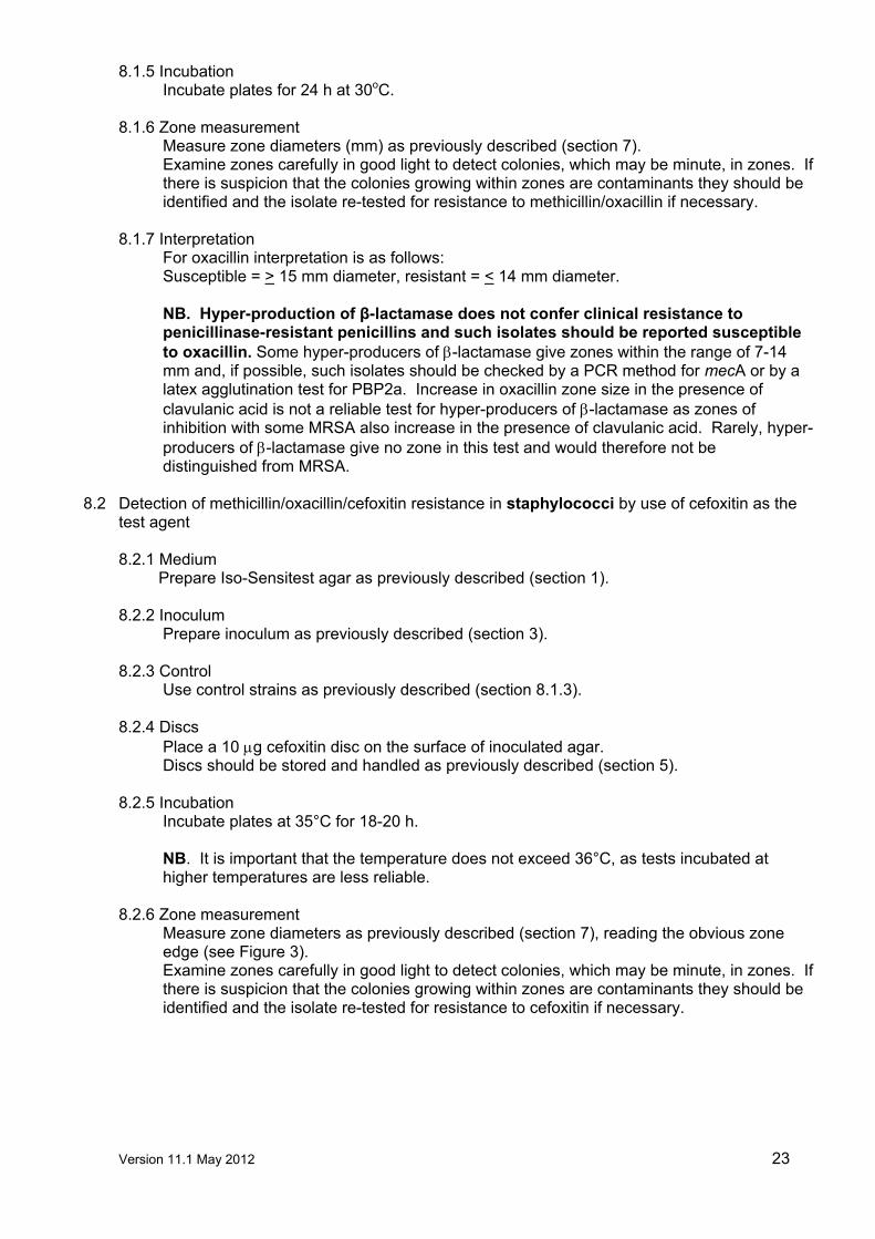
8.1.5 Incubation Incubate plates for 24 h at 30oC.
8.1.6 Zone measurement Measure zone diameters (mm) as previously described (section 7). Examine zones carefully in good light to detect colonies, which may be minute, in zones. If there is suspicion that the colonies growing within zones are contaminants they should be identified and the isolate re-tested for resistance to methicillin/oxacillin if necessary.
8.1.7 Interpretation For oxacillin interpretation is as follows: Susceptible = > 15 mm diameter, resistant = < 14 mm diameter. NB. Hyper-production of β-lactamase does not confer clinical resistance to penicillinase-resistant penicillins and such isolates should be reported susceptible to oxacillin. Some hyper-producers of β-lactamase give zones within the range of 7-14 mm and, if possible, such isolates should be checked by a PCR method for mecA or by a latex agglutination test for PBP2a. Increase in oxacillin zone size in the presence of clavulanic acid is not a reliable test for hyper-producers of β-lactamase as zones of inhibition with some MRSA also increase in the presence of clavulanic acid. Rarely, hyper-producers of β-lactamase give no zone in this test and would therefore not be distinguished from MRSA.
8.2 Detection of methicillin/oxacillin/cefoxitin resistance in staphylococci by use of cefoxitin as the
test agent
8.2.1 Medium Prepare Iso-Sensitest agar as previously described (section 1).
8.2.2 Inoculum Prepare inoculum as previously described (section 3).
8.2.3 Control Use control strains as previously described (section 8.1.3).
8.2.4 Discs
Place a 10 µg cefoxitin disc on the surface of inoculated agar. Discs should be stored and handled as previously described (section 5).
8.2.5 Incubation Incubate plates at 35°C for 18-20 h. NB. It is important that the temperature does not exceed 36°C, as tests incubated at higher temperatures are less reliable.
8.2.6 Zone measurement Measure zone diameters as previously described (section 7), reading the obvious zone edge (see Figure 3). Examine zones carefully in good light to detect colonies, which may be minute, in zones. If there is suspicion that the colonies growing within zones are contaminants they should be identified and the isolate re-tested for resistance to cefoxitin if necessary.

Version 11.1 May 2012 24
Figure 3: Reading cefoxitin zones of inhibition with staphylococci
8.2.7 Interpretation: For S. aureus
Susceptible = >22 mm diameter, resistant = <21 mm diameter For S. saprophyticus Susceptible = >20 mm diameter, resistant = <19 mm diameter For coagulase staphylococci other than S. saprophyticus Susceptible = >27 mm diameter, intermediate = 22-26 mm, resistant = <21 mm diameter NB. Hyper -production of β-lactamase does not confer clinical resistance to penicillinase-resistant penicillins and such isolates should be reported susceptible to cefoxitin. Hyper-producers of β-lactamase give zones within the ranges of the susceptible population.
Obvious zone to be measured
Inner zone NOT to be measured
Examine this area for minute colonies

Version 11.1 May 2012
25
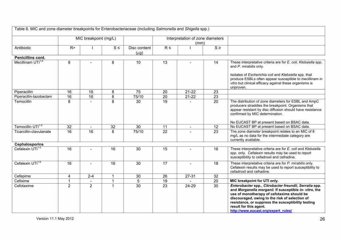
Table 6. MIC and zone diameter breakpoints for Enterobacteriaceae (including Salmonella and Shigella spp.)
The identification of Enterobacteriaceae to species level is essential before applying Expert Rules for the interpretation of susceptibility. Comments 1-5 relate to urinary tract infections (UTIs) only. 1UTI recommendations are for organisms associated with uncomplicated urinary infections only. For complicated UTI systemic recommendations should be used. 2If an organism is isolated from multiple sites, for example from blood and urine, interpretation of susceptibility should be made with regard to the systemic site (e.g., if the blood isolate is
resistant and the urine isolate susceptible, both should be reported resistant irrespective of the results obtained using interpretative criteria for urine isolates). 3For agents not listed, criteria given for systemic isolates may be used for urinary tract isolates. Intermediate susceptibility infers that the infection may respond as the agent is concentrated at
the site of infection. 4Direct susceptibility tests on urine samples may be interpreted only if the inoculum gives semi-confluent growth.
5 In the absence of definitive organism identification, use the recommendations most appropriate for the presumptive identification, accepting that on some occasions the interpretation may be incorrect. A more cautious approach is to use the systemic recommendations.
Table 6. MIC and zone diameter breakpoints for Enterobacteriaceae (including Salmonella and Shigella spp.) MIC breakpoint (mg/L) Interpretation of zone diameters
(mm)
Antibiotic R> I S ≤ Disc content(µg)
R ≤ I S ≥
Aminoglycosides Amikacin 16 16 8 30 15 16-18 19 Gentamicin 4 4 2 10 16 17-19 20 Tobramycin 4 4 2 10 17 18-20 21
Salmonella spp. should be reported resistant to these agents, irrespective of susceptibility testing result, as they are inactive against Salmonella spp. in vivo. Individual aminoglycoside agents must be tested; susceptibility to other aminoglycosides cannot be inferred from the gentamicin result and vice versa.
Penicillins Amoxicillin 8 - 8 10 14 - 15 Ampicillin 8 - 8 10 14 - 15
Species that have chromosomal penicillinases (Klebsiella spp.) or those that typically have inducible AmpC enzymes (e.g. Enterobacter spp., Citrobacter spp. and Serratia spp.) are intrinsically resistant to ampicillin/amoxicillin.
Co-amoxiclav Systemic 8 - 8 20/10 20 - 21 Co-amoxiclav UTI1-5 32 - 32 20/10 12 - 13
Species that typically have inducible AmpC enzymes (e.g. Enterobacter spp., Citrobacter spp. and Serratia spp.) are intrinsically resistant to co-amoxiclav. Zone diameter based on a 2:1 ratio of amoxicillin: clavulanate are currently under review to establish correlation with an MIC breakpoint with a fixed concentration of clavulanate.
Version 11.1 May 2012
26
Table 6. MIC and zone diameter breakpoints for Enterobacteriaceae (including Salmonella and Shigella spp.) MIC breakpoint (mg/L) Interpretation of zone diameters
(mm)
Antibiotic R> I S ≤ Disc content(µg)
R ≤ I S ≥
Penicillins cont. Mecillinam UTI1-5 8 - 8 10 13 - 14 These interpretative criteria are for E. coli, Klebsiella spp.
and P. mirabilis only. Isolates of Escherichia coli and Klebsiella spp. that produce ESBLs often appear susceptible to mecillinam in vitro but clinical efficacy against these organisms is unproven.
Piperacillin 16 16 8 75 20 21-22 23 Piperacillin-tazobactam 16 16 8 75/10 20 21-22 23 Temocillin
8 - 8 30 19 - 20 The distribution of zone diameters for ESBL and AmpC producers straddles the breakpoint. Organisms that appear resistant by disc diffusion should have resistance confirmed by MIC determination. No EUCAST BP at present based on BSAC data.
Temocillin UTI1-5 32 - 32 30 11 - 12 No EUCAST BP at present based on BSAC data. Ticarcillin-clavulanate 16 16 8 75/10 22 - 23 The zone diameter breakpoint relates to an MIC of 8
mg/L as no data for the intermediate category are currently available.
Cephalosporins Cefalexin UTI1-5 16 - 16 30 15 - 16 These interpretative criteria are for E. coli and Klebsiella
spp. only. Cefalexin results may be used to report susceptibility to cefadroxil and cefradine.
Cefalexin UTI1-5 16 - 16 30 17 - 18 These interpretative criteria are for P. mirabilis only. Cefalexin results may be used to report susceptibility to cefadroxil and cefradine.
Cefepime 4 2-4 1 30 26 27-31 32 Cefixime 1 - 1 5 19 - 20 MIC breakpoint for UTI only. Cefotaxime 2 2 1 30 23 24-29 30 Enterobacter spp., Citrobacter freundii, Serratia spp.
and Morganella morganii. If susceptible in- vitro, the use of monotherapy of cefotaxime should be discouraged, owing to the risk of selection of resistance, or suppress the susceptibility testing result for this agent. http://www.eucast.org/expert_rules/

Version 11.1 May 2012
27
Table 6. MIC and zone diameter breakpoints for Enterobacteriaceae (including Salmonella and Shigella spp.) MIC breakpoint (mg/L) Interpretation of zone diameters
(mm)
Antibiotic R> I S ≤ Disc content(µg)
R ≤ I S ≥
Cephalosporins cont. Cefoxitin (AmpC screen)
30 23 This is an epidemiological “cut off” for AmpC detection which has high sensitivity, but poor specificity as susceptibility is also affected by permeability.
Cefpodoxime (ESBL screen)
1 - 1 10 19 - 20 If screening for ESBLs is required for infection control or epidemiological purposes, Enterobacteriaceae isolates should be screened with cefpodoxime or both cefotaxime (or ceftriaxone) and ceftazidime. The presence of ESBLs should be confirmed with a specific test.
Ceftazidime 4 2-4 1 30 22 23-26 27 Ceftriaxone 2 2 1 30 23 24-27 28
Enterobacter spp., Citrobacter freundii, Serratia spp. and Morganella morganii. If susceptible to ceftazidime or ceftriaxone in- vitro, the use of monotherapy of ceftazidime or ceftriaxone should be discouraged, owing to the risk of selection of resistance, or suppress the susceptibility testing result for this agent. http://www.eucast.org/expert_rules/
Cefuroxime (axetil) UTI1-5 only
8 - 8 30 19 - 20
Cefuroxime (parenteral) 8 - 8 30 19 - 20
Salmonella spp. should be reported resistant to these agents, irrespective of susceptibility testing result, as they are inactive in-vivo. For parenteral cefuroxime the breakpoint relates to a dosage of 1.5 g three times a day and to E. coli, Klebsiella spp. and P. mirabilis only.
Carbapenems Doripenem 4 2-4 1 10 18 19-23 24 Ertapenem 1 1 0.5 10 15 16-27 28 Imipenem 8 4-8 2 10 16 17-20 21 Meropenem 8 4-8 2 10 19 20-26 27
Detection of carbapenem resistance is difficult. Guidance on detection is given at http://www.hpa.org.uk/web/HPAwebFile/HPAweb_C/1294740725984 Proteus spp. and Morganella morganii are considered poor targets for imipenem.
Other β-Lactams Aztreonam 4 2-4 1 30 22 23-27 28
Version 11.1 May 2012
28
Table 6. MIC and zone diameter breakpoints for Enterobacteriaceae (including Salmonella and Shigella spp.) MIC breakpoint (mg/L) Interpretation of zone diameters
(mm)
Antibiotic R> I S ≤ Disc content(µg)
R ≤ I S ≥
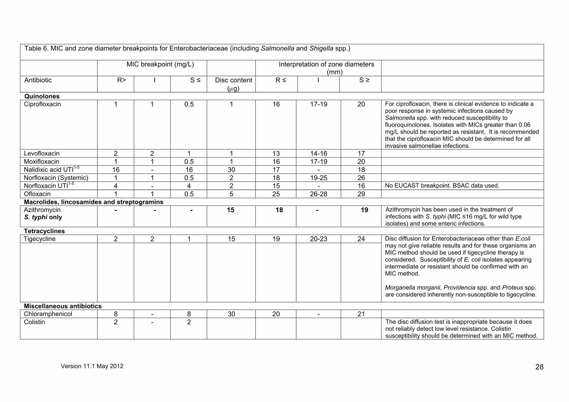
Quinolones Ciprofloxacin 1 1 0.5 1 16 17-19 20 For ciprofloxacin, there is clinical evidence to indicate a
poor response in systemic infections caused by Salmonella spp. with reduced susceptibility to fluoroquinolones. Isolates with MICs greater than 0.06 mg/L should be reported as resistant. It is recommended that the ciprofloxacin MIC should be determined for all invasive salmonellae infections.
Levofloxacin 2 2 1 1 13 14-16 17 Moxifloxacin 1 1 0.5 1 16 17-19 20 Nalidixic acid UTI1-5 16 - 16 30 17 - 18 Norfloxacin (Systemic) 1 1 0.5 2 18 19-25 26 Norfloxacin UTI1-5 4 - 4 2 15 - 16 No EUCAST breakpoint. BSAC data used. Ofloxacin 1 1 0.5 5 25 26-28 29 Macrolides, lincosamides and streptogramins Azithromycin S. typhi only
- - - 15 18 - 19 Azithromycin has been used in the treatment of infections with S. typhi (MIC ≤16 mg/L for wild type isolates) and some enteric infections.
Tetracyclines Tigecycline 2 2 1 15 19 20-23 24
Disc diffusion for Enterobacteriaceae other than E.coli may not give reliable results and for these organisms an MIC method should be used if tigecycline therapy is considered. Susceptibility of E. coli isolates appearing intermediate or resistant should be confirmed with an MIC method. Morganella morganii, Providencia spp. and Proteus spp. are considered inherently non-susceptible to tigecycline.
Miscellaneous antibiotics Chloramphenicol 8 - 8 30 20 - 21 Colistin 2 - 2 The disc diffusion test is inappropriate because it does
not reliably detect low level resistance. Colistin susceptibility should be determined with an MIC method.
Version 11.1 May 2012
29
Table 6. MIC and zone diameter breakpoints for Enterobacteriaceae (including Salmonella and Shigella spp.) MIC breakpoint (mg/L) Interpretation of zone diameters
(mm)
Antibiotic R> I S ≤ Disc content(µg)
R ≤ I S ≥
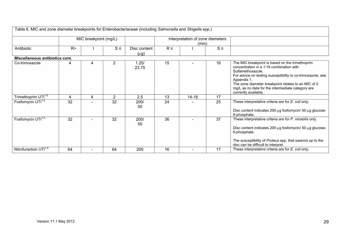
Miscellaneous antibiotics cont. Co-trimoxazole
4 4 2 1.25/ 23.75
15 - 16 The MIC breakpoint is based on the trimethoprim concentration in a 1:19 combination with Sulfamethoxazole. For advice on testing susceptibility to co-trimoxazole, see Appendix 1. The zone diameter breakpoint relates to an MIC of 2 mg/L as no data for the intermediate category are currently available.
Trimethoprim UTI1-5 4 4 2 2.5 13 14-16 17 Fosfomycin UTI1-5 32 - 32 200/
50 24 - 25 These interpretative criteria are for E. coli only.
Disc content indicates 200 µg fosfomycin/ 50 µg glucose-6-phosphate.
Fosfomycin UTI1-5 32 - 32 200/ 50
36 - 37 These interpretative criteria are for P. mirabilis only. Disc content indicates 200 µg fosfomycin/ 50 µg glucose-6-phosphate. The susceptibility of Proteus spp. that swarms up to the disc can be difficult to interpret.
Nitrofurantoin UTI1-5 64 - 64 200 16 - 17 These interpretative criteria are for E. coli only.

Version 11.1 May 2012
30
Table 7. MIC and zone diameter breakpoints for Acinetobacter spp.
MIC breakpoint (mg/L) Interpretation of zone diameters
(mm)
Antibiotic R > I S ≤ Disc content (µg)
R ≤ I S ≥ Comment
Aminoglycosides Amikacin 16 16 8 30 18 19-20 21 Gentamicin 4 - 4 10 19 - 20 Penicillins Piperacillin-tazobactam 16 16 8 75/10 19 20-21 22 No EUCAST MIC BP as there is insufficient
clinical evidence. BSAC data used. Carbapenems Doripenem 4 2-4 1 10 14 15-21 22 Imipenem 8 4-8 2 10 13 14-24 25 Meropenem 8 4-8 2 10 12 13-19 20 Quinolones Ciprofloxacin 1 - 1 1 20 - 21 Tetracyclines Tigecycline No EUCAST MIC BP as there is insufficient
clinical evidence. For determining susceptibility an MIC method should be used and the EUCAST Non-Species specific MIC BP of S = 0.25 mg/L, R = > 0.5 mg/L applied to interpret susceptibility.
Miscellaneous antibiotics Colistin 2 - 2 - - - - Disc diffusion susceptibility testing is
unreliable. An MIC method is therefore recommended.

Version 11.1 May 2012
31
Table 8. MIC and zone diameter breakpoints for Pseudomonas spp.
Table 8. MIC and zone diameter breakpoints for Pseudomonas spp.
MIC breakpoint (mg/L) Interpretation of zone diameters (mm)
Antibiotic R > I S ≤ Disc content (µg)
R ≤ I S ≥ Comment
Aminoglycosides Amikacin 16 16 8 30 15 16-21 22
Gentamicin 4 - 4 10 17 - 18 Netilmicin 4 - 4 10 13 - 14 Tobramycin 4 - 4 10 19 - 20 Penicillins
Piperacillin 16 - 16 75 24 - 25 Piperacillin-tazobactam 16 - 16 75/10 24 - 25 Ticarcillin 16 - 16 75 19 - 20 Ticarcillin-clavulanate 16 - 16 75/10 19 - 20 Cephalosporins Ceftazidime 8 - 8 30 23 - 24 Carbapenems Doripenem 4 2-4 1 10 24 25-31 32 Imipenem 8 8 4 10 16 17-22 23
The detection of resistance mediated by carbapenemases is

Version 11.1 May 2012
32
Table 8. MIC and zone diameter breakpoints for Pseudomonas spp.
MIC breakpoint (mg/L) Interpretation of zone diameters (mm)
Antibiotic R > I S ≤ Disc content (µg)
R ≤ I S ≥ Comment
Meropenem 8 4-8 2 10 15 16-19 20 difficult, particularly if resistance is not fully expressed. For epidemiological or cross infection purposes consideration should be given to testing isolates resistant to ceftazidime and a carbapenem for the presence of carbapenemases (http://www.hpa.org.uk/web/HPAwebFile/HPAweb_C/1294740725984)
Other β-Lactams Aztreonam 16 2-16 1 30 19 20-35 36 Relates only to isolates from
patients with cystic fibrosis given high dosage therapy to treat P. aeruginosa.
Quinolones Ciprofloxacin 1 1 0.5 1 12 13-22 23 Ciprofloxacin 1 1 0.5 5 19 20-29 30 Levofloxacin 2 2 1 5 16 17-21 22 No EUCAST MIC BP as there is
insufficient clinical evidence. EUCAST non-species specific MIC breakpoint and BSAC data used.
Miscellaneous antibiotics Colistin
4 - 4 The disc diffusion test is unreliable. Colistin susceptibility should be determined with an MIC method.

Version 11.1 May 2012
33
Table 9. MIC and zone diameter breakpoints for Stenotrophomonas maltophilia
MIC breakpoint (mg/L) Interpretation of zone diameters (mm)
Antibiotic R > I S ≤ Disc content (µg)
R ≤ I S ≥ Comment
Co-trimoxazole 4 - 4 1.25/23.75 19 - 20 For Stenotrophomonas maltophilia, susceptibility testing is not recommended except for co-trimoxazole (see www.bsac.org.uk BSA Standardized Susceptibility Testing Method, Additional Methodology, Stenotrophomonas maltophilia) The MIC breakpoint is based on the trimethoprim concentration in a 1:19 combination with sulfamethoxazole.

Version 11.1 May 2012
34
Table 10. MIC and zone diameter breakpoints for staphylococci
Comments 1-3 relate to urinary tract infections (UTI) only. 1 These recommendations are for organisms associated with uncomplicated urinary tract infections only. For complicated infections and infections caused by Staphylococcus aureus and Staphylococcus epidermidis, which are associated with more serious infections, systemic recommendations should be used. 2 If an organism is isolated from multiple sites, for example from blood and urine, interpretation of susceptibility should be made with regard to the systemic site (e.g., if the blood isolate is resistant and the urine isolate susceptible, both should be reported resistant irrespective of the results obtained using interpretative criteria for urine isolates). 3 Direct susceptibility tests on urine samples may be interpreted only if the inoculum gives semi-confluent growth.
Table 10. MIC and zone diameter breakpoints for staphylococci
MIC breakpoint (mg/L) Interpretation of zone diameters (mm)
Antibiotic R > I S ≤ Disc content
(µg)
R ≤ I S ≥ Comment
Aminoglycosides Amikacin for Staphylococcus aureus
16 16 8 30 15 16-18 19
Amikacin for coagulase-negative staphylococci
16 16 8 30 21 22-24 25
Gentamicin 1 - 1 10 19 - 20 Tobramycin for Staphylococcus aureus
1 - 1 10 20 - 21

Version 11.1 May 2012
35
Tobramycin for coagulase-negative staphylococci
1 - 1 10 29 - 30
Neomycin
- - - 10 16 - 17 For topical use only. The zone diameter breakpoint distinguishes the “wild type” susceptible population from isolates with reduced susceptibility.
Table 10. MIC and zone diameter breakpoints for staphylococci β-Lactams Most staphylococci are penicillinase-producers. The benzylpenicillin will mostly, but not unequivocally, separate β-lactamase producers. Isolates positive for β-lactamase are resistant to benzylpenicillin, phenoxymethylpenicillin, amino-,carboxy-and ureidopenicillins. Isolates negative for β-lactamase and susceptible to cefoxitin (cefoxitin is used to screen for “methicillin resistance”) can be reported susceptible to these drugs. Isolates positive for β-lactamase and susceptible to cefoxitin are susceptible to penicillin- β-lactamase inhibitor combinations and penicillinase-resistant penicillins (oxacillin, cloxacillin, dicloxacillin and flucloxacin). Isolates resistant to cefoxitin are methicillin resistant and resistant to β-lactam agents, including β-lactamase inhibitor combinations, except for cephalosporins with approved anti-MRSA activity and clinical breakpoints. MIC breakpoint (mg/L) Interpretation of zone diameters
(mm)
Antibiotic R > I S ≤ Disc content
(µg)
R ≤ I S ≥ Comment
Ampicillin UTI1-3
Staphylococcus saprophyticus - - - 25 25 - 26
Cefoxitin Staphylococcus aureus (Screen)
4 - - 10 21 - 22
Cefoxitin S. saprophyticus (Screen)
- - - 10 19 - 20
Cefoxitin coagulase-negative staphylococci (Screen)
4 10 21 22-26 27
Staphylococci exhibiting resistance to oxacillin/cefoxitin should be regarded as resistant to other penicillins, cephalosporins, carbapenems and combinations of β-lactam and β-lactamase inhibitors. For coagulase negative staphylococci with cefoxitin zone diameters of 22-26 mm, PCR for mecA is required to determine susceptibility for treatment of deep seated infection with any β-lactam.

Version 11.1 May 2012
36
Oxacillin (Screen)
2 1 14 - 15
Penicillin
0.12 - 0.12 1 unit 24 - 25
For oxacillin tests on Mueller–Hinton or Columbia agars with 2% NaCl: Some hyper-producers of β-lactamase give zones within the range of 7-14 mm and if possible, should be checked by a PCR method for mecA or a latex agglutination test for PBP2a. Increase in oxacillin zone size in the presence of clavulanic acid is not a reliable test for hyper-producers of β-lactamase as zones of inhibition with some MRSA also increase in the presence of clavulanic acid. Rarely, hyper-producers of β-lactamase give no zone in this test and would therefore not be distinguished from MRSA. For S. saprophyticus there is very little data for resistant mecA strains. With penicillin check for a heaped zone edge which indicates β-lactamase mediated resistance.
Table 10. MIC and zone diameter breakpoints for staphylococci
MIC breakpoint (mg/L) Interpretation of zone diameters (mm)
Antibiotic R > I S ≤ Disc content
(µg)
R ≤ I S ≥ Comment
Quinolones Ciprofloxacin 1 - 1 1 13 - 14 MIC breakpoints relate to high-dose therapy (750 mg
BD). Ciprofloxacin UTI1-3
Staphylococcus saprophyticus 1 - 1 1 17 - 18
Moxifloxacin 1 1 0.5 1 15 16-19 20 Ofloxacin 1 - 1 5 27 - 28 Glycopeptides Teicoplanin Staphylococcus aureus
2 - 2 - - - -
Teicoplanin Coagulase negative staphylococci
4 - 4 - - - -
Vancomycin Staphylococcus aureus
2 - 2 - - - -
Disc diffusion for staphylococci does not give reliable results. An MIC method should be used to determine susceptibility, positive results requiring confirmation. Population analysis is the most reliable method for confirming resistance and for distinguishing susceptible, hetero-GISA and GISA isolates. If, on clinical grounds, resistance to vancomycin is

Version 11.1 May 2012
37
Vancomycin Coagulase negative staphylococci
4
-
4
- - - - suspected, it is recommended that the organism be sent to a specialist laboratory, such as Southmead Hospital in Bristol1 or the Antibiotic Research Laboratory in Cardiff2.
(http://www.bsac.org.uk/Resources/BSAC/Use%20of%20gradient%20tests.pdf)
Macrolides, lincosamides and streptogramins Azithromycin 2 2 1 15 19 - 20 The zone diameter breakpoint relates to an MIC of 1
mg/l as no data for the intermediate category are currently available.
Clarithromycin 2 2 1 2 14 15-17 18
Table 10. MIC and zone diameter breakpoints for staphylococci MIC breakpoint (mg/L) Interpretation of zone diameters
(mm)
Antibiotic R > I S ≤ Disc content
(µg)
R ≤ I S ≥ Comment
Macrolides, lincosamides and streptogramins cont. Clindamycin 0.5 0.5 0.25 2 22 23-25 26 Erythromycin can be used to determine the
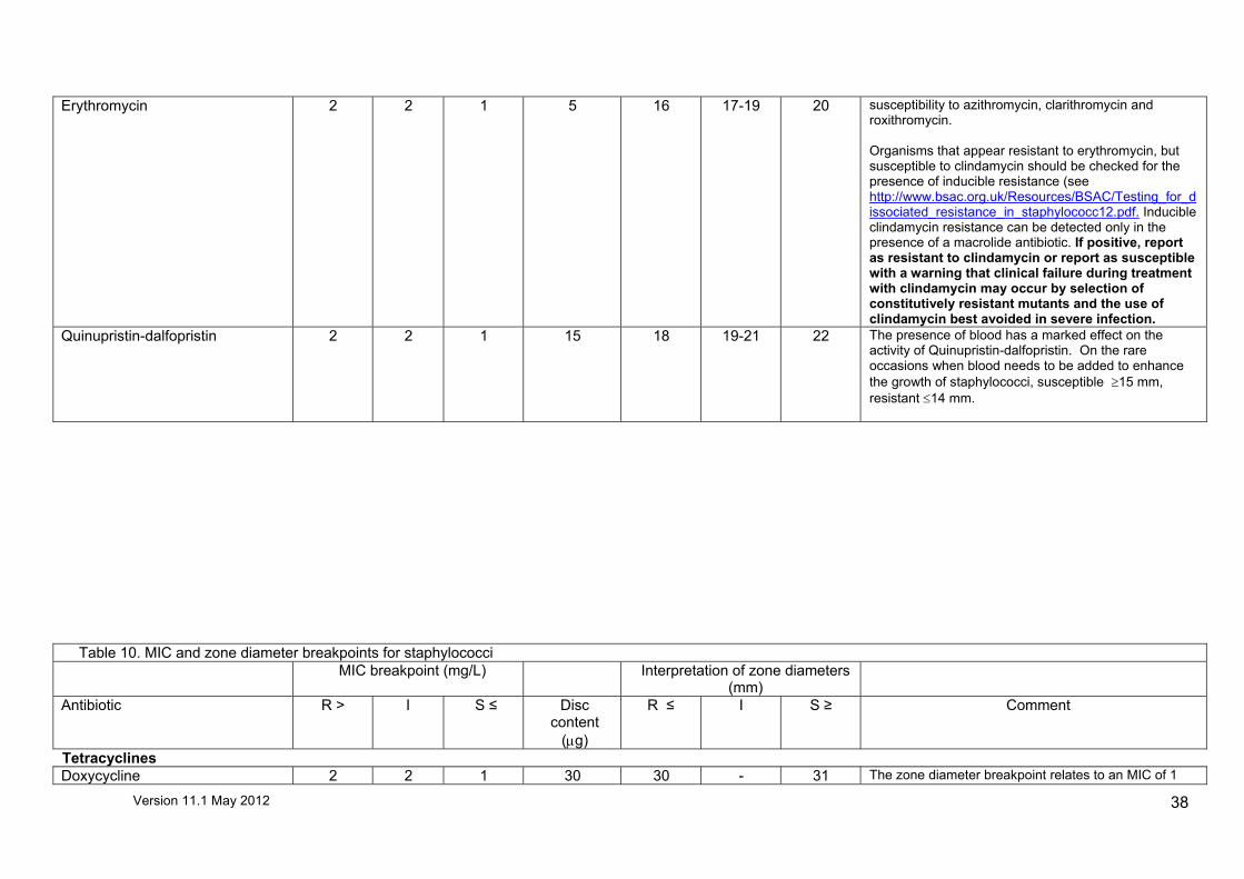
Version 11.1 May 2012
38
Erythromycin 2 2 1 5 16 17-19 20 susceptibility to azithromycin, clarithromycin and roxithromycin. Organisms that appear resistant to erythromycin, but susceptible to clindamycin should be checked for the presence of inducible resistance (see http://www.bsac.org.uk/Resources/BSAC/Testing_for_dissociated_resistance_in_staphylococc12.pdf. Inducible clindamycin resistance can be detected only in the presence of a macrolide antibiotic. If positive, report as resistant to clindamycin or report as susceptible with a warning that clinical failure during treatment with clindamycin may occur by selection of constitutively resistant mutants and the use of clindamycin best avoided in severe infection.
Quinupristin-dalfopristin 2 2 1 15 18 19-21 22 The presence of blood has a marked effect on the activity of Quinupristin-dalfopristin. On the rare occasions when blood needs to be added to enhance the growth of staphylococci, susceptible ≥15 mm, resistant ≤14 mm.
Table 10. MIC and zone diameter breakpoints for staphylococci
MIC breakpoint (mg/L) Interpretation of zone diameters (mm)
Antibiotic R > I S ≤ Disc content
(µg)
R ≤ I S ≥ Comment
Tetracyclines Doxycycline 2 2 1 30 30 - 31 The zone diameter breakpoint relates to an MIC of 1

Version 11.1 May 2012
39
mg/l as no data for the intermediate category are currently available.
Minocycline 1 1 0.5 30 27 - 28 The zone diameter breakpoint relates to an MIC of 0.5 mg/l as no data for the intermediate category are currently available.
Tetracycline 2 2 1 10 19 - 20 The zone diameter breakpoint relates to an MIC of 1 mg/l as no data for the intermediate category are currently available. Staphylococci susceptible to tetracycline are also susceptible to doxycycline and minocycline. Some staphylococci resistant to tetracycline may be susceptible to minocycline and doxycycline.
Tigecycline 0.5 - 0.5 15 25 - 26 Strains with MIC values above the susceptible breakpoint are very rare or not yet reported. The identification and antimicrobial susceptibility tests on any such isolate must be repeated and if the result is confirmed the isolate must be sent to a reference laboratory. Until there is further evidence regarding clinical response for confirmed isolates with MIC above the current resistant breakpoint they should be reported as resistant.
Table 10. MIC and zone diameter breakpoints for staphylococci MIC breakpoint (mg/L) Interpretation of zone diameters
(mm)
Antibiotic R > I S ≤ Disc content
(µg)
R ≤ I S ≥ Comment
Miscellaneous antibiotics Daptomycin 1 - 1 - - - - Strains with MIC values above the susceptible

Version 11.1 May 2012
40
breakpoint are very rare or not yet reported. The identification and antimicrobial susceptibility tests on any such isolate must be repeated and if the result is confirmed the isolate sent to a reference laboratory. Until there is evidence regarding the clinical response for confirmed isolates with MIC above the current resistant breakpoint they should be reported resistant. Susceptibility testing by disc diffusion is not reliable. Susceptibility should be determined using a broth dilution method with Mueller Hinton broth or by an MIC method on Mueller Hinton agar.
Chloramphenicol 8 - 8 10 14 - 15 Co-trimoxazole 4 4 2 1.25/23.75 13 14-16 17 For advice on testing susceptibility to co-trimoxazole
see Appendix 1. The MIC breakpoint is based on the trimethoprim concentration in a 1:19 combination with sulfamethoxazole.
Trimethoprim
1 - 1 5 19 - 20 Breakpoints are epidemiological “cut-offs” based on distributions for the “wild type” population. However, there is no clear evidence correlating these breakpoints with clinical efficacy.
Trimethoprim UTI1-3
Staphylococcus saprophyticus 4 4 2 2.5 12 13-14 15
Fosfomycin (IV) 32 - 32 200/50 33 - 34 Disc content indicates 200 µg fosfomycin/50 µg glucose-6-phosphate
Fusidic acid 1 - 1 10 29 - 30 Linezolid 4 - 4 10 19 - 20
Table 10. MIC and zone diameter breakpoints for staphylococci MIC breakpoint (mg/L) Interpretation of zone diameters
(mm)
Antibiotic R > I S ≤ Disc content
(µg)
R ≤ I S ≥ Comment
Miscellaneous antibiotics cont. Mupirocin 256 2-256 1 20 6 7-26 27

Version 11.1 May 2012
41
Nitrofurantoin UTI1-3
Staphylococcus saprophyticus 64 - 64 200 19 - 20
Rifampicin 0.5 0.12-0.5 0.06 2 23 24-29 30
1 = Department of Microbiology, Lime Walk Building, Southmead Hospital Westbury–on-Trym, Bristol, BS10 5NB. 2 = Public Health Wales, Microbiology Cardiff, University Hospital of Wales, Heath Park, Cardiff, CF14 4XW.
Table 11. MIC and zone diameter breakpoints for Streptococcus pneumoniae
Table 11. MIC and zone diameter breakpoints for Streptococcus pneumoniae MIC breakpoint (mg/L) Interpretation of zone
diameters (mm)
Antibiotic R > I S ≤ Disc content (µg)
R ≤ I S ≥ Comment
Most MIC values for penicillin, ampicillin, amoxicillin and piperacillin (with or without a β-lactamase inhibitor) differ by no more than one dilution step and isolates fully susceptible to

Version 11.1 May 2012
42
Table 11. MIC and zone diameter breakpoints for Streptococcus pneumoniae MIC breakpoint (mg/L) Interpretation of zone
diameters (mm)
Antibiotic R > I S ≤ Disc content (µg)
R ≤ I S ≥ Comment
benzlpenicillin (MIC ≤0.06 mg/L; susceptible by oxacillin disc screen) can be reported susceptible to β-lactam agents that have been given breakpoints. Penicillins Penicillin 2 0.12-2 0.06 Oxacillin1 10 11-19 20 Cephalosporins Cefaclor 0.5 0.06-0.5 0.03 - - - - Cefotaxime 2 1-2 0.5 - - - - Cefpodoxime 0.5 0.5 0.25 - - - - Ceftriaxone 2 1-2 0.5 - - - - Cefuroxime 1
1
0.5
- - - -
Reduced susceptibility to penicillin in Streptococcus pneumoniae is most reliably detected with an oxacillin 1 µg disc; confirm resistance with a penicillin MIC determination. Infections with organisms with a penicillin MIC ≤ 2mg/L may be effectively treated if adequate doses are used except in infections of the central nervous system. In addition, cefotaxime or ceftriaxone MIC determination is advised for isolates from meningitis or other invasive infections. Isolates categorised as susceptible with the oxacillin 1 µg disc can be reported susceptible to cefepime, cefotaxime, cefpodoxime, ceftriaxone, cefuroxime ± axetil and cefaclor. Isolates with MIC values above the S/I breakpoint for cefotaxime or ceftriaxone are very rare. The identification and antimicrobial susceptibility tests on any such isolate must be repeated and if the result is confirmed the isolate sent to a reference laboratory. Until there is evidence regarding clinical response for confirmed isolates with MIC above the current resistant breakpoint they should be reported resistant.
Carbapenems Ertapenem 0.5 - 0.5 - - - - Imipenem 2 - 2 - - - -
Screen for β-lactam resistance with the oxacillin 1 µg disc. Isolates categorised as susceptible can be
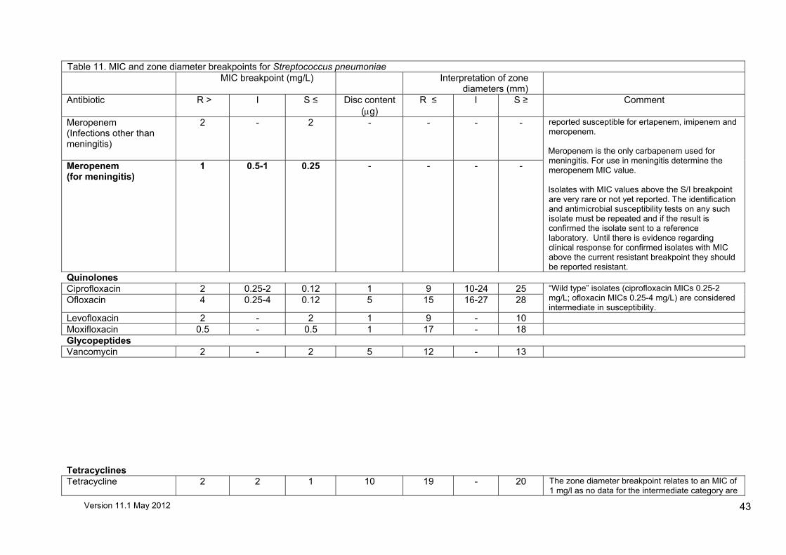
Version 11.1 May 2012
43
Table 11. MIC and zone diameter breakpoints for Streptococcus pneumoniae MIC breakpoint (mg/L) Interpretation of zone
diameters (mm)
Antibiotic R > I S ≤ Disc content (µg)
R ≤ I S ≥ Comment
Meropenem (Infections other than meningitis)
2 - 2 - - - -
Meropenem (for meningitis)
1 0.5-1 0.25 - - - -
reported susceptible for ertapenem, imipenem and meropenem. Meropenem is the only carbapenem used for meningitis. For use in meningitis determine the meropenem MIC value. Isolates with MIC values above the S/I breakpoint are very rare or not yet reported. The identification and antimicrobial susceptibility tests on any such isolate must be repeated and if the result is confirmed the isolate sent to a reference laboratory. Until there is evidence regarding clinical response for confirmed isolates with MIC above the current resistant breakpoint they should be reported resistant.
Quinolones Ciprofloxacin 2 0.25-2 0.12 1 9 10-24 25 Ofloxacin 4 0.25-4 0.12 5 15 16-27 28
“Wild type” isolates (ciprofloxacin MICs 0.25-2 mg/L; ofloxacin MICs 0.25-4 mg/L) are considered intermediate in susceptibility.
Levofloxacin 2 - 2 1 9 - 10 Moxifloxacin 0.5 - 0.5 1 17 - 18 Glycopeptides Vancomycin 2 - 2 5 12 - 13 Tetracyclines Tetracycline 2 2 1 10 19 - 20 The zone diameter breakpoint relates to an MIC of
1 mg/l as no data for the intermediate category are
Version 11.1 May 2012
44
Table 11. MIC and zone diameter breakpoints for Streptococcus pneumoniae MIC breakpoint (mg/L) Interpretation of zone
diameters (mm)
Antibiotic R > I S ≤ Disc content (µg)
R ≤ I S ≥ Comment
currently available. Isolates susceptible to tetracycline are also susceptible to doxycycline and minocycline. Some isolates resistant to tetracycline may be susceptible to minocycline and /or doxycycline.
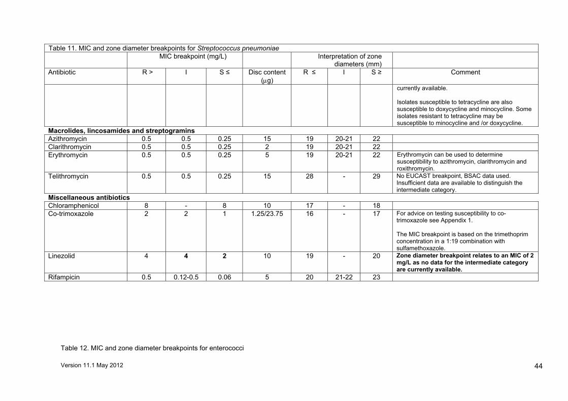
Macrolides, lincosamides and streptogramins Azithromycin 0.5 0.5 0.25 15 19 20-21 22 Clarithromycin 0.5 0.5 0.25 2 19 20-21 22 Erythromycin 0.5 0.5 0.25 5 19 20-21 22 Erythromycin can be used to determine
susceptibility to azithromycin, clarithromycin and roxithromycin.
Telithromycin 0.5 0.5 0.25 15 28 - 29 No EUCAST breakpoint, BSAC data used. Insufficient data are available to distinguish the intermediate category.
Miscellaneous antibiotics Chloramphenicol 8 - 8 10 17 - 18 Co-trimoxazole 2 2 1 1.25/23.75 16 - 17 For advice on testing susceptibility to co-
trimoxazole see Appendix 1. The MIC breakpoint is based on the trimethoprim concentration in a 1:19 combination with sulfamethoxazole.
Linezolid 4 4 2 10 19 - 20 Zone diameter breakpoint relates to an MIC of 2 mg/L as no data for the intermediate category are currently available.
Rifampicin 0.5 0.12-0.5 0.06 5 20 21-22 23
Table 12. MIC and zone diameter breakpoints for enterococci

Version 11.1 May 2012
45
Comments 1-3 relate to urinary tract infections (UTIs) only. 1 UTI recommendations are for organisms associated with uncomplicated urinary tract infections only. For complicated urinary tract infections, systemic recommendations should be used. 2 If an organism is isolated from multiple sites, for example from blood and urine, interpretation of susceptibility should be made with regard to the systemic site (e.g., if the blood isolate is resistant and the urine isolate susceptible, both should be reported resistant irrespective of the results obtained using interpretative criteria for urine isolates). 3 Direct susceptibility tests on urine samples may be interpreted only if the inoculum gives semi-confluent growth. NB. For isolates from endocarditis the MIC should be determined and interpreted according to national endocarditis guidelines (Elliott TS et al. Guidelines for the antibiotic treatment of endocarditis in adults: report of the Working Party of the British Society for Antimicrobial Chemotherapy. J Antimicrob Chemother. 2004; 54: 971-81).
Table 12. MIC and zone diameter breakpoints for enterococci
MIC breakpoint (mg/L) Interpretation of zone diameters (mm)
Antibiotic R > I S ≤ Disc content (µg)
R ≤ I S ≥ Comment
Aminoglycosides Gentamicin
128 - 128 200 14 - 15 High-level gentamicin-resistant enterococci usually give no zone or only a trace of inhibition around gentamicin 200 µg discs. Occasionally, however, the plasmid carrying the resistance gene may be unstable and the resistance is seen as a zone of inhibition with a few small colonies within the zone. Retesting of resistant colonies results in growth to the disc or increased numbers of colonies within the zone. Zones should be carefully examined to avoid missing such resistant organisms. If in doubt, isolates may be sent to a reference laboratory for confirmation.
Streptomycin 128 - 128 300 23 - 24 The EUCAST breakpoint is 512 mg/L tested on Mueller- Hinton agar which correlates with the MIC breakpoint of 128 mg/L on Iso-Sensitest agar and the zone criteria given.
Penicillins Ampicillin 8 8 4 10 19 - 20 The MIC breakpoint has changed but a review of the
data indicates that no adjustment of the zone diameter breakpoints is necessary. Co-amoxiclav susceptibility can be inferred from the ampicillin result.
Carbapenems Imipenem 8 8 4 10 16 17-18 19 Recommendations for E. faecalis only. Glycopeptides Teicoplanin 2 - 2 30 19 - 20 To ensure that microcolonies indicating reduced
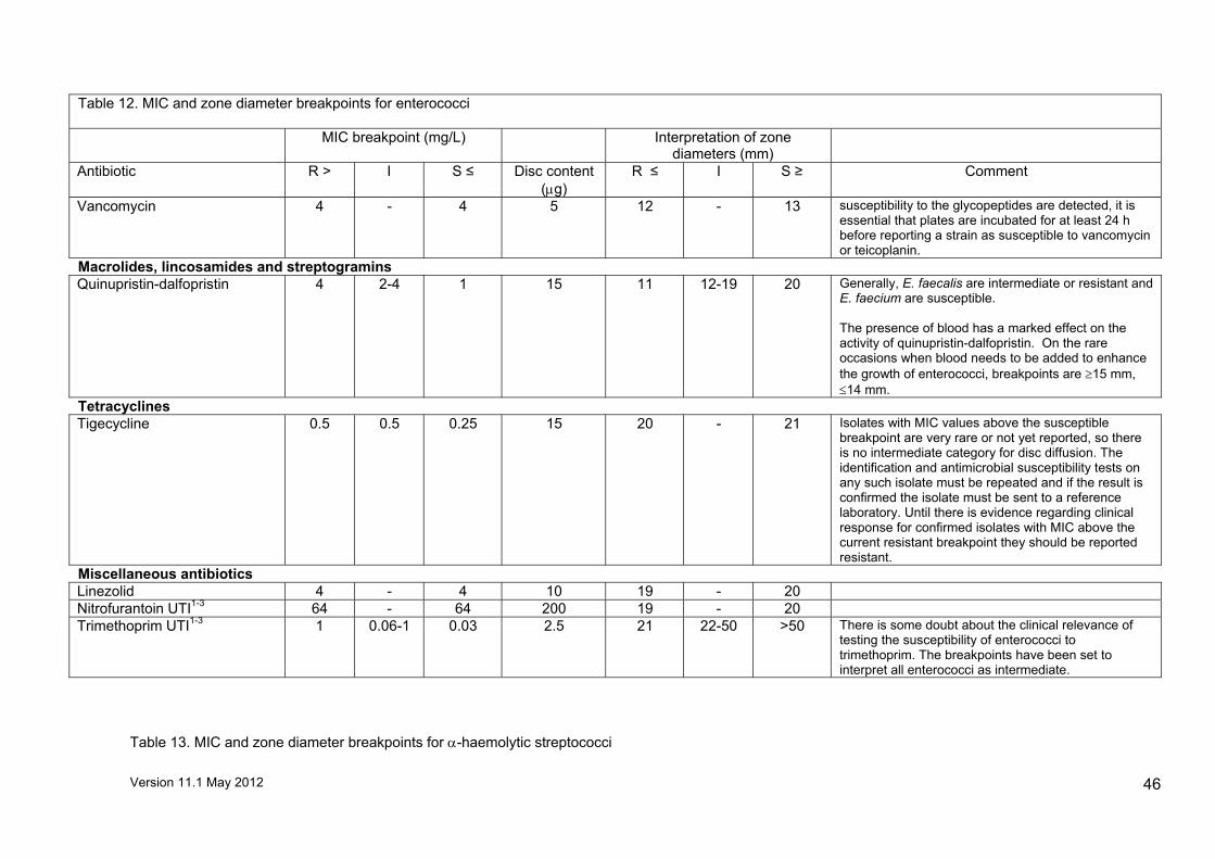
Version 11.1 May 2012
46
Table 12. MIC and zone diameter breakpoints for enterococci
MIC breakpoint (mg/L) Interpretation of zone diameters (mm)
Antibiotic R > I S ≤ Disc content (µg)
R ≤ I S ≥ Comment
Vancomycin 4 - 4 5 12 - 13 susceptibility to the glycopeptides are detected, it is essential that plates are incubated for at least 24 h before reporting a strain as susceptible to vancomycin or teicoplanin.
Macrolides, lincosamides and streptogramins Quinupristin-dalfopristin 4 2-4 1 15 11 12-19 20 Generally, E. faecalis are intermediate or resistant and
E. faecium are susceptible. The presence of blood has a marked effect on the activity of quinupristin-dalfopristin. On the rare occasions when blood needs to be added to enhance the growth of enterococci, breakpoints are ≥15 mm, ≤14 mm.
Tetracyclines Tigecycline 0.5 0.5 0.25 15 20 - 21 Isolates with MIC values above the susceptible
breakpoint are very rare or not yet reported, so there is no intermediate category for disc diffusion. The identification and antimicrobial susceptibility tests on any such isolate must be repeated and if the result is confirmed the isolate must be sent to a reference laboratory. Until there is evidence regarding clinical response for confirmed isolates with MIC above the current resistant breakpoint they should be reported resistant.
Miscellaneous antibiotics Linezolid 4 - 4 10 19 - 20 Nitrofurantoin UTI1-3 64 - 64 200 19 - 20 Trimethoprim UTI1-3 1 0.06-1 0.03 2.5 21 22-50 >50 There is some doubt about the clinical relevance of
testing the susceptibility of enterococci to trimethoprim. The breakpoints have been set to interpret all enterococci as intermediate.
Table 13. MIC and zone diameter breakpoints for α-haemolytic streptococci

Version 11.1 May 2012
47
N.B. For isolates from endocarditis the MIC should be determined and interpreted according to national endocarditis guidelines (Elliott TS et al. Guidelines for the antibiotic treatment of endocarditis in adults: report of the Working Party of the British Society for Antimicrobial Chemotherapy. J Antimicrob Chemother. 2004; 54: 971-81).
Table 13. MIC and zone diameter breakpoints for α-haemolytic streptococci MIC breakpoint (mg/L) Interpretation of zone diameters (mm)
Antibiotic R > I S ≤ Disc content (µg)
R ≤ I S ≥ Comment
Penicillins Amoxicillin 2 1-2 0.5 2 14 15-23 24 Penicillin 2 0.5-2 0.25 1 unit 10 11-16 17 Cephalosporins Cefotaxime 0.5 - 0.5 5 22 - 23 Glycopeptides Teicoplanin 2 - 2 30 15 - 16 Vancomycin 2 - 2 5 13 - 14
Macrolides, lincosamides and streptogramins Clindamycin 0.5 - 0.5 2 19 - 20 Erythromycin
2 - 2 5 19 - 20 Organisms that appear resistant to erythromycin, but susceptible to clindamycin should be checked for the presence of inducible MLSB resistance (see http://www.bsac.org.uk/Resources/BSAC/Testing_for_dissociated_resistance_in_staphylococc12.pdf). Inducible clindamycin resistance can be detected only in the presence of a macrolide antibiotic. If positive, report as susceptible to clindamycin with a warning that resistance may develop during treatment. No EUCAST MIC breakpoint for erythromycin as there is insufficient clinical evidence. BSAC data used.
Miscellaneous antibiotics Linezolid
2 - 2 10 19 - 20 No EUCAST MIC breakpoint as there is insufficient clinical evidence. BSAC data used.
Table 14. MIC and zone diameter breakpoints for β-haemolytic streptococci

Version 11.1 May 2012
48
Comments 1-3 relate to urinary tract infections (UTIs) only. 1 UTI recommendations are for organisms associated with uncomplicated urinary tract infections only. For complicated urinary tract infections and infections systemic recommendations should be used. 2 If an organism is isolated from multiple sites, for example from blood and urine, interpretation of susceptibility should be made with regard to the systemic site (e.g., if the blood isolate is resistant and the urine isolate susceptible, both should be reported resistant irrespective of the results obtained using interpretative criteria for urine isolates). 3 Direct susceptibility tests on urine samples may be interpreted only if the inoculum gives semi-confluent growth.
Table 14. MIC and zone diameter breakpoints for β-haemolytic streptococci MIC breakpoint (mg/L) Interpretation of zone
diameters (mm)
Antibiotic R > I S ≤ Disc content (µg)
R ≤ I S ≥ Comment
Penicillins Penicillin
0.25 - 0.25 1 unit 19 - 20 Susceptibility to other penicillins, carbapenems and cephalosporins can be inferred from the penicillin result.
Macrolides, lincosamides and streptogramins Azithromycin 0.5 0.5 0.25 15 19 20-21 22
Clarithromycin 0.5 0.5 0.25 2 19 20-21 22 Clindamycin 0.5 - 0.5 2 16 - 17 Organisms that appear resistant to
erythromycin, but susceptible to clindamycin should be checked for the presence of inducible MLSB resistance (see http://www.bsac.org.uk/Resources/BSAC/Testing_for_dissociated_resistance_in_staphylococc12.pdf). If positive, report as susceptible to clindamycin with a warning that resistance may develop during treatment.
Erythromycin 0.5 0.5 0.25 5 19 20-21 22 Telithromycin
0.5 0.5 0.25 15 25 - 26 Zone diameter breakpoints relate to the “wild type” susceptible population as no data are available for the non-susceptible population.
Tetracyclines Tetracycline
2 2 1 10 19 - 20 Isolates susceptible to tetracycline are also susceptible to doxycycline and minocycline.
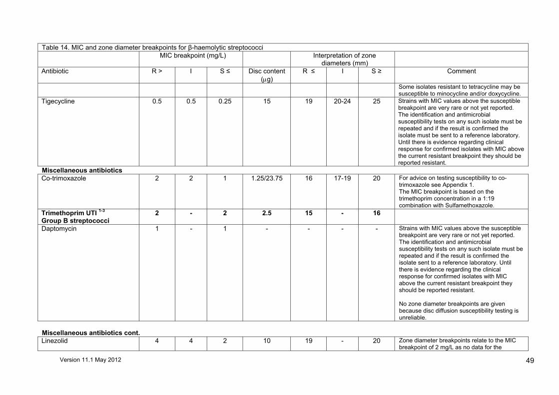
Version 11.1 May 2012
49
Table 14. MIC and zone diameter breakpoints for β-haemolytic streptococci MIC breakpoint (mg/L) Interpretation of zone
diameters (mm)
Antibiotic R > I S ≤ Disc content (µg)
R ≤ I S ≥ Comment
Some isolates resistant to tetracycline may be susceptible to minocycline and/or doxycycline.
Tigecycline
0.5 0.5 0.25 15 19 20-24 25 Strains with MIC values above the susceptible breakpoint are very rare or not yet reported. The identification and antimicrobial susceptibility tests on any such isolate must be repeated and if the result is confirmed the isolate must be sent to a reference laboratory. Until there is evidence regarding clinical response for confirmed isolates with MIC above the current resistant breakpoint they should be reported resistant.
Miscellaneous antibiotics Co-trimoxazole 2 2 1 1.25/23.75 16 17-19 20 For advice on testing susceptibility to co-
trimoxazole see Appendix 1. The MIC breakpoint is based on the trimethoprim concentration in a 1:19 combination with Sulfamethoxazole.
Trimethoprim UTI 1-3 Group B streptococci
2 - 2 2.5 15 - 16
Daptomycin 1 - 1 - - - - Strains with MIC values above the susceptible breakpoint are very rare or not yet reported. The identification and antimicrobial susceptibility tests on any such isolate must be repeated and if the result is confirmed the isolate sent to a reference laboratory. Until there is evidence regarding the clinical response for confirmed isolates with MIC above the current resistant breakpoint they should be reported resistant. No zone diameter breakpoints are given because disc diffusion susceptibility testing is unreliable.
Miscellaneous antibiotics cont. Linezolid 4 4 2 10 19 - 20 Zone diameter breakpoints relate to the MIC
breakpoint of 2 mg/L as no data for the

Version 11.1 May 2012
50
Table 14. MIC and zone diameter breakpoints for β-haemolytic streptococci MIC breakpoint (mg/L) Interpretation of zone
diameters (mm)
Antibiotic R > I S ≤ Disc content (µg)
R ≤ I S ≥ Comment
intermediate category are currently available. Nitrofurantoin UTI1-3
Group B Streptococci 64 - 64 200 18 - 19
Table 15. MIC and zone diameter breakpoints for Moraxella catarrhalis
Table 15. MIC and zone diameter breakpoints for Moraxella catarrhalis

Version 11.1 May 2012
51
MIC breakpoint (mg/L) Interpretation of zone diameters (mm) Antibiotic R > I S ≤ Disc content
(µg) R ≤ I S ≥ Comment
Penicillins Ampicillin
- - - - - - - Resistance to ampicillin by production of β- lactamase (BRO-1/2 β-lactamase) may be misidentified by disk diffusion technique and, because β-lactamase production is slow, may give weak results with in vitro tests. Since >90% of M. catarrhalis strains produce β-lactamase, testing of penicillinase production is discouraged and isolates reported resistant to ampicillin and amoxicillin.
Co-amoxiclav 1 - 1 2/1 18 - 19 Cephalosporins Cefaclor 0.12 - 0.12 30 37 - 38 MIC breakpoints render all M. catarrhalis
resistant to cefaclor. Cefuroxime
8
8
4
5 16 - 17 Zone diameter breakpoints relate to the MIC breakpoint of 4 mg//L as no data for the intermediate category are currently available.
Cefuroxime axetil 4 0.25-4 0.12 5 16 17-34 35 Carbapenems Ertapenem 0.5 - 0.5 10 34 - 35 Quinolones Ciprofloxacin 0.5 - 0.5 1 17 - 18 Levofloxacin 1 - 1 1 19 - 20 Moxifloxacin 0.5 - 0.5 1 17 - 18 Nalidixic acid (Screen) - - - 30 17 - 18 Ofloxacin 0.5 - 0.5 5 34 - 35
Quinolone resistance is most reliably detected with nalidixic acid discs.
Macrolides, lincosamides and streptogramins Clarithromycin 0.5 0.5 0.25 2 19 20-21 22 Erythromycin 0.5 0.5 0.25 5 27 - 28 Zone diameter breakpoints relate to the MIC
breakpoint of 0.25 mg/L as no data for the intermediate category are currently available. Erythromycin can be used to determine susceptibility to azithromycin and clarithromycin.
Macrolides, lincosamides and streptogramins cont. Telithromycin 0.5 0.5 0.25 15 29 - 30
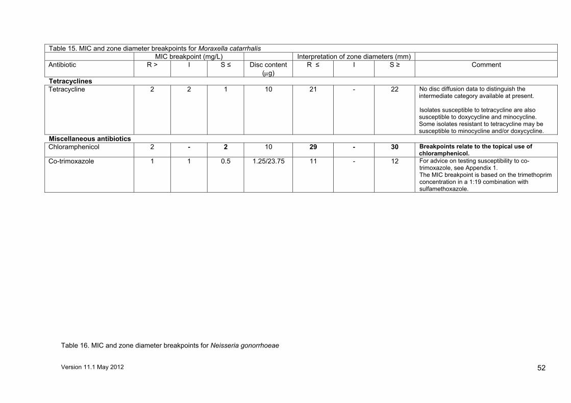
Version 11.1 May 2012
52
Table 15. MIC and zone diameter breakpoints for Moraxella catarrhalis MIC breakpoint (mg/L) Interpretation of zone diameters (mm) Antibiotic R > I S ≤ Disc content
(µg) R ≤ I S ≥ Comment
Tetracyclines Tetracycline
2 2 1 10 21 - 22 No disc diffusion data to distinguish the intermediate category available at present. Isolates susceptible to tetracycline are also susceptible to doxycycline and minocycline. Some isolates resistant to tetracycline may be susceptible to minocycline and/or doxycycline.
Miscellaneous antibiotics Chloramphenicol 2 - 2 10 29 - 30 Breakpoints relate to the topical use of
chloramphenicol. Co-trimoxazole
1 1 0.5 1.25/23.75 11 - 12 For advice on testing susceptibility to co-trimoxazole, see Appendix 1. The MIC breakpoint is based on the trimethoprim concentration in a 1:19 combination with sulfamethoxazole.
Table 16. MIC and zone diameter breakpoints for Neisseria gonorrhoeae

Version 11.1 May 2012
53
Table 16. MIC and zone diameter breakpoints for Neisseria gonorrhoeae MIC breakpoint (mg/L) Interpretation of zone diameters
(mm)
Antibiotic R > I S ≤ Disc content (µg)
R ≤ I S ≥ Comment
Penicillins Penicillin 1 0.12-1 0.06 1 unit 17 18-25 26 Always test for β-lactamase. If positive report resistant to
penicillin. Cephalosporins Cefixime 0.12 - 0.12 5 34 - 35 Cefotaxime 0.12 - 0.12 5 29 - 30 Ceftriaxone 0.12 - 0.12 5 34 - 35 Cefuroxime (Screen) - - - 5 19 - 20
Results for isolates with reduced zones around ceftriaxone, cefotaxime and cefixime discs should be confirmed by MIC determinations. Although cefuroxime is not recommended for clinical use, it can be used as an indicator antibiotic to detect reduced susceptibility to other oxyimino cephalosporin.
Quinolones Ciprofloxacin 0.06 0.06 0.03 1 28 - 29
Nalidixic acid - - - 30 9 10-31 32
For ciprofloxacin the zone diameter breakpoints relate to the MIC breakpoint of 0.03mg/L as no data for the intermediate category are currently available. Quinolone resistance is generally reliably detected with nalidixic acid; however there are a few isolates that are resistant to ciprofloxacin yet susceptible to nalidixic acid in disc diffusion tests. The mechanism of resistance and the prevalence of these isolates in the UK is still under investigation. Isolates with reduced susceptibility to fluoroquinolones normally have no zone of inhibition with a 30 µg nalidixic acid disc. For organisms with nalidixic acid zone diameters 10-31 mm a ciprofloxacin MIC should be determined if the patient is to be treated with this agent.
Macrolides, lincosamides and streptogramins Azithromycin 0.5 0.5 0.25 15 27 - 28 Zone diameter breakpoints relate to the MIC breakpoints
of >0.5 mg/L as disc diffusion testing will not reliably differentiate between the intermediate and susceptible populations.
Tetracyclines Tetracycline 1 1 0.5 10 26 27-31 32 The tetracycline result may be used to infer susceptibility
to doxycycline. Isolates susceptible to tetracycline are also susceptible to minocycline, but some isolates resistant to tetracycline may be susceptible to minocycline.

Version 11.1 May 2012
54
Table 16. MIC and zone diameter breakpoints for Neisseria gonorrhoeae MIC breakpoint (mg/L) Interpretation of zone diameters
(mm)
Antibiotic R > I S ≤ Disc content (µg)
R ≤ I S ≥ Comment
Miscellaneous antibiotics Spectinomycin 64 - 64 25 13 - 14

Version 11.1 May 2012
55
Table 17. MIC and zone diameter breakpoints for Neisseria meningitidis
Table 17. MIC and zone diameter breakpoints for Neisseria meningitidis MIC breakpoint (mg/L) Interpretation of zone
diameters (mm)
Antibiotic R > I S ≤ Disc content (µg)
R ≤ I S ≥ Comment
Penicillins Ampicillin - - - 2 31 - 32 Amoxicillin - - - 2 29 - 30
Ampicillin and amoxicillin are used as indicator antibiotics to detect reduced susceptibility to penicillin. The recommendations given are for this purpose only; ampicillin and amoxicillin should not be used therapeutically. EUCAST MIC breakpoints are S ≤ 0.12 mg/L, R > 1 mg/L. Currently there are no BSAC MIC breakpoints and zone diameter breakpoints relating to the presence of specific mutations in the penA gene.
Penicillin 0.25 0.12-0.25 0.06 1 unit 14 15-28 29 Cephalosporins
Cefotaxime 0.12 - 0.12 5 39 - 40 Ceftriaxone 0.12 - 0.12 5 39 - 40
Quinolones
Ciprofloxacin 0.06 0.06 0.03 1 31 - 32 Quinolone resistance is most reliably detected in tests with nalidixic acid. Isolates with reduced susceptibility to fluoroquinolones have no zone of inhibition with 30 µg nalidixic acid discs. Zone diameter breakpoints relate to the MIC breakpoint of 0.03 mg/L as no data for the intermediate category are currently available.
Miscellaneous antibiotics
Chloramphenicol 4 4 2 10 19 - 20 Zone diameter breakpoints relate to the MIC breakpoint of 2 mg/L as insufficient data to distinguish the intermediate category are currently available.
Rifampicin 0.25 - 0.25 2 29 - 30 Epidemiological breakpoint based on an MIC breakpoint of 0.25 mg/L.

Version 11.1 May 2012
56
Table 18. MIC and zone diameter breakpoints for Haemophilus influenzae
Table 18. MIC and zone diameter breakpoints for Haemophilus influenzae MIC breakpoint (mg/L) Interpretation of zone diameters
(mm)
Antibiotic R > I S ≤ Disc content (µg)
R ≤ I S ≥ Comment
Penicillins Amoxicillin 2 - 2 2 13 - 14 Ampicillin
1 - 1 2 17 - 18 Always test for β-lactamase; β-lactamase positive isolates should be reported resistant Breakpoints apply to β-lactamase negative isolates only. Strains may be resistant to penicillins, aminopenicillins and/or cephalosporins due to changes in PBPs (BLNAR, β-lactamase negative ampicillin resistant) and a few strains have both resistance mechanisms (BLPACR, β-lactamase positive, amoxicillin-clavulanate resistant). Isolates susceptible to ampicillin/amoxicillin are also susceptible to piperacillin and piperacillin-tazobactam and isolates susceptible to amoxicillin-clavulanate are also susceptible to piperacillin-tazobactam. Susceptibility to amoxicillin can be inferred from ampicillin.
Co-amoxiclav 2 - 2 2/1 13 - 14
Cephalosporins
Cefaclor
0.5 - 0.5 30 14 - 15 See Appendix 2. MIC breakpoints render most H. influenzae resistant for cefaclor. The disc diffusion test can be used to screen for BLNAR. Isolates with zone diameters<15 mm should be checked for ampicillin and cephalosporin resistance.
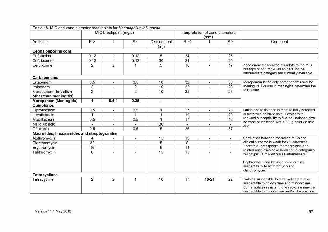
Version 11.1 May 2012
57
Table 18. MIC and zone diameter breakpoints for Haemophilus influenzae MIC breakpoint (mg/L) Interpretation of zone diameters
(mm)
Antibiotic R > I S ≤ Disc content (µg)
R ≤ I S ≥ Comment
Cephalosporins cont.
Cefotaxime 0.12 - 0.12 5 24 - 25 Ceftriaxone 0.12 - 0.12 30 24 - 25 Cefuroxime 2 2 1 5 16 - 17 Zone diameter breakpoints relate to the MIC
breakpoint of 1 mg//L as no data for the intermediate category are currently available.
Carbapenems Ertapenem 0.5 - 0.5 10 32 - 33 Imipenem 2 - 2 10 22 - 23 Meropenem (Infection other than meningitis)
2 - 2 10 22 - 23
Meropenem (Meningitis) 1 0.5-1 0.25 - - - -
Meropenem is the only carbapenem used for meningitis. For use in meningitis determine the MIC value.
Quinolones Ciprofloxacin 0.5 - 0.5 1 27 - 28 Levofloxacin 1 - 1 1 19 - 20 Moxifloxacin 0.5 - 0.5 1 17 - 18 Nalidixic acid - - - 30 - - - Ofloxacin 0.5 - 0.5 5 26 - 37
Quinolone resistance is most reliably detected in tests with nalidixic acid. Strains with reduced susceptibility to fluoroquinolones give no zone of inhibition with a 30µg nalidixic acid disc.
Macrolides, lincosamides and streptogramins Azithromycin 4 - - 15 19 - - Clarithromycin 32 - - 5 8 - - Erythromycin 16 - - 5 14 - - Telithromycin 8 - - 15 15 - -
Correlation between macrolide MICs and clinical outcome is weak for H. influenzae. Therefore, breakpoints for macrolides and related antibiotics have been set to categorize “wild type” H. influenzae as intermediate. Erythromycin can be used to determine susceptibility to azithromycin and clarithromycin.
Tetracyclines Tetracycline 2 2 1 10 17 18-21 22 Isolates susceptible to tetracycline are also
susceptible to doxycycline and minocycline. Some isolates resistant to tetracycline may be susceptible to minocycline and/or doxycycline.
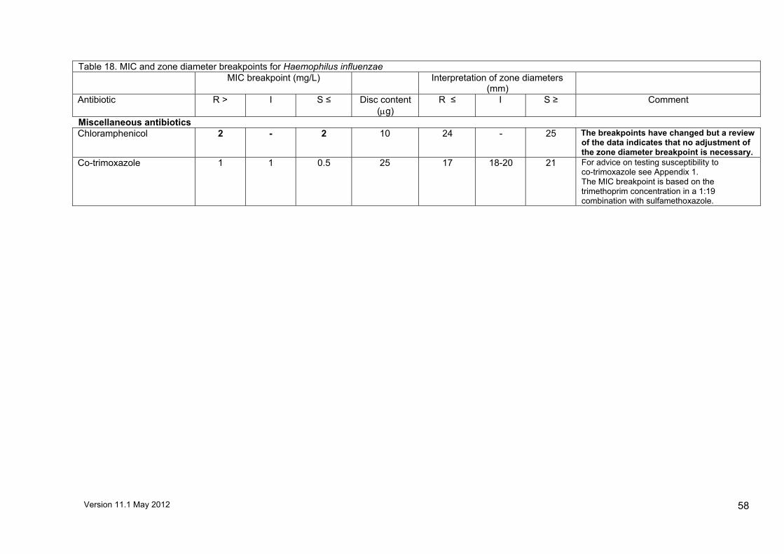
Version 11.1 May 2012
58
Table 18. MIC and zone diameter breakpoints for Haemophilus influenzae MIC breakpoint (mg/L) Interpretation of zone diameters
(mm)
Antibiotic R > I S ≤ Disc content (µg)
R ≤ I S ≥ Comment
Miscellaneous antibiotics Chloramphenicol 2 - 2 10 24 - 25 The breakpoints have changed but a review
of the data indicates that no adjustment of the zone diameter breakpoint is necessary.
Co-trimoxazole 1 1 0.5 25 17 18-20 21 For advice on testing susceptibility to co-trimoxazole see Appendix 1. The MIC breakpoint is based on the trimethoprim concentration in a 1:19 combination with sulfamethoxazole.

Version 11.1 May 2012
59
Table 19. MIC and zone diameter breakpoints for Pasteurella multocida
MIC breakpoint (mg/L) Interpretation of zone diameters (mm) Antibiotic R > I S ≤ Disc content
(µg) R ≤ I S ≥ Comment
Penicillins Ampicillin 1 - 1 10 29 - 30 Penicillin 0.12 - 0.12 1 unit 21 - 22 Cephalosporins Cefotaxime 1 - 1 5 33 - 34 Quinolones Ciprofloxacin 1 - 1 1 28 - 29 Nalidixic acid - - - 30 27 - 28
Quinolone resistance is most reliably detected in tests with nalidixic acid discs.
Tetracyclines Tetracycline 1 - 1 10 25 - 26

Version 11.1 May 2012
60
Table 20. MIC and zone diameter breakpoints for Campylobacter spp.
MIC breakpoint (mg/L) Interpretation of zone diameters (mm) Antibiotic R > I S ≤ Disc content
(µg) R ≤ I S ≥ Comment
Quinolones Ciprofloxacin 1 1 0.5 1 25 - 26 Nalidixic acid - - - 30 19 - 20
Quinolone resistance is most reliably detected in tests with nalidixic acid discs. The zone diameters for ciprofloxacin relate to an MIC breakpoint of 0.5 mg/L as no data for the intermediate category are currently available.
Macrolides, lincosamides and streptogramins Erythromycin 4 - 4 5 21 - 22 The susceptibility of clarithromycin can be
inferred from the erythromycin result.

Version 11.1 May 2012 61
Table 21. MIC and zone diameter breakpoints for Coryneform organisms
MIC breakpoint (mg/L) Interpretation of zone diameters (mm)
Antibiotic R > I S ≤ Disc content (µg)
R ≤ I S ≥ Comment
Penicillins Penicillin 0.12 - 0.12 1 unit 19 - 20 Quinolones Ciprofloxacin 1 1 0.5 1 11 12-16 17 The zone diameters relate to an MIC breakpoint
of 0.5 mg/L as no data for the intermediate category are currently available.
Glycopeptides Vancomycin 8 8 4 5 19 - 20 The zone diameters relate to an MIC breakpoint
of 4 mg/L as no data for the intermediate category are currently available.

Version 11.1 May 2012 62
Table 22. MIC and zone diameter breakpoints for Gram-negative anaerobes Table 22. MIC and zone diameter breakpoints for Gram-negative anaerobes
MIC breakpoint (mg/L) Interpretation of zone diameters (mm)
Antibiotic R > I S ≤ Disc content (µg)
R ≤ I S ≥ Comment
Penicillins Ampicillin 2 1-2 0.5 - - - -
Amoxicillin 2 1-2 0.5 - - - -
Co-amoxiclav 8 8 4 30 20 21-28 29 Zone diameter breakpoints are for B. fragilis only.
Penicillin 0.5 - 0.25 - - - - Susceptibility to ampicillin, amoxicillin and piperacillin ± tazobactam can be inferred from the susceptibility to penicillin. B. fragilis is inherently resistant to penicillin.
Piperacillin 16 - 16 - - - - Piperacillin-tazobactam 16 16 8 75/10 26 - 27 Zone diameter breakpoints are for B. fragilis
only. The breakpoints are based on the “wild type” susceptible population as there are few clinical data relating MIC to outcome. Organisms that appear resistant in disc diffusion tests should have resistance confirmed by MIC determination and resistant isolates should be sent to the Anaerobe Reference Laboratory in Cardiff. The zone diameter breakpoint relates to an MIC of 8 mg/L as no data for the intermediate category are currently available.
Ticarcillin 16 - 16 - - - - Ticarcillin-clavulanate 16 16 8 - - - - Carbapenems Doripenem 1 - 1 - - - - Ertapenem 1 - 1 - - - -
Imipenem 8 4-8 2 - - - -
Meropenem 8 4-8 2 10 18 19-25 26 Zone diameter breakpoints are for B. fragilis and B. thetaiotaomicron only.

Version 11.1 May 2012 63
Table 22. MIC and zone diameter breakpoints for Gram-negative anaerobes MIC breakpoint (mg/L) Interpretation of zone diameters
(mm)
Antibiotic R > I S ≤ Disc content (µg)
R ≤ I S ≥ Comment
Macrolides, lincosamides and streptogramins Clindamycin 4 - 4 2 9 - 10 Zone diameter breakpoints are for B. fragilis
and B. thetaiotaomicron only. The breakpoints are based on the “wild type” susceptible population as there are few clinical data relating MIC to outcome. Organisms that appear resistant in disc diffusion tests should heave resistance confirmed by MIC determination and resistant isolates should be sent to the Anaerobe Reference Laboratory in Cardiff.
Miscellaneous antibiotics Chloramphenicol 8 - 8 - - - - Metronidazole 4 - 4 5 17 - 18 Zone diameter breakpoints are for B. fragilis
and B. thetaiotaomicron only.

Version 11.1 May 2012 64
Table 23. MIC and zone diameter breakpoints for Gram-positive anaerobes except Clostridium difficile Table 23. MIC and zone diameter breakpoints for Gram-positive anaerobes except Clostridium difficile
MIC breakpoint (mg/L) Interpretation of zone diameters (mm)
Antibiotic R > I S ≤ Disc content (µg)
R ≤ I S ≥ Comment
Penicillins Ampicillin 8 8 4 - - - -
Amoxicillin 8 8 4 - - - -
Co-amoxiclav 8 8 4 30 31 - 32 The zone diameter breakpoints are for C. perfringens only. The zone diameter breakpoint relates to an MIC of 4 mg/L as no data for the intermediate category are currently available.
Penicillin 0.5 0.5 0.25 1 unit 22 - 23 The zone diameter breakpoints are for C. perfringens only. The breakpoints are based on the “wild type” susceptible population as there are few clinical data relating MIC to outcome. Organisms that appear resistant in disc diffusion tests should have resistance confirmed by MIC determination and resistant isolates should be sent to the Anaerobe Reference Laboratory in Cardiff. For penicillin the zone diameter breakpoint relates to an MIC of 0.25 mg/L as no data for the intermediate category are currently available. Susceptibility to ampicillin, amoxicillin and piperacillin ± tazobactam can be inferred from susceptibility to penicillin.

Version 11.1 May 2012 65
Table 23. MIC and zone diameter breakpoints for Gram-positive anaerobes except Clostridium difficile MIC breakpoint (mg/L) Interpretation of zone diameters
(mm)
Antibiotic R > I S ≤ Disc content (µg)
R ≤ I S ≥ Comment
Penicillins cont. Piperacillin 16 16 8 - - - - Piperacillin-tazobactam 16 16 8 75/10 29 - 30 The zone diameter breakpoints are for C.
perfringens only. The breakpoints are based on the “wild type” susceptible population as there are few clinical data relating MIC to outcome. Organisms that appear resistant in disc diffusion tests should have resistance confirmed by MIC determination and resistant isolates should be sent to the Anaerobe Reference Laboratory in Cardiff. For piperacillin-tazobactam the zone diameter breakpoint relates to an MIC of 8 mg/L as no data for the intermediate category are currently available.
Ticarcillin 16 16 8 - - - - Ticarcillin-clavulanate 16 16 8 - - - - Carbapenems Doripenem 1 - 1 - - - - Ertapenem 1 - 1 - - - -
Imipenem 8 4-8 2 - - - -
Meropenem 8 4-8 2 10 18 19-25 26 Zone diameter breakpoints are for C. perfringens only.
Glycopeptides Vancomycin 2 - 2 - - - -

Version 11.1 May 2012 66
Table 23. MIC and zone diameter breakpoints for Gram-positive anaerobes except Clostridium difficile MIC breakpoint (mg/L) Interpretation of zone diameters
(mm)
Antibiotic R > I S ≤ Disc content (µg)
R ≤ I S ≥ Comment
Macrolides, lincosamides and streptogramins Clindamycin 4 - 4 2 9 - 10 Zone diameter breakpoints are for C.
perfringens only. The breakpoints are based on the “wild type” susceptible population as there are few clinical data relating MIC to outcome. Organisms that appear resistant in disc diffusion tests should heave resistance confirmed by MIC determination and resistant isolates should be sent to the Anaerobe Reference Laboratory in Cardiff.
Miscellaneous antibiotics Chloramphenicol 8 - 8 - - - - Metronidazole 4 - 4 5 17 - 18 Zone diameter breakpoints are for C.
perfringens only. There is no evidence for changing the epidemiological zone diameter breakpoint in line with the change in MIC breakpoint.

Version 11.1 May 2012 67
Table 24. MIC and zone diameter breakpoints for Clostridium difficile Table 24. MIC and zone diameter breakpoints for Clostridium difficile
MIC breakpoint (mg/L) Interpretation of zone diameters (mm)
Antibiotic R > I S ≤ Disc content (µg)
R ≤ I S ≥ Comment
Daptomycin 4 - - - - - - Not used clinically. May be tested for epidemiological purposes only. MIC breakpoint based on the ECOFF for the “wild type” population.
Fusidic acid 2 - - - - - - Not used clinically. May be tested for epidemiological purposes only. MIC breakpoint based on the ECOFF for the “wild type” population.
Metronidazole 2 - 2 - - - - The breakpoints are based on epidemiological “cut-off” values (ECOFFs) which distinguish “wild-type” isolates from those with reduced susceptibility.
Moxifloxacin 4 - - - - - - Not used clinically. May be tested for epidemiological purposes only. MIC breakpoint based on the ECOFF for the “wild type” population.
Tigecycline 0.25 - - - - - - Not used clinically. May be tested for epidemiological purposes only. MIC breakpoint based on the ECOFF for the “wild type” population.
Rifampicin 0.004 - - - - - - Not used clinically. May be tested for epidemiological purposes only. MIC breakpoint based on the ECOFF for the “wild type” population.
Vancomycin 2 - 2 - - - - The breakpoints are based on epidemiological “cut-off” values (ECOFFs) which distinguish “wild-type” isolates from those with reduced susceptibility.

Version 11.1 May 2012 68
Appendix 1: Advice on testing the susceptibility to co-trimoxazole Breakpoints for testing susceptibility to co-trimoxazole are provided. However, the following recommendations from the UK Committee on the Safety of Medicines (CSM) should be noted.
”Co-trimoxazole should be limited to the role of drug of choice in Pneumocyctis carinii pneumonia, it is also indicated for toxoplasmosis and nocardiasis. It should now only be considered for use in acute exacerbations of chronic bronchitis and infections of the urinary tract when there is good bacteriological evidence of sensitivity to co-trimoxazole and good reason to prefer this combination to a single antibiotic; similarly it should only be used in acute otitis media in children when there is good reason to prefer it. Review of the safety of co-trimoxazole using spontaneous adverse drug reaction data has indicated that the profile of reported adverse reactions with trimethoprim is similar to that with co-trimoxazole; blood and generalised skin disorders are the most serious reactions with both drugs and predominantly have been reported to occur in elderly patients. A recent large post-marketing study has demonstrated that such reactions are very rare with co-trimoxazole; the study did not distinguish between co-trimoxazole and trimethoprim with respect to serious hepatic, renal, blood or skin disorders.”
Version 11.1 May 2012 69
Appendix 2: Efficacy of cefaclor in the treatment of respiratory infections caused by Haemophilus influenzae
Concerns have been expressed, particularly by laboratories moving from Stokes’ method to the BSAC disc diffusion method, about the interpretation of susceptibility of Haemophilus influenzae to cefaclor. When using Stokes’ method the majority of isolates appeared susceptible; but with the BSAC disc diffusion method most isolates are now reported resistant. The following comments explain the BSAC rationale for interpretation of cefaclor susceptibility. Cefaclor pharmacokinetics Cefaclor is dosed at 250-500 mg TDS po: 250 mg TDS is probably the most common dose but data is absent to confirm this. The expected Cmax for 250 mg is 5-10 mg/L and 10-20 mg/l for 500 mg; the half life is 1 h; drug concentration in blood is <1 mg/L at 4 h and the protein binding is 25-50%. Tissue penetration is similar to other β-lactams. Cefaclor potency against Haemophilus influenzae Data from the BSAC surveillance programme 2003-2004 (n= 899) indicates that the cefaclor MIC range is 0.12-128 mg/L; MIC50 2 mg/L; MIC90 8 mg/L. Pharmacodynamics An average patient with an Haemophilus influenzae infection will have a free drug Time>MIC of 25% with 250 mg dosing and 37% with 500 mg dosing. A conservative Time>MIC target for cephalosporins in community practice is 40-50%, but this is not achieved with cefaclor. Therefore, it is likely that cefaclor will have at best borderline activity against Haemophilus influenzae. Conclusion The pharmacodynamic data indicate that cefaclor has borderline activity against Haemophilus influenzae, even for community use. The outcome of infection will be difficult to predict and susceptibility testing is likely to be of limited value.

Version 11.1 May 2012 70
Acknowledgment The BSAC acknowledges the assistance of the Swedish Reference Group for Antibiotics (SRGA) in supplying some breakpoint data for inclusion in this document. References 1. Moosdeen, F., Williams, J.D. & Secker, A. (1988). Standardization of inoculum
size for disc susceptibility testing: a preliminary report of a spectrophotometric method. J. Antimicrob Chemother 21, 439-43.
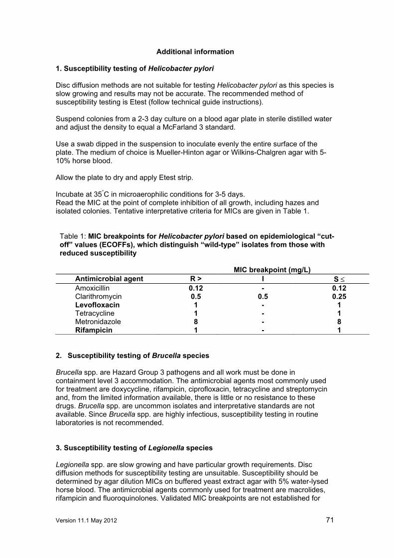
Version 11.1 May 2012 71
Additional information 1. Susceptibility testing of Helicobacter pylori Disc diffusion methods are not suitable for testing Helicobacter pylori as this species is slow growing and results may not be accurate. The recommended method of susceptibility testing is Etest (follow technical guide instructions). Suspend colonies from a 2-3 day culture on a blood agar plate in sterile distilled water and adjust the density to equal a McFarland 3 standard. Use a swab dipped in the suspension to inoculate evenly the entire surface of the plate. The medium of choice is Mueller-Hinton agar or Wilkins-Chalgren agar with 5-10% horse blood. Allow the plate to dry and apply Etest strip. Incubate at 35°C in microaerophilic conditions for 3-5 days. Read the MIC at the point of complete inhibition of all growth, including hazes and isolated colonies. Tentative interpretative criteria for MICs are given in Table 1. Table 1: MIC breakpoints for Helicobacter pylori based on epidemiological “cut-off” values (ECOFFs), which distinguish “wild-type” isolates from those with reduced susceptibility
MIC breakpoint (mg/L)
Antimicrobial agent R > I S ≤ Amoxicillin 0.12 - 0.12 Clarithromycin 0.5 0.5 0.25 Levofloxacin 1 - 1 Tetracycline 1 - 1 Metronidazole 8 - 8 Rifampicin 1 - 1
2. Susceptibility testing of Brucella species Brucella spp. are Hazard Group 3 pathogens and all work must be done in containment level 3 accommodation. The antimicrobial agents most commonly used for treatment are doxycycline, rifampicin, ciprofloxacin, tetracycline and streptomycin and, from the limited information available, there is little or no resistance to these drugs. Brucella spp. are uncommon isolates and interpretative standards are not available. Since Brucella spp. are highly infectious, susceptibility testing in routine laboratories is not recommended. 3. Susceptibility testing of Legionella species Legionella spp. are slow growing and have particular growth requirements. Disc diffusion methods for susceptibility testing are unsuitable. Susceptibility should be determined by agar dilution MICs on buffered yeast extract agar with 5% water-lysed horse blood. The antimicrobial agents commonly used for treatment are macrolides, rifampicin and fluoroquinolones. Validated MIC breakpoints are not established for

Version 11.1 May 2012 72
Legionella spp. If results for test isolates are within range of the normal wild type distribution, given in table 2, clinical susceptibility may be assumed. Table 2: MIC ranges for wild type Legionella spp.
Antimicrobial agent MIC range for wild-type Legionella spp. (mg/L)
Erythromycin 0.06-0.5 Clarithromycin 0.004-0.06 Rifampicin 0.004-0.06 Ciprofloxacin 0.016-0.06
4. Susceptibility testing Listeria spp. For susceptibility testing Listeria spp. an MIC determination is advised on Iso-Sensitest agar with incubation at 35-370C in air. If a gradient method is used the test should be undertaken following the manufacturer’s instructions. In Table 3 the MIC ranges and cut offs for “wild type” strains are shown and these can be used as an aid to interpreting susceptibility. Table 3: MIC ranges for “wild type” Listeria spp.
Antimicrobial agent
MIC range (mg/L)
MIC cut off (mg/L)
Comment
Ampicillin 0.12-4 ≤1 Penicillin 0.015-2 ≤1
No resistance described
Meropenem - ≤1 Daptomycin 1-4 ≤4 Erythromycin 0.12-1 ≤1 Resistance very rare ≤ 0.5% Gentamicin 0.06-1 ≤1 Linezolid 1-4 ≤4 Tetracycline 0.06-1 ≤1 Resistance rare 0% Trimethoprim 0.06-1 ≤1 Co-trimoxazole - ≤ 0.06 The MIC breakpoint is based on the
trimethoprim concentration in a 1:19 combination with sulfamethoxazole. For advice on testing susceptibility to co-trimoxazole, see Appendix 1.
Vancomycin 0.5-4 ≤4
5. Susceptibility testing of topical antibiotics MIC breakpoints specifically for topical antibiotics are not given because there are no pharmacological, pharmacodynamic or clinical response data on which to base recommendations. [Relevant data would be gratefully received].

Version 11.1 May 2012 73
6. Development of MIC and zone diameter breakpoints All breakpoints are subject to review in the light of additional data and any data relating to breakpoints, control zone ranges or any other aspect of antimicrobial susceptibility testing would be welcome (contact the Working Party secretary or any member listed at the front of this document). The BSAC is part of the European Committee on Antimicrobial Susceptibility Testing (EUCAST) and is actively involved in the process of harmonization of MIC breakpoints in Europe. This process will undoubtedly lead to some small breakpoint adjustments, and these will be incorporated into the BSAC method as European breakpoints are agreed. The BSAC has a mechanism to modify and publish changes to breakpoints on an annual basis via the BSAC www site (www.bsac.org.uk). Any changes will be dated. Ad hoc modifications to breakpoints by users are not acceptable.
Version 11.1 May 2012 74
Control of Antimicrobial Susceptibility Testing 1. Control strains Control strains include susceptible strains to monitor test performance (not for the interpretation of susceptibility), and resistant strains to confirm that the method will detect particular mechanisms of resistance, for example, Haemophilus influenzae ATCC 49247 is a β-lactamase negative, ampicillin resistant strain (see table 2 of Disc Diffusion Method). Tables 2-6 provide zone diameters for recommended control organisms under a range of test conditions. Control strains can be purchased from the National Collection of Type Cultures (NCTC; HPA Centre for Infections, 61 Colindale Avenue, London NW9 5HT). Alternatively, some may be obtained commercially (see section on suppliers) 2. Maintenance of control strains Store control strains by a method that minimises the risk of mutations, for example, at -700C, on beads in glycerol broth. Ideally, two vials of each control strain should be stored, one as an ”in-use” supply, the other for archiving. Every week a bead from the ”in-use” vial should be subcultured on to appropriate non-selective media and checked for purity. From this pure culture, prepare one subculture for each of the following 7 days. Alternatively, for fastidious organisms that will not survive on plates for 7 days, subculture the strain daily for no more than 6 days. 3. Calculation of control ranges for disc diffusion tests The acceptable ranges for the control strains have been calculated by combining zone diameter data from `field studies' and from multiple centres supplying their daily control data, from which cumulative distributions of zones of inhibition have been prepared. From these distributions, the 2.5 and 97.5 percentiles were read to provide a range that would contain 95% of observations. If distributions are normal, these ranges correspond to the mean ± 1.96 SD. The percentile ranges obtained by this method are, however, still valid even if the data do not show a normal distribution. 4. Frequency of routine testing with control strains When the method is first introduced, daily testing is required until there are acceptable readings from 20 consecutive days (this also applies when new agents are introduced or when any test component changes). This provides sufficient data to support once weekly testing. 5. Use of control data to monitor the performance of disc diffusion tests Use a reading frame of 20 consecutive results (remove the oldest result when adding a new one to make a total of 20) as illustrated in Figure 1. Testing is acceptable if no more than1 in every 20 results is outside the limits of acceptability. If 2 or more results fall out of the acceptable range this requires immediate investigation. Look for trends within the limits of acceptability e.g. tendency for zones to be at the limits of acceptability; tendency for zones to be consistently above or below the mean;

Version 11.1 May 2012 75
gradual drift in zone diameters. Quality Assurance will often pick up trends before the controls go out of range.
6. Recognition of atypical results for clinical isolates
Atypical results with clinical isolates may indicate problems in testing that may or may not be reflected in zone diameters with control strains.
An organism with inherent resistance appears susceptible e.g. Proteus spp. susceptible to colistin or nitrofurantoin. Resistance is seen in an organism when resistance has previously not been observed, e.g. penicillin resistance in Group A streptococci. Resistance is seen in an organism when resistance is rare or has not been seen locally, e.g. vancomycin resistance in Staphylococcus aureus. Incompatible susceptibilities are reported, e.g. a methicillin resistant staphylococcus reported susceptible to a β-lactam antibiotic.
In order to apply such rules related to atypical results it is useful to install an `expert’ system for laboratory reporting to avoid erroneous interpretation. 7. Investigation of possible sources of error If the control values are found to be outside acceptable limits on more than one occasion during a reading frame of twenty tests, investigation into the possible source of error is required. Possible problem areas are indicated in table 1.

Version 11.1 May 2012 76
Table 1: Potential sources of error in disc diffusion antimicrobial susceptibility testing.
Possible source of error
Detail to check
Test conditions Excessive pre-incubation before discs applied Excessive pre-diffusion before plates incubated Incorrect incubation temperature Incorrect incubation atmosphere Incorrect incubation time Inadequate illumination of plates when reading Incorrect reading of zone edges
Medium Required susceptibility testing agar not used Not prepared as required by the manufacturer’s instructions Batch to batch variation Antagonists present (e.g. with sulphonamides and trimethoprim) Incorrect pH Incorrect divalent cation concentration Incorrect depth of agar plates Agar plates not level Expiry date exceeded
Antimicrobial discs Wrong agent or content used Labile agent possibly deteriorated Light sensitive agent left in light Incorrect storage leading to deterioration Disc containers opened before reaching room temperature Incorrect labelling of disc dispensers Expiry date exceeded
Control strains Contamination Mutation Incorrect inoculum density Uneven inoculation Old culture used
8. Reporting susceptibility results when controls indicate problems
Microbiologists must use a pragmatic approach, as results from repeat testing are not available on the same day. If results with control strains are out of range the implications for test results need to be assessed. Control results out of range
If control zones are below range but test results are susceptible, or control zones are above range but test results are resistant, investigate possible sources of error but report the test results. Otherwise it may be necessary to suppress reports on affected agents, investigate and retest.
Atypical results If results are atypical with clinical isolates, the purity of the isolate and identification should be confirmed and the susceptibility repeated. Suppress the results for individual agents and retest.

Version 11.1 May 2012 77
Table 2: Acceptable zone diameter (mm) ranges for control strains on Iso-Sensitest agar, plates incubated at 35-37 0C in air for 18-20 h.
Escherichia coli Pseudomonas
aeruginosa Staphylococcus
aureus Enterococcus
faecalis Antimicrobial agent
Disc
content (µg
unless stated)
NCTC 10418
ATCC 25922
NCTC 115601
NCTC 10662
ATCC 27853
NCTC 6571
ATCC 25923
ATCC 29212
Amikacin 30 24-27 23-27 - 21-30 26-32 25-30 25-29 - Ampicillin 10 21-26 16-22 - - - - - 26-35 Ampicillin 25 24-30 21-28 - - - 42-50 40-46 - Amoxicillin 10 20-24 13-18 - - - - - - Aztreonam 30 39-44 36-40 - 27-30 26-30 - - - Azithromycin 15 - - - - - 27-33 25-30 15-21 Carbenicillin 100 - - - 20-25 18-23 - - - Cefamandole 30 32-36 35-39 - - - - - - Cefepime 30 38-43 37-42 - - - - - - Cefepime- clavulanic acid
30/10 38-43 37-42 - - - - - -
Cefixime 5 32-36 27-30 - - - - - - Cefoxitin 30 28-33 26-30 - - - - - - Cefotaxime 30 36-45 34-44 - 20-29 20-24 - - - Cefotaxime-clavulanic acid
30/10 39-44 37-42 - - - - - -
Cefotetan 30 36-41 34-38 - - - - - - Cefpodoxime 10 29-36 25-31 - - - - - - Cefpodoxime- clavulanic acid
10/1 29-36 25-31 - - - - - -
Cefpirome 30 34-43 36-43 - - - - - - Ceftazidime 30 32-40 31-39 - 29-37 27-35 - - - Ceftazidime-clavulanic acid
30/10 31-39 30-36 - - - - - -
Ceftizoxime 30 44-49 40-44 - - - - - - Ceftriaxone 30 41-46 37-42 - - - - - - Cefuroxime 30 25-32 24-29 - - - - - - Cefalexin 30 21-28 16-21 - - - - - -

Version 11.1 May 2012 78
Escherichia coli Pseudomonas aeruginosa
Staphylococcus aureus
Enterococcus faecalis
Antimicrobial agent
Disc
content (µg
unless stated)
NCTC 10418
ATCC 25922
NCTC 115601
NCTC 10662
ATCC 27853
NCTC 6571
ATCC 25923
ATCC 29212
Cefradine 30 19-25 16-22 - - - - - - Cephalothin 30 22-26 17-21 - - - - - - Chloramphenicol 10 21-27 20-29 - - - 20-26 19-27 - Ciprofloxacin 1 31-40 31-37 - 21-28 24-30 25-32 17-22 14-19 Ciprofloxacin 5 - - - 29-37 31-37 - - 21-27 Clarithromycin 2 - - - - - 25-30 24-28 - Clindamycin 2 - - - - - 30-35 26-33 No zone Co-amoxiclav 3 - - - - - 32-38 27-32 - Co-amoxiclav 30 18-31 20-26 12-18 - - 42-50 37-44 - Colistin 25 15-19 16-20 - 17-20 16-20 - - - Cotrimoxazole 25 33-38 28-34 - - - - 31-35 - Cotrimoxazole incubation @ 300C
25 35-39 31-34 - - - - - -
Doripenem 10 - - - 33-37 41-45 - - - Doxycycline 30 - - - - - 35-40 33-37 - Ertapenem 10 35-41 35-39 - - - - - - Erythromycin 5 - - - - - 22-31 22-29 - Fosfomycin trometamol/G6P
200/50 29-33 36-41 - - - 25-32 25-30 27-31
Fusidic acid 10 - - - - - 32-40 30-37 - Gentamicin 10 21-27 21-27 - 20-26 22-28 24-30 22-29 - Gentamicin 200 - - - - - - - 22-27 Imipenem 10 32-37 33-37 - 20-27 23-28 - - 28-32 Levofloxacin 1 30-33 28-34 - - - - - - Levofloxacin 5 - - - 22-29 23-29 - - - Linezolid 10 - - - - - 26-33 26-30 24-29 Mecillinam 10 34-39 30-35 - - - - - - Meropenem 10 38-42 27-39 - 26-33 32-39 - - 22-28 Mezlocillin 75 31-36 27-32 - - - - - - Minocycline 30 - - - - - 34-39 33-36 -

Version 11.1 May 2012 79
Escherichia coli Pseudomonas aeruginosa
Staphylococcus aureus
Enterococcus faecalis
Antimicrobial agent
Disc
content (µg
unless stated)
NCTC 10418
ATCC 25922
NCTC 115601
NCTC 10662
ATCC 27853
NCTC 6571
ATCC 25923
ATCC 29212
Moxifloxacin 1 31-35 29-33 - - - 33-40 33-38 - Moxifloxacin 5 - - - 19-24 23-27 - - - Mupirocin 5 - - - - - 26-35 24-34 - Mupirocin 20 - - - - - 30-38 27-35 - Nalidixic acid 30 28-36 26-32 - - - - - - Neomycin 10 - - - - - 18-22 21-27 - Netilmicin 10 22-27 22-26 - 17-20 20-24 - 22-28 - Nitrofurantoin 200 25-30 23-27 - - - 21-25 20-26 - Norfloxacin 2 34-37 32-36 - - - - - - Ofloxacin 5 31-37 31-38 - 18-26 18-25 - - Penicillin 1 unit - - - - - 32-40 28-36 - Piperacillin 75 30-35 27-32 - 27-35 27-34 - - - Pip/tazobactam 85 30-35 26-31 - 28-35 28-35 - - 26-32 Quinupristin- Dalfopristin
15 - - - - - 27-31 - 12-19
Rifampicin 2 - - - - - 27-39 29-36 - Streptomycin 10 18-24 17-22 - - - - - - Streptomycin 300 - - - - - - - 20-24 Teicoplanin 30 - - - - - 17-23 16-20 19-25 Tetracycline 10 23-29 22-28 - - - 31-40 26-35 9-13 Ticarcillin 75 32-35 27-30 - 24-28 23-27 - - - Ticarcillin- clavulanic acid
85 33-37 27-31 - 25-29 24-27 - - -
Tigecycline 15 29-32 28-32 - - - 29-34 27-30 26-31 Tobramycin 10 24-27 23-27 - 23-30 26-32 26-31 29-35 - Trimethoprim 2.5 30-37 25-31 - - - 25-30 20-28 28-35 Trimethoprim 5 - - - - - 24-34 - - Vancomycin 5 - - - - - 14-20 13-17 13-19
1 = β-Lactamase producing strain

Version 11.1 May 2012 80
Table 3: Acceptable zone diameter (mm) ranges for control strains on Iso-Sensitest agar supplemented with 5% defibrinated horse blood, with or without the addition of NAD, plates incubated at 35-370C in air for 18-20 h.
Staphylococcus aureus Group A streptococci Antimicrobial agent Disc content (µg unless stated) NCTC 6571 ATCC 25923 NCTC 8198 ATCC 19615
Amoxicillin 2 25-29 - - - Cefuroxime 5 20-27 - - - Chloramphenicol 10 17-23 - - - Clindamycin 2 - - 25-28 29-35 Co-amoxiclav 2/1 29-36 - - - Erythromycin 5 26-33 23-29 - - Nalidixic acid 30 6-9 - - - Penicillin 1 unit 30-41 27-35 - - Tetracycline 10 30-38 28-36 - -
Table 4: Acceptable zone diameter ranges for control strains for detection of methicillin/oxacillin/cefoxitin resistance in staphylococci (methicillin/oxacillin incubated at 300C; cefoxitin incubated at 350C). Staphylococcus aureus Antimicrobial agent
Medium Disc content
(µg) NCTC 6571 ATCC
25923 NCTC 12493a
Methicillin Columbia/Mueller Hinton agar + 2% NaCl 5 18-30 18-28 No zone Oxacillin Columbia/Mueller Hinton agar + 2% NaCl 1 19-30 19-29 No zone Cefoxitin ISA 10 26-31 24-29 10-20
a Methicillin/oxacillin/cefoxitin- resistant strain.
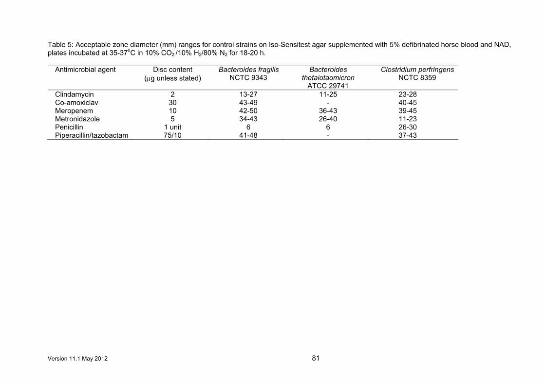
Version 11.1 May 2012 81
Table 5: Acceptable zone diameter (mm) ranges for control strains on Iso-Sensitest agar supplemented with 5% defibrinated horse blood and NAD, plates incubated at 35-370C in 10% CO2 /10% H2/80% N2 for 18-20 h.
Antimicrobial agent Disc content (µg unless stated)
Bacteroides fragilis NCTC 9343
Bacteroides thetaiotaomicron
ATCC 29741
Clostridium perfringens NCTC 8359
Clindamycin 2 13-27 11-25 23-28 Co-amoxiclav 30 43-49 - 40-45 Meropenem 10 42-50 36-43 39-45 Metronidazole 5 34-43 26-40 11-23 Penicillin 1 unit 6 6 26-30 Piperacillin/tazobactam 75/10 41-48 - 37-43

Version 11.1 May 2012 82
Table 6: Acceptable zone diameter (mm) ranges for control strains on Iso-Sensitest agar supplemented with 5% defibrinated horse blood with or
without the addition of NAD, plates incubated at 35-370C in 4-6% CO2 for 18-20 h.
Pasteurella multocida
Neisseria gonorrhoeae
(with NAD)
Staphylococcus aureus
Haemophilus influenzae (with NAD)
Streptococcus pneumoniae
Antimicrobial agent
Disc content ( µg unless
stated) NCTC 8489 NCTC 12700 NCTC 6571
ATCC 25923
NCTC 11931
ATCC 49247a
ATCC 49619
Amoxicillin 2 - - 29-34 - 20-26 No zone - Ampicillin 2 - - - - 22-30 6-13 - Ampicillin 10 32-37 - - - - - - Azithromycin 15 - 30-40 - - 28-32 23-27 25-30 Cefaclor 30 - - - - 29-38 No zone 26-33 Cefixime 5 - 33-44 - - - - - Cefotaxime 5 35-41 32-44 26-32 - 33-45 27-38 27-35 Ceftazidime 30 - - - - 39-46 36-41 - Ceftizoxime 30 - - - - - - 36-44 Ceftriaxone 5 - 33-47 - - 47-54 38-44 - Ceftriaxone 30 - - - - - - 38-47 Cefuroxime 5 - 23-32 22-29 24-29 22-28 6-16 - Chloramphenicol 10 - - 21-26 - 30-40 30-38 21-29 Ciprofloxacin 1 31-37 40-50 22-29 18-23 32-40 33-44 14-21 Clarithromycin 2 - - - - 6-10 No zone 26-31 Clindamycin 2 - - 21-25 - - - - Co-amoxiclav 3 - - 29-36 - 20-27 10-20 - Co-trimoxazole 25 - - - - 40-47 38-42 21-25 Ertapenem 10 - - - - 30-38 25-34 35-40 Erythromycin 5 - 20-29 25-29 - 12-23 9-16 23-36 Imipenem 10 - - - - 32-39 31-36 - Levofloxacin 1 - - - - 38-43 35-41 17-21 Linezolid 10 - - 22-26 - - - - Meropenem 10 - - - - 38-45 33-39 - Moxifloxacin 1 - - - - 36-42 33-39 24 Nalidixic acid 30 - 32-40 9-17 9-17 33-38 33-39 - Ofloxacin 5 - - - - 39-49 38-44 21-26 Oxacillin 1 - - - - - - 8-16 Penicillin 1 unit 24-28 12-20 37-44 29-36 - - -

Version 11.1 May 2012 83
Pasteurella multocida
Neisseria gonorrhoeae
(with NAD)
Staphylococcus aureus
Haemophilus influenzae (with NAD)
Streptococcus pneumoniae
Antimicrobial agent
Disc content ( µg unless
stated) NCTC 8489 NCTC 12700 NCTC 6571
ATCC 25923
NCTC 11931
ATCC 49247a
ATCC 49619
Quinupristin- Dalfopristin
15 - - - - - - 21-29
Rifampicin 2 - 26-34 32-37 - - - - Rifampicin 5 - - - - - - 28-35 Spectinomycin 25 - 17-23 - - - - - Teicoplanin 30 - - 14-19 - - - - Telithromycin 15 - - - - 26-31 22-26 33-40 Tetracycline 10 29-34 27-35 33-40 27-34 27-35 9-14 26-36 Tigecycline 15 - - 27-30 24-28 - - 26-30 Trimethoprim 2.5 - - - - 30-40 28-36 - Vancomycin 5 - - 12-16 - - - -
a β-Lactamase-negative, ampicillin-resistant strain

Version 11.1 May 2012 84
9. Control of MIC determination Tables 7-10 provide target MIC (mg/L) values for recommended control strains by BSAC methodology.1,2 MICs should be within one two-fold dilution of the target values i.e. target MIC 1 mg/L acceptable range 0.5 – 2 mg/L. Table 7: Target MICs (mg/L) for Haemophilus influenzae, Enterococcus faecalis,
Streptococcus pneumoniae, Bacteroides fragilis and Neisseria gonorrhoeae control strains by BSAC methods
Haemophilus influenzae
Enterococcus faecalis
Streptococcus pneumoniae
Bacteroides fragilis
Neisseria gonorrhoeae
NCTC ATCC ATCC ATCC NCTC ATCC
Antimicrobial
agent 11931 49247 29212 49619 9343 49226
Amikacin - - 128 - - - Amoxicillin 0.5 4 0.5 0.06 32 0.5 Ampicillin - - 1 0.06 32 - Azithromycin 2 2 - 0.12 - - Azlocillin - - - - 4 - Aztreonam - - >128 - 2 - Cefaclor - 128 >32 2 >128 - Cefamandole - - - - 8 - Cefixime 0.03 0.25 - 1 64 - Cefotaxime - 0.25 32 0.06 4 - Cefoxitin - - - - 4 - Cefpirome 0.06 0.5 16 16 - Cefpodoxime 0.12 0.5 >32 0.12 32 - Ceftazidime 0.12 - >32 - 8 - Ceftriaxone - - >32 0.06 4 - Cefuroxime 2 16 >32 0.25 32 - Cephadroxil - - >32 - 32 - Cephalexin - - >32 - 64 - Cephalothin - - 16 - - - Chloramphenicol - - 4 4 4 - Ciprofloxacin 0.008 0.008 1 1 2 0.004 Clarithromycin 8 4 - 0.03 0.25 0.5 Clindamycin - - 8 0.12 0.5 - Co-amoxiclav 0.5 8 0.5 0.06 0.5 0.5 Cotrimoxazole - 1 2 4 - - Dalfopristin/ quinupristin
- - 1 0.5 16 -
Enoxacin - - - - 1 - Ertapenem 0.12 0.5 - 0.12 0.25 - Erythromycin 8 8 4 0.12 1 0.5 Faropenem - - - 0.06 1 - Fleroxacin - - - - 4 - Flucloxacillin - - - - 16 - Fucidic acid - - 2 - - - Gatifloxacin 0.008 - 0.25 0.25 0.5 0.004 Gemifloxacin 0.12 - 0.03 0.03 0.25 0.002 Gentamicin - - 8 - 128 - Grepafloxacin - 0.004 - 0.25 - - Imipenem - - 0.5 - 0.06 - Levofloxacin 0.008 0.015 1 0.5 0.5 0.008 Linezolid - - 1 2 4 - Loracarbef - 128 >32 2 >128 - Mecillinam - - >128 - >128 - Meropenem - - 2 - 0.06 -
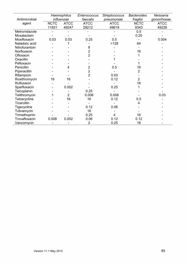
Version 11.1 May 2012 85
Haemophilus influenzae
Enterococcus faecalis
Streptococcus pneumoniae
Bacteroides fragilis
Neisseria gonorrhoeae
NCTC ATCC ATCC ATCC NCTC ATCC
Antimicrobial
agent 11931 49247 29212 49619 9343 49226
Metronidazole - - - - 0.5 - Moxalactam - - - - 0.25 - Moxifloxacin 0.03 0.03 0.25 0.5 - 0.004 Naladixic acid - 1 - >128 64 - Nitrofurantoin - - 8 - - - Norfloxacin - - 2 - 16 - Ofloxacin - - 2 - 1 - Oxacillin - - - 1 - - Pefloxacin - - - - 1 - Penicillin - 4 2 0.5 16 - Piperacillin - - 2 - 2 - Rifampicin - - 2 0.03 - - Roxithromycin 16 16 - 0.12 2 - Rufloxacin - - - - 16 - Sparfloxacin - 0.002 - 0.25 1 - Teicoplanin - - 0.25 - - - Telithromycin 1 2 0.008 0.008 - 0.03 Tetracycline - 16 16 0.12 0.5 - Ticarcillin - - - - 4 - Tigecycline - - 0.12 0.06 - - Tobramycin - - 16 - - - Trimethoprim - - 0.25 4 16 - Trovafloxacin 0.008 0.002 0.06 0.12 0.12 - Vancomycin - - 2 0.25 16 -

Version 11.1 May 2012 86
Table 8: Target MICs (mg/L) for Escherichia coli, Pseudomonas aeruginosa and Staphylococcus aureus control strains by BSAC methods
Escherichia coli Pseudomonas
aeruginosa Staphylococcus aureus
NCTC ATCC NCTC ATCC NCTC ATCC ATCC
Antimicrobial agent
10418 25922 10662 27853 6571 25923 29213 Amikacin 0.5 1 2 2 1 - 2 Amoxicillin 2 4 >128 >128 0.12 0.25 0.5 Ampicillin 2 4 >128 >128 0.06 - - Azithromycin - - - - 0.12 0.12 0.12 Azlocillin 4 - 4 - 0.25 - - Aztreonam 0.03 0.25 4 2 >128 - >128 Carbenicillin 2 - 32 - 0.5 - - Cefaclor 1 2 >128 >128 1 - 1 Cefamandole 0.25 - >128 >128 0.25 - - Cefixime 0.06 0.25 16 - 8 8 16 Cefotaxime 0.03 0.06 8 8 0.5 - 1 Cefotetan 0.06 - >128 >128 4 - - Cefoxitin 4 - >128 >128 2 2 - Cefpirome 0.03 0.03 4 1 0.25 - 0.5 Cefpodoxime 0.25 0.25 128 >128 1 4 2 Ceftazidime 0.06 0.25 1 1 4 - 8 Ceftizoxime 0.008 - - - 2 - - Ceftriaxone 0.03 0.06 8 8 1 - 2 Cefuroxime 2 4 >128 >128 0.5 1 1 Cephadroxil 8 8 >128 >128 1 - 2 Cephalexin 4 8 >128 >128 1 - 4 Cephaloridine - - >128 >128 0.06 - - Cephalothin 4 8 >128 >128 0.5 - 0.25 Cephradine - - >128 >128 2 - - Chloramphenicol 2 4 128 - 2 - 2 Ciprofloxacin 0.015 0.015 0.25 0.25 0.12 0.5 0.5 Clarithromycin - - - - 0.12 0.12 0.12 Clindamycin - - - - 0.06 0.12 0.06 Co-amoxiclav 2 4 >128 128 0.12 0.12 0.25 Colistin 0.5 - 2 4 128 - - Cotrimoxazole 0.25 0.25 - - - - 2 Dalfopristin/ Quinupristin
- - - - 0.12 0.25 0.25
Daptomycin Mueller Hinton
- - - - 1 2 -
Dirythromycin - - - - 1 - 1 Doripenem 0.008 0.008 0.5 0.25 - - - Doxycycline - - - - 0.06 0.12 - Enoxacin 0.25 - 1 - 0.5 - - Ertapenem 0.008 0.015 - - - - - Erythromycin - - - - 0.12 0.5 0.25 Farapenem 0.25 - >128 >128 0.12 - - Fleroxacin 0.06 0.12 1 - 0.5 - - Flucloxacillin - - >128 >128 0.06 - - Flumequine 2 - >128 >128 - - - Fosfomycin 4 - >128 >128 8 - - Fusidic acid >128 - - - 0.06 0.12 0.06 Gatifloxacin 0.015 0.015 1 1 0.03 0.12 0.12 Gemifloxacin 0.008 0.008 0.25 0.25 0.015 0.03 0.03 Gentamicin 0.25 0.5 1 1 0.12 0.25 0.25 Grepafloxacin 0.03 0.03 0.5 - 0.03 - - Imipenem 0.06 0.12 2 1 0.015 - 0.015

Version 11.1 May 2012 87
Escherichia coli Pseudomonas aeruginosa
Staphylococcus aureus
NCTC ATCC NCTC ATCC NCTC ATCC ATCC
Antimicrobial agent
10418 25922 10662 27853 6571 25923 29213 Kanamycin 1 - 1 - 2 - - Levofloxacin 0.03 0.03 0.5 0.5 0.12 0.25 0.25 Linezolid - - - - 0.5 1 - Lomefloxacin - - - - 0.5 - - Loracarbef 0.5 1 >128 >128 0.5 - 1 Mecillinam 0.12 0.12 8 - 8 - 64 Meropenem 0.015 0.008 2 0.25 0.03 - 0.06 Methicillin - - >128 >128 1 2 2 Mezlocillin 2 - 8 - 0.5 - - Minocycline - - - - 0.06 0.06 - Moxalactam 0.03 - 8 - 8 - - Moxifloxacin 0.03 0.03 2 2 0.06 0.06 0.06 Mupirocin - - - - 0.25 0.25 0.12 Nalidixic acid 2 4 >128 >128 >128 128 128 Neomycin - - 32 - 0.12 - - Netilmicin - - 1 0.5 - - - Nitrofurantoin 4 8 - - 8 - 16 Norfloxacin 0.06 0.06 1 1 0.25 - 1 Ofloxacin 0.06 0.03 1 1 0.25 - 0.5 Oxacillin - - >128 >128 0.25 0.25 0.5 Pefloxacin 0.06 - 0.5 - 0.25 - - Penicillin - - >128 >128 0.03 0.03 0.12 Piperacillin 0.5 2 4 2 0.25 - 1 Piperacillin/ tazobactam
0.5 2 4 4 - - -
Rifampicin 16 - - - 0.004 0.015 0.004 Roxithromycin - - - - 0.25 0.5 0.5 Rufloxacin 0.5 - 8 - 1 - - Sparfloxacin 0.015 0.015 0.5 0.5 0.03 - - Sulphonamide 16 - >128 >128 64 - - Sulphamethoxazole 0.06 0.12 - - - - - Teicoplanin - - - - 0.25 1 1 Telithromycin - - - - 0.03 0.06 0.06 Temocillin 2 - >128 - 128 - - Tetracycline 1 2 - 32 0.06 0.06 0.5 Ticarcillin 1 - 16 - 0.5 - - Ticarcillin/ 4mg/L clavulanate
- - 32 16 - - -
Tigecycline 0.12 0.12 - - 0.12 - - Tobramycin 0.25 0.5 0.5 0.5 0.12 - 0.5 Trimethoprim 0.12 0.25 32 - 0.25 - 0.5 Trovafloxacin 0.015 0.015 0.5 0.5 0.015 0.03 0.03 Vancomycin - - - - 0.5 0.5 1

Version 11.1 May 2012 88
Table 9: Target MICs (mg/L) for Pasteurella multocida control strain by BSAC methods
Pasteurella multocida
NCTC Antimicrobial agents
8489 Ampicillin 0.12 Cefotaxime 0.004 Ciprofloxacin 0.008 Penicillin 0.12 Tetracycline 0.25
Table 10: Target MICs (mg/L) for anaerobic control strains by BSAC methods on Iso-Sensitest agar supplemented with 5% defibrinated horse blood and 20 mg/L NAD
Antimicrobial agent
Bacteroides
fragilis NCTC 9343
Bacteroides
thetaiotaomicron ATCC 29741
Clostridium perfringens NCTC 8359
Clindamycin 0.5 2 0.06 Co-amoxiclav (2:1 ratio)
0.5 0.5 ≤ 0.06
Meropenem 0.06 0.12 ≤ 0.015 Metronidazole 0.5 4 8 Penicillin 16 16 0.06 Piperacillin/tazobactam (fixed 4 mg/L tazobactam)
≤ 0.12 8 0.5
Table 11: Target MICs (mg/L) for Group A streptococci control strains by BSAC methods
Group A streptococci Antimicrobial agent
NCTC 8198
ATCC 19615
Clindamycin 0.03 0.06

Version 11.1 May 2012 89
References
1. Andrews, J.M. Determination of minimum inhibitory concentrations. Journal of Antimicrobial Chemotherapy, Suppl S1 to Volume 48 July 2001.
2. Andrews, J. M., Jevons, G., Brenwald, N. and Fraise, A. for the BSAC Working Party
on Sensitivity Testing. Susceptibility testing Pasteurella multocida by BSAC standardized methodology. Journal of Antimicrobial Chemotherapy.
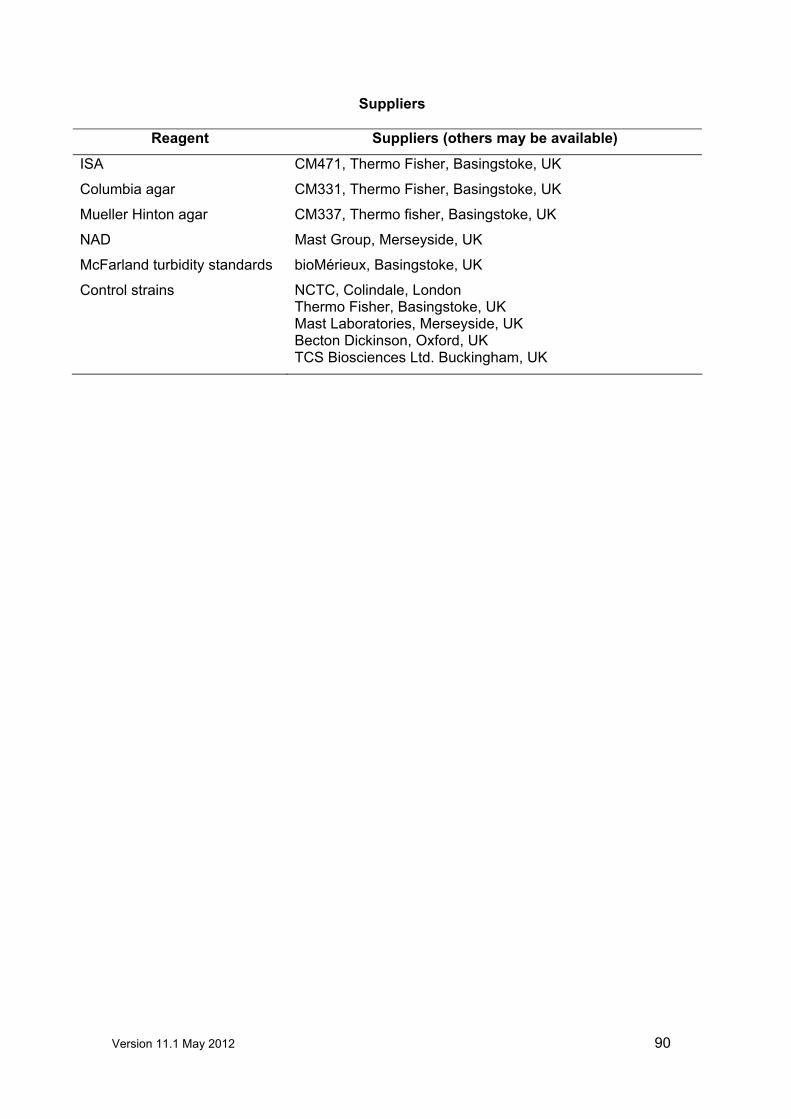
Version 11.1 May 2012 90
Suppliers
Reagent Suppliers (others may be available)
ISA CM471, Thermo Fisher, Basingstoke, UK Columbia agar CM331, Thermo Fisher, Basingstoke, UK
Mueller Hinton agar CM337, Thermo fisher, Basingstoke, UK
NAD Mast Group, Merseyside, UK McFarland turbidity standards bioMérieux, Basingstoke, UK
Control strains NCTC, Colindale, London Thermo Fisher, Basingstoke, UK Mast Laboratories, Merseyside, UK Becton Dickinson, Oxford, UK TCS Biosciences Ltd. Buckingham, UK

Version 11.1 May 2012 91
Useful web sites
BSAC British Society for Antimicrobial Chemotherapy http://www.bsac.org.uk
SRGA The Swedish Reference Group for Antibiotics http://www.srga.org
CDC Centre for Disease Control (Atlanta, USA) http://www.cdc.gov
WHO World Health Organisation (Geneva,
Switzerland)
http://www.who.int
CLSI Clinical and Laboratory Standards Institute http://www.clsi.org
NEQAS National External Quality Assessment Scheme http://www.ukneqas.org.uk
NCTC National Collection of Type Cultures http://www.ukncc.co.uk
JAC The Journal of Antimicrobial Chemotherapy http://www.jac.oupjournals.org
EUCAST European Committee on Antimicrobial
Susceptibility Testing
http://www.eucast.org