ventilator associated pneumonia and central line … · ventilator associated pneumonia and central...
TRANSCRIPT

Ventilator Associated Pneumonia and Central Line Infection Prevention Toolkit Critical Care Secretariat February 2012

Version 1.0 February 2012
2
For information regarding this toolkit contact: Critical Care Secretariat Phone: 416-340-4800 x.5577 Email: [email protected]

Table of Contents
Acknowledgements 5 Executive Summary 6 Ontario’s Critical Care Strategy 8
Background 8 Moving Towards Best Practices 9
The Ventilator Associated Pneumonia and Central Line Infection Prevention Toolkit 9 A Focus on Quality 12 VAP and CLI Surveillance and Audit 16
Provincial Case Definitions 17 Best Practices: Updates on VAP and CLI Prevention 20
VAP Prevention 21 CLI Prevention 25 Vascular Access 30 Antimicrobial Stewardship 30 Using Services for Best Practice Implementation 33 Appendices 34
Appendix A: Change Concepts Template 35 Appendix B: Barriers and Solutions to Best Practice Uptake 37 Appendix C: ICU Daily Goals Checklist and Plan of Care 38 Appendix D: VAP and CLI Data Entry Process into CCIS 39 Appendix E: Rate Calculations – VAP and CLI 40 Appendix F: Using Statistical Process Control to Review Infection Data 41 Appendix G: Communication Tool for Surveillance and Improvement Practices 46 Appendix H: The Audit Process 48 Appendix I: VAP Surveillance Data Form 49 Appendix J: Needs Assessment and Survey of Current ICU Practices 50 Appendix K: Literature Review Process for Best Practice Recommendations 51 Appendix L: Mouth Care Protocol 52 Appendix M: Example of an Antimicrobial Stewardship Program in Ontario 54 References 55
References Related to Background 55 References Related to Quality 55 References Related to Surveillance and Audit 56 References Related to Best Practices 56

Ventilator Associated Pneumonia and
Central Line Infection Prevention Toolkit
Disclaimer: The contents of this toolkit may change over time. Clinicians should use judgment for individual patient encounters. CCBPSC and the Critical Care Secretariat will not be making absolute recommendations on a standardized bundle but will strongly recommend practices for which there is substantial evidence in the literature. To inform about the most recent evidence, this toolkit will be periodically updated as additional information becomes available.

Acknowledgements
This toolkit is the result of collaborative efforts between the Critical Care Secretariat, led by Dr. Bernard Lawless and Julie Trpkovski, and the Ontario Critical Care Best Practice Steering Committee (CCBPSC), led by Dr. John Muscedere. We wish to thank all members of the CCBPSC for their support and guidance in the development, review of best practices, and publication of this document: Debra Carew (Director of Operations for the Trauma, Emergency & Critical Care Program, Sunnybrook Health Sciences Centre) Dr. Howard Clasky (Director of Intensive Care, The Scarborough Hospital) Dr. Nick Daneman (Clinical Scientist, Division of Infectious Diseases, Sunnybrook Health Sciences Centre) Sue Jones (Charge Respiratory Therapist, Royal Victoria Hospital) Kari Kostiw (Clinical Coordinator Heart Function Clinic, Health Sciences North) Dr. Camille Lemieux (Associate Director, Infection Prevention and Control Unit, University Health Network) Olga Livshits (Physiotherapist, Mount Sinai Hospital) Dale Mann (Manager, Respiratory Therapy, Grand River Hospital) Anne Marie Marsigliese (Nurse Practitioner, Hotel Dieu Grace Hospital) Dr. John Muscedere (Critical Care LHIN Leader South East LHIN, Intensivist Kingston General Hospital) Brenda Morgan (Clinical Nurse Specialist, London Health Sciences Centre) Fran Priestap (Epidemiologist, London Health Sciences Centre) Dr. Damon Scales (Intensivist, Sunnybrook Health Sciences Centre) Carol Shelton (ICU Nurse Manager, The Scarborough Hospital) Elisa Vincencio (Epidemiologist, Infection Prevention and Control, University Health Network)

Version 1.0 February 2012
6
Executive Summary
Ventilator Associated Pneumonia (VAP) and Central Line Infections (CLI) are the most common Hospital Acquired Infections (HAI) in the critical care environment. These infections are associated with high levels of morbidity and mortality as well as increased costs and ICU length of stay. As such, national and international campaigns have focused on targeted reductions of VAP and CLI rates. In Ontario, hospitals are required to report their rates of VAP and CLI as part of the initiative to promote transparency and to improve patient safety.
Reducing VAP and CLI rates requires an organized process that is consistent with best evidence-based practices and which meets local and organizational needs. Thus, this toolkit was developed to support hospitals in their goal of reducing VAP and CLI and to provide them with an opportunity to revisit best practice in surveillance, prevention and improvement implementation. The toolkit also provides local examples of successful tools and strategies that could help guide healthcare providers with their VAP and CLI improvement initiatives. It is intended for use by frontline healthcare providers, Unit Managers, Nursing Administration and Medical Directors as well as Quality Improvement Teams who are directly or indirectly involved in the care of critical care patients. The toolkit was developed around four guiding principles which also define the vision and scope for the document, including:
Quality: This section aims to align VAP and CLI improvement work with broader quality improvement initiatives in individual hospitals and across the province. It also highlights process and implementation challenges faced by healthcare professionals throughout their improvement journeys, and provides tools, strategies and references which can help anticipate and mitigate against these challenges.
Surveillance and Audit: This section provides a summary of the Provincial Infectious Diseases Advisory Committee’s (PIDAC) recommended steps in surveillance of healthcare associated infections and how they are applied in VAP and CLI improvement work. Tools and references relevant to each recommended step including provincial case definitions, rate formulas, data entry process into CCIS, sample surveillance data form, data analysis methods, communication tools, and audit tools are also provided to help facilitate improvements in surveillance practices.
Best Practices: This section summarizes the overall strategy of reducing infections in a critical care setting and provides evidence based recommendations on several prevention practices for VAP and CLI in a table format. Tools and references for additional prevention strategies including those related to vascular access and antimicrobial stewardship are also provided.
Services and Tools: This section describes services that critical care units may utilize in their VAP and CLI improvement work. In addition, specific local tools that may be utilized or adapted to meet individual organizational needs are provided in this section.
It is hoped that this toolkit not only serves as an information resource for VAP and CLI improvement work but that it also cultivates a culture of ongoing accountability and performance improvement as well as encourages healthcare providers to employ and share innovative approaches to achieve quality benchmarks in critical care services.

Version 1.0 February 2012
7
1: Background

Version 1.0 February 2012
8
Ontario’s Critical Care Strategy
Background Following Ontario’s battle with Severe Acute Respiratory Syndrome (SARS), the Ministry of Health and Long-Term Care (MOHLTC) asked a group of system leaders to conduct a comprehensive review of the province’s critical care services. This process culminated in the release of the Ontario Critical Care Steering Committee’s Final Report in March 2005 (available at: www.health.gov.on.ca/criticalcare) which sets out a blueprint for the transformation of Ontario’s critical care services. Four of the report’s thirty-three recommendations put forward an approach for improving the performance of the province’s critical care system. Acting on this report, in January 2006, the MOHLTC, announced Ontario’s Critical Care Strategy, a seven- fold strategy to improve access, quality and system integration (see figure 1). The strategy has expanded over time to incorporate programs related to critical care, including neurosurgery, trauma and burns, transplant, and chronic ventilation. Figure 1. Ontario’s Critical Care Strategy
As a further evolution of the recommendations by the Ontario Critical Care Steering Committee, the Performance Improvement Collaborative (PIC) was established to support work related to Quality Improvement (QI) and Performance Improvement (PI) initiatives in critical care. There are four main projects under the umbrella of the PIC: 1) development of a critical care balanced scorecard as a system measurement and performance tool, 2) provision of education, conferences and workshops related to QI and PI in the critical care environment, 3) identification and spread of literature based on best practices and local leading practices to support critical care teams in their QI and PI planning, and, 4) provision of tools and training programs to critical care service providers including support of the Provincial Patient Safety Indicators.
Improve Access Improve Quality Work as a
System
Critical Care Response
Team
Surge Planning &
Capacity Management
Performance Improvement Collaborative
System-Level Training
HHR Investments
Critical Care Information
System
Ethical Issues of Access

Version 1.0 February 2012
9
Moving Towards Best Practices
In May 2008, the MOHLTC announced the Provincial Patient Safety Initiative which evolved to include public reporting requirements on nine patient safety indicators. The aim of this initiative is to provide valuable data on which to base effective benchmarks and best practices and foster patient safety improvements across the province's health care system (available at: http://www.health.gov.on.ca/patient_safety/). Two of the nine indicators are related to the critical care environment, namely VAP and CLI. Generally, VAP is an infection that occurs in patients requiring, intermittently or continuously, mechanical ventilation through a tracheostomy or endotracheal tube for more than 48 hours. VAP incidences are significantly associated with prolonged duration of mechanical ventilation, ICU stay and hospitalization, and increased resource utilization (Muscedere et al, 2008). In addition, it has been estimated in the literature using Canadian data that physician expenses incurred as a direct result of this infection are approximately $11,450 per patient (Muscedere et al, 2008). CLI is an infection that spreads from a central venous line to the bloodstream and is associated with the insertion or maintenance of the central line. Attributable mortality from CLI is estimated between 2% to 18% (Pittett, 1994) and this infection has been found to increase ICU length of stay by approximately 7 days (Soufir, 1999) as well as incur direct costs for hospitals ranging from $34,508 to $56,000 (U.S. data) per infection (CDC, 2002). Due to the morbidity, mortality and increased costs associated with these infections, targeted reduction campaigns have been part of patient safety collaborative efforts for many years, both nationally through Safer Healthcare Now! (SHN), currently operated and owned by the Canadian Patient Safety Institute (CPSI), and internationally as part of the 100,000 Lives Campaign promoted through the Institute for Health Care Improvement (IHI) in the USA, and the National Health Service (NHS) in the UK. In line with recommendations from the Ontario Critical Care Steering Committee’s Final Report and in keeping with other jurisdictions, MOHLTC’s Critical Care Secretariat formed a Critical Care Best Practice Steering Committee (CCBPSC) in January 2009. This group was tasked with gathering or building processes and tools that could help stakeholders respond to and implement best practices with an initial focus on VAP and CLI.
The Ventilator Associated Pneumonia and Central Line Infection Prevention Toolkit
This toolkit was developed to summarize best practice recommendations and provide local examples of successful tools and strategies that could help guide Ontario’s healthcare providers with their VAP and CLI improvement initiatives. It is intended for use by frontline healthcare providers, Unit Managers, Nursing Administration and Medical Directors who are directly or indirectly involved with patient care in a critical care environment. Additionally, the hospital Quality Improvement Teams who are involved in VAP and CLI initiatives in the critical care environment may find this toolkit helpful. The toolkit was developed around four guiding principles which were identified by the CCBPSC as critical elements to include in the toolkit. These principles provide a vision for the document, guide the CCBPSC to define the scope of this toolkit and outline the framework for the toolkit (see Table 1).

Version 1.0 February 2012
10
Table 1. Guiding Principles for Toolkit Development
Principle 1 Quality: The primary goal with measuring VAP and CLI is to improve patient safety and quality at a hospital level.
Principle 2 Surveillance and Audit: The VAP and CLI data will be used to drive changes in critical care units across Ontario sites and will focus surveillance practices in units that report into CCIS. However, it is recommended that hospitals follow patients with pneumonia and central line activities and infections throughout the organization, especially those coming into and out of critical care units.
Principle 3 Best Practices: The CCBPSC and the MOHLTC will compile, sort and provide information on available/tested best practices and benchmarks from the literature but will not make absolute recommendations on a standardized bundle. Hospitals will set their own targets for improvement initiatives.
Principle 4 Services and Tools: Individual hospitals are responsible for assessing best practices related to VAP and CLI prevention. Nevertheless, the Critical Care Secretariat will provide a number of services and tools to help hospitals in their improvement work.
Version 1.0 February 2012
11
2: Quality

Version 1.0 February 2012
12
A Focus on Quality
Quality was selected as the first principle because it is the primary driver of not only system solutions but also the foundation for organizations to continuously improve and provide the best care for patients first and every time.
Hence, CCBPSC’s recommendations are based solely on practices that will achieve this objective.
Quality is comprised of outcomes, processes and balancing measures and is considered the primary driver in system solutions to challenges in healthcare. In June 2010, the Ontario government passed the Excellent Care for All Act (ECFAA). This legislation defines quality as “accessible, appropriate, effective, efficient, equitable, integrated, patient centered, population health focused, and safe” (ECFAA, 2010). The ECFAA legislation has resulted in a number of accountabilities and support initiatives around quality, including a Quality Improvement Plan Guidance Document (available at: http://www.health.gov.on.ca/en/ms/ecfa/pro/updates/qualityimprov/qip_guide.pdf). This document recommends that VAP and CLI rates be included as core indicators in a hospital’s quality agenda. As part of the ECFAA accountabilities, hospitals are required to submit and publicly post their quality improvement plans. Different hospitals will have different priorities depending on their VAP and CLI rates and it is not required that all hospitals include reducing VAP and CLI rates as part of their targets. Critical care teams are encouraged to understand the significance of VAP and CLI rates given their patient populations, what quality improvement tools and techniques are used in their organization and how their quality initiatives are integrated into the hospital plans. With increased focus on quality in healthcare services and associated accountabilities, critical care units need to have in place structured processes for planning and executing a continuous flow of improvements to be able to provide health care that meets or exceeds expectations. Nevertheless, there are several challenges inherent in implementing QI initiatives, including:
Achieving Ongoing and Sustainable Improvement in the Organization: To foster ongoing improvement, healthcare organizations are encouraged to adopt a combination of top-down and bottom up approaches. A top-down approach involves setting corporate objectives, having an informed leadership team, developing plans and policies, navigating approval processes, rolling out plans to units and programs, having in place resources for staff and physician education around QI, as well as Senior Management providing advocacy for QI in the organization. The bottom-up approach, which is key to establishing a sustainable culture, involves staff engagement through involving inter-professional teams in generating ideas for improvement, employing effective communication strategies, and providing the healthcare team with the information they need to know to understand the issue and make changes. Well-tested approaches to spread and sustainability can be found at in IHI’s guidance document “How-to Guide: Sustainability and Spread” at: http://www.ihi.org/knowledge/Pages/Tools/HowtoGuideSustainabilitySpread.aspx. For additional information on frontline engagement strategies, refer to the guidance document published by IHI entitled “Transforming Care at the Bedside How-to Guide: Engaging Front-Line Staff in Innovation and Quality Improvement”, available at: http://www.ihi.org/knowledge/Pages/Tools/TCABHowToGuideEngagingStaff.aspx. Additionally, to learn more about physician engagement strategies, refer to the white paper published by IHI entitled “Physician Engagement in Quality and Safety”, available at: http://www.ihi.org/knowledge/Pages/IHIWhitePapers/default.aspx.

Version 1.0 February 2012
13
Ease of Implementation: Improvement initiatives are not always easy to implement. Therefore it is important for units to identify and prioritize initiatives that will result in the most visible improvement in outcomes. Change concepts are helpful in establishing priorities and generating ideas that lead to improvement. Combining these change concepts with knowledge about specific subjects can help generate ideas for testing change. An example of change concepts related to Sepsis treatment is provided in Appendix A. This tool can be adapted to help critical care units prioritize improvement initiatives related to VAP and CLI. In addition to change concepts, John Kotter’s change model highlights eight steps that need to be in place for your unit’s change implementation to be successful (Adams, 2003). These include: 1. Establishing a sense of urgency by identifying potential challenges, and developing
alternative solutions, examining opportunities for improvement, and providing convincing evidence for your argument.
2. Creating a guiding coalition by identifying the true leaders in your organization, asking for commitments from key leaders, and ensuring the coalition includes representatives from diverse departments and disciplines in your hospital.
3. Developing a vision and strategy by clearly communicating what you are trying to achieve and providing directives as necessary.
4. Communicating the change vision by frequently speaking about the change vision, openly and honestly address peoples' concerns, and leading by example.
5. Empowering employees for broad-based action by removing barriers to change, changing systems or structures that undermine the vision, and encouraging risk taking and nontraditional ideas, activities, and actions in your unit.
6. Generating short-term wins by establishing visible performance targets in addition to long-term goals and rewarding individuals who contribute to these wins.
7. Consolidating gains and producing more change by encouraging persistence, ongoing change, and progress reporting as well as highlighting achieved and future milestones.
8. Anchoring new approaches in the culture by linking the connections between the new behaviors and your unit’s success.
For more in-depth information about change management principles refer to the article by John Adams (2003) or to Kotter International website (available at: http://www.kotterinternational.com/kotterprinciples/changesteps)
Knowledge-to-Action (KTA) Gap: Research support for care practices is available but those practices are not always readily adopted by healthcare professionals. Appendix B provides a summary of barriers and recommended solutions related to best practice uptake in healthcare organizations.
Numerous Improvement Tools Available: Healthcare professionals are often left with the question, “What type of improvement methodology is right for success in my organization or unit?” The answer to this question depends on the knowledge and comfort level of those who are participating in the improvement work with the methodologies under consideration. Table 2 provides a brief summary of improvement strategies commonly used in healthcare settings. While the CCBPSC committee does not endorse one method over another, MOHLTC’s Quality Improvement Plan Guidance Document recommends IHI’s Model for Improvement developed by Associates for Process Improvement (see Table 2).

Version 1.0 February 2012
14
Table 2: Improvement Strategies
Improvement Strategy
Description Reference
IHI’s Model for Improvement
Improvement process driven by three fundamental questions: 1. What are we trying to accomplish? 2. How will we know that a change is an improvement? 3. What changes can we make that will result in an improvement?
The model can be used for the ongoing improvement of almost anything and it contains the following four continuous steps: Plan, Do, Study and Act.
1. Plan - Develop a plan for improving quality at a process level 2. Do - Execute the plan, first on a small scale basis 3. Study - Evaluate feedback to confirm or to adjust the plan 4. Act - Make the plan permanent or study the adjustments
Institute for Healthcare Improvement: http://www.ihi.org/knowledge/Pages/HowtoImprove/default.aspx
LEAN Strategy focused on improving processes, reducing waste, synchronizing work flows, and managing variability in production flows.
Key elements are quality, staff and physician engagement, willingness to change, and effective communication.
Involves distinguishing value added steps (activities that benefit patients) from non-value-added steps, and eliminating waste so that ultimately every step adds value to the process.
http://www.lean.org/
Six Sigma Used in healthcare on a limited basis.
Evaluates whether a process can be performed error free, where error is defined as anything that results in patient (customer) dissatisfaction.
Usually follows the Define-Measure-Analyze-Improve-Control (DMAIC) steps to problem solving:
1. Define the problem and scope of the work of the project team using hypothesis statement
2. Measure the current process or performance 3. Analyze the current performance to isolate the problem using quantitative and
qualitative analysis 4. Improve the problem by targeting its root cause 5. Control the improved process or product performance to ensure the target(s) are met
Martin W.F., Quality Models: Selecting the Best Model to Deliver Results. (2007). Available at: http://www.ilr.cornell.edu/laborPrograms/events/upload/Quality-Models-Selecting-the-Best-Model.pdf
Collaboratives “Learning by doing” approach to improvement where multi-disciplinary improvement teams participate in a series of face-to-face learning sessions and action periods. Between learning sessions, ideas are tested locally. Successful changes are adopted and the cycle is repeated until the overall improvement goal is reached.
Create workshops and provide opportunities for face-to-face contact.
Provide passive opportunities, such as email, forums and group discussions, for nurturing newly established relationships.
Provide training in knowledge translation.
Canadian Patient Safety Institute – Safer Healthcare Now!: http://www.saferhealthcarenow.ca/EN/Pages/default.aspx For other innovative collaboratives visit: http://www.ihi.org/

Version 1.0 February 2012
15
3: Surveillance & Audit

Version 1.0 February 2012
16
VAP and CLI Surveillance and Audit
Surveillance and audit were selected as the second principle in this toolkit because these practices highlight behaviors that contribute to infectious disease outbreak and spread. Surveillance and audit
practices should be part of any successful infection prevention program and as such are emphasized in this toolkit to help critical care professionals minimize the risk of VAP and CLI incidence in their ICUs.
Surveillance is the systematic and ongoing data collection, collation and analysis with timely communication of information to those who require it in order to take action. The actions usually relate to improvements in prevention or control of the condition (PIDAC, 2008). In 2008, PIDAC released the Best Practices for Surveillance of Health Care-Associated Infections in Patient and Resident Populations (Available at: http://www.health.gov.on.ca/patient_safety/pro/cdad/toolkit_ricn/rep_pidac_hai_best_prac.pdf). In this document, PIDAC outlines the general steps required to establish a surveillance program that can be followed by healthcare entities, including ICUs. A summary of PIDAC recommendations and how they could apply to VAP and CLI Surveillance in critical care settings is provided in Table 3 below:
Table 3: PIDAC Recommended Steps in Surveillance of Healthcare Associated Infections and Application to VAP and CLI in Critical Care Settings
PIDAC Step Recommended Actions
Assess the population to be surveyed
In the context of VAP, patients must be invasively ventilated for 48 hours before the diagnosis of VAP. This is in order to exclude pneumonias present at the time of mechanical ventilation initiation. In order to report unit attributable rates, only infections that are documented after Day 2 of admission to your critical care unit should be included.
In the context of CLI, patients must have had a central line in place before the diagnosis of the infection. In order to report unit attributable rates, only infections that are documented after Day 2 of admission to your critical care unit should be included.
For the context of this toolkit adhere to and report into CCIS using provincial case definitions for VAP and CLI (see below).
Select the outcome(s) for surveillance
Data sets assisting in the selection of infections for monitoring could include rates of the specified infection. For additional data sets to serve as outcome measures for your surveillance, refer to PIDAC (2008).
Establish case definitions for infection
In the context of VAP and CLI, use provincial case definitions (see below).
Collect the surveillance data
Enter data into CCIS on a daily basis (see Appendix D for example). See also Appendix I for a locally developed VAP surveillance data form using the provincial definition of VAP and SHN Interventions.
Use the 7 days post discharge time or the 2 weeks prior to public reporting deadlines to correct errors.
Calculate and analyze surveillance rates
Review your unit specific VAP and CLI rates released via the Critical Care Information System (CCIS) Quarterly Reports. Data is verified through an established review process (for information on how rates are calculated, refer to Appendix E).
Use control charts (see Appendix F) to look at trends and special cause variation and investigate accordingly.

Version 1.0 February 2012
17
PIDAC Step Recommended Actions
Interpret Hospital Acquired Infection rates
Understand your unit’s rates and share this information with your key stakeholders.
Review your unit’s data to see whether there are real differences in comparison to past data (see Appendix G for additional instructions on how to interpret your unit’s rates).
Communicate and use surveillance information to improve practice
Set targets and benchmarks for your future rates and use these to improve practice.
Use the tools provided in Appendix G to enhance communication pertaining to VAP and CLI in your ICU.
Use this toolkit and other successful communication methods that have worked well in the past to improve practice.
Evaluate the surveillance system
This toolkit recommends two options for evaluating your surveillance system: 1) Audit outlined in Appendix H.
2) Using Model for Improvement’s Plan-Do-Study-Act (PDSA) (Available at: http://www.ihi.org/knowledge/Pages/HowtoImprove/default.aspx). If your unit decides to use this method of evaluation, it is important that you course correct and act on your findings immediately.
Provincial Case Definitions
This section describes VAP and CLI as defined in the Critical Care Information System. These are the provincial definitions used for the reporting of VAP and CLI and could be used to develop an audit checklist as part of the VAP and CLI surveillance process in your unit. In auditing your infection rates, a starting point in your unit would be to assess whether your unit’s diagnosis of VAP and CLI is consistent with the definitions described below. It should be noted, however, that there may be a difference between what is picked up by the reporting definition and what is seen at the bedside clinically which may be treated as VAP. Please refer to Appendix E for VAP and CLI rate calculations.
VAP Definition
In a patient who has been invasively mechanically ventilated for greater than 48 hours, the diagnostic criteria for ventilator-associated pneumonia are as follows:
New, worsening or persistent infiltrate consolidation or cavitation on CXR compatible with pneumonia and 1 of:
White Blood Cells ≥ 12,000 or < 4,000
Temperature greater than 38 degrees Celsius or less than 36 degrees Celsius with no other recognized cause
And both of the following:
New onset of purulent sputum, or change in character of sputum, or increase in respiratory secretions or increase in suctioning requirements
Worsening gas exchange (e.g., increasing oxygen requirements, worsening PaO2/FiO2 ratio, increasing in minute ventilation)
AND
The patient is being treated with antibiotics for ventilator-associated pneumonia

Version 1.0 February 2012
18
CLI Definition
Include only ICU patients
A Blood Steam Infection (BSI) is considered to be associated with a central line if the line was in place during the 48-hour period before the development of the BSI. If the time interval between the onset of infection and device use is greater than 48 hours, there should be compelling evidence that the infection is related to the central line.
Laboratory-Confirmed Bloodstream Infection must meet at least one of the following criteria:
Criterion 1: Patient has a central line and has a recognized pathogen (e.g. Staphylococcus aureus; Enterococcus species, Escherichia coli, Klebsielle species, Enterobacter spp, Pseudomonas species, Candida species cultured from one or more blood cultures, and the pathogen cultured from the blood is not related to an infection or pathology from another site.
Criterion 2: Patient has at least one of the following signs or symptoms: fever (100.4 Fahrenheit [38 degrees Celsius]), chills, or hypotension, and signs and symptoms and these are not related to an infection at another site, and at least one of the following:
1. In association with a central line:
a. A common skin contaminant [e.g., Corynebacterium sp. (formerly diphtheroids), Bacillus sp., Propionibacterium sp., coagulase-negative staphylococci, or micrococci] isolated from two or more blood cultures drawn separately (at least one from a venipuncture).
b. A common skin contaminant [e.g. Corynebacterium sp. (formerly diphtheroids), Bacillus sp., Propionibacterium sp., coagulase-negative staphylococci, or micrococci] is cultured from at least one blood culture (from the line or a venipuncture) from a patient with an intravascular line, and the physician institutes appropriate antimicrobial therapy.
c. Positive antigen test on blood (e.g., H. influenzae, S. pneumoniae, N. meningitidis, or Group B streptococcus).
Note: Blood cultures should be drawn if a patient develops any of the following*: hypothermia or hyperthermia, increase or decrease in white blood cell count, hypotension.
* These apply only if they are unexplained or there is no other source for these findings.

Version 1.0 February 2012
19
4: Best Practices

Version 1.0 February 2012
20
Best Practices: Updates on VAP and CLI Prevention
Evidence based healthcare is the conscientious, explicit, and judicious use of current best evidence in decision making about the care of individual patients. The practice of evidence based medicine means
integrating individual clinical expertise with the best available external clinical evidence from systematic research. The CCBPSC recognizing that many aspects of infection prevention practices are still
undergoing debate, sought to clarify infection prevention practices that are supported by rigorous scientific evidence and result in improved VAP and CLI outcomes.
Best practices pertaining to VAP and CLI are summarized in this section to help critical care healthcare professionals achieve quality infection prevention and control practices. Table 5 below defines the recommendation categories used in the subsequent tables of VAP and CLI prevention recommendations. In addition to these tables, a needs assessment survey of current ICU VAP and CLI prevention practices has been provided in Appendix J so that units can track improvement initiatives and focus on areas that require further attention. Table 5. Recommendation Categories and Interpretation
Recommendation Level Interpretation
Strong Recommendation
This prevention activity has enough evidence to support it as a strong consideration for your unit. The CCBPSC would strongly recommend that this be part of your team’s prevention practice.
Special Circumstance Recommendation
Prevention is associated with a particular subgroup or situation. Particular attention should be paid if subgroups of patients or the described clinical situation is encountered in your patient populations.
Consideration Evidence is not strong enough for a strong recommendation but may be considered as an option, especially if your prevention practice includes all the basics and your unit’s rates are not declining.
No Recommendation There is not enough evidence at this time to make a recommendation or there is evidence of harm rather than benefit, or there is inconclusive evidence in the literature.

Version 1.0 February 2012
21
VAP Prevention
Recommendations for preventing VAP are summarized in Table 6. These recommendations have built on the updated guidelines released by Muscedere et al. (2008) in which the authors used MEDLINE, EMBASE, CINAHL, and the Cochrane Database of Systematic Reviews and Register of Controlled Trials to look for all relevant randomized, controlled trials and systematic reviews on VAP in adults (see Appendix K for additional information on the literature review process). Table 6. VAP Prevention Recommendations
Prevention Practice CCBPSC Recommendation
(references)
CCBPSC Conclusions Additional Information to Consider
Closed Endotracheal Suctioning System
Strong Recommendation
(38,78, 49)
Strong recommendation for the use of closed endotracheal suctioning systems.
Daily Trials of Spontaneous Breathing/Weaning Protocols
Strong Recommendation
(40, 44, 48, 55, 62, 72, 82, 88)
Daily trials of spontaneous breathing/weaning protocols are strongly recommended as a best practice in general. Reduction of time on a ventilator reduces time at risk of VAP. Spontaneous breathing trials/weaning protocols form part of a focused assessment of the respiratory system and are helpful at reducing time to successful discontinuation of ventilatory support.
Endotracheal Tubes with Subglottic Secretion Drainage (SSD)
Strong Recommendation
(32, 33, 52, 56, 57, 59, 63, 73, 79, 90, 91)
Thirteen studies have found that VAP occurred significantly less frequently with endotracheal tubes incorporating SSD than those without SSD. Endotracheal tubes with SSD should be utilized in patients who are expected to remain invasively ventilated long enough to put them at risk for VAP.
It is sometimes a challenge to predict ventilation duration.
Frequency of Change of Airway Humidification
Strong Recommendation
(68, 69)
Changes of heat and moisture exchangers with each patient, every 5-7 days and as clinically indicated.
Although manufacturers may recommend more frequent changes, those recommendations are not necessarily based on clinical evidence.
Frequency of Change of Endotracheal Suctioning System
Strong Recommendation
(68, 69)
Closed endotracheal suctioning system should be changed for each patient and as clinically indicated.
Frequency of Ventilator Circuit Changes
Strong Recommendation
(46)
New circuits for each patient, and changes if the circuits become soiled or damaged, but no scheduled ventilator circuit changes.
Non-Invasive Ventilation (NIV)
Strong Recommendation
(40, 44, 48,50, 55, 62, 72, 82, 88)
Use NIV as a Best Practice in General. Use NIV when possible to reduce requirement for invasive mechanical ventilation. However, there is no direct linkage to VAP prevention with the exception of less time on the ventilator reduces exposure time.
NIV should be considered in appropriate patients as suggested by current NIV guidelines to prevent ET intubation and re-intubation.

Version 1.0 February 2012
22
Prevention Practice CCBPSC Recommendation
(references)
CCBPSC Conclusions Additional Information to Consider
Oral Care with Chlorhexidine
Strong Recommendation
(30, 31, 35, 37, 41, 65, 70, 71, 74, 80, 86)
Structured oral care using chlorhexidine solution should be carried out routinely on mechanically ventilated patients.
Clear guidelines of oral care with monitoring and education should be utilized for nursing care.
There are no recommendations detailing the frequency, concentration and protocols for oral care regimen.
See Appendix L for a specific checklist related to mouth care assessment and documentation.
Oral route of Endotracheal Intubation
Strong Recommendation
(68, 69)
Orotracheal route of intubation should be used when intubation is necessary and there are no contra-indications to the oral route of intubation.
Positive End Expiratory Pressure (PEEP)
Strong Recommendation
(60)
Low levels of PEEP compared to no PEEP in non-hypoxemic patients reduces VAP incidence.
Decreased rates are most prominent for early-onset VAP.
Low PEEP levels have other benefits (e.g., reduction of atelectasis) and are well tolerated and physiologic. Thus, a PEEP of at least 5 cm of H2O should be used for all intubated patients.
Semi-recumbent Positioning
Strong Recommendation
(28, 43,45,67,73,81,83, 84, 85)
Best evidence supports the head of the bed to be elevated to 45° where possible.
Although some of the literature recommends 30° or greater, the best evidence is for 45°.
Silver Coated Endotracheal Tubes
Special Circumstance Recommendation
(53, 54)
One trial demonstrated effectiveness but unclear as to their role in general populations.
Could be considered in populations who are at high risk or where there is a very high incidence of VAP or who may be at very high risk from VAP such as immuno-compromised patients. Unknown if they are more effective than tubes with Subglottic Secretion Drainage (SSD) and they are more expensive.
If your unit is doing everything else and rates are still high, consider the option.
Cost of tubes may be prohibitive if used routinely.
It is important to be able to understand and identify high risk populations.

Version 1.0 February 2012
23
Prevention Practice CCBPSC Recommendation
(references)
CCBPSC Conclusions Additional Information to Consider
Small Bowel Feeding vs. Gastric
Special Circumstance Recommendation
(36, 47)
In units where obtaining small bowel access is feasible, the routine use of small bowel feedings is recommended.
In units where obtaining access involves more logistical difficulties, small bowel feedings should be considered for patients at high risk for intolerance to EN (on inotropes, continuous infusion of sedatives, paralytic agents, or patients with high nasogastric drainage) or at high risk for regurgitation and aspiration (nursed in supine position).
In units where obtaining small bowel access is not feasible (no access to fluoroscopy or endoscopy and blind techniques are not reliable), small bowel feedings should be considered for those select patients who repeatedly demonstrate high gastric residual volumes and are not tolerating adequate amounts of enteral nutrition delivered into the stomach.
Prophylactic Instillation of Saline
Consideration
(34)
Saline instillation prior to all tracheal suctioning of intubated patients was demonstrated to reduce VAP in one trial and should be considered, as it is low cost and relatively benign.
Good health care provider hygiene should also be practiced (including proper hand washing and use of gloves when manipulating airways and handling respiratory secretions).
Bacterial filters No Recommendation Good health care provider hygiene should also be practiced (includes proper hand washing and use of gloves when manipulating airways and handling respiratory secretions).
Probiotics No Recommendation Meta-analysis of 5 RCTs found that probiotics decreased VAP incidence, though the studies reviewed were based on small sample sizes, thus CCBPSC is not making recommendations on this practice at this time.
In these studies no adverse effects associated with probiotic administration were found.

Version 1.0 February 2012
24
Prevention Practice CCBPSC Recommendation
(references)
CCBPSC Conclusions Additional Information to Consider
Prone Positioning No Recommendation Prone positioning was associated with a reduced risk of VAP in 5 trials but was not associated with a decrease in ventilator days, ICU length of stay or mortality. Improved oxygenation may be a beneficial effect of prone positioning.
Semi-recumbent positioning at 45° should be considered before resorting to prone positioning for the prevention of VAP.
Given associated difficulties (i.e. labor intensive, potential danger to patients) and conflicting evidence for VAP prevention, there is no role for prone positioning in VAP prevention.
Prone positioning may be used to treat patients with severe hypoxemia with threshold of PaO2/FiO2 = 140 mmHg.
6 trials have shown increased risk of pressure ulcers with prone positioning.
Rotational Beds No Recommendation There is insufficient evidence addressing the patient population that would benefit most from kinetic therapy; in addition there is a lack of evidence on effective rotation parameters.
Kinetic therapy is also not associated with reduction in mortality, duration of mechanical ventilation or length of stay.
Systematic Search for Maxillary Sinusitis
No Recommendation Although a systematic search for maxillary sinusitis in patients who are intubated by the nasotracheal route may decrease the incidence of VAP, no evidence supports this practice in patients who are intubated by the orotracheal route.
Timing of tracheostomy No Recommendation Based on current evidence, CCSBSC concludes that there is no difference in the incidence of VAP between early and late tracheostomy.
Type of Airway Humidification
No Recommendation There is no difference in the incidence of VAP between patients whose airways are humidified using a heat and moisture exchanger and those whose airways are
humidified using a heated humidifier.
Type of Cuff on Endotracheal Tubes
No Recommendation There is inconclusive evidence as to the best type of cuff that should be utilized for the prevention of VAP.
No RCT level of evidence in human beings at this time.

Version 1.0 February 2012
25
CLI Prevention
Recommendations for preventing CLI are summarized in in Table 7. These recommendations have been built on guidelines from the Centre for Disease Control and Prevention (CDC) (2011) and Marschall et al. (2008) (see Appendix K for additional information on the literature review process). Table 7. CLI Prevention Recommendations
Prevention Practice CCBPSC Recommendation CCBPSC Conclusions Additional Information to Consider
Insertion Barrier Precautions Strong Recommendation
(96) Use of maximal sterile barrier precautions, including a cap, mask, sterile gown, sterile gloves, and a sterile full body drape, for the insertion of Central Venous Catheters (CVCs), Peripherally Inserted Central Catheters (PICCs), or guide-wire catheter exchange. Use a sterile sleeve to protect pulmonary artery catheters during insertion
Hand Hygiene Strong Recommendation (96)
Proper hand hygiene practices prior to catheter insertion or during maintenance care, combined with proper aseptic technique during catheter manipulation provides protection against bloodstream infections.
Perform hand hygiene procedures either by washing hands with conventional soap and water or with alcohol-based hand rubs (CDC, 2011).
Site of Insertion Strong Recommendation (96)
Optimal site selection remains unchanged in most references: internal jugular and sub-clavian vein are preferred sites. A catheter with the least number of dedicated lumens should be selected, with unneeded lumens being permanently closed or considered for catheter removal.
Special consideration should be given to obese patients requiring insertion of temporary dialysis catheters with jugular site being preferred. In addition, while the sub-clavian site may be associated with fewer infections, site selection should be determined on a case-by-case basis, taking into consideration factors such as pneumothorax risk, coagulopathy, vascular patency and operator skill. The femoral site should not be used outside of resuscitation. Some emerging literature suggests no difference in infection rates between jugular and femoral sites when proper sterile precautions are used. Some also found the use of the sub-clavian site is associated with higher incidence of complications, and is contra-indicated in many patients.
Skin Antisepsis-Solution Type and Application
Strong Recommendation (92, 120, 121, 96)
Use of 2% chlorhexidine in 70% alcohol has been shown to be more effective than povidone-iodine in preventing catheter colonization and infection.
Allow the antiseptic solution to dry for two minutes before puncturing skin.

Version 1.0 February 2012
26
Prevention Practice CCBPSC Recommendation CCBPSC Conclusions Additional Information to Consider
Ultrasound Guidance of Central Venous Catheters
Strong Recommendation (96, 103)
Ultrasound guidance of central venous catheters is associated with enhanced ease of catheter insertion and decreased mechanical complications. Insertion of a central line using ultrasound guidance reduces the time for catheter insertion, and decreases complications including: puncture failures, arterial puncture, and pneumothorax but there is no direct relationship to infection.
Ultrasound guidance should only be used by those
fully trained in its technique (CDC, 2011).
Coated / Impregnated Catheters
Special Circumstance Recommendation (94, 96, 98,100,108, 117)
Research has shown mixed results in the effectiveness of silver ion/alloy catheters in preventing hospital-acquired infections. The use of chlorhexidine and silver sulfadiazine catheters in reducing Blood Stream Infection (BSI) is questionable. More large scale trials are needed. Pooled research results demonstrate that a reduction in the risk of Catheter-Related Bloodstream Infections (CRBSI) is associated with minocycline/rifampicin coatings, and also that these types of catheters are more effective in preventing CRBSI than silver-platinum-carbon-coated CVCs.
Catheter Securement
No Recommendation Suturing and securement devices are more effective in preventing dislodgement than tape; however, there is no conclusive evidence that suturing, used to secure non-tunneled central venous catheters, contributes to central line infection.
Silver Impregnated Subcutaneous Cuff
No Recommendation Research has shown mixed results with respect to the effectiveness of these cuffs.

Version 1.0 February 2012
27
Prevention Practice CCBPSC Recommendation CCBPSC Conclusions Additional Information to Consider
Maintenance Avoid Replacement of Catheters
Strong Recommendation (96, 97)
Studies demonstrate that a substantial proportion of patients with catheter-related bloodstream infection revealed a recurrent infection after catheter reinsertion. Central line reinsertion after initial catheter-related bloodstream infection should be avoided especially if organism is fungal.
It is suggested that catheters not be routinely changed unless signs of infection are apparent. No recommendation is made regarding replacement of peripheral catheters in adults only when clinically indicated (CDC, 2011).
Replace midline catheters only when there is a specific indication (CDC, 2011). Do not routinely replace CVCs, PICCs, hemodialysis catheters, or pulmonary artery catheters to prevent catheter-related infections (CDC, 2011). Do not routinely replace arterial catheters to prevent catheter-related infections (CDC, 2011).
Changing Dressings Strong Recommendation (96)
Frequency of dressing change dependent on type of dressing. For transparent dressings – up to 7 days, and for gauze dressings - every 2 days. Change dressing more frequently if soiled or occlusivity disrupted.
Monitor for evidence of skin breakdown if used.
Replace dressings used on short-term CVC sites at least every 7 days for transparent dressings, except in those pediatric patients in which the risk for dislodging the catheter may outweigh the benefit of changing the dressing (CDC, 2011).
Replace transparent dressings used on tunneled or implanted CVC sites no more than once per week (unless the dressing is soiled or loose), until the insertion site has healed (CDC, 2011).
Replace catheter site dressing if the dressing becomes damp, loosened, or visibly soiled (CDC, 2011).
Hand Hygiene Strong Recommendation (96, 130)
Proper hand hygiene practices prior to catheter insertion or during maintenance care, combined with proper aseptic technique during catheter manipulation provides protection against bloodstream infections.
Perform hand hygiene procedures either by washing hands with conventional soap and water or with alcohol-based hand rubs (CDC, 2011).
Parenteral Fluids Strong Recommendation (96, 115, 128)
Administration of parenteral fluids is associated with a higher rate of infectious complications. Routine cultures of administered fluids in patients with Gram-negative (GNR) bacteremia can increase the safety of Intravenous (IV) therapy.
Do not administer dextrose-containing solutions or parenteral nutrition fluids through the pressure monitoring circuit (CDC, 2011).
Preparation/Quality of IV Admixtures
Strong Recommendation
Admix of all routine parenteral fluids in the pharmacy in a
Do not use any container of parenteral fluid that has visible turbidity, leaks, cracks, or particulate

Version 1.0 February 2012
28
Prevention Practice CCBPSC Recommendation CCBPSC Conclusions Additional Information to Consider
laminar-flow hood using aseptic technique.
matter or if the manufacturer's expiration date has passed (CDC, 2002). Use single-dose vials for parenteral additives or medications when possible (CDC, 2002). Do not combine the leftover content of single-use vials for later use (CDC, 2002). If multidose vials are used: 1. Refrigerate multidose vials after they are opened if recommended by the manufacturer (CDC, 2002). 2. Cleanse the access diaphragm of multidose vials with 70% alcohol before inserting a device into the vial (CDC, 2002). 3. Use a sterile device to access a multidose vial and avoid touch contamination of the device before penetrating the access diaphragm (CDC, 2002). 4. Discard multidose vial if sterility is compromised (CDC, 2002).
Review Necessity of Line and Remove if Non-essential
Strong Recommendation (96, 110, 112)
Need for intravascular access should be assessed on a daily basis during multidisciplinary rounds. Non-essential catheters should be removed.
Weigh the risks and benefits of placing a central venous device at a recommended site to reduce infectious complications against the risk for mechanical complications (e.g., pneumothorax, subclavian artery puncture, subclavian vein laceration, subclavian vein stenosis, hemothorax, thrombosis, air embolism, and catheter misplacement (CDC, 2011).
Promptly remove any intravascular catheter that is no longer essential (CDC, 2011).
Antibiotic Lock Prophylaxis
Special Circumstance Recommendation (96, 113, 114, 129)
Data only supports use in long term, tunneled silicone catheters, such as PICC lines and those used for hemodialysis. There is conflicting data regarding effect of ethanol on polyurethane catheters.
Although antibiotic lock solution may be associated with decreased infection rates, it also provides a selection pressure which may increase rates of drug-resistant pathogens in the ICU. Use prophylactic antimicrobial lock solution in patients with long-term catheters who have a history of multiple CRBSI despite optimal maximal adherence to aseptic technique.
Bathing Special Circumstance Recommendation (93, 96, 99, 101, 104)
Chlorhexidine bathing is an effective agent for elimination of skin bacteria, thus reducing the chance of acquiring catheter-related bloodstream infection. Chlorhexidine gluconate has broad antimicrobial activity, a prolonged residual effect and is superior to iodophor skin preparations.

Version 1.0 February 2012
29
Prevention Practice CCBPSC Recommendation CCBPSC Conclusions Additional Information to Consider
Replacement of Administration Sets/Tubing
Special Circumstance Recommendation (96, 115, 124)
Replacement of administration sets not used for blood, blood products, or lipids at intervals no longer than 96 hours.
No recommendation can be made regarding the frequency for replacing intermittently used administration sets (CDC, 2011).
No recommendation can be made regarding the frequency for replacing needles to access implantable ports (CDC, 2011).
Replace tubing used to administer blood, blood products, or fat emulsions (those combined with amino acids and glucose in a 3-in-1 admixture or infused separately) within 24 hours of initiating the infusion (CDC, 2011). Replace tubing used to administer propofol infusions every 6 or 12 hours, when the vial is changed, per the manufacturer’s recommendation (CDC, 2011).
Type of Dressing Special Circumstance Recommendation (96, 109, 124)
In immune-compromised populations, the use of a chlorhexidine impregnated dressing should be strongly considered. If the incidence of catheter-related infection remains high despite adherence to other best practice guidelines and recommended measures, the use of chlorhexidine impregnated dressings should be considered. Based on randomized controlled trial (RCT) evidence, use of chlorhexidine gluconate-impregnated sponges may decrease colonization at the CVC insertion site.
There is no consensus in the group around this due
to cost factor (sponges more expensive).
As per CDC (2011), use chlorhexidine-impregnated
sponge dressing for temporary short-term
catheters in patients older than 2 months of age if
the CLI rate is not decreasing despite adherence to
basic prevention measures, including education
and training, appropriate use of chlorhexidine for
skin antisepsis, and maximal sterile barrier.
No recommendation is made for other types of chlorhexidine dressings.
Use of Positive Pressure Needleless Connectors
Special Circumstance Recommendation (96, 111, 127)
There is evidence indicating increased bloodstream infection rates are temporally associated with switching to needleless connectors due to many possible causes such as lack of education on usage and inadequate disinfection. As such, CCBPSC recommends that a thorough assessment of risks, benefits, and education regarding proper use of this device is conducted prior to the decision to use it.
For specifics on what to consider when using these connectors please refer to Hall et al. (2004).
Antibiotic/Antiseptic Ointments
No Recommendation
Povidone-iodine or polysporin ointment should be applied to hemodialysis catheter insertion sites in patients with a history of recurrent Staphylococcus aureus
Use povidone iodine antiseptic ointment or bacitracin/gramicidin/polymyxin B ointment at the hemodialysis catheter exit site after catheter insertion and at the end of each dialysis session only if this ointment does not interact with the

Version 1.0 February 2012
30
Prevention Practice CCBPSC Recommendation CCBPSC Conclusions Additional Information to Consider
CLI. One RCT suggested that mupirocin ointment should not be applied to the catheter insertion site due to the risks of mupirocin resistance and damage to polyurethane catheters.
material of the hemodialysis catheter per manufacturer’s recommendation (CDC, 2011).
Antimicrobial Prophylaxis
No Recommendation No recommendation for short-term or tunnelled catheter insertion or while catheters are in site due to lack of evidence of the effectiveness of antimicrobial prophylaxis in preventing catheter related infections. No recommendation for systemic antimicrobial prophylaxis.
In-line Filters No Recommendation No recommendation due to lack of evidence substantiating benefit of in-line filters in reducing infection, phlebitis or sepsis.
Vascular Access
In addition to the prevention practices in Table 6, minimum training levels for physicians and nursing staff that perform central line and peripheral line procedures as well as adherence to practice standards are encouraged to reduce the risk of CLI. While the CCBPSC has not made any recommendations specifically related to Vascular Access, the Adult Vascular Access Device (VAD) policy and VAD Dressing Change policy by The Johns Hopkins Hospital (available at: http://www.hopkinsmedicine.org/bin/y/d/AdultVADpolicy.pdf) is a highly recommended resource pertaining to this topic. The policy delineates responsibilities of physicians and nursing staff in ensuring compliance with practice standards and presents the training levels required for physicians and nursing staff to perform central line and peripheral line procedures. The policy also provides guidance on the equipment, procedures and documentation practices for nursing personnel who perform central VAD dressing changes.
Antimicrobial Stewardship
Antimicrobial stewardship is broadly defined as a practice that ensures the optimal selection, dose and duration of antimicrobials and leads to the best clinical outcome for the treatment or prevention of infection while producing the fewest possible side effects and the lowest risk for subsequent resistance (Gerding, 2001). Overuse of antibiotics in critical care has been associated with increased levels of antimicrobial resistance and consequent negative impacts on patient mortality, length of stay, and costs.

Version 1.0 February 2012
31
As a result, efforts have been made to improve utilization of antibiotics through standardized procedures and protocols with some hospitals implementing Antimicrobial Stewardship Programs (ASP) to mitigate antimicrobial resistance. This is further encouraged by Accreditation Canada, who are considering incorporation of ASP as a Required Organizational Practice for patient safety, as well as Public Health Ontario launching the Ontario ASP (available at: http://www.oahpp.ca/services/antimicrobial-stewardship-program.html).
Given antimicrobials are used heavily in critical care, ASP is particularly applicable to this setting (George & Morris, 2010). Furthermore, despite the approach being relatively new, there is growing evidence that rigorous programs can contribute to reduced incidence of resistance to antimicrobials in critical care units, with corresponding benefits in decreased length of stay (refer to Appendix M for information on one such program in the province).

Version 1.0 February 2012
32
5: Services & Tools

Version 1.0 February 2012
33
Using Services for Best Practice Implementation
In addition to this toolkit, units have available a number of resources and services developed or currently undergoing development by the MOHLTC and the Critical Care Secretariat, to implement improvement work in VAP and CLI. These include:
Quality Improvement Plan Guidance Document: developed by MOHLTC’s ECFAA strategy, this guidance document provides assistance to health care organizations in their efforts to complete a Quality Improvement Plan. ICUs are encouraged to review this document and align their improvement initiatives with their organization’s objectives. The document is available at: http://www.health.gov.on.ca/en/ms/ecfa/pro/updates/qualityimprov/qip_guide.pdf.
Health Quality Ontario: a government mandated agency which monitors and reports to the people of Ontario on access to publicly funded health services, health human resources in publicly funded health services, population health status, and health system outcomes. In addition, its website (available at: http://www.ohqc.ca/) includes a number of tools and guidance documents pertaining to quality improvement, particularly in healthcare.
Networks and Collaboratives: Safer Health Care Now! (available at: http://www.saferhealthcarenow.ca/EN/Pages/default.asp/), IHI (see: http://www.ihi.org/), and Critical Care Canada Forum (see: www.criticalcarecanada.com) are some examples of opportunities to network, share knowledge, and learn about leading practices.
Critical Care Experts: The Critical Care Secretariat assigns these experts to provide regular educational webinars and workshops on best practice topics, including those related to VAP and CLI, to ICUs across the province. For more information contact The Critical Care Secretariat at: [email protected]
Critical Care High Performer Checklist: The Critical Care Secretariat is currently developing this checklist to complement the critical care balanced scorecard. This checklist summarizes best practices of high performing critical care units so that ICUs across the province are able to compare their initiatives with those of other high performers and identify areas where they need further development.

Version 1.0 February 2012
34
Appendices
In addition to recommendations provided in previous sections of this toolkit, as well as services provided by the province, ICU’s may use and adapt the tools provided in this section. Tools and resources related to Quality
Appendix A: Change Concepts Template
Appendix B: Barriers and Solutions to Best Practice Uptake
Appendix C: ICU Daily Goals Sheet and Plan of Care
Tools and resources related to Surveillance and Audit
Appendix D: VAP and CLI Data Entry Process in CCIS
Appendix E: Rate Calculations – VAP and CLI
Appendix F: Using Statistical Process Control to Review VAP and CLI Data
Appendix G: Communication Tools for Surveillance and Improvement Practices
Appendix H: The Audit Process
Appendix I: VAP Surveillance Data Form
Tools and resources related to Best Practices
Appendix J: Needs Assessment and Survey of Current ICU Practices
Appendix K: Literature Review Process for Best Practice Recommendations
Appendix L: Mouth Care Protocol
Appendix M: Example of an Antimicrobial Stewardship Program in Ontario

Version 1.0 February 2012
35
Appendix A: Change Concepts Template
Contact Dr. Claudio Martin, Chair, Canadian ICU Collaborative ([email protected]) for information regarding this appendix. An example of change concepts related to Sepsis treatment is provided below. This tool can be adapted to help your unit prioritize improvement initiatives related to VAP and CLI as well as brainstorm creative and scientifically supported ideas for improvement work in VAP and CLI.
Change Concept Underlying Science Sample Ideas for Change Determined by the ICU team and based on experience and
underlying science
Improve Workflow
Synchronize activities
Schedule into multiple processes
Minimize handoffs
Move steps in the process close together
Find and remove bottlenecks
Use automation
Smooth workflow
Do tasks in parallel
Consider people as in the same system
Use multiple processes
Adjust to peak demand
Change order of process steps
Houck et al. (2004). Timing of antibiotics administration and outcomes. Archives of Internal Medicine (164). Bates et al. (2003). Resource utilization among pts. with sepsis syndrome. Infection Control (24). Kotter. (2005). Leading Change: Why Transformation efforts fail. HBR. Kumar (2006). Duration of hypotension before initiation of anti-microbial therapy in the critical determinant in human sepsis shock, CCM, 34(6) 6 A’s: awareness, ABCs, antibiotics, adrenals, APC and all other general recommendations (e.g., tight glucose controls, VAP bundles, etc.)
Streamline checklists or protocols so that all components are relevant Reassess use of checklist/protocols to determine compliance, redundancy, and areas for improvement Consider formalizing sepsis management (e.g., protocol, pre-printed orders, etc.) Identify key aspects of sepsis management to be prioritized and easily accessed (e.g., fluid, lactates, early antibiotics, APC, adrenal support, etc.) Establish multidisciplinary group to “own” the change and ongoing evaluation Bundle care activities into logical groups
Eliminate Waste
Eliminate things that are not used
Eliminate multiple entry
Reduce or eliminate overkill
Reduce controls on the system
Recycle or reuse
Use substitution
Reduce classifications
Remove intermediaries
Match the amount to need
Use sampling
Change targets or set points
Establish reliable processes (e.g., access to antibiotics that will work 24/7, etc.) Streamline definitions of sepsis, severe sepsis, and septic shock Align antibiotics according to suspected source of infection (e.g., body system)
Optimize Inventory
Match inventory to predicted demand
Use pull systems
Reduce choices of features
Reduce multiple brands of same item
Timely access to antibiotics reduces mortality (Kollef et al., 1999; Kumar, 2005) Houck et al. (2004). Timing of antibiotics administration and outcomes. Archives of Internal Medicine (164). Bates et al. (2003). Resource utilization among patients with sepsis syndrome. Infection Control (24).
Ensure in unit or on ward access to broad spectrum antibiotics Suggest combinations of coverage for common clinical presentations (e.g., abdominal sepsis, community acquired pneumonia, etc.) Establish working relationships so that the patient is “pulled’ to the ICU (e.g., we want the patient) versus ED or wards having to “push” for a bed Look at bringing the “sepsis expert” staff to the patient versus the patient to the staff (ICU presence in the ED, etc.)
Change the Work Environment
Give people access to information
Use proper measurements
Take care of basics
Reduce demotivating aspects of system
Education and cross training
Invest more resources improvement
Focus on core processes and purpose (aim from Charter)
Share risks
Emphasize natural and logical consequences
Develop alliances and cooperative/collaborative relationships
Minimize steps
Tucker et al. (2003). Why hospitals don’t learn from failures. California Management Review. Grimshaw et al. (2001). Changing provider behavior: An overview of systematic reviews of interventions. Rivers et al. (2005). Early and innovative interventions for severe sepsis and septic shock. CMAJ. IHI (2006). Only 2 ways to improve a process. IHI Website – Improvement stories.
Apply best science Use goal directed therapy Develop operational definitions (what is SIRS, severe sepsis and septic shock) Share results from PDSAs (both good and bad) with both care providers and administration who are responsible for the successes/failures Provide timely feedback Focus on key aspects of sepsis management Determine where sepsis “hotspots” are (e.g., via ED, wards) and develop relationships with these stakeholders Share results/feedback with other stakeholders

Version 1.0 February 2012
36
Change Concept Underlying Science Sample Ideas for Change Determined by the ICU team and based on experience and
underlying science
Producer/Customer Interface
Listen to customers
Coach customers to use product/service
Focus on outcome to customer
Use a coordinator
Reach agreement on expectations
Outsource for “free”
Optimize level of inspection
Work with suppliers
Michie et al. (2005). Making psychological theory useful for implementing evidence based practice. Quality Safer Health Care (14) Kotter. (1995). Leading Change. HBR. Tucker et al. (2003). Why hospitals don’t learn from failures. California Management Review.
Link sepsis management to a previous/notable case in your area (e.g., missed dx, young person who died of sepsis) Pilot the sepsis management plan (e.g., protocol, checklist, pre-printed orders, etc.) with small group of patients – obtain feedback from bedside staff (e.g., what works, what is confusing, etc.) – then INCORPORATE Ask the question “What will make it easier for the bedside staff to manage severe sepsis/septic shock” and “What systems make it hard to implement best practice?”
Focus on Time
Reduce start up or set up time
Set up timing to use discounts
Optimize maintenance
Extend specialist’s time
Reduce wait times
Tucker et al. (2003). Why hospitals don’t learn from failures. California Management Review. Early antibiotics (Kollef)
Reach agreement for definitions of severe sepsis/septic shock – and provide this information to front line staff (e.g., triage in ED, ICU staff, etc.) Establish standing orders based on above definition (to expedite care)
Focus on Variation
Standardize (create a formal process)
Stop tampering
Develop operational definitions
Improve predictions
Develop contingency plans
Sort product into grades
Exploit variation
Use checklists
Gao et al. (2005). The impact of compliance with 6 and 24 hour sepsis bundles on hospital mortality in pts. with severe sepsis: a prospective observational study. Critical Care (9)
Develop systems to ensure the desired practice is the easiest to accomplish – make it harder to do it incorrect Formalize the checklist between departments (ED, pharmacy, etc.)
Mistake Proof
Use reminders
Use differentiation
Use constraints
Use affordances
Shapiro et al. (2005). A blueprint for a sepsis protocol. Academic Emergency Medicine, (12). Grimshaw et al. (2001)
Use checklists to standardize care Use multiple strategies to reinforce concepts of sepsis management
Focus on product or service
Mass customize
Offer product/service anytime
Offer product/service anyplace
Emphasize intangibles
Influence or take advantage of trends
Reduce the # of component parts
Disguise problems
Differentiate product using quality dimensions (access, quality, efficiency, outcome, etc.)
Ensure key aspects of sepsis management are accessible 24/7 (e.g., antibiotics, etc.) Simplify checklists and protocols – make it more user friendly

Version 1.0 February 2012
37
Appendix B: Barriers and Solutions to Best Practice Uptake
Contact Dr. Tasnim Sinuff, Critical Care and Respirology ([email protected]) at Sunnybrook Health Sciences Centre for information about this appendix. Ambiguities section adapted from Gurses et al. (2008) referenced in the reference section of the toolkit
This appendix will help your unit identify and anticipate some of the barriers to uptake of best practices and recommends solutions.
Barrier Solution
Ambiguities related to task, expectations, responsibilities, and methods.
Manage task ambiguity (e.g., uncertainty surrounding which guidelines are applicable for a particular patient, what tasks have been completed and which are outstanding) by:
Using design and implementation of IT solutions
Providing process-oriented information tools (e.g., one-page forms describing the status of CVCs for each patient).
Manage expectation ambiguity (e.g., understanding what is expected of oneself on an individual level and a unit-based level) by:
Incorporating innovative and more participatory approaches to infection control education. Manage responsibilities ambiguity by:
Having supervisory physician and nursing staff of the care setting holding care providers
responsible for non-compliance with guidelines.
Facilitating decisions regarding guideline deviations; decision-support tools should be
established.
Manage methods ambiguity by:
Having items such as supplies, equipment and copies of guidelines readily available and accessible to care providers. As well this ambiguity can be reduced by infection control professionals consulting and assisting the team when required.
High volume of guidelines impacting the clinician work load and cost of implementation.
Manage volume by:
Careful selection of the QI initiatives in your ICU (How many can our ICU handle?)
Prioritization: which will you implement at any given time? (see change concepts tool as an example).
Complexity inherent in some of the available guidelines lies in their impracticality and the work load and time required to implement the recommendations.
Simplify by:
Developing master content such as pre-printed orders, checklists and bundles.
Tailoring interventions according to the gaps in your ICU.
Lack of resources for delivery of guideline recommendations.
Maximize delivery by:
Implementing improvement at point of care (checklists, daily goals) 1. Daily goals sheet by Pronovost et al (2003) provides sections on some evidence-based
prevention interventions which prompt users to use other checklists to complement the daily goals checklist including diagnosis checklists, prevention checklists, treatment checklists and monitoring checklist.
2. See Appendix C for a local daily goals sheet pertaining to VAP and CLI as well as other plans of care in the ICU
Going electronic (e-repositories on bedside computers, Intranet, Internet, email).
Inertia can occur due to ICU culture and readiness to change. Reasons to resist change vary but could include: comfort level with previous practices and lack of familiarity with recommended guidelines, lack of agreement on best practices within the care team, and skepticism related to outcomes
Manage change by:
Evaluating your ICU’s culture, readiness to change (motivation, fear, agreement, skills, and intra-team collaboration patterns).
Educating staff and physicians. This can be done through shadowing and mentoring, morning briefings, daily goals and learning from defects – assessing and correcting on a continuous basis.
Engaging staff and physicians using checklists at point of care. This provides motivation to improve and has the benefit of immediacy of feedback.
Executing by setting goals and implementing.
Evaluating your work through audit/feedback.
Engaging staff through setting benchmarks.

Version 1.0 February 2012
38
Appendix C: ICU Daily Goals Checklist and Plan of Care
Contact Lily Waugh, Nurse Manager Intensive Care Unit and CCRT ([email protected]) at St. Joseph’s Healthcare Hamilton for information regarding this appendix. This appendix illustrates an ICU daily goals checklist and plan of care. Daily goals sheets are designed to capture some of the key patient management requirements and to present them in a form that could be used and accessed by the entire multidisciplinary team. They provide a way to capture decisions made during rounds so there is no misunderstanding about day-to-day diagnostic outcomes and intended treatments. The daily goals checklist below can be adapted to suit your ICU’s patient care needs.

Version 1.0 February 2012
39
Appendix D: VAP and CLI Data Entry Process into CCIS
The text and associated figures below provides a brief review of how VAP and CLI are currently captured in CCIS. Data entry into CCIS should be used as part of the data collection component of your unit’s surveillance. Refer to the CCIS VAP and CLI Reference Guide available at: https://www.ccis-criticall.ca/portal/Home.aspx for a more detailed description.
To capture the number of patients who are admitted to the critical care unit with CLI, the unit first needs to establish whether the patient has a Central Line in place upon admission to the critical care unit. Figure a below illustrates this on the CCIS admission page. Once this is done, the unit is asked whether the patient is being admitted with an existing CLI. This question is answered by clicking yes or no (figure b).
Figure a. Capturing Number of Patients Admitted with Central Line
Figure b. Capturing Number of Patients Admitted with CLI
To acquire VAP, a patient must be receiving mechanically invasive ventilation. Invasive mechanical ventilation is a lifesaving intervention for patients with respiratory failure. The most commonly used modes of mechanical ventilation are assist-control, synchronized intermittent mandatory ventilation, and pressure support ventilation. To acquire CLI, the patient must have or had a Central Venous Line/ Central line. Central line is a catheter placed into a large vein in the internal jugular vein, external jugular vein, subclavian vein, axillary vein or femoral vein. Central line and ventilator exposures in the critical care unit are captured on the LSI (NEMS) data entry page in the CCIS as shown in Figure c below. New occurrences of VAP and CLI in your ICU are entered on the LSI (NEMS) page in the CCIS as shown in figure d.
Figure c. Capturing Mechanical Invasive Ventilation and Central Venous Line Exposure
Figure d. Capturing New Incidences of VAP and CLI

Version 1.0 February 2012
40
Appendix E: Rate Calculations – VAP and CLI
This appendix illustrates how VAP and CLI are calculated in the province.
VAP Rate Calculation
VAP Infection Rate: (Total number of VAP cases after Day 2 of admission in patients ≥ 18 years old / Total number of Ventilator Days for ICU patients 18 years and older) X 1,000
CLI Rate Calculation
CLI Infection Rate: (Total number of BSI in ICU patients or cases of BSI after Day 2 in patients admitted to ICU
with a central line in patients ≥ 18 years old Total number of Central Line Days for ICU patients 18 years or older) X 1,000

Version 1.0 February 2012
41
Appendix F: Using Statistical Process Control to Review Infection Data
One option in trending is to plot your data over time using a control chart format with statistical limits to analyze patterns in infection rates. Do rates differ seasonally? Are they significantly different week to week and perhaps related to different healthcare provider practice? Is the pattern a “normal” fluctuation over but not trending downward as expected? The control chart, created by Walter Shewhart, has been proven as a simple and effective means of understanding patterns in data.
Statistical process control charts graphically illustrate ICU process performance. They are designed to identify which type of variation exists within a process. There are two types of variation: common cause variation and special cause variation. Common cause variation occurs as the result of natural or ordinary causes and results in a process that is predictable. Special cause variation occurs due to irregular or unnatural causes that are not inherent in the design of the process. It results in an ‘unstable’ process that is not predictable. Two of the most common statistical process charts are the run chart and the control chart. A run chart is a plot of data over time with the unit of time always plotted on the x-axis and the indicator (the key quality characteristic) always plotted on the y-axis. It is a useful tool to identify the types of variations existing in a process. Rules that could be applied to run charts for determining different types of variation in the process include (NHS):
Number of Runs: Are there too few or too many runs in the process?
Shift: Is the number of successive useful observations that fall on the same side of the centerline (median), greater than 7?
Trend: Is the number of successive useful observations that either increase or decrease, greater than 7?
Zig-Zag: Is the number of useful observations that decrease and increase alternately (creating a zig-zag pattern), greater than 14?
Wildly different: Is a useful observation deemed as wildly different from the other observations?
Cyclical Pattern: Is a regular pattern occurring over time (e.g. seasonality effect)? Similar to run charts, control charts, are graphic dynamic displays of process variation over time but in comparison to run charts, control charts are more sensitive to special cause variation (Peden & Rooney, 2009). This tool is mainly used to avoid two mistakes related to data analysis, namely, false alarm (interpreting routine variation as a signal of change in the underlying process), and missed opportunity (believing that a signal of change in an underlying process is routine variation). There are several statistical packages and software that can be purchased to create control charts, or the ICU could enlist the help of decision support.
The test rules used to statistically evaluate data are control limits set at 1, 2, and 3 standard deviations above and below the mean. Shewhart’s seven criteria that signal a special cause include:
1 beyond the limits (3 sigma)
2 of 3 above 2 sigma
2 of 3 below 2 sigma
4 of 5 above 1 sigma
4 of 5 below 1 sigma
8 above centerline
8 below centerline

Version 1.0 February 2012
42
Flags are indicated on the control chart wherever these ‘trends’ occur and help to readily identify any special cause variation that should be further investigated.
Step 1: Create a table showing the data To create a control chart, various types of data must first be entered into a table in Microsoft Excel. Columns for this table should include:
Date
The data
Sample mean
Sample standard deviation and
Control limits

Version 1.0 February 2012
43
The Data, control limits, sample mean and standard deviation:
Date
Number of
Incidences of VAP
Sample
Mean
Sample
Standard
Deviation
Lowest
Control
Limit
Lower
Control Limit
Low
Control
Limit
High
Control
Limit
Higher
Control Limit
Highest
Control Limit
Oct-10 6 6.85 5.49 2.28 3.80 5.32 8.37 9.89 11.41
Nov-10 7 6.85 5.49 2.28 3.80 5.32 8.37 9.89 11.41
Dec-10 22 6.85 5.49 2.28 3.80 5.32 8.37 9.89 11.41
Jan-11 6 6.85 5.49 2.28 3.80 5.32 8.37 9.89 11.41
Feb-11 5 6.85 5.49 2.28 3.80 5.32 8.37 9.89 11.41
Mar-11 9 6.85 5.49 2.28 3.80 5.32 8.37 9.89 11.41
Apr-11 6 6.85 5.49 2.28 3.80 5.32 8.37 9.89 11.41
May-11 2 6.85 5.49 2.28 3.80 5.32 8.37 9.89 11.41
Jun-11 2 6.85 5.49 2.28 3.80 5.32 8.37 9.89 11.41
Jul-11 6 6.85 5.49 2.28 3.80 5.32 8.37 9.89 11.41
Aug-11 12 6.85 5.49 2.28 3.80 5.32 8.37 9.89 11.41
Sep-11 6 6.85 5.49 2.28 3.80 5.32 8.37 9.89 11.41
Oct-11 0 6.85 5.49 2.28 3.80 5.32 8.37 9.89 11.41
The sample mean and standard deviation take into account the entire set of data. There are six control limits—three below the sample mean, and three above
the sample mean. Each control limit is one standard error apart from the next.

Version 1.0 February 2012
44
The table below displays tips on calculating mean and standard deviation. “Cell reference” refers to the cell or cell range (e.g., A1:A10, B3:D3) in which the data is entered.
Data Notes
Sample mean Microsoft Excel uses the formula “=AVERAGE (cell reference)” to calculate mean.
Sample standard deviation Microsoft Excel uses the formula “=STDEV (cell reference)” to calculate standard deviation.
Control limits are helpful in determining whether the data needs to be further investigated due to controlled or uncontrolled variation. Thus, these figures should also be marked on the control chart. When calculating the limits, it is helpful to remember that:
Standard error = Standard Deviation divided by the square root of the sample size
Lower limits are 1, 2 and 3 standard errors below the sample mean, and;
Higher limits are 1, 2 and 3 standard errors above the sample mean
Note: that control limits are often classified as “lower” and “upper” and usually apply to the outermost limits. For the purposes of this table however, use the terms “low”, “lower”, “lowest”, “high”, “higher” and “highest” to clearly differentiate between the six types of control limits.
Step Two: Create a control chart to display the data In Microsoft Excel, highlight the entire table, click on “Insert” and select “Chart”. On the Standard Types tab, click on “Line” under Chart Type and select “Line with markers” under Chart Sub-Type. When complete, click “Next” (see figure a). Next, click on the “Series” tab. Under “Series” are all of the column headings listed from the table. Remove “Sample Standard Deviation” from the Series box by highlighting it and clicking “Remove”. Then click next (see figure b). Figure a. Chart Creation
Figure b. Chart Creation
Next, click on the “Series” tab. Under “Series” are all of the column headings listed from the table. Remove “Sample Standard Deviation” from the Series box by highlighting it and clicking “Remove”.

Version 1.0 February 2012
45
Then click next (see figure c). On the Titles tab, enter the chart title and names of axis as appropriate. Make adjustments as necessary on the “Axes“, “Gridlines“, “Legend“, “Data Labels“ and “Data Table“ tabs. Click “Finish “when complete (see figure d). Figure c. Chart Creation
Figure d. Chart Creation
The chart should then appear. Make any final adjustments as necessary and flag any “trends”. 3 sigma-interval limits will include ~99% of the sample means. Any observation that falls outside these limits (illustrated in figure e below as “Above highest control limit” and “Below lowest control limit” are special cause and require immediate investigation. Figure e. Sample Control Chart – VAP Incidences
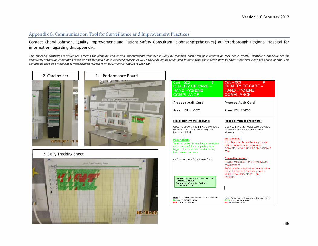
Version 1.0 February 2012
46
Appendix G: Communication Tool for Surveillance and Improvement Practices
Contact Cheryl Johnson, Quality Improvement and Patient Safety Consultant ([email protected]) at Peterborough Regional Hospital for information regarding this appendix. This appendix illustrates a structured process for planning and linking improvements together visually by mapping each step of a process as they are currently, identifying opportunities for improvement through elimination of waste and mapping a new improved process as well as developing an action plan to move from the current state to future state over a defined period of time. This can also be used as a means of communication related to improvement initiatives in your ICU.
1. Performance Board 2. Card holder
3. Daily Tracking Sheet

Version 1.0 February 2012
47
At Peterborough Regional Hospital, a performance Board (1) located in a visible area of the unit is used to identify corporate and unit based measures to front-line staff and physicians. Card holder (2) for the cards allows a visual check on how well they are doing throughout the day. Visual audit cards were developed aimed at validating processes critical in the support of the corporate objectives. Cards are designed for frontline staff and clinical leadership working within the ICU. All nursing staff complete one card daily – returning the completed card to a holder with either the green side (indicates process pass) or red side (failed process) showing. All staff can see at a glance how they are performing daily with each critical process. Progress is tracked monthly visually through frequency graphs – identify trends track process improvements. Daily tracking sheet (3) allows staff to identify success and problems in real time and action plans are posted to demonstrate to staff the status of their corrective actions. Performance huddles allows meaningful dialogue between all members of the ICU team. The team also consistently reviews and celebrates successes, identifies issues, and populates the action plans for further improvement.

Version 1.0 February 2012
48
Appendix H: The Audit Process
This appendix illustrates the process related to chart audit related to surveillance activities in your unit.
In order to look for all possible cases of infection, a retrospective or a prospective audit is recommended. Retrospective audit is generally based on review of records of discharged patients. Prospective audit is based on the collection of information about patients during their process of care. It permits more reliable and complete clinical data collection since the data required is pre-defined and errors can be corrected while the data collection takes place.
A chart audit is a multi-step process to determine the effectiveness of patient care provided at a particular institution and to correct any errors for future references by comparing patient’s data with standards held to be adequate (Gregory et al, 2008). Regardless of whether your unit performs retrospective or prospective chart audit, the following steps are vital parts of any audit process:
Step General Description How this applies to VAP and CLI in your unit/hospital
1. Select a topic Your topic should study issues that are high frequency and/or high risk. You should also ensure that the objective is clear, neither too narrow nor too broad, and measurable using data available in the medical record.
VAP and CLI are part of the provincial patient safety initiative and are required reporting.
2. Identify measures At this step you need to define exactly what you will measure. Specific guidelines need to be outlined as to what should be counted as a “yes” (criteria met) and what should be counted as a “no” (not met).
The standard for diagnosing CLI and VAP is the provincial definition of CLI and VAP as defined in the CCIS. In addition, a sample VAP surveillance data collection tool is provided in Appendix I.
3. Identify patient population
In order to determine which records to review, you need to define the population you want to assess by defining inclusion and exclusion criteria.
Patients who are 18 years or older and have been on mechanical ventilation in your unit for more than 48 hours. Patient must have had a central line in place continuously or intermittently in a 48 hour period before the onset of the infection. If the time interval between the onset of infection and device use is greater than 48 hours, there should be compelling evidence that the infection is related to the central line.
4. Determine sample size
Because the audit of all eligible charts would in most cases be time-consuming and infeasible, you will need to determine of sample of your patients for which audits will be conducted.
Employ rigorous sampling procedures for more statistically valid samples related to VAP and CLI infections in your ICU.
5. Create an audit tool Your audit tool allows you to record your findings. The data should be collected in such way that all individual records are kept separately yet could easily be complied together.
The gold standard would be criteria for diagnosis as defined by the province.
6. Collect data Select the period during which you will collect data. Depending on your sampling methodology, this will determine the length of period for which data needs to be collected. For example, if your sampling revealed that you need to look at 100 charts, then you need to ensure that you have 100 charts to review. For some organizations this may mean a year’s charts and it would mean 100% of the charts. For others however, it could mean pulling a sample from a quarter.
7. Summarize results Summarize the results incorporating in table format the following:
Total charts reviewed
Percentage of patients who met your diagnosis or other issue you want to study (e.g. CLI)
Each infection studied should have its own table – the results should not be pooled.
8. Analyze and apply results
Once you have compiled your data and calculated the results, you can compare them to an established benchmark. You should take into account the differences between your population and those you're comparing it with, as appropriate. You may wish to set a performance target and apply improvement methodologies to help you reach your goal.
For example while your unit reported that 5% of your patients with Central Line were diagnosed with CLI, a chart audit shows 7%. That means error in your unit’s surveillance methods. You may wish to improve the accuracy of your surveillance to 100% and set this as a benchmark that will drive your improvement work and for future auditing of your surveillance.

Version 1.0 February 2012
49
Appendix I: VAP Surveillance Data Form
Contact Lily Waugh, Nurse Manager Intensive Care Unit and CCRT ([email protected]) at St. Joseph’s Healthcare Hamilton for information regarding this appendix. This appendix illustrates a locally developed VAP surveillance data form using provincial definition of VAP and SHN formulated interventions. This form can be used to aid your unit’s surveillance activities. Alternatively, you could develop your own checklist using the provincial definitions of the infection you would like to audit.

Version 1.0 February 2012
50
Appendix J: Needs Assessment and Survey of Current ICU Practices
Adapted from the Critical Care Best Practices Project, this survey will help your unit identify the strategies that your unit already employs regarding VAP and CLI prevention as well as related practices. This tool also helps identify practices that need to be further reinforced in the unit.
* ‘Rounds’ is defined as the systematic review of the status of each patient within the unit at regular intervals conducted by a multidisciplinary team of healthcare professionals.
Best Practice How relevant do you think
this intervention is to your patient population?
Which of the following strategies are currently in place for this intervention?
What is your level of interest in working on this best practice on a scale of 1-5? (1 = Not of interest, 5 = High interest)
What level of impact do you think this will have on your ICU on a scale of 1-5?
(1 = Low impact, 5 = High impact)
How difficult do you feel it will be to implement this intervention in your ICU?
PREVENTION OF CATHETER-BASED BLOODSTREAM INFECTIONS
□ Extremely relevant □ Very relevant □ Somewhat relevant □ Minimally relevant □ Not at all relevant
□ Pre-printed orders □ Established guidelines □ Daily checklist □ Reminder systems □ Other: _____________ □ No specific strategy in place as of yet
□ Extremely difficult □ Very difficult □ Somewhat difficult □ Minimally difficult □ Not at all difficult Please expand on anticipated barriers:
PREVENTION OF VENTILATOR-ASSOCIATED PNEUMONIA
□ Extremely relevant □ Very relevant □ Somewhat relevant □ Minimally relevant □ Not at all relevant
□ Pre-printed orders □ Established guidelines □ Daily checklist □ Reminder systems □ Other: _____________ □ No specific strategy in place as of yet
□ Extremely difficult □ Very difficult □ Somewhat difficult □ Minimally difficult □ Not at all difficult
Please expand on anticipated barriers:
Current Approaches
1 – Does your ICU employ a multidisciplinary ‘rounds’* system? Yes □ / No □ i. Please indicate discipline of all staff involved in multidisciplinary patient rounds: Physicians □ Nurses □ Charge Nurse □ Dietician □ Respiratory Therapist □ Pharmacist □ Educator □ Speech Language Pathologist □ Others (specify): _________________________________________________________ ii. Please indicate frequency of multidisciplinary patient rounds in your ICU: ____________________________ times per week iii. If a format other than ‘rounds’ is used in your ICU, please elaborate: _____________________________________________________________
2 – Does your ICU currently use pre-printed/standardized admission orders? Yes □ / No □
3 – Is your ICU currently involved in any other best-practice initiatives/studies? Critical Care Collaborative □ Safer HealthCare Now! □ Other: ________________________________________________________

Version 1.0 February 2012
51
Appendix K: Literature Review Process for Best Practice Recommendations
This appendix describes the literature review process conducted by the CCBPSC prior to the development of final recommendations. For additional information regarding this process, contact Dr. John Muscedere at Kingston General Hospital.
The following databases were searched to identify relevant literature: CINAHL, EMBASE, PubMed and the Cochrane Database of Systematic Reviews. Limits were applied to the search strategy in order to retrieve articles published in English, and spanning the period 2004-2010 for CLI and 2008-2010 for VAP. The final list was determined based on the need to update the last known references. The chair of the CCBPSC in partnership with the CCS coordinator reviewed these and other documents to determine whether there were new interventions for which there was RCT evidence and which interventions simply required an update from the last set of published guidelines. In order to facilitate an in-depth review of literature on needed updates, members of the CCBPSC were subdivided into a VAP group and a CLI group based on their area of expertise and/or interest. Each individual was provided with specific references relevant to their topic, as well as information regarding the review of articles based on the Scottish Intercollegiate Guidelines Network (SIGN) (2010) criteria. The adoption of SIGN guidelines ensured that all reviewers maintained a consistent method for assessing the quality of the literature retrieved. Individuals were instructed to perform further literature searches if required, and to compile the information gathered on a document template that was used to facilitate discussion with the group. Once the information was reviewed and compiled, each of the groups met to discuss the evidence via webinar. In addition to integrating the evidence provided by the committee, the CCS coordinator contributed and gave context to the meeting by reviewing the purpose of the toolkit, intended audience, and some principles to facilitate compliance with best practice guidelines. Notes were compiled during these meetings and then summarized into a spreadsheet outlining the main recommendations for each intervention. These notes were then sent out to all participants to give feedback. They were asked to document agreement or disagreement with the recommendations and provide additional comments as required.

Version 1.0 February 2012
52
Appendix L: Mouth Care Protocol
Contact Elizabeth Gordon, Advanced Practice Nursing Educator, MSICU ([email protected]) at the University Health Network for information about this document. This appendix illustrates a checklist related to mouth care assessment and documentation. The CCBPSC has strongly recommended proper mouth care as a VAP prevention practice.
University Health Network Policy & Procedure Manual
Critical Care Nursing – Oral Care Policy The provision of oral hygiene is a standard of care in the intensive care setting. The nurse will assess the level of oral care by completing an Oral Assessment Guide each shift and prn. Oral assessment is to be done once per shift. The Oral Assessment Guide will document and measure the level of oral care required, and identify potential risk factors. Intervention is determined based on one of three levels of care: basic, advanced or extensive. Procedure Basic Oral Care: Score 5 (to include tracked patients) 1. Independent Care: · Brush teeth q 12 hours. · Provide patient with the following items:
a. toothbrush b. toothpaste c. towel – to protect gown and to wipe face d. cup of room temperature water e. kidney basin or Yankauer and suction source
2. Non-independent Care: · Brush teeth q 12 hours with a soft brush. · Oral freshening q 2-4 hours with foam stick and water. · Suction excess secretions with oral Yankauer. · Ice Chips P.R.N. after consultation with physician or speech pathologist. · Encourage patient to wear dentures when possible, and remove them during evening care. Note: Brush off any debris before soaking over night. Advanced Oral Care: Score 6-8 (and to include all intubated patients) 1. Brush teeth with soft toothbrush bid (i.e., 06/ 18 or 08/20 or 10/22 hours). 2. Chlorhexidine 0.12% mouth rinses q bid. · Pour 15 mL of chlorhexidine 0.12% into a medicine cup. · Soak foam stick in chlorhexidine 0.12% until saturated. · Rub soaked foam stick along buccal, gingival, tongue and tooth surfaces in a circular motion. · Discard foam stick after each use. · Avoid any other oral agents for 30 minutes after chlorhexidine rinse. 3. Mouth freshening every 2-4 h and P.R.N. with either
a. foam stick and water b. foam stick and 1.5 % hydrogen peroxide
4. Lubrication of lips and oral mucous every 2-4 hours with a water-soluble ointment. 5. Do not use nystatin 2 hours before or after the use of chlorhexidine solution.

Version 1.0 February 2012
53
Chlorhexidine 0.12% may discolour teeth, but can be reversed with professional dental cleaning. Extensive Oral Care: Score 9 or greater (individualized based on assessment, diagnosis or physician specific directives) 1. Dry mucosa/tongue: obtain order for artificial saliva replacements. 2. Excessive bleeding: gentle mouth rinses with foam stick and water every hour and prn. 3. Assess for pain, ulceration, infection, bleeding gingival or altered saliva consistency. References
1. Evans, G. (2001). A rationale for oral care. Nursing Standard. 15(43), 33-36. 2. Grap, M.J., Munro, C.L., Ashtiani, B., & Bryant, S. (2003). Oral care interventions in critical care: frequency
and documentation. American Journal of Critical Care.12 (2), 113-118. 3. Royal Free Hampstead HNS Trust. (2000). Guidelines for oral care. pp. 1-14. 4. Houston, S., Hougland, P., Anderson, J. J., LaRocco, M., Kennedy, V., & Gentry, L. O. (2002). Effectiveness
of 0.12% chlorhexidine gluconate oral rinse in reducing prevalence of nosocomial pneumonia in patients undergoing heart surgery. American Journal of Critical Care. 11(6), 567-570.
5. Pearson, L.S. (1996). A comparison of the ability of foam swabs and toothbrushes to remove dental plaque; implications for nursing practice. Journal Advanced Nursing.23, 62-69.
6. Roberts, J. (2000). Developing an oral assessment and intervention tool for older people: 2. British Journal of Nursing. 9(18), 1124-1127.
7. Schleder, B., Stott, K. & Lloyd, R. C. (2002). The effect of a comprehensive oral care protocol on patients at risk for ventilator – associated pneumonia. Journal of Advocate Health Care.4 (1), 27-30.

Version 1.0 February 2012
54
Appendix M: Example of an Antimicrobial Stewardship Program in Ontario
In Ontario, Mount Sinai Hospital (MSH) and University Health Network (UHN) began a partnership in 2009 to implement an ASP program in their respective institutions (Morris, 2011). The ASP team is comprised of infectious diseases physicians, infectious diseases pharmacists, infection control professionals, microbiologists, informatics analysts, data analysts and hospital epidemiologists, as well as project management professionals. The key elements of this program are (Morris, 2011):
Prospective audit with intervention and feedback: The feedback is performed by an infection control professional and provided directly to the prescriber.
Educational profiling to influence prescribing behavior: This process involves reviewing all patients who are on antimicrobials for a particular service, meeting with the healthcare team, discussing the clinical scenario and providing treatment recommendations. The advice offered by the ASP team is consultative, and, as such, the decision regarding whether or not to follow through with the ASP team’s recommendations is at the discretion of the critical care team.
Collaboration: The ASP team works collaboratively with hospital infection control teams, pharmacy and specific departments such as critical care to reduce the impact of infectious diseases.
Since the implementation of the ASP program, susceptibility to Candidaemia (a form of CLI) in ICUs, as well as antimicrobial utilization and associated costs have significantly declined in both institutions. However, the impact on mortality and length of stay has yet to be determined. The ASP team has published several articles around the program. For further information about the MSH-UHN ASP, refer to the article published by Morris et al (2010). Additionally a template for developing a business case for ASP in your organization as well as additional resources and educational materials on the topic are available at: http://www.idologist.com/Docs.html.

Version 1.0 February 2012
55
References
References Related to Background
1. Centres for Disease Control and Prevention. (2002). Guidelines for the Prevention of Intravascular Catheter-Related Infections, 2002. Retrieved from: http://www.cdc.gov/mmwr/preview/mmwrhtml/rr5110a1.htm.
2. Muscedere, J., Dodek, P., Keenan, S., Fowler, R., Cook, D., Heyland, D. et al. (2008).Comprehensive
evidence-based clinical practice guidelines for ventilator-associated pneumonia: Prevention. Journal of Critical Care, 23(1):138-147.
3. Ontario Critical Care Steering Committee’s Final Report (2005). Retrieved from:
www.health.gov.on.ca/criticalcare 4. Pittett, D. (1994). Nosocomial pneumonia: incidence, morbidity and mortality in the intubated-
ventilated patient. Schweiz Med Wochenschr, 124(6):227-235.
5. Provincial Patient Safety Initiative: Retrieved from: http://www.health.gov.on.ca/patient_safety/ 6. Soufir, L., Timsit, J.F., Mahe, C., Carlet, J., Regnier, B., & Chevret, S. (1999). Attributable morbidity
and mortality of catheter-related septicemia in critically ill patients: a matched, risk-adjusted, cohort study. Infect Control Hosp Epidemiol, 20(6):396-401.
References Related to Quality
7. Adams, J. (2003). Successful change: paying attention to the intangibles. Practitioner, 35(4):3-7.
8. Canadian Patient Safety Institute – Safer Healthcare Now!. Retrieved from: http://www.saferhealthcarenow.ca/EN/Pages/default.aspx
9. ECFFA Quality Improvement Plan Guidance Document. Retrieved from: http://www.health.gov.on.ca/en/ms/ecfa/pro/updates/qualityimprov/qip_guide.pdf
10. How-to Guide: Sustainability and Spread. Retrieved from IHI: http://www.ihi.org/knowledge/Pages/Tools/HowtoGuideSustainabilitySpread.aspx
11. Gurses, A.P., Seidl, K. L., Vaidya, V., Bochicchio, G., Harris, A.D., Hebden, J., et al. (2008). Systems ambiguity and guideline compliance: a qualitative study to how intensive care units follow evidence-based guidelines to reduce healthcare-associated infections. Quality and Safety in Health Care, 17: 351-359.
12. LEAN. Retrieved from: http://www.lean.org/ 13. Peden, C.J., & Rooney, K.D. (2009). The science of improvement as it relates to quality and safety in
the ICU. The Intensive Care Society, 10(4):260-265.

Version 1.0 February 2012
56
14. Pronovost, P., Berenholtz, S., Dorman, T., Lipsett, P., Simmonds, T., & Haraden, C. Improving communication in the ICU using daily goals. (2003). J Crit Care, 18 (2):71–75.
15. Six Sigma: Retrieved from: http://www.ilr.cornell.edu/laborPrograms/events/upload/Quality-Models-Selecting-the-Best-Model.pdf
16. The Model for Improvement. Retrieved from IHI: http://www.ihi.org/knowledge/Pages/HowtoImprove/default.aspx
17. The 8-step Process for Leading Change. Retrieved from Kotter International: http://www.kotterinternational.com/kotterprinciples/changesteps
18. Transforming Care at the Bedside How-to Guide: Engaging Front-Line Staff in Innovation and Quality
Improvement. Retrieved from IHI: http://www.ihi.org/knowledge/Pages/Tools/TCABHowToGuideEngagingStaff.aspx.
References Related to Surveillance and Audit
19. Best Practices for Surveillance of Health Care-Associated Infections in Patient and Resident
Populations. Retrieved from PIDAC: http://www.health.gov.on.ca/patient_safety/pro/cdad/toolkit_ricn/rep_pidac_hai_best_prac.pdf).
20. Gregory, B. H., Van Horn, C., & Kaprielian, V.S. (2008). Eight steps to a chart audit for quality. Fam Pract Manag, 15 (7): A3-A8.
21. Statistical Monitoring Quality in Healthcare Process Clinical Indicators Support Team Control. Retrieved from NHS Scotland: http://www.indicators.scot.nhs.uk/SPC/Statistical_Process_Control_Tutorial_Guide_010207.pdf
22. The Model for Improvement’s Plan-Do-Study-Act (PDSA). Retrieved from IHI: http://www.ihi.org/knowledge/Pages/HowtoImprove/default.aspx
References Related to Best Practices
23. Edmond, M.B., &Wenzel, R.P. Organization for infection control. In: Principles and Practice of Infectious Diseases, 6th ed. Mandell, GL, Bennett, JE, Dolin, R, (Eds), Churchill Livingstone, Philadelphia, PA 2005, p.3323.
24. George, P., & Morris, A.M. (2010). Pro/con debate: should antimicrobial stewardship programs be adopted universally in the intensive care unit? Critical Care, 14 (1), 205.
25. Gerding, D. N. (2001). The search for good antimicrobial stewardship program. JT Comm J Qual Improv, 27(8): 403-404.
26. Morris, A. M., Stewart, T .E., Shandling, M., McIntaggart, S., & Liles, C. (2010). Establishing an antimicrobial stewardship program. Healthcare Quarterly. 13 (2): 65-70.

Version 1.0 February 2012
57
27. Provincial hand hygiene initiative. Retrieved from:
http://www.health.gov.on.ca/en/ms/handhygiene/
VAP
28. Alexiou, V.G., Ierodiakonou, V., Dimopoulos, G., & Falagas, M.E. (2009). Impact of patient position on the incidence of ventilator-associated pneumonia: a meta-analysis of randomized controlled trials. Journal of Critical Care, 24 (4):152 -153.
29. Barruad, D., Blard, C., Hein, F., Marcon, O., Cravoisy, A., Nace, L., et al. (2010). Probiotics in the critically ill patient: A double blind, randomized, placebo-controlled trial. Intensive Care Medicine, 36: 1540-1547.
30. Beraldo, C.C., & Andrade, D. (2008). Oral hygiene with chlorhexidine in preventing pneumonia associated with mechanical ventilation. Jornal Brasileiro de Pneumologia. 34(9): 707-714.
31. Berry, A.M., Davidson, P.M., Masters, J., & Rolls, K. (2007). Systematic literature review of oral hygiene practices for intensive care patients receiving mechanical ventilation. American Journal of Critical Care, 16 (6), 552-563.
32. Bo, H., He, L., & Qu, J. (2000). Influence of the subglottic secretion drainage on the morbidity of ventilator associated pneumonia in mechanically ventilated patients. Zhonghua Jie He He Hu Xi Za Zhi, 23 (8): 472-474.
33. Bouza, E., Pérez, M.J., Muñoz, P., Rincón, C., Barrio, J.M., & Hortal, J. (2008). Continuous aspiration of subglottic secretions in the prevention of ventilator-associated pneumonia in the postoperative period of major heart surgery. Chest, 134 (5): 938-946.
34. Caruso, P., Denari, S., Soraia, A.L., Sergio, R., Demarzo, E., & Deheinzelin, D. (2009). Saline instillation before tracheal suctioning decreases the incidence of ventilator-associated pneumonia. Critical Care Medicine, 37 (1): 32-
35. Chan, E.Y., Ruest, A., Meade, M.O., & Cook, D.J. (2007). Oral decontamination for prevention of pneumonia in mechanically ventilated adults: Systematic review and meta-analysis. British Medical Journal. 334 (7599), 889. [Epub ahead of print]. Retrieved from: http://www.bmj.com/content/334/7599/889.full.pdf.
36. Chen, YC. (2009). Critical analysis of the factors associated with enteral feeding in preventing VAP: A systematic review. Journal of the Chinese Medical Association, 72 (4): 171-178.
37. Chlebicki, M.P., & Safdar, N. (2007). Topical chlorhexidine for prevention of ventilator-associated pneumonia: a meta-analysis. Critical Care Medicine, 35 (2): 595-602.
38. David, D., Samuel, P., David, T., Keshava, S.N., Irodi, A., & Peter, J.V. (2010). An open-labeled randomized controlled trial comparing costs and clinical outcomes of open endotracheal suctioning with closed endotracheal suctioning in mechanically ventilated medical intensive care patients. Journal of Critical Care: Epub ahead of print.
Version 1.0 February 2012
58
39. Doyle, A., Joshi, M., Frank, P., Craven, T., Moondi, P., &Young, P. (2011). A change in humidification can eliminate endotracheal tube occlusion. Journal of Critical Care: Epub ahead of print.
40. Ferrer, M., Sellarés, J., Valencia, M., Carrillo, A., Gonzalez, G., Badia, JR,. et al. (2009). Non-invasive ventilation after extubation in hypercapnic patients with chronic respiratory disorders: Randomized controlled trial. Lancet, 374 (9695): 1082-1088.
41. Fields, L.B. (2008). Oral care intervention to reduce incidence of ventilator-associated pneumonia in the neurologic intensive care unit. The Journal of Neuroscience Nursing 40(5), 291-298.
42. Forestier, C., Guelon, D, Cluytens, V., Gillart, T., Sirot, J., & De Champs, C. (2008). Oral probiotic and prevention of Pseudomonas aeruginosa infections: a randomized, double-blind, placebo-controlled pilot study in intensive care unit patients. Critical Care 2008, 12: R69.
43. Gattinoni, L., & Protti, A. (2008). Ventilation in the prone position: For some but not for all? Canadian Medical Association Journal, 178 (9), 1174-1176.
44. Girard, T.D., & Ely, E.W. (2008). Protocol-driven ventilator weaning: Reviewing the evidence. Clinics
in Chest Medicine, 29(2): 241-252.
45. Goldhill, D.R., Imhoff, M., McLean, B., & Waldmann, C. (2007). Rotational Bed Therapy to Prevent and Treat Respiratory Complications: A Review and Meta-Analysis. American Journal of Critical Care, 16 (1): 50-61.
46. Han, J. & Liu, Y. (2010). Effect of ventilator circuit changes on ventilator-associated pneumonia: a
systematic review and meta-analysis. Respiratory Care, 255 (4): 467-74.
47. Hsu, C.W., Sun, S.F., Lin, S.L., Kang, S.P., Chu, K.A., Lin, CH., et al. (2009). Duodenal versus gastric feeding in medical intensive care unit patients: a prospective, randomized, clinical study. Critical Care Medicine, 37(6), 1866-1872.
48. Jacobi, J., Fraser, G.L., Coursin, D.B., Riker, R.R. Fontaine, D. Wittbrodt, et al. (2002). Clinical practice guidelines for the sustained use of sedatives and analgesics in the critically ill adult. Critical Care Medicine. 30 (1): 119–141.
49. Jongerden, I., Buiting, A.G., Leverstein-van Hall, Maurine. A., Speelberg, B., Zeidler, S et al. (2011). Effect of open and closed endotracheal suctioning on cross-transmission with Gram-negative bacteria: A prospective crossover study. Critical Care Medicine, 39 (6): 1313-1321.
50. Keenan, S.P., Sinuff, T., Burns, K.E.A., Muscedere, J., Kutsogiannis, J. Mehta, S. et al. (2011). Clinical practice guidelines for the use of non-invasive positive-pressure ventilation and noninvasive continuous positive airway pressure in the acute care setting. CMAJ, 183 (3): E195 – E214.
51. Kelly, M., Gillies, D., Todd, D. A., & Lockwood, C. (2010). Heated humidification versus heat and moisture exchangers for ventilated adults and children. Cochrane Database of Systematic Reviews, 14 (4): CD004711.

Version 1.0 February 2012
59
52. Kollef, M.H. (1999). The prevention of ventilator-associated pneumonia. New England Journal of Medicine, 40 (8): 627-34.
53. Kollef, M.H., Afessa, B., Anzueto, A., Veremakis, C., Kerr, K.M., Margolis, B.D. et al. (2008). Silver-coated endotracheal tubes and incidence of ventilator-associated pneumonia. JAMA, 300 (7): 805-813.
54. Kollef, M.H., Skubas, N.J., & Sundt, TM. (1999). A randomized clinical trial of continuous aspiration of subglottic secretions in cardiac surgery patients. Chest, 116 (5): 1339-1346.
55. Krishnan, J.A., Moore, D., Robeson, C., Rand, C.S., & Fessler, H.E. (2004). A prospective, controlled trial of a protocol-based strategy to discontinue mechanical ventilation. American Journal of Respiratory and Critical Care Medicine, 169 (6):673-678.
56. Lacherade, J.C., De Jonghe, B., Guezennec, P., Debbat, K., Hayon, J., Monsel, A., et al. (2010). Intermittent subglottic secretion drainage and ventilator-associated pneumonia: A multicenter trial. American Journal of Respiratory and Critical Care Medicine, 182 (7): 910-917.
57. Liu, SH., Yan, XX., Cao, SQ., An, SC., & Zhang, LJ. (2006). The effect of subglottic secretion drainage on prevention of ventilator-associated lower airway infection. Zhonghua Jie He He Hu Xi Za Zhi, 29 (1): 19-22.
58. Lorente, L., Lecuona, M., Jiménez, A., Mora, M.L., & Sierra, A. (2007). Influence of an endotracheal tube with polyurethane cuff and subglottic secretion drainage on pneumonia. American Journal of Respiratory and Critical Care Medicine, 176 (11): 1079-1083.
59. Mahul, P., Auboyer, C., Jospe, R., Ros, A., Guerin, C., el Khouri, Z. et al. (1992). Prevention of nosocomial pneumonia in intubated patients: respective role of mechanical subglottic secretions drainage and stress ulcer prophylaxis. Intensive Care Medicine, 18 (1): 20-25.
60. Manzano, F., Fernández-Mondéjar, E., Colmenero, M., Poyatos, M.E., Riveira, R., Machado, J., et al. (2008). Positive-end expiratory pressure reduces incidence of ventilator-associated pneumonia in nonhypoxemic patients. Critical Care Medicine, 36 (80): 2225-2231.
61. MacIntyre, N.R., Cook, D.J., Ely, E.W., Jr., Epstein, S.K., Fink, J.B., Heffner, J.E. et al. Evidence-based guidelines for weaning and discontinuing ventilatory support: a collective task force facilitated by the American College of Chest Physicians; the American Association for Respiratory Care; and the American College of Critical Care Medicine. (2001). Chest, 120 (6 Suppl): 375S-395S.
62. McLean, S, E., Jensen, L, A., Schroeder, D, G., Gibney, N,R., & Skjodt, N,M. (2006). Improving adherence to a mechanical ventilation weaning protocol for critically ill adults: Outcomes after an implementation program. American Journal of Critical Care, 15 (3): 299-309.
63. Metz, C., Linde, H.J., Gobel, L., Gobel, F., & Taeger, K. Influence of intermittent subglottic lavage on subglottic colonisation and ventilator associated pneumonia. (1998). Clinical Intensive Care, 9: 20–24.

Version 1.0 February 2012
60
64. Miller, M.A., Arndt, J.L., Konkle, M.A., Chenoweth, C.E., Iwashyna, T.J., et al. (2010). A polyurethane cuffed endotracheal tube is associated with decreased rates of ventilator-associated pneumonia. Journal of Critical Care. [Epub ahead of print]. Retrieved from: http://www.sciencedirect.com/science?_ob=MImg&_imagekey=B7590-50KVG6P-2-3&_cdi=12940&_user=1166899&_pii=S0883944110001632&_origin=gateway&_coverDate=07%2F23%2F2010&_sk=999999999&view=c&wchp=dGLbVzW-zSkzS&md5=8aa166bc02321a1358397186fd0761c5&ie=/sdarticle.pdf
65. Mori, H., Hirasawa, H., Oda, S., Shiga, H., Matsuda, K., & Nakamura, M. (2006). Oral care reduces incidence of ventilator-associated pneumonia in ICU populations. Intensive Care Medicine, 32 (2): 230-236.
66. Morrow, L. E., Kollef, M.H.m., & Casale, T.B. (2010). Probiotic prophylaxis of Ventilator-associated
Pneumonia: A blinded, randomized, controlled trial. American Journal of Respiratory and Critical Care Medicine, 182: 1058-1064.
67. Mounier, R., Adrie, C., Français, A., Garrouste-Orgeas, M., Cheval, C., Allaouchiche, B., et al. (2010). Study of prone positioning to reduce ventilator-associated pneumonia in hypoxaemic patients. The European Respiratory Journal, 35 (4): 795-804.
68. Muscedere, J., Dodek, P., Keenan, S., Fowler, R., Cook, D., & Heyland, D. (2008).Comprehensive evidence-based clinical practice guidelines for ventilator-associated pneumonia: Diagnosis and treatment. Journal of Critical Care. 23 (1): 26 - 137.
69. Muscedere, J., Dodek, P., Keenan, S., Fowler, R., Cook, D., Heyland, D. et al. (2008).Comprehensive evidence-based clinical practice guidelines for ventilator-associated pneumonia: Prevention. Journal of Critical Care. 23(1): 138-147.
70. Panchabhai, T.S., Dangayach, N.S., Krishnan, A., Kothari, V.M., & Karnad, D. R. (2009). Oropharyngeal cleansing with 0.2% chlorhexidine for prevention of nosocomial pneumonia in critically ill patients: An open-label randomized trial with 0.01% potassium permanganate s control. The American College of Chest Physicians, 135 (5): 1150-1156.
71. Powers, J., Brower, A., & Tolliver, S. (2007). Impact of oral hygiene on prevention of ventilator-associated pneumonia in neuroscience patients. Journal of Nursing Care Quality, 22 (4): 316-321.
72. Quenot, J.P., Ladoire, S., Devoucoux, F., Doise, J.M., Cailliod, R., Cunin N., et al. (2007). Effect of a nurse-implemented sedation protocol on the incidence of ventilator-associated pneumonia. Critical Care Medicine, 35 (9): 2031-2036.
73. Rewa, O., & Muscedere, J. (2011). Ventilator-associated pneumonia: Update on etiology, prevention and management. Current Infectious Disease Reports. 13 (3): 287-95. Retrieved from: http://www.springerlink.com/content/gk024v72um517036/
74. Ross, A., & Crumpler, J. (2007). The impact of an evidence-based practice education program on the role of oral care in the prevention of ventilator-associated pneumonia. Intensive and Critical Care Nursing, 23 (3): 132-136.

Version 1.0 February 2012
61
75. Sackett, D.L., Rosenberg, W.M.C., Gray, J., Haynes, R.B., & Richardson W.S. (1996). Evidence based medicine: what it is and what it isn't. BMJ, 13: 71-72.
76. Schultz, M.J., Haas, Lenneke, E. (2011). Antibiotics or probiotics as preventive measures against ventilator-associated pneumonia: a literature review. Critical Care, 15 (1): (R18).
77. Siempos, I.I., Ntaidou, T.K., & Falagas, M.E. (2010). Impact of the administration of probiotics on the incidence of ventilator-associated pneumonia: A meta-analysis of randomized controlled trials. Critical Care Medicine, 38 (3): 954-962.
78. Siempos, I. I., Vardakas, K. Z., & Falagas, M. E. (2008). Closed tracheal suction systems for prevention of ventilator-associated pneumonia. British Journal of Anaesthesia, 100 (3): 299-306.
79. Smulders, K., Van Der Hoeven, H., Weers-Pothoff, I, & Vandenbroucke-Grauls, C. (2002). A randomized clinical trial of intermittent subglottic secretion drainage in patients receiving mechanical ventilation. Chest, 121(3): 858-862.
80. Sona, C.S., Zack, J.E., Schallom, M.E., McSweeney, M., McMullen K, Thomas J, et al (2009). The impact of a simple, low-cost oral care protocol on ventilator-associated pneumonia rates in a surgical intensive care unit. Journal of Intensive Care Medicine, 24(1): 54-62.
81. Staudinger, T., Bojic, A., Holzinger, U., Meyer, B., Mallner, F., Schellongowski, P., et al. (2010). Continuous lateral rotation therapy to prevent ventilator-associated pneumonia. Critical Care Medicine, 38 (2): 486-490.
82. Stoller, J.K. (2004). The effectiveness of respiratory care protocols. Respiratory Care, 49(7): 761-765.
83. Sud, S., Friedrich, J.O., Taccone, P., Polli, F., Adhikari, N.K.J., Latini, R. et al. (2010). Prone ventilation reduces mortality in patients with acute respiratory failure and severe hypoxemia: Systematic review and meta-analysis. Intensive Care Medicine, 36 (4): 585-599.
84. Sud, S., Sud, M., Friedrich, J.O., & Adhikari, N.K. (2008). Effect of mechanical ventilation in the prone position of clinical outcomes in patients with acute hypoxemic respiratory failure: a systematic review and meta-analysis. Canadian Medical Association Journal, 178 (9): 1174-1176.
85. Taccone, P., Pesenti, A., Latini, R., Polli, F., Vagginelli, F., Mietto, C., et al. (2009). Prone positioning in patients with moderate and severe acute respiratory distress syndrome: A randomized controlled trial. The Journal of American Medical Association, 302 (18): 1977-1984.
86. Tantipong, H., Morkchareonpong, C., Jaiyindee, S., & Thamlikitkul, V. (2008). Randomized controlled trial and meta-analysis of oral decontamination with 2% chlorhexidine solution for the prevention of ventilator-associated pneumonia. Infection Control and Hospital Epidemiology, 29 (2), 131-136.
87. Terragni P.P., Antonelli, M., Fumagalli, R., Faggiano, C., Berardino, M., Pallavicini, F.B. et al. (2010) Early versus late tracheotomy for prevention of pneumonia in mechanically ventilated adult ICU patients: a randomized controlled trial. JAMA. 303 (15): 1483-1489.

Version 1.0 February 2012
62
88. Tuma, R.S. (2007). American Thoracic Society (ATS) 103rd International Conference:Daily breathing and awakening tests speed weaning from mechanical ventilation. Retrieved from Medscape Today website: http://www.medscape.com/viewarticle/557233
89. Updated Recommendations: Canadian Clinical Practice Guidelines - Summary of Topics and Recommendations (2009). Retrieved from: http://www.criticalcarenutrition.com/docs/cpg/srrev.pdf
90. Vallés, J., Artigas, A., Rello, J., Bonsoms, N., Fontanals, D., Blanch, L., Fernández., et al. (1995). Continuous aspiration of subglottic secretions in preventing ventilator-associated pneumonia. Annals of Internal Medicine, 122 (3), 179-186.
91. Yang, C.S., Qiu, H.B., Zhu, Y.P., Huang, Y.Z., Xu, X.T., & Gao L. (2008). Effect of continuous aspiration
of subglottic secretions on the prevention of ventilator-associated pneumonia in mechanically ventilated patients: a prospective, randomized, controlled clinical trial. Zhonghua Nei Ke Za Zhi, 47 (8): 625-629.
CLI
92. Balamongkhon, B., & Thamlikitkul, V. (2007). Implementation of chlorhexidine gluconate for central venous catheter site care at Siriraj Hospital, Bangkok, Thailand. American Journal of Infection Control, 35 (9): 585-588.
93. Bleasdale, S.C., Trick, W.E., Gonzalez, I.M., Lyles, R.D., Hayden, M.K., & Weinstein, RA. (2007). Effectiveness of chlorhexidine bathing to reduce catheter-associated bloodstream infections in medical intensive care unit patients. Archives of Internal Medicine, 167 (19): 2073-2079.
94. Bonawitz, S.C., Hammell, E.J., & Kirkpatrick, J.R. (1991). Prevention of central venous catheter sepsis: a prospective randomized trial. The American Journal of Surgery, 57 (10): 618-623.
95. Centres for Disease Control and Prevention. (2002). Guidelines for the Prevention of Intravascular Catheter-Related Infections, 2002. Retrieved from: http://www.cdc.gov/mmwr/preview/mmwrhtml/rr5110a1.htm.
96. Centres for Disease Control and Prevention. (2011). Guidelines for the Prevention of Intravascular Catheter-Related Infections, 2011. Retrieved from: http://www.cdc.gov/hicpac/pdf/guidelines/bsi-guidelines-2011.pdf.
97. Chin, B.S., Han, S.H., Lee, H.S., Jeong, S.J., Choi, H., Kim, C.O., et al. (2010). Risk factors for recurrent catheter-related infections after catheter-related bloodstream infections. International Journal of Infectious Diseases, 14 (1): e16-21.
98. Cicalini, S., Palmieri, F., & Petrosillo N. (2004). Clinical review: New technologies for prevention of intravascular catheter-related infections. Critical Care, 8 (3): 157-162.
99. Climo, M.W., Sepkowitz, K.A., Zuccotti, G., Fraser, V.J., Warren, D.K., Perl, T.M., et al. (2009). The effect of daily bathing with chlorhexidine on the acquisition of methicillin-resistant Staphylococcus aureus, vancomycin-resistant Enterococcus, and healthcare-associated bloodstream infections: Results of a quasi-experimental multicenter trial. Critical Care Medicine: 37 (6), 2097-2098

Version 1.0 February 2012
63
100. Dahlberg, P.J., Agger, W.A., Singer, J.R., Yutuc, W.R., Newcomer, K.L., Schaper, A., et al. (1995).
Subclavian hemodialysis catheter infections: A prospective, randomized trial of an attachable silver-impregnated cuff for prevention of catheter-related infections. Infection Control and Hospital Epidemiology, 16 (9): 506-511.
101. Evans, H.L., Dellit, T.H., Chan, J., Nathens, A.B., Maier, R.V., & Cuschieri, J. (2010). Effect of
chlorhexidine whole-body bathing on hospital-acquired infections among trauma patients. Archives of Surgery, 145 (3): 240-246.
102. Foster, J., Richards, R., & Showell, M. (2006). Intravenous in-line filters for preventing morbidity
and mortality in neonates. Cochrane Database Systematic Reviews. (2). Art. No.: CD005248. DOI: 10.1002/14651858.CD005248.pub2
103. Frasca, D., Dahyot-Fizelier, C., & Mimoz, O. (2010). Prevention of central venous catheter-
related infection in the intensive care unit. Critical Care, 14 (2): 212 – 220.
104. Fraser, T.G., Fatica, C., Scarpelli, M., Arroliga, A.C., Guzman, J., Shrestha, N.K., et al. (2010). Decrease in Staphylococcus aureus colonization and hospital-acquired infection in a medical intensive care unit after institution of an active surveillance and decolonization program. Infection Control and Hospital Epidemiology, 31 (8): 779-783.
105. Frey, A.M., & Schears, G.J. (2006). Why are we stuck on tape and suture? Journal of Infusion
Nursing, 29 (1): 34-38.
106. Gabriel, J. (2001). PICC securement: Minimising potential complications. Nursing Standard, 15 (43); 42-44.
107. Graf, J.M., Newman, C.D., & McPherson, M.L. (2006). Techniques, material devices: Sutured
securement of peripherally inserted central catheters yields fewer complications in pediatric patients. Journal of Parenteral and Enteral Nutrition, 30 (6): 532- 535.
108. Groeger, J.S., Lucas, A.B., Coit, D., LaQuaglia, M., Brown, A.E., Turnbull, A., et al. (1995). A
prospective, randomized evaluation of the effect of silver impregnated subcutaneous cuffs for preventing tunneled chronic venous access catheter infections in cancer patients. Annals of Surgery, 218 (2): 206-210.
109. Ho, K.M. & Litton, E. (2006). Use of chlorhexidine-impregnated dressing to prevent vascular and
epidural catheter colonization and infection: A meta-analysis. Journal of Antimicrobial Chemotherapy, 58 (2): 281-287.
110. Hota, B., Harting, B., Weinstein, RA., Lyles, RD., Bleasdale, SC., Trick, W. (2010). Electronic
algorithmic prediction of central vascular catheter use. Infection Control and Hospital Epidemiology, 31(4): 4-11.
111. Jarvis, W.R., Murphy, C., Hall, K.K., Fogle, P.J., Karchmer, T.B., Harrington, G., et al. (2009).
Health care-associated bloodstream infections associated with negative – or positive – pressure

Version 1.0 February 2012
64
or displacement mechanical valve needleless connectors. Clinical Infectious Diseases, 49 (12): 1821-1827.
112. Khalifa, R., Dahyot-Fizelier, C., Laksiri, L., Ragot, S., Petitpas, F., Nanadoumgar, H., et al. (2008).
Indwelling time and risk of colonization of peripheral arterial catheters in critically ill patients. Intensive Care Medicine, 34 (10): 1820-1826.
113. Levin, A., Mason A.J., Jindal, K.K., Fong, I.W., & Goldstein, M.B. (1991). Prevention of
hemodialysis subclavian vein catheter infections by topical providone-iodine. Kidney International, 40: 934–938.
114. Lok, C.E., Stanley, K.E., Hux, J.E., Richardson, R., Tobe, S.W., & Conly, J. (2003). Hemodialysis
Infection Prevention with Polysporin Ointment. J Am Soc Nephrol, 14: 169-179.
115. Macias, A.E., Huertas, M., De Leon, S.P., Munoz, J.M., Chavez, A.R., & Sifuentes-Osornio, J. (2010). Contamination of intravenous fluids: A continuing cause of hospital bacteremia. American Journal of Infection Control, 38 (3): 217-221.
116. Maiefski, M., Rupp, M.E., & Hermsen, ED. (2009). Ethanol lock technique: Review of the
literature. Infection Control and Hospital Epidemiology, 30 (11): 1096-1108.
117. Maki, D.G., Kluger, D.M., & Crnich, C.J. (2006). The risk of bloodstream infection in adults with different intravascular devices: A systematic review of 200 published prospective studies. Mayo Clinic Proceedings, 81: 1159-1171.
118. Marschall, J., Mermel L.A., Classen, D., Arias K.M. Podgorny K., Anderson, D.J., et al. (2008). Strategies to prevent central line–associated bloodstream infections in acute care hospitals (2008). Infection Control and Hospital Epidemiology, 29 (S1): S22 – S30.
119. McKee, R., Dunsmuir, R., Whitby, M., & Garden, O.J. (1985). Does antibiotic prophylaxis at the time of catheter insertion reduce the incidence of catheter- related sepsis in intravenous nutrition? Journal of Hospital Infection, 6 (4): 419 – 425.
120. Mimoz, O., Villeminey, S., Ragot, S., Dahyot-Fizelier, C., Laksiri, L., Petitpas, F., & Debaene, B. (2007). Chlorhexidine-based antiseptic solution vs. alcohol-based providone-iodine for central venous catheter care. Archives of Internal Medicine, 167 (19): 2066-2072.
121. Vallés, J., Fernández, I., Alcaraz, D., Chacón, E., Cazorla, A., Canals, M., et al. (2008). Prospective randomized trial of 3 antiseptic solutions for prevention of catheter colonization in an intensive care unit for adult patients. Infection Control and Hospital Epidemiology, 29 (9): 847-853.
122. Van Den Hoogen, A., Krediet, T.G., Uiterwaal, C.S., Bolenius, J.F., Gerards, L.J., & Fleer, A. (2006).
In-line filters in central venous catheters in a neonatal intensive care unit. Journal of Perinatal Medicine, 34 (1): 71-74.
123. Van de Wetering, M.D., van Woensel, J.B.M., Kremer, L.C.M., & Caron, H.N. (2005). Prophylactic antibiotics for preventing early Gram-positive central venous catheter infections in oncology patients, a Cochrane systematic review. Cancer Treatment Reviews, 31 (3): 186-196.

Version 1.0 February 2012
65
124. Walz, J., Memtsoudis, S., & Heard, S. (2010). Prevention of central venous catheter bloodstream
infections. Journal of Intensive Care Medicine: 25(3): 131 – 138.
125. Wang, C., & Xie, X. (2010). Treatment of an unraveled intracerebral coil. Catheterization and Cardiovascular Interventions, 76: 746–750.
126. Yamamoto, A.J., Solomon, J.A., Soulen, M.C., Tang, J., Parkinson, K., Lin, R., et al. (2002). Sutureless securement device reduces complications of peripherally inserted central venous catheters. Journal of Vascular and Interventional Radiology, 13 (1): 77-88.
127. Yébenes, J.C., Sauca, G., Solsona, M., Martinez, R., Serra-Prat, M., Gil, Pet., al. (2008). Safety of positive-pressure valve connectors in arterial catheters inserted into critically ill patients. Journal of Hospital Infections, 70 (4): 341-345.
128. Yilmaz, G., Koksal, I., Aydin, K., Caylan, R., Sucu, N., & Aksoy, F. (2007). Risk factors of catheter-related bloodstream infections in parenteral nutrition catheterization. Journal of Parenteral and Enteral Nutrition, 31(4): 284-287.
129. Zakrzewska-Bode, A., Muytjens, H.L., Liem, K.D., & Hoogkamp-Korstanje, J.A. (1995). Mupirocin resistance in coagulase-negative staphylococci after topical prophylaxis for the reduction of colonization of central venous catheters. J Hosp Infect, 31: 189–193.
130. Zingg, W., Imhof, A., Maggiorini, M., Stocker, R., Keller, E., & Ruef, C. (2009). Impact of a prevention strategy targeting hand hygiene and catheter care on the incidence of catheter-related bloodstream infections. Critical Care Medicine, 37 (7): 2167-2173.

Version 1.0 February 2012