varicella-zoster virus epithelial keratitis4eyes.gr/images/4eyes/pdf/cornea/varicell zoster virus...
TRANSCRIPT


Varicella-Zoster Virus Epithelial Keratitis in Herpes Zoster Ophthalmicus


Helena M. Tabery
Varicella-Zoster Virus Epithelial Keratitis in Herpes Zoster Ophthalmicus
In Vivo Morphology in the Human Cornea

ISBN 978-3-642-14486-8 e-ISBN 978-3-642-14487-5DOI 10.1007/978-3-642-14487-5Springer Heidelberg Dordrecht London New York
© Springer-Verlag Berlin Heidelberg 2011
This work is subject to copyright. All rights are reserved, whether the whole or part of the material is concerned, specifically the rights of translation, reprinting, reuse of illustrations, recitation, broadcasting, reproduction on microfilm or in any other way, and storage in data banks. Duplication of this publication or parts thereof is permitted only under the provisions of the German Copyright Law of September 9, 1965, in its current version, and permission for use must always be obtained from Springer. Violations are liable to prosecution under the German Copyright Law.The use of general descriptive names, registered names, trademarks, etc. in this publication does not imply, even in the absence of a specific statement, that such names are exempt from the relevant protective laws and regulations and therefore free for general use.Product liability: The publishers cannot guarantee the accuracy of any information about dosage and appli-cation contained in this book. In every individual case the user must check such information by consulting the relevant literature.
Cover design: eStudioCalamar, Figueres/Berlin
Printed on acid-free paper
Springer is part of Springer Science+Business Media (www.springer.com)
Helena M. TaberyÖgonkliniken UMAS20502 Malmö[email protected]

v
Preface
This book treats varicella-zoster virus (VZV) caused corneal epithelial changes cap-tured in high-magnification photographs in herpes zoster ophthalmicus (HZO). The images highlight the typical substructure of VZV lesions clinically presenting in a large variety of shapes and sizes, both in conjunction with and in the absence of typi-cal HZO rash; the accompanying case reports illustrate the varying clinical features of the disease, ranging between typical and rare ones.
In addition, the book shows serial photographs capturing the dynamic features of VZV impact on the corneal epithelial architecture. The opportunity was unique, not only because the corneal epithelium is the only one in the human body in which mor-phological changes can be directly observed and followed without intervention, and highlighted by in vivo staining, but also because the follow-up was not terminated by treatment. Contrary to expectations, the at that time recommended antiviral drug (acy-clovir or valacyclovir) showed no detectable effect, neither on the morphology nor on the dynamics of the epithelial disease.
In the interpretation of the disturbances of the epithelial architecture, this book partly relates to the morphology of herpes simplex virus (HSV) caused changes, for reasons extending beyond differential diagnostics. The point is that it is not only the impact of the infection that has to be taken in account, but also epithelial healing responses. When the similarities between the two viruses are sorted out, very different reparative patterns emerge; these patterns indicate that after having reached the cor-neal epithelium via the same route, the two viruses strongly diverge in their behaviour. Because all this is reflected in the individual lesions, the comparison between them can explain at least some mechanisms behind their appearance.
With this book I intended to fill a void in the literature by adding high-magnification in vivo images that capture several aspects of an intriguing disease so far defying attempts to be reproduced in laboratory animals. I hope I have done that.
Malmö, Sweden Helena M. TaberyJanuary 2010


vii
Contents
1 The Morphology of Varicella-Zoster Virus Epithelial Keratitis in Herpes Zoster Ophthalmicus . . . . . . . . . . . . . . . . . . . . . . . . . . . . . . . . 1
VZV Cytopathic Effect in Cell Cultures . . . . . . . . . . . . . . . . . . . . . . . . . . . . 2VZV Cytopathic Effect in the Living Human Corneal Epithelium . . . . . . . . 3VZV Epithelial Keratitis: Surface Elevations and Disruptions . . . . . . . . . . . 4VZV Epithelial Keratitis: Dynamics of Fluorescein Sodium Staining . . . . . 5VZV Epithelial Keratitis: Surface Plaques . . . . . . . . . . . . . . . . . . . . . . . . . . 6VZV Epithelial Keratitis and Epithelial Edema . . . . . . . . . . . . . . . . . . . . . . 8Epithelial Erosion: A Sequela of VZV Epithelial Keratitis . . . . . . . . . . . . . . 11Subepithelial Opacity: A Sequela of VZV Epithelial Keratitis (1) . . . . . . . . 12Subepithelial Opacity: A Sequela of VZV Epithelial Keratitis (2) . . . . . . . . 13Inflammatory Cells on the Endothelium in VZV Epithelial Keratitis (1) . . . 14Inflammatory Cells on the Endothelium in VZV Epithelial Keratitis (2) . . . 15
2 The Dynamics of Varicella-Zoster Virus Epithelial Keratitis in Herpes Zoster Ophthalmicus . . . . . . . . . . . . . . . . . . . . . . . . . 17
Case 1: Changing Shapes of a Large VZV Lesion . . . . . . . . . . . . . . . . . . . . 18Case 2: Changing Shapes of a Smaller VZV Lesion . . . . . . . . . . . . . . . . . . . 22Case 3: Appearance and Disappearance of VZV
Corneal Epithelial Lesions . . . . . . . . . . . . . . . . . . . . . . . . . . . . . . . . 30Development of VZV Corneal Epithelial Lesions in the Same Location . . . . . . . . . . . . . . . . . . . . . . . . . . . . . . . . . . . . 39
3 Recurrent VZV Epithelial Keratitis in HZO; HZO Sine Herpete . . . . . 43
Case 1: Recurrent VZV Epithelial Keratitis in HZO . . . . . . . . . . . . . . . . . . 44Case 2: Recurrent VZV Epithelial Keratitis in HZO . . . . . . . . . . . . . . . . . . 46Case 3: Recurrent VZV Epithelial Keratitis in HZO . . . . . . . . . . . . . . . . . . 52Case 1: VZV Epithelial Keratitis in HZO Sine Herpete . . . . . . . . . . . . . . . . 53Case 2: VZV Epithelial Keratitis in HZO Sine Herpete . . . . . . . . . . . . . . . . 54Case 3: VZV Epithelial Keratitis in HZO Sine Herpete . . . . . . . . . . . . . . . . 62

viii Contents
4 Three Rare Cases of Ocular Surface Involvement in Acute HZO . . . . . 65
Case 1: HZO, Epithelial Edema, and (Presumed) VZV Epithelial Keratitis . . . . . . . . . . . . . . . . . . . . . . . . . . . . . . . . . . . . . . . . . 66
Case 2: HZO and Corneal Epithelial Cysts . . . . . . . . . . . . . . . . . . . . . . . . . . 70Case 3: HZO, VZV Epithelial Keratitis, and VZV
Conjunctival Lesions . . . . . . . . . . . . . . . . . . . . . . . . . . . . . . . . . . . . 72
5 Comparison of HSV and VZV Epithelial Keratitis . . . . . . . . . . . . . . . . . 75
Swollen Epithelial Cells; Surface Ulceration (HSV). . . . . . . . . . . . . . . . . . . 76Subsurface Changes, Surface Elevations . . . . . . . . . . . . . . . . . . . . . . . . . . . . 77Light-Reflecting Properties . . . . . . . . . . . . . . . . . . . . . . . . . . . . . . . . . . . . . . 78Light-Reflecting Properties and Staining Features . . . . . . . . . . . . . . . . . . . . 79Various Aspects of an HSV Lesion . . . . . . . . . . . . . . . . . . . . . . . . . . . . . . . . 80Various Aspects of a VZV Lesion . . . . . . . . . . . . . . . . . . . . . . . . . . . . . . . . . 81The Origin of HSV Dendrites and VZV Pseudodendrites. . . . . . . . . . . . . . . 82Fluorescein Staining of HSV Dendrites and VZV Pseudodendrites . . . . . . . 83Rose Bengal Staining of HSV Dendrites and VZV Pseudodendrites . . . . . . 84Addendum. Interplay of Destructive and Healing Forces
in HSV Epithelial Keratitis . . . . . . . . . . . . . . . . . . . . . . . . . . . . . . . . 86Final Remark . . . . . . . . . . . . . . . . . . . . . . . . . . . . . . . . . . . . . . . . . . . . . . . . . 88
Bibliography . . . . . . . . . . . . . . . . . . . . . . . . . . . . . . . . . . . . . . . . . . . . . . . . . . . . 89
Index . . . . . . . . . . . . . . . . . . . . . . . . . . . . . . . . . . . . . . . . . . . . . . . . . . . . . . . . . . 91

ix
About Herpes Zoster Ophthalmicus
Infection with varicella-zoster virus (VZV) causes varicella (chickenpox), a disease that manifests as a disseminated vesicular body rash. After that, the virus remains latent in the sensory ganglia; it reactivates later on and causes new symptoms – herpes zoster (HZ).
In herpes zoster ophthalmicus (HZO), the reactivated virus descends from the trigeminal ganglion through the first division of the fifth nerve, the nervus ophthal-micus, which via its different branches supplies the skin of the forehead, the lids, the nose, and the eye. HZO is a very common disease affecting the elderly; it is rare in chil-dren and young adults. At all ages, immunosuppression is a predisposing factor. HZO might severely damage any eye structure and even result in a destruction of the eye.
The HZO diagnosis is clinical. It is easy in patients presenting with a typical vesic-ular rash, challenging when mimicked by vesicles caused by herpes simplex virus (HSV), and may be missed when skin eruptions are lacking (zoster sine herpete). The problem is that almost all HZO ocular manifestations are per se unspecific and often indistinguishable from those occurring for other causes in general and those caused by HSV infections in particular. Yet, there is one exception – VZV epithelial keratitis. Clinically, it is the least troublesome of VZV ocular manifestations, but it occupies an outstanding position because of its typical features. VZV epithelial keratitis may pre-cede the rash, accompany it, develop later on, and recur; in some patients, it may be the only clue revealing the true cause of their disease.


xi
The photographs presented in this book have been chosen to show
The • in vivo morphology of VZV corneal epithelial lesions in patients with HZO, accompanying signs and sequelae (Chap. 1)The • dynamic features of VZV corneal epithelial lesions in patients with HZO (Chap. 2)The morphological and dynamic features of VZV epithelial lesions in • HZO sine herpete and of recurrent VZV epithelial lesions (Chap. 3)Three rare cases• of ocular surface involvement in HZO (Chap. 4)A • comparison of (HZO) VZV and (recurrent) HSV corneal epithelial lesions (Chap. 5)
The photographs were taken by non-contact in vivo photomicrography, a method that requires neither contact with the epithelium nor the use of anesthetics. By this method structures that optically differ from their regularly organized surroundings are visual-ized; a normal corneal epithelium or stromal cells cannot be discerned. As there is no contact with the ocular surface, the architecture of epithelial changes is not disturbed by the examination, and there is no risk of spreading infections. The technique allows the use of various illumination modes to complement each other and a free applica-tion of diagnostic dyes to expand the information, e.g., 1% fluorescein sodium and 1% rose bengal (preservative-free solutions). These dyes are commonly used in clini-cal practice.
The diagnosis was clinical; in some cases, it was verified by PCR.The photographs of cell cultures were taken by the same method.The bars indicate 200 mm throughout the book.
About This Book


xiii
CPE Cytopathic effectFluorescein Fluorescein sodiumIOP Intraocular pressureHIV Human immunodeficiency virusHSV Herpes simplex virusHZO Herpes zoster ophthalmicusKCS Keratoconjunctivitis siccaPCR Polymerase chain reactionVZV Varicella-zoster virus
Abbreviations

H. M. Tabery, Varicella-Zoster Virus Epithelial Keratitis in Herpes Zoster Ophthalmicus DOI: 10.1007/978-3-642-14487-5_1, © Springer-Verlag Berlin Heidelberg 2011
Before the introduction of newer methods, the gold standard of detection and identification of viruses was virus isolation test in cell culture. In living cells, virus replication causes cell swelling and rounding (a phe-nomenon termed the virus cytopathic effect, CPE), fol-lowed by cell bursting and disappearance.
When the multilayered living human corneal epithe-lium in situ becomes infected with varicella-zoster virus (VZV), the virus CPE generates secondary phe-nomena: Subsurface cell swelling causes volume increase resulting in surface elevations and disruptions; later on, degenerating and dead cells appear on the sur-face from which they are shed. The surface debris has propensity to confluence resulting, probably with mucus contribution, in plaque-like formations. Surface ulcerations (in the sense of missing substance) are not a morphologic feature of VZV lesions but might occur as a sequela (see below). With the exception of the rare patient seen very early after onset, VZV lesions usually show both incipient and more advanced changes in adjacent areas.
The shapes of VZV lesions vary greatly. Those appear-ing as branching figures have been termed pseudoden-drites to differentiate them from branching figures caused by herpes simplex virus (HSV) infections. The resemblance between the two is only superficial; their substructures differ from each other (Chap. 5). It is
only during the very early stage, i.e. the stage showing subsurface cell swelling and surface elevations, in which the impact of the two viruses appears similar. In clinical practice, such situation in the absence of other clues seems rare. (I happened to see it only once. The following day, the diagnosis was clear – HZO sine her-pete, Chap. 3.)
As accompanying signs, anterior uveitis with keratic precipitates on the endothelium is frequently seen con-currently with epithelial keratitis; a concurrent epithe-lial edema (often associated with elevated intraocular pressure) is occasionally encountered.
A sequela, or complication, of VZV epithelial kerati-tis might be epithelial erosions resulting from sloughing-off of whole involved areas. This occurs infrequently, in corneae probably predisposed by a poor quality of the epithelium. Another sequelae of epithelial keratitis, developing in some but not all corneae, are subepithelial opacities showing abnormal cells located about the level of the epithelial basement membrane. The exact nature of these cells is not clear, but their persistence, in some patients for several months, implies invading inflammatory cells possibly attracted by the virus anti-gen. In the photographs, such cells are per se indistin-guishable from virus-damaged ones; it is their presence, and persistence, under a restored surface that implies their different nature (cf. also Chap. 2).
Chapter 1
The Morphology of Varicella-Zoster Virus Epithelial Keratitis in Herpes Zoster Ophthalmicus

2 Chapter 1 The Morphology of Varicella-Zoster Virus Epithelial Keratitis in Herpes Zoster Ophthalmicus
VZV Cytopathic Effect in Cell Cultures
a
b
Fig. 1.1 VZV cytopathic effect in cultured cells. (a) This culture shows swollen/rounded cells, individual (arrowhead) or aggregated (straight arrow). Cell death and detachment from the underlying surface has resulted in cell-devoid areas (bowed arrow) (GMK, green monkey kidney). (b) Also in this cell culture are visible swollen/rounded cells (arrowhead) and cell-devoid areas (bowed arrow). Additionally, there is a propensity to cell confluence (straight arrows) (A549, human lung cell carcinoma). (Adapted from [7])

VZV Cytopathic Effect in the Living Human Corneal Epithelium 3
VZV Cytopathic Effect in the Living Human Corneal Epithelium
ba
dc
Fig. 1.2 a–d VZV cytopathic effect in the living human corneal epithelium. In all photographs are visible swollen/rounded cells (arrowheads) distributed at random; in (b) is additionally visible a corneal nerve (arrows), and in (d) a more advanced light-reflecting lesion (arrow)
ba
Fig. 1.3 VZV cytopathic effect in the living human cornea epithelium. (a) This lesion contains many swollen/rounded cells (arrowheads). (b) shows a lesion in which swollen/rounded cells (arrowheads) are visible at the edges but difficult to see in the area indicated by arrow; whether the cells are confluent or obscured by overlying debris cannot be discerned

4 Chapter 1 The Morphology of Varicella-Zoster Virus Epithelial Keratitis in Herpes Zoster Ophthalmicus
VZV Epithelial Keratitis: Surface Elevations and Disruptions
a
b
Fig. 1.5 (a) In the tear film stained green with fluorescein sodium, incipient VZV lesions located below an intact surface (arrows) appear dark. In the right lesion are visible grouped swollen/rounded cells (arrowhead). (b) In this part of a larger lesion, protruding swollen/rounded cells appear as dark dots (arrowheads) in the tear film stained green with fluorescein sodium
Fig. 1.4 Incipient foci of VZV corneal epithelial infection (arrows) visualized with fluorescein sodium and blue filter. Elevated foci with intact surfaces appear dark; bright fluorescein staining indicates surface disruptions

VZV Epithelial Keratitis: Dynamics of Fluorescein Sodium Staining 5
VZV Epithelial Keratitis: Dynamics of Fluorescein Sodium Staining
a
b
c
Fig. 1.6 The same part of a larger VZV corneal epithelial lesion. (The arrows are placed in corresponding locations)
(a) Shortly after the application of fluorescein sodium, the staining shows a broken pattern; some areas appear intensively green, others only weakly. The white arrow indicates an area of a small incipient lesion, the black arrows point to small cysts
(b) After a short while, with ongoing diffusion, the staining is more pro-nounced; the individual parts float together, which gives rise to an impres-sion of a continuous, branching figure
(c) A few minutes later is visible a smooth, green stained branching fig-ure with no discernible details except for some cysts (brightly green dots). Rose bengal reveals diseased surface cells and cell debris; in places, the red staining is confluent

6 Chapter 1 The Morphology of Varicella-Zoster Virus Epithelial Keratitis in Herpes Zoster Ophthalmicus
VZV Epithelial Keratitis: Surface Plaques
ba
dc

VZV Epithelial Keratitis: Surface Plaques 7
Rose Bengal Staining of Surface Plaques
Fig. 1.7 (Opposite page) Shows the same area (a) before staining, (b and c) after staining with fluorescein and (d) with addi-tion of rose bengal. Surface plaques (arrows) are strongly light reflecting, stain yellow with (adherent) fluorescein and red with rose bengal. In addition, this series shows an enlargement with fluorescein diffusion of the visible area of damage (cf. Fig. 1.6). (The arrows are placed in corresponding locations)
Fig. 1.8 Low-magnification photograph of VZV epithelial keratitis visualized with rose bengal. The area indicated by circular frame is shown in Fig. 1.9 (right), and that in rectan-gular frame in Fig. 1.10 (below)
Fig. 1.10 The plaque-like rose bengal staining of these VZV epithelial lesions ranges between a dense (black arrows) and a weak or a barely perceptible one (white arrows). Some diseased areas do not stain (arrowhead). (Composed photograph)
Fig. 1.9 Bizarre appearance of a VZV epithelial lesion stained with rose bengal

8 Chapter 1 The Morphology of Varicella-Zoster Virus Epithelial Keratitis in Herpes Zoster Ophthalmicus
VZV Epithelial Keratitis and Epithelial Edema
Fig. 1.11 (Right) Composed low-magnification photograph showing a part of a large VZV pseudodendrite. The branching configuration mim-ics an HSV dendrite. The epithelium was edematous over the whole cornea and two days later suffered a large erosion. Details and staining features are shown in Fig. 1.12 (below), Figs. 1.13 and 1.14 (opposite page), and Fig. 1.15 (overleaf); the erosion is shown in Fig. 1.16
ba
Fig. 1.12 Central part of the VZV epithelial lesion shown in Fig. 1.11. (a) Before staining, the epithelium shows light-reflect-ing (plaque-like) structures (long arrows) that (b) stain red with rose bengal. Brightly green staining with fluorescein sodium reveals surface disruptions in an additional area of damage (short arrow). (The arrows are placed in corresponding locations). The area indicated in (b) by circular frame is shown in Fig. 1.13 and that within rectangular frame in Fig. 1.14, opposite page

VZV Epithelial Keratitis and Epithelial Edema (cont.) 9
VZV Epithelial Keratitis and Epithelial Edema (cont.)
ba
Fig. 1.13 The part of the lesion indicated by circular frame in Fig. 1.12b. (a) The light-reflecting areas (arrow) (b) stain red with rose bengal. In (b), in the in-between areas, are additionally visible superficial damaged/diseased surface cells staining red with rose bengal (arrowhead). Cf. also Fig. 1.14 (below) and Fig. 1.15 (overleaf). (The arrows are placed in corresponding locations)
ba
Fig. 1.14 The part of the lesion indicated by rectangular frame in Fig. 1.12b. (a) Yellow staining of the lesion’s surface (arrows) with (adherent) fluorescein corresponds to (b) red staining with rose bengal. The arrowhead in (b) indicates red-stained diseased surface cells in the surrounding epithelium. Cf. also Fig. 1.15 (overleaf). (The arrows are placed in corresponding locations)

10 Chapter 1 The Morphology of Varicella-Zoster Virus Epithelial Keratitis in Herpes Zoster Ophthalmicus
VZV Epithelial Keratitis and Epithelial Edema (cont.)
Fig. 1.15 Upper part of the large VZV epithelial lesion shown in Fig. 1.11. The mottled appearance of the surrounding epi-thelium is caused by large numbers of diseased/damaged surface cells staining red with rose bengal and seen against a back-ground of a diffuse green staining with fluorescein sodium of edematous epithelium. VZV lesions (long arrows) stain heavily with rose bengal. An additional area of damage appears as a brightly green channel (short arrows) that seems to be connecting the rose bengal–stained patches

Epithelial Erosion: A Sequela of VZV Epithelial Keratitis 11
Epithelial Erosion: A Sequela of VZV Epithelial Keratitis
Fig. 1.16 Sequela of VZV epithelial keratitis. A part of a large epithelial erosion (bowed arrow) surrounded by edematous epithelium staining green (long arrow). The detached epithelium is partly folded at the edge (short arrow). The adjacent sur-face shows diseased/damaged surface cells staining red with rose bengal (arrowhead). (The same cornea as shown in Figs. 1.11–1.15, two days later)
Addendum
The patient suffered from diabetes; after the keratitis episode, KCS was diagnosed in both eyes.
Fig. 1.17 For comparison with Fig. 1.15, two VZV lesions seen against a background of a normal epithelium. Fluorescein has disappeared from the tear film. The lesions show patches of cell debris staining red with rose bengal (arrows); the green stain-ing with fluorescein is limited to the lesions (cf. inset)

12 Chapter 1 The Morphology of Varicella-Zoster Virus Epithelial Keratitis in Herpes Zoster Ophthalmicus
Subepithelial Opacity: A Sequela of VZV Epithelial Keratitis (1)
ba
c
Fig. 1.18 This subepithelial opacity captured 8 weeks after the onset of VZV epithelial keratitis is (a) light reflecting, has a granular structure, and (b) contains abnormal cells (arrowheads). (c) shows a close view of the area in frame in (b). (The arrowheads in (b) and (c) are placed in corresponding locations)

Subepithelial Opacity: A Sequela of VZV Epithelial Keratitis (2) 13
Subepithelial Opacity: A Sequela of VZV Epithelial Keratitis (2)
a b
Fig. 1.19 a-b A subepithelial opacity 12 months after VZV epithelial keratitis. In (b) are visible abnormal cells (arrowheads)
Fig. 1.20 For comparison with Fig. 1.18 (opposite page), inflammatory cells (arrowheads) attached to the endothelium captured in anterior uveitis occurring concurrently with VZV epithelial keratitis

14 Chapter 1 The Morphology of Varicella-Zoster Virus Epithelial Keratitis in Herpes Zoster Ophthalmicus
Inflammatory Cells on the Endothelium in VZV Epithelial Keratitis (1)
a
b
Fig. 1.21 a–b These patients had anterior uveitis concurrently with VZV epithelial keratitis. The majority of inflammatory cells adhering to the endothelium (white arrowheads) are round; some appear fusiform (black arrowheads)

Inflammatory Cells on the Endothelium in VZV Epithelial Keratitis (2) 15
Inflammatory Cells on the Endothelium in VZV Epithelial Keratitis (2)
a
b
Fig. 1.22 a–b Two additional examples of rounded (white arrowheads) and fusiform (black arrowheads) inflammatory cells adhering to the endothelium in patients with VZV epithelial keratitis accompanied by anterior uveitis


H. M. Tabery, Varicella-Zoster Virus Epithelial Keratitis in Herpes Zoster Ophthalmicus DOI: 10.1007/978-3-642-14487-5_2, © Springer-Verlag Berlin Heidelberg 2011
The morphology of an individual VZV lesion reflects a sequence of events triggered by the virus impact on corneal epithelial cells. When seen in time perspective, it becomes evident that the morphology of the lesions at each moment is a result of an ongoing, highly dynamic process that involves not only the destructive action of the virus but also an action of natural healing forces. Serial observations reveal that the shapes of the lesions change rapidly (as fast as within 24 hours). This feature is relatable to two phenomena: partly a disappearance of dis-eased/damaged cells in some locations and, as judged by the absence of ulcerations, their substitution by fresh ones, and partly an appearance of new sites of damage in adjacent areas. The first phenomenon implies that in some locations the virus noxious action has ceased and the second one that new infec-tions in new locations have occurred. In larger lesions, this process results in their changing shapes; in smaller lesions located at some distance from each other the same is demonstrated by their coming and going.
The VZV lesions presented in this chapter have been captured in patients treated with an antiviral drug in vitro arresting the virus replication (acyclovir, 800mg five times a day) administered for a week. It is notable that: (a) new lesions continued to develop after the treatment was started; these lesions were morphologically indistinguish-able both from those which had developed before that and from those developing after the treatment was stopped; and (b) that the features of VZV epithelial kera-titis in treated patients were indistinguishable from the natural course of the disease observed in patients not treated with antiviral drugs (cf. Chap. 3). And the same applied to lesions treated with topical acyclovir (Chap. 3).
In the absence of a detectable effect of treatment (so clearly visible in HSV infections) in conjunction with the absence of knowledge on how rapidly invading abnormal (inflammatory?) cells appear, it is possible that in patients subsequently developing subepithelial opacities in the same areas some images captured both virus-damaged and invading cells (Case 2, Figs. 2.12–2.13 and Case 3, Figs. 2.21–2.22). Before the surface is restored, a distinction between the two is not possible.
Chapter 2
The Dynamics of Varicella-Zoster Virus Epithelial Keratitis in Herpes Zoster Ophthalmicus

18 Chapter 2 The Dynamics of Varicella-Zoster Virus Epithelial Keratitis in Herpes Zoster Ophthalmicus
Case 1: Changing Shapes of a Large VZV Lesion
Day 1
Fig. 2.1 Day 1 (before treatment). The lower part of a large VZV lesion (pseudodendrite, inset) consists of several smaller lesions located close to each other. Left: Tear fluid stained green with fluorescein sodium penetrates into the lesions through surface disruptions. Right: A couple of minutes later and after application of rose bengal. With ongoing diffusion, the lesions appear larger but the pattern is preserved. Diseased surface elements stain red with rose bengal. (The area within frame is shown at higher magnification in Fig. 2.4, overleaf)
area of observation
Case Report
An 82-year-old healthy man with typical HZO rash in the left side of the forehead and blisters on the upper lid. The symptoms had started 3 days previously. The left eye was injected, and the cornea showed a large VZV pseudodendrite. He was treated with acyclovir, 800 mg five times a day, for a week. The photographs were taken on day 1 (at presentation) and on days 4 and 7.
Fig. 2.3 (Opposite page) Day 7 (6 days of acyclovir treatment). The lesion has changed shape again (inset). Left: This photo-graph taken shortly after the application of fluorescein sodium captured the yellow (adherent) fluorescein staining of the light-reflecting plaques (white arrows) present concurrently with incipient diffusion into the lesion (green staining). The dye additionally visualizes fine wavy lines close to the lesion (arrowhead) and a cyst (black arrow). Right: After a couple of minutes and application of rose bengal. As on days 1 and 4, the lesion appears larger because of the green staining. Rose bengal stains the same surface plaques as adherent fluorescein. Also in this picture, the fine wavy lines (arrowhead) are visible. For further details see Fig. 2.7, overleaf. (The markers are placed in corresponding locations)

Changing Shapes of a Large VZV Lesion (Case 1, cont.) 19
Day 7
Day 4
Fig. 2.2 Day 4 (3 days of acyclovir treatment). The pseudodendrite has changed shape (inset). As before, it consists of several adjacent smaller lesions and shows the same staining features (cf. Fig. 2.1). Left: Tear fluid stained green with fluorescein sodium penetrates into the lesions; confluent surface debris appears as strongly light-reflecting plaques (arrows). Right: A couple of minutes later and after application of rose bengal. The lesions appear larger, but the pattern is preserved. Fusiform cells parallel the lesions (arrowhead). For details, see Fig. 2.5 (rectangular frame) and Fig. 2.6 (oval frame), overleaf. (The arrows are placed in corresponding locations.) Comment: In the inset is visible that the lesion is slightly raised above the sur-rounding epithelium; the green staining adjacent to its lower part is caused by pooling of green stained tear fluid in a tear meniscus
Changing Shapes of a Large VZV Lesion (Case 1, cont.)

20 Chapter 2 The Dynamics of Varicella-Zoster Virus Epithelial Keratitis in Herpes Zoster Ophthalmicus
VZV Corneal Epithelial Lesions (Case 1, cont.)
ba
Fig. 2.4 Day 1. Close view of the area indicated by frame in Fig. 2.1, (a) shortly after the application of fluorescein and (b) a minute or two later. An incipient VZV lesion (white arrows) close to the main one shows swollen/rounded cells (arrow-heads) and small cysts (black arrows). (The markers are placed in corresponding locations)
a b
Fig. 2.5 Day 4. (a) This part of the VZV lesion (indicated by rectangular frame in Fig. 2.2) shows fluorescein diffusion into the epithelium (green) and degenerating surface cells/cell debris staining red with rose bengal (arrowheads). (b) Also this area, located distally from the main VZV lesion, shows diseased surface cells (arrowheads) and fluorescein diffusion into their surroundings

VZV Corneal Epithelial Lesions (Case 1, cont.) 21
Fig. 2.7 Day 7. Wavy lines (arrows) showing green stained cystic spaces. Left: shortly after the application of fluorescein sodium. Right: a minute or two later; there is no fluorescein diffusion around these lines. The origin of these lines is uncertain. (The arrows are placed in corresponding locations)
Fig. 2.6 Day 4. Close view of the area indicated by oval frame in Fig. 2.2. Green fluorescein staining (arrow-heads) visualizes fusiform cells paralleling the lesion
VZV Corneal Epithelial Lesions (Case 1, cont.)
Addendum
Two weeks after onset of the first (cutaneous) symptoms, all VZV lesions were gone. The cornea showed a faint subepithelial shadow. Four weeks after onset, when last seen, the cornea appeared normal.

22 Chapter 2 The Dynamics of Varicella-Zoster Virus Epithelial Keratitis in Herpes Zoster Ophthalmicus
Case 2: Changing Shapes of a Smaller VZV Lesion
Day 1 Day 2 Day 3
Fig. 2.8 This and opposite page. Day 1 (before treatment) – day 10. Survey at low magnification of changing shapes of a pseudodendrite captured on days 1, 2, 3, and (opposite page) days 4, 8 and 10. The upper row shows the figure stained with fluorescein sodium, the lower row with addition of rose bengal
area of observation
Case Report
A 66-year-old man with left-sided headaches for a week and redness of the left eye for 3 days. The left side of the forehead and of the nose showed incipi-ent vesicles. The left eye was slightly injected, and a VZV pseudodendrite was present in the nasal part of the cornea. Acyclovir p.o. (800 mg five times a day for a week) was started. On the following day, the pseudodendrite was still present, and a new, small one appeared in the nasal upper part of the cornea. The anterior chamber showed a few cells which disappeared within a few days. The photographs were taken on day 1 (at presentation), and days 2, 3, 4, 8, 10, 15, 24, and after 7 weeks.
Already after 24 h, the “same” lesion located in the nasal part of the cornea is not the same. Its shape changes but, in the absence of reference points others than the figures themselves, the exact locations of

Changing Shapes of a Smaller VZV Lesion (Case 2, cont.) 23
Day 4 Day 8 Day 10
Fig. 2.9 (Right). Survey of changing shapes of the same figure at the same occasions as in Fig. 2.8 (days 1, 2, 3, 4, 8, and 10), shown without staining (upper row) and stained with rose bengal only (lower row)
Changing Shapes of a Smaller VZV Lesion (Case 2, cont.)
preceding and subsequent changes cannot be pinpointed (cf. Fig. 2.26). On day 10, the appearance of the lesion is still compatible with a VZV infection. For some details see Figs. 2.10–2.13. (Day 1 and day 3, lower row, are composed photographs.) (Day 1, upper row, adapted from [9], day 1–day 4, lower row, adapted from [7])

24 Chapter 2 The Dynamics of Varicella-Zoster Virus Epithelial Keratitis in Herpes Zoster Ophthalmicus
VZV Corneal Epithelial Lesions (Case 2, cont.)
ba
Day 1
Fig. 2.10 Day 1, before treatment. (a) Individual swollen/rounded cells (arrowheads) present close to the left upper part of the lesion (arrow). (b) Surface elevations (dark, bowed arrows) in apposition to the lesion (straight arrow) staining yellow with fluorescein

VZV Corneal Epithelial Lesions (Case 2, cont.) 25
VZV Corneal Epithelial Lesions (Case 2, cont.)
ba
Day 4
c d
Fig. 2.11 Day 4, 3 days of acyclovir treatment. (a) shows light-reflecting surface plaques (arrows) before staining, (b) an early staining with fluorescein sodium revealing an additional, incipient lesion (short white arrow); (c) swollen/rounded cells (arrow-heads) and a surface elevation (bowed arrow) in apposition to the lesion (protruding in the green stained tear film refreshed after a blink), and (d) rose bengal staining of surface plaques; in the lower part is visible an additional green fluorescein staining con-necting the lesions (black arrow). (The long white arrows are placed in corresponding locations; (c) composed photograph)
26 Chapter 2 The Dynamics of Varicella-Zoster Virus Epithelial Keratitis in Herpes Zoster Ophthalmicus
Comment
Whether, at this stage, the light-reflecting dots represent virus-infected cells or abnormal (inflammatory?) ones, or both, cannot be decided. Cf. Figs. 2.13 and Figs. 2.21–2.22
VZV Corneal Epithelial Lesions (Case 2, cont.)
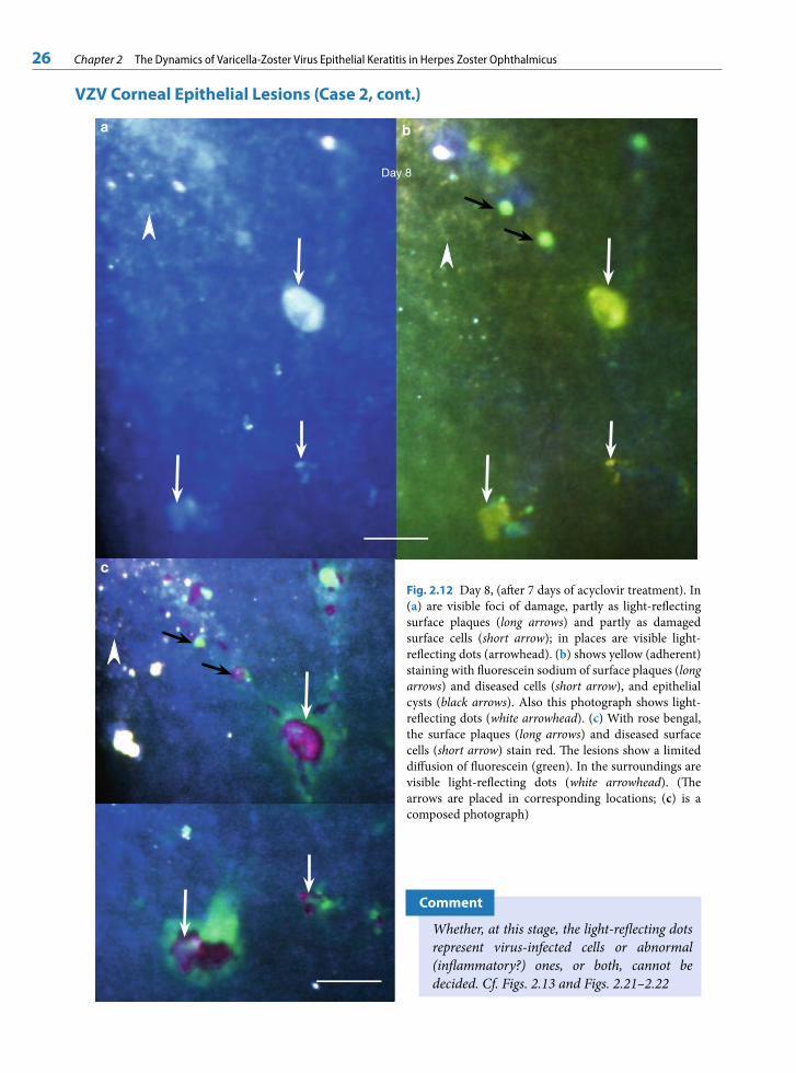
Fig. 2.12 Day 8, (after 7 days of acyclovir treatment). In (a) are visible foci of damage, partly as light-reflecting surface plaques (long arrows) and partly as damaged surface cells (short arrow); in places are visible light- reflecting dots (arrowhead). (b) shows yellow (adherent) staining with fluorescein sodium of surface plaques (long arrows) and diseased cells (short arrow), and epithelial cysts (black arrows). Also this photograph shows light-reflecting dots (white arrowhead). (c) With rose bengal, the surface plaques (long arrows) and diseased surface cells (short arrow) stain red. The lesions show a limited diffusion of fluorescein (green). In the surroundings are visible light-reflecting dots (white arrowhead). (The arrows are placed in corresponding locations; (c) is a composed photograph)
ba
Day 8
c

VZV Corneal Epithelial Lesions (Case 2, cont.) 27
VZV Corneal Epithelial Lesions (Case 2, cont.)
a
b
c
Day 10Fig. 2.13 This figure, captured on day 10 (3 days after the treatment was stopped), is still compat-ible with a result of VZV infection
(a) Before staining are visible many light-reflecting dots, agglomerated or individual (arrowheads)
(b) Fluorescein sodium visualizes diseased surface cells and cell debris (yellow staining) and small cystic spaces (arrow)
(c) Diseased surface cells and cell debris are clearly visible after the application of rose bengal. The staining is in places confluent, and there is still a limited diffusion of green stained tear fluid (white arrow). Also this photograph captured light-reflecting not-stainable dots outside the figure (arrowheads). (The black arrow points to the same cyst as in [b])

28 Chapter 2 The Dynamics of Varicella-Zoster Virus Epithelial Keratitis in Herpes Zoster Ophthalmicus
Subepithelial Opacities: A Sequela of VZV Epithelial Keratitis (Case 2, cont.)
Day 15 Day 24 7 weeks
Fig. 2.14 All surface changes have disappeared. Abnormal cells situated about the level of the basement membrane (present on day 15, 24, and 7 weeks after onset in the area previously showing VZV epithelial lesions) appear as fine dots (shown at higher magnification in Figs. 2.15 and 2.16)
Day 24
Fig. 2.15 Abnormal cells (arrowheads), individual or grouped, on day 24
Subepithelial Opacities: A Sequela of VZV Epithelial Keratitis (Case 2, cont.) 29
Subepithelial Opacities: A Sequela of VZV Epithelial Keratitis (Case 2, cont.)
Addendum
The skin lesions disappeared within a week leaving no scars. Three months after onset, the patient had no com-plaints, and the cornea showed no sequelae.
a b
7 weeks
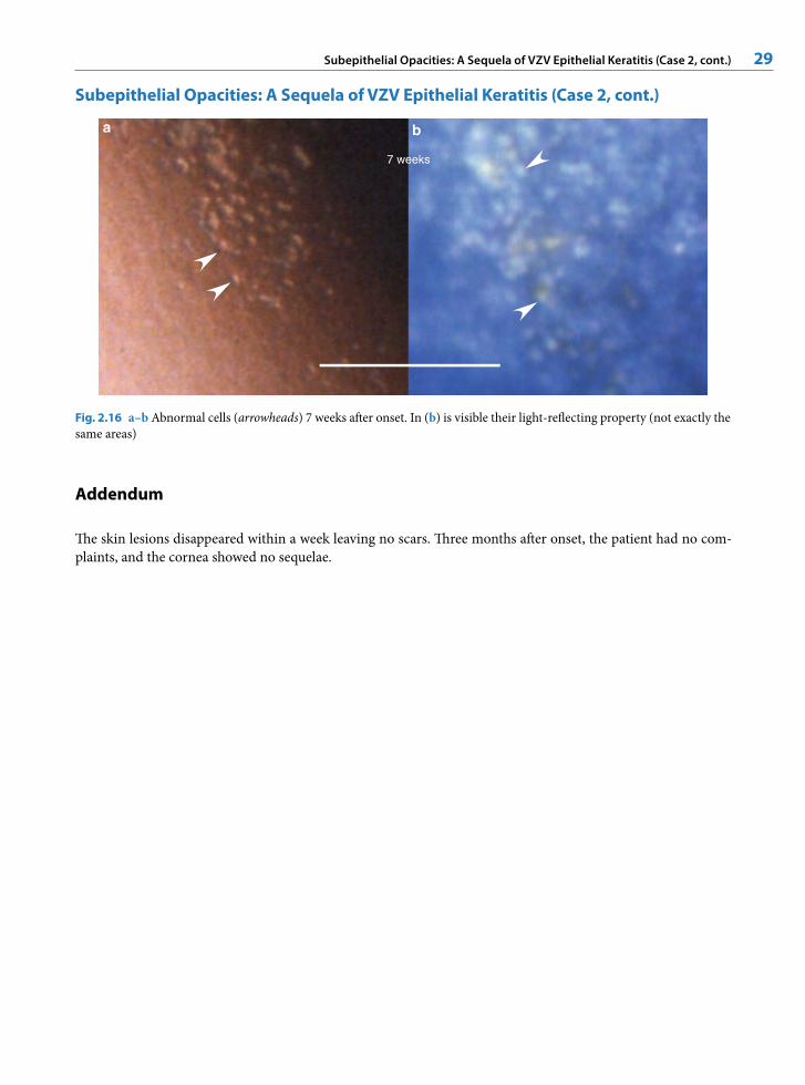
Fig. 2.16 a–b Abnormal cells (arrowheads) 7 weeks after onset. In (b) is visible their light-reflecting property (not exactly the same areas)
30 Chapter 2 The Dynamics of Varicella-Zoster Virus Epithelial Keratitis in Herpes Zoster Ophthalmicus
Case 3: Appearance and Disappearance of VZV Corneal Epithelial Lesions
Day 1
Day 3
Case Report
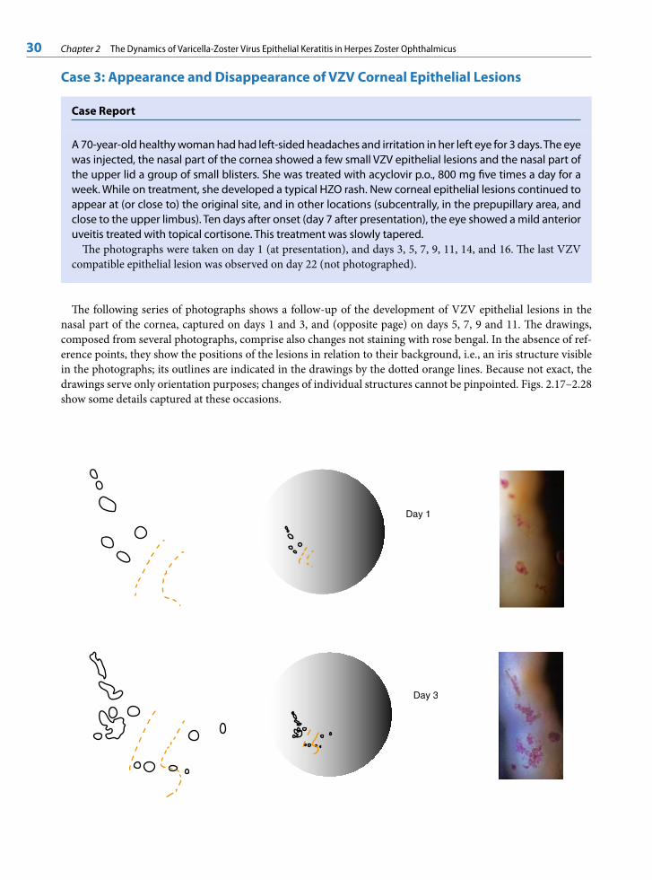
A 70-year-old healthy woman had had left-sided headaches and irritation in her left eye for 3 days. The eye was injected, the nasal part of the cornea showed a few small VZV epithelial lesions and the nasal part of the upper lid a group of small blisters. She was treated with acyclovir p.o., 800 mg five times a day for a week. While on treatment, she developed a typical HZO rash. New corneal epithelial lesions continued to appear at (or close to) the original site, and in other locations (subcentrally, in the prepupillary area, and close to the upper limbus). Ten days after onset (day 7 after presentation), the eye showed a mild anterior uveitis treated with topical cortisone. This treatment was slowly tapered.
The photographs were taken on day 1 (at presentation), and days 3, 5, 7, 9, 11, 14, and 16. The last VZV compatible epithelial lesion was observed on day 22 (not photographed).
The following series of photographs shows a follow-up of the development of VZV epithelial lesions in the nasal part of the cornea, captured on days 1 and 3, and (opposite page) on days 5, 7, 9 and 11. The drawings, composed from several photographs, comprise also changes not staining with rose bengal. In the absence of ref-erence points, they show the positions of the lesions in relation to their background, i.e., an iris structure visible in the photographs; its outlines are indicated in the drawings by the dotted orange lines. Because not exact, the drawings serve only orientation purposes; changes of individual structures cannot be pinpointed. Figs. 2.17–2.28 show some details captured at these occasions.

Appearance and Disappearance of VZV Corneal Epithelial Lesions (Case 3, cont.) 31
Day 5
Day 7
Day 9
Day 11
Appearance and Disappearance of VZV Corneal Epithelial Lesions (Case 3, cont.)
32 Chapter 2 The Dynamics of Varicella-Zoster Virus Epithelial Keratitis in Herpes Zoster Ophthalmicus
VZV Corneal Epithelial Lesions (Case 3, cont.)
ba
dc
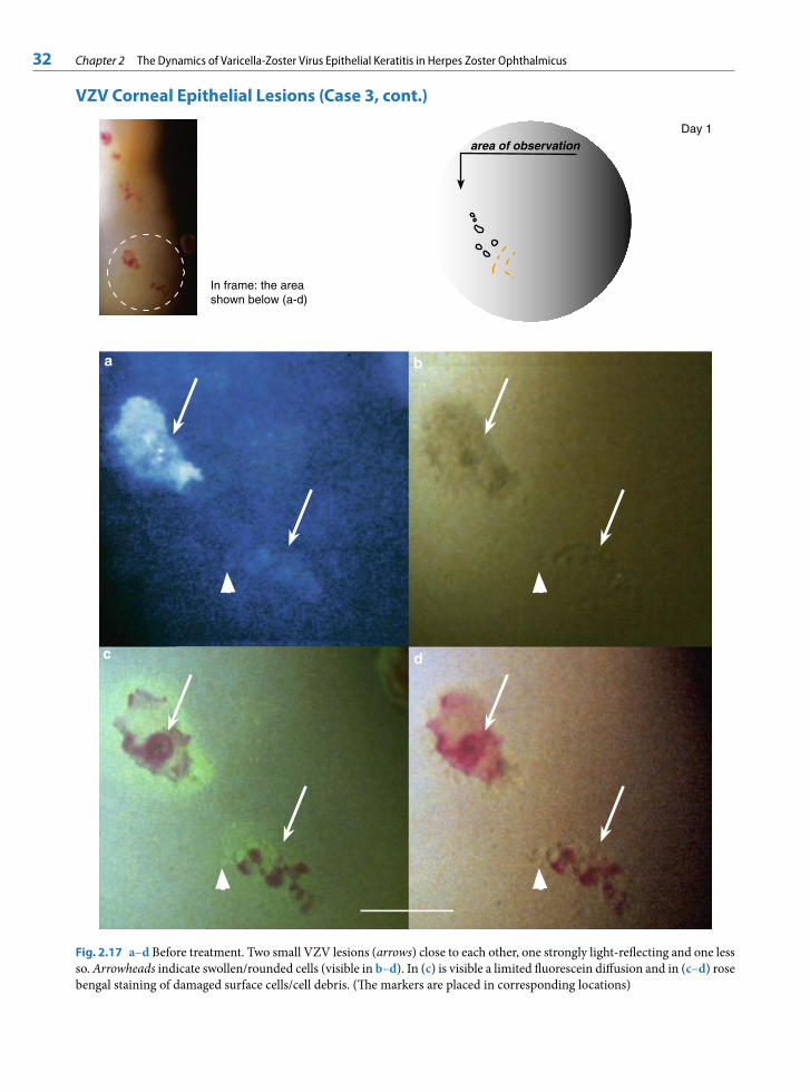
Fig. 2.17 a–d Before treatment. Two small VZV lesions (arrows) close to each other, one strongly light-reflecting and one less so. Arrowheads indicate swollen/rounded cells (visible in b–d). In (c) is visible a limited fluorescein diffusion and in (c–d) rose bengal staining of damaged surface cells/cell debris. (The markers are placed in corresponding locations)
In frame: the areashown below (a-d)
Day 1area of observation

VZV Corneal Epithelial Lesions (Case 3, cont.) 33
c
d
a
b
Fig. 2.18 After 2 days of acyclovir treatment. (a) The light-reflecting lesions (arrows) stain (b) green with fluorescein sodium. There is no diffusion into the surroundings. (The arrows are placed in corresponding locations.) (c) A rounded lesion (long arrow) present subcentrally stains green with fluorescein sodium and, in the center, red with rose bengal. Close to it is visible an incipient lesion (short arrow) that does not stain. (d) shows a light-reflecting lesion (white arrow) and a corneal nerve (black arrow)
VZV Corneal Epithelial Lesions (Case 3, cont.)
In frame: the areashown below (a-b)
Day 3area of observation

34 Chapter 2 The Dynamics of Varicella-Zoster Virus Epithelial Keratitis in Herpes Zoster Ophthalmicus
VZV Corneal Epithelial Lesions (Case 3, cont.)
In frame: the areashown below in (a-b)
Day 5
area of observation
dc
ba
Fig. 2.19 After 4 days of acyclovir treatment. (a) Adjacent incipient lesions (arrows) containing swollen/rounded cells (arrow-heads); (b) the left lesion shows fluorescein diffusion and a red staining of surface cells/cell debris. (The arrows are placed in corresponding locations.) (c–d) shows incipient lesions (arrows) that developed in a new (subcentral) location; they show the same staining as in (a–b). In (d) are visible swollen/rounded cells (arrowhead, cf. also inset). (The arrows are placed in corresponding locations)

VZV Corneal Epithelial Lesions (Case 3, cont.) 35
VZV Corneal Epithelial Lesions (Case 3, cont.)
In frame: the area shown below(a-d). The long white arrow indicates the same location in allphotographs
Day 7
area of observation
ba
dc
Fig. 2.20 After 6 days of acyclovir treatment. The lower part of the lesion shown above shows a VZV typical mixture of features: (a) light-reflecting surface plaques (long white arrow) appear (b) bright in the tear film stained green with fluorescein, and (c–d) stain red with rose bengal. Fluorescein visualizes protruding swollen/rounded cells (b, arrowhead), surface elevations (b and c, bowed arrows), and diffusion into the tissues (c, short white arrow). In (d) are additionally visible swollen/rounded cells (arrowhead). (Adapted from [7])

36 Chapter 2 The Dynamics of Varicella-Zoster Virus Epithelial Keratitis in Herpes Zoster Ophthalmicus
VZV Corneal Epithelial Lesions (Case 3, cont.)
In frame: the areashown below in (a).The black arrowheadsare placed in the samelocation in all photo-graphs.
Day 9
area of observation
a b
d
c
e
Fig. 2.21 (a) shows red staining of damaged cells (black arrowheads), surface plaques (long white arrows), and penetration of green stained tear fluid into the epithelium (short white arrow; composed photograph). In the pair of photographs (b and c) are visible swollen/rounded cells (white arrowheads) present in areas staining green in (a) and larger rounded structures (black arrow), probably cysts. (d and e) This pair of photographs shows an area to the left from the main lesion; it contains heaped-up abnormal cells (white arrowhead) but it does not stain. Cf. Fig. 2.22 and Figs. 2.23–2.24 (overleaf)
Comment
Similarly to Case 2 (Fig. 2.12–2.13), it is impossible, at this stage, to differentiate virus-infected cells from abnormal (inflammatory?) ones.
VZV Corneal Epithelial Lesions (Case 3, cont.) 37
VZV Corneal Epithelial Lesions (Case 3, cont.)
ba
c d
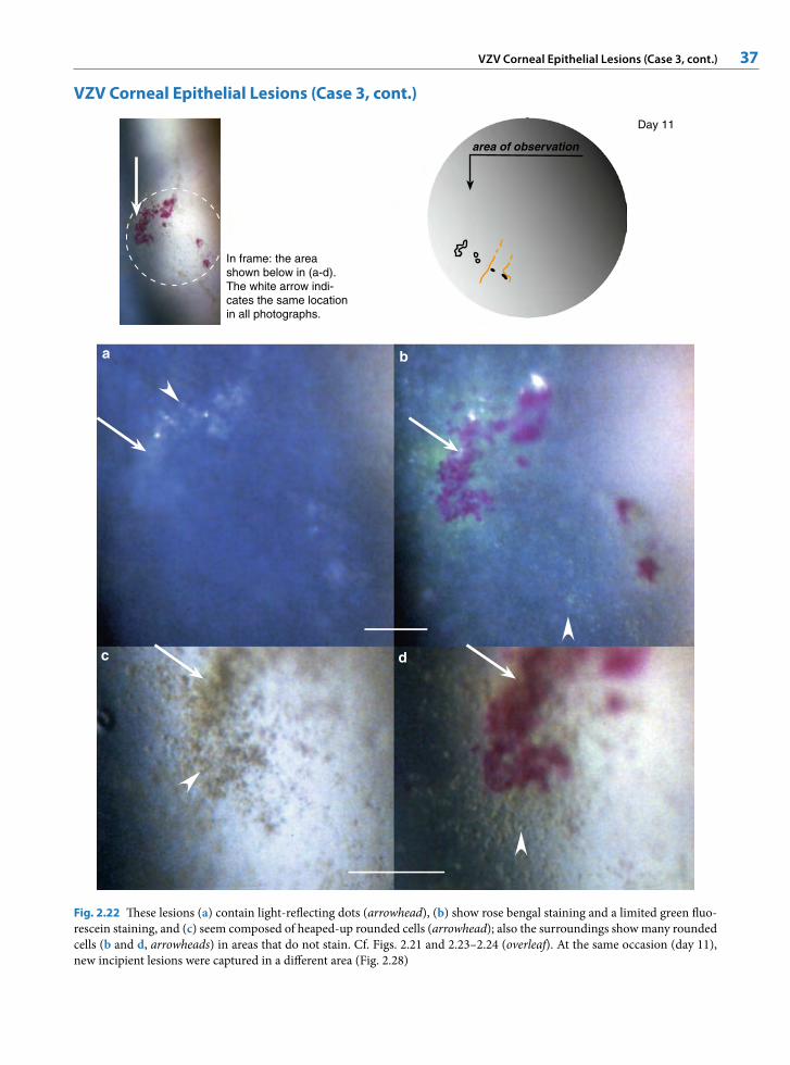
Fig. 2.22 These lesions (a) contain light-reflecting dots (arrowhead), (b) show rose bengal staining and a limited green fluo-rescein staining, and (c) seem composed of heaped-up rounded cells (arrowhead); also the surroundings show many rounded cells (b and d, arrowheads) in areas that do not stain. Cf. Figs. 2.21 and 2.23–2.24 (overleaf). At the same occasion (day 11), new incipient lesions were captured in a different area (Fig. 2.28)
Day 11
In frame: the areashown below in (a-d).The white arrow indi-cates the same locationin all photographs.
area of observation

38 Chapter 2 The Dynamics of Varicella-Zoster Virus Epithelial Keratitis in Herpes Zoster Ophthalmicus
VV Corneal Epithelial Lesions (Case 3, cont.)
A few patches of surface debrisstaining red with rosebengal are still present
Day 14
In frame: the area shown inFig. 2.23 a, below
Day 16
In this area, rose bengalsurface staining has disap-peared
In frame: the area shown inFig. 2.24, below
a
b
Fig. 2.23 (a) In this area, surface changes have disappeared. The rounded cells (arrowhead) probably represent abnormal cells situated about the level of the basement membrane. (b) For comparison, inflammatory cells (arrowhead) attached to the endothelium during anterior uveitis (captured on day 7)
Fig. 2.24 Abnormal cells (arrowhead) situated about the level of the basement membrane
area of observation
Fig. 2.25 (right) At the same occasion (day 16), the upper part of the cornea showed new VZV lesions
Development of VZV Corneal Epithelial Lesions in the Same Location (Case 3, cont.) 39
Development of VZV Corneal Epithelial Lesions in the Same Location (Case 3, cont.)
Day 7 Day 9 Day 11
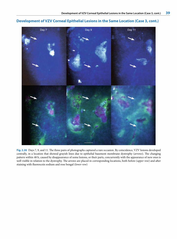
Fig. 2.26 Days 7, 9, and 11. The three pairs of photographs captured a rare occasion. By coincidence, VZV lesions developed centrally, in a location that showed grayish lines due to epithelial basement membrane dystrophy (arrows). The changing pattern within 48 h, caused by disappearance of some lesions, or their parts, concurrently with the appearance of new ones is well visible in relation to the dystrophy. The arrows are placed in corresponding locations, both before (upper row) and after staining with fluorescein sodium and rose bengal (lower row)

40 Chapter 2 The Dynamics of Varicella-Zoster Virus Epithelial Keratitis in Herpes Zoster Ophthalmicus
VZV Corneal Epithelial Lesions (Case 3, cont.)
a
b
Fig. 2.27 Day 7, after 6 days of acyclovir treatment. (a) This lesion (arrow) located subcentrally shows swollen/rounded cells at the edges (arrowhead); similar cells are visible in the upper right corner (arrowhead). (b) shows the light-reflecting property of the same lesion. (The arrows are placed in corresponding locations.) Four days later, lesions with similar features were captured in a different area (cf. Fig. 2.28, opposite page)

VZV Corneal Epithelial Lesions (Case 3, cont.) 41
VZV Corneal Epithelial Lesions (Case 3, cont.)
a
b
Fig. 2.28 Day 11, 3 days after acyclovir treatment was stopped. This subcentral area shows (a) an incipient lesion (bowed arrow), a more advanced one (long arrow), and swollen/rounded cells (arrowheads); the gray arrow points to a small cyst. In (b), fluorescein has disappeared from the tear film: The incipient lesion (bowed arrow) does not stain; the more advanced lesion (long arrow) shows a limited green fluorescein staining, and rose bengal staining; to the right are visible swollen/rounded cells (arrowhead) and a small cyst (gray arrow). (The arrows are placed in corresponding locations)
Addendum
Three months after onset, when last seen, the patient was symptom-free. The cornea showed no sequelae of the infection; the only finding was epithelial basement membrane dystrophy.

H. M. Tabery, Varicella-Zoster Virus Epithelial Keratitis in Herpes Zoster Ophthalmicus DOI: 10.1007/978-3-642-14487-5_3, © Springer-Verlag Berlin Heidelberg 2011
In some patients with HZO, VZV epithelial keratitis recurs. In the present patients, recurrent lesions were indistinguishable from those observed during the HZO episode; both their morphology and their dynamics appeared identical. Such recurrences are less well known but might be more common than suspected. I have seen several patients in whom VZV epithelial keratitis developed between appointments without causing any symptoms that would prompt the patient to attend the Clinic. If developing silently and leaving no traces, many a recurrence probably passes unobserved.
HZO sine herpete is a well-known occurrence that causes considerable diagnostic difficulties because of the absence of rash. In some patients presenting with anterior uveitis, and/or elevation of the intraocular pressure, or stromal keratitis of unknown origin, a sud-den appearance of VZV epithelial keratitis reveals the underlying cause.
In patients with HZO sine herpete presented in this chapter, the epithelial keratitis was indistinguishable from that occurring during acute HZO. Because none of the other HZO manifestations show clinical features that would allow a clear distinction between virus-caused
damage and associated immune phenomena, it cannot be decided whether the epithelial keratitis represented a new episode of virus shedding or was an additional manifestation of an ongoing one. The existence of a pro-longed virus shedding may be suspected in patients with HZO, whether classical or sine herpete, in whom VZV epithelial lesions persist and “migrate” over the surface for several weeks.
Finally, there is one more phenomenon to be men-tioned: zoster mucus plaques keratitis, delayed corneal mucus plaques, late adherent mucus plaques, and delayed pseudodendrites. These terms have been applied interchangeably to epithelial keratitis developing months or years after a known episode of HZO. Since VZV could not be proven (by cell culture), alternative mechanisms had been proposed; later on, however, VZV DNA in such lesions has been proven by PCR. It seems that the most probable mechanisms behind them are recurrent episodes of virus shedding, i.e., episodes of HZO sine herpete.
In five of the six patients presented in this chapter, acyclovir ointment 3% was used for some time during the course of the disease. It did not bring about any detectable morphological change.
Chapter 3
Recurrent VZV Epithelial Keratitis in HZO; HZO sine Herpete

44 Chapter 3 Recurrent VZV Epithelial Keratitis in HZO; HZO sine Herpete
Recurrent VZV Epithelial Keratitis in HZO. Case 1
Case Report
An 86-year-old woman with right-sided HZO. Five days after onset of the rash, the right eye showed a fine epithelial edema and a mild anterior uveitis. Eight months later, while on one drop of steroid a day, the cornea showed several VZV compatible lesions that disappeared within a week. About 16 months after the onset of the first symptoms, the cornea showed a large pseudodendrite. It was scraped off and the material sent to the laboratory. Within 2 days, the epithelial defect was closed but the same site showed two new small lesions, and another one appeared also in the superior temporal quadrant. Acyclovir ointment 3% five times a day was added. All lesions disappeared within a week.
area of observation
a
b
Fig. 3.1 a–b One of several small VZV compatible lesions developing 8 months after HZO. The lesion (arrow) is visible against a background showing a fine texture of basal epithelial edema (arrowheads). (The markers are placed in corresponding locations)

Recurrent VZV Epithelial Keratitis in HZO (Case 1, cont.) 45
Recurrent VZV Epithelial Keratitis in HZO (Case 1, cont.)
area of observation
Fig. 3.2 A branching VZV lesion (pseudodendrite, arrow) developed 16 months after HZO
Addendum
Virus isolation test from the sample was unsuccessful. During the next ten years, no further recurrences of epithelial keratitis were observed but the cornea suffered repeated epithelial erosions.

46 Chapter 3 Recurrent VZV Epithelial Keratitis in HZO; HZO sine Herpete
Recurrent VZV Epithelial Keratitis in HZO. Case 2
a b c
Fig. 3.4 Fresh VZV corneal epithelial lesions showing (a) light-reflecting plaques (arrows), (b) a limited green fluorescein stain-ing, and (c) swollen/rounded cells (arrowhead); such cells are also visible in (b) as protruding round, dark dots (arrowhead). (The markers are placed in corresponding locations)
Case Report
A 90-year-old healthy women presented with a 3–4 days history of discomfort around the right eye. The skin of the forehead showed about 5 pap-ulae but no vesicles. The right eye was slightly injected, and the lower part of the cornea showed a few grouped VZV epithelial lesions. She was treated with valacyclovir p.o., 1 g three times a day for a week. Within 2 days, a few skin vesicles devel-oped. All skin lesions resolved within one week and the corneal lesions within 2 weeks. After that, the eye showed epithelial and stromal corneal edema, keratic precipitates, a mild anterior uveitis, and a slight elevation of the intraocular pressure; she was treated with topical steroid, cycloplegics, and acetazolamide p.o.
area of observation
Fig. 3.3 Survey of VZV corneal epithelial lesions in the lower part of the cornea. The frame indicates the area shown at higher magnification in Fig. 3.4a–c (below)

Recurrent VZV Epithelial Keratitis in HZO (Case 2, cont.) 47
a
b
*
*
Day 1
Fig. 3.5 a–b Recurrence of VZV corneal epithelial lesions 3 weeks after resolution of acute HZO epithelial keratitis. Day 1. Incipient lesions (bowed arrows) cause surface elevations (dark in the green stained tear film). More advanced ones (straight arrows) show fluorescein (yellow) stained surface plaques and swollen/rounded cells (arrowhead); the asterisk indicates the same location as in (b) (composed photograph). (b) The left part of the lesion shows surface elevation (bowed arrow)
Recurrent VZV Epithelial Keratitis in HZO (Case 2, cont.)
Three weeks after the resolution of epithelial keratitis, while on topical steroid four times a day, new VZV lesions appeared in the lover part of the cornea. PCR showed VZV DNA. The steroid was slowly tapered.
At this occasion, photographs were taken on day 1 (at presentation), day 8, and day 15.
area of observation
48 Chapter 3 Recurrent VZV Epithelial Keratitis in HZO; HZO sine Herpete
Recurrent VZV Epithelial Keratitis in HZO (Case 2, cont.)
a
b
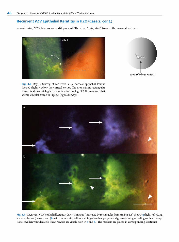
Fig. 3.7 Recurrent VZV epithelial keratitis, day 8. This area (indicated by rectangular frame in Fig. 3.6) shows (a) light-reflecting surface plaques (arrows) and (b) with fluorescein, yellow staining of surface plaques and green staining revealing surface disrup-tions. Swollen/rounded cells (arrowheads) are visible both in a and b. (The markers are placed in corresponding locations)
Day 8
Fig. 3.6 Day 8. Survey of recurrent VZV corneal epithelial lesions located slightly below the corneal vertex. The area within rectangular frame is shown at higher magnification in Fig. 3.7 (below) and that within circular frame in Fig. 3.8 (opposite page)
A week later, VZV lesions were still present. They had “migrated” toward the corneal vertex.
area of observation

Recurrent VZV Epithelial Keratitis in HZO (Case 2, cont.) 49
Recurrent VZV Epithelial Keratitis in HZO (Case 2, cont.)
Fig. 3.9 Recurrent VZV epithelial keratitis, day 8. The subcentral cornea shows several incipient lesions (dark in the green stained tear film, bowed arrows) and more advanced ones (straight arrow). Swollen/rounded cells appear as dark dots (arrowheads)
ba
Fig. 3.8 a–b Recurrent VZV epithelial keratitis, day 8. The area indicated by circular frame in Fig. 3.6 shows swollen/rounded cells (arrowheads), a cyst (short arrow), and a yellow stained surface plaque (long arrow). (The markers are placed in corre-sponding locations.) See also Fig. 3.10 (overleaf)

50 Chapter 3 Recurrent VZV Epithelial Keratitis in HZO; HZO sine Herpete
Recurrent VZV Epithelial Keratitis in HZO (Case 2, cont.)
a
c
b
Fig. 3.10 a–c Recurrent VZV epithelial keratitis, day 8. In this area, the epithelium shows light-reflecting surface plaques (straight arrows) staining yellow with fluorescein sodium, swollen/rounded cells (arrowheads), and incipient lesions causing surface elevations (dark in the green stained tear film, bowed arrows). (The straight arrows and the arrowheads are placed in corresponding locations)

Recurrent VZV Epithelial Keratitis in HZO (Case 2, cont.) 51
Recurrent VZV Epithelial Keratitis in HZO (Case 2, cont.)
Day 15
Fig. 3.11 Recurrent VZV epithelial keratitis, day 15. The epithelium still shows many swollen/rounded cells (arrowheads). (Composed photograph; adapted from [9])
area of observation
After a further week (now 2 weeks after onset of the recurrence), the central cornea showed a large VZV lesion.
Addendum
Three weeks after onset of the recurrence, acyclovir ointment 3% five times a day was added. VZV lesions were still present after a week’s treatment but disappeared within a further 2 weeks. The patient was followed for 5 years. The eye continued to show a mild anterior uveitis and epithelial edema; both recurred each time an attempt was made to stop the steroids. No further episodes of VZV epithelial lesions were observed.

52 Chapter 3 Recurrent VZV Epithelial Keratitis in HZO; HZO sine Herpete
Recurrent VZV Epithelial Keratitis in HZO. Case 3
a b
Fig. 3.12 Recurrent VZV epithelial keratitis, 5 months after HZO. (a) The lesions (arrows) show light-reflecting surface mate-rial (diseased cells/cell debris) that (b) stains red with rose bengal. The green staining is caused by fluorescein diffusion into epithelium of poor quality. (The arrows are placed in corresponding locations)
area of observation
Case Report
A 68-year-old man with Waldenstrom’s macroglobulinemia, treated elsewhere for right-sided HZO (with valacyclovir, 1 g three times a day), developed eye symp-toms 1 day after the treatment was stopped. At presentation, 3 days later, the cor-nea showed several small VZV typical epithelial lesions that were treated with acyclovir ointment 3% five times a day. Within 9 days, they were gone; at that occasion, the cornea showed stromal edema, keratic precipitates, and a mild ante-rior uveitis.
Five months after onset, while on topical steroid three times a day, the cornea showed a new, small, VZV typical epithelial lesion. PCR was positive for VZV DNA. The patient continued to use topical steroid three times a day; the lesion disappeared within a week.
Addendum
After that, the cornea showed fine epithelial edema, diffuse stromal opacities, and a few keratic precipitates. A mild form of dry eye was diagnosed in both eyes. About a year after onset appeared diffuse stromal opacities also in the fellow eye. During the following 6 years, all attempts to withdraw the steroid resulted in recurrences of uveitis and epithelial edema. No further recurrences of VZV epithelial lesions were observed.
Fig. 3.13 (Opposite page) Recurrent VZV epithelial keratitis and anterior uveitis, 11 months after HZO sine herpete. (a) A part of a large pseudodendrite. The surface of the lesion stains yellow with fluorescein sodium; the brightly green staining indicates incipient diffusion into the surroundings. (b–c) Inflammatory cells adhering to the endothelium; some of them (arrowheads) appear fusiform (cf. also Figs. 1.21, 1.22, and 2.23)

VZV Epithelial Keratitis in HZO Sine Herpete. Case 1 53
VZV Epithelial Keratitis in HZO Sine Herpete. Case 1
a b
c
area of observation
Case Report
A 71-year-old woman with polyarthritis and malignant lymphoma pre-sented 6 months after an episode of left-sided HZ oticus (geniculate zoster). The left eye showed corneal epithelial edema, keratic precipitates, a mild anterior uveitis, and elevated IOP (43 mmHg). Three weeks later, while on topical steroid four times a day, the cornea showed several small VZV typi-cal pseudodendrites close to the limbus. They became confluent and per-sisted for about one month.
The patient did not turn up for follow-up visits. When next seen, about 6 months later, the eye was injected, the cornea edematous, the pupil wide, and the IOP elevated (50 mmHg). These symptoms subsided with antiglau-comatous and topical steroid treatment. Eleven months after the onset of the first ocular symptoms (about a year and a half after the initial HZ oti-cus), a large VZV pseudodendrite was present in the nasal part of the cor-nea. She was treated with acyclovir ointment 3% five times a day in addition to topical steroid. The pseudodendrite disappeared within 2 weeks. The patient died shortly thereafter.

54 Chapter 3 Recurrent VZV Epithelial Keratitis in HZO; HZO sine Herpete
VZV Epithelial Keratitis in HZO Sine Herpete. Case 2
a b
Day 1
Fig. 3.14 Day 1. The lower one of two VZV lesions present in the lower nasal corneal quadrant (cf. drawing) shows (a) dam-aged surface cells/cell debris staining yellow with fluorescein sodium (long white arrow), a limited diffusion of fluorescein into the surroundings (brightly green, short white arrow), and adjacent surface elevations (dark areas, bowed arrows). (b) shows swollen/rounded subsurface cells (arrowheads), here visible in the area of the surface elevation captured in (a) at the left lower edge of the lesion (left bowed arrow) and at the edge of the main lesion (straight arrow). (The straight arrow and the left bowed arrow are placed in corresponding locations)
area of observation
Case Report
A 79-year-old man with no history of HZ or eye disease presented with redness of the left eye for a “few days.” The eye was injected, and the temporal part of the cornea showed stromal edema and keratic precipitates; 4 days later, the eye showed a mild anterior uveitis and elevated intraocular pressure (50 mmHg). The symptoms rapidly subsided with topical steroid and acetazolamide p.o.
Seven weeks after onset of the first symptoms, while on topical steroid three times a day, two small VZV lesions appeared in the lower nasal quadrant of the cornea. The photographs, taken at that occasion (referred to as day 1) and on days 2, 3, 8, and 16, show the lesions’ changing shapes and their “migration” in the lower nasal quadrant.
The duration of this episode of epithelial keratitis was about 3 weeks. PCR was positive for VZV DNA.
Fig. 3.15 (Opposite page) Day 2. (a) Survey of changes present in the lower nasal corneal quadrant. The lesions indicated by frames are shown in (b–d) and (e–h). (The flecks visible outside them are caused by keratic precipitates.) (b–d) The lesion in rectangular frame in (a) shows (b) a broken pattern of light-reflecting material that (c) stains yellow with fluorescein sodium and (d) red with rose bengal. In (c–d) is visible a limited fluorescein diffusion (brightly green) into the surroundings. The light-reflecting structure (b and c, arrows) is an incipient lesion (d, inset) located below a still intact surface. The arrowhead in (d) indicates a swollen/rounded cell and the black arrow (b–d) a small cyst. (The markers are placed in corresponding locations.) (e–h) The lesion indicated by circular frame in (a) shows (e) light-reflecting material that stains (g and h) in a similar way as that shown in (b–d). In (f) are visible swollen/rounded cells within the lesion (arrowhead). The arrows (e–h) point to an adja-cent area of incipient cell swelling. (The arrows are placed in corresponding locations; 3.14a and 3.15d adapted from [9])

VZV Epithelial Keratitis in HZO Sine Herpete (Case 2, cont.) 55
e f
g h
a b
c d
Day 2

56 Chapter 3 Recurrent VZV Epithelial Keratitis in HZO; HZO sine Herpete
ba
dc
e f
Day 3
Fig. 3.16 Day 3. (a) Survey. (The flecks outside the lesions indicated by frames are caused by keratic precipitates.) (b–f) show the area indicated in (a) by circular frame. The lesion is (b) light reflecting, (c) contains swollen/rounded cells (arrowheads), (d) the light-reflecting parts stain yellow with fluorescein and (f) red with rose bengal. Fluorescein diffusion (brightly green) increases with time (d–e) but remains limited to the immediate surroundings. Green dots in the surroundings are probably caused by toxic effect of rose bengal. Arrows (placed in corresponding locations) indicate a small satellite lesion
VZV Epithelial Keratitis in HZO Sine Herpete (Case 2, cont.)

VZV Epithelial Keratitis in HZO Sine Herpete (Case 2, cont.) 57
a
b
c
Fig. 3.17 The optical and staining properties of the lesion indicated by rectangular frame in Fig. 3.16a are the same as of that shown in Fig. 3.16b–f. Swollen/rounded cells (arrowheads) are visible within the lesion (b) and outside it within an area that does not stain (an incipient lesion, arrow in a–c and inset in a). (The arrows are placed in corresponding locations; 3.17a and c adapted from [9])
VZV Epithelial Keratitis in HZO Sine Herpete (Case 2, cont.)

58 Chapter 3 Recurrent VZV Epithelial Keratitis in HZO; HZO sine Herpete
VZV Epithelial Keratitis in HZO Sine Herpete (Case 2, cont.)
Fig. 3.18 a–h (This and opposite page). Day 8. (a) Survey. (Flecks visible outside the lesions are keratic precipitates.) The lesions (b–d, rectangular frame) and (e–h, circular frame) show new shapes but the same optical and staining features as before (cf. Figs. 3.15–3.17). In (e–h), the right white arrow indicates an incipient lesion and the black arrow a cyst. (The mark-ers are placed in corresponding locations in each of the lesions; 3.18d adapted from [9])
e
ba
c d
Day 8

VZV Epithelial Keratitis in HZO Sine Herpete (Case 2, cont.) 59
f
g
h

60 Chapter 3 Recurrent VZV Epithelial Keratitis in HZO; HZO sine Herpete
VZV Epithelial Keratitis in HZO Sine Herpete (Case 2, cont.)
a
b
Day 16
Fig. 3.19 Day 16. This part of a large lesion shows (a) strongly light-reflecting plaques (arrows); (b) at their edges are visible swollen/rounded cells (arrowheads). (The arrows are placed in corresponding locations)

VZV Epithelial Keratitis, Anterior Uveitis, and Keratic Precipitates (Case 2, cont.) 61
VZV Epithelial Keratitis, Anterior Uveitis, and Keratic Precipitates (Case 2, cont.)
a
b
Fig. 3.20 a–b Keratic precipitates (arrows) and inflammatory cells (arrowheads) on the endothelium captured on day 3. (b) Some inflammatory cells appear round (white arrowheads), others fusiform (black arrowheads). (Cf. Figs. 1.21, 1.22, and 3.13)
Addendum
Within 3 weeks after the first episode shown here, a new, large, VZV pseudodendrite developed in the lower nasal part of the cornea, and a week later a few small lesions appeared in the upper temporal quadrant. All lesions disappeared within a further 3 weeks. During the following year, topical steroid was stopped and the eye was quiet for several months. A mild form of dry eye was diagnosed in both eyes. Fifteen months after the onset of the first symptoms, keratic precipitates and a mild anterior uveitis recurred.
62 Chapter 3 Recurrent VZV Epithelial Keratitis in HZO; HZO sine Herpete
VZV Epithelial Keratitis in HZO Sine Herpete. Case 3
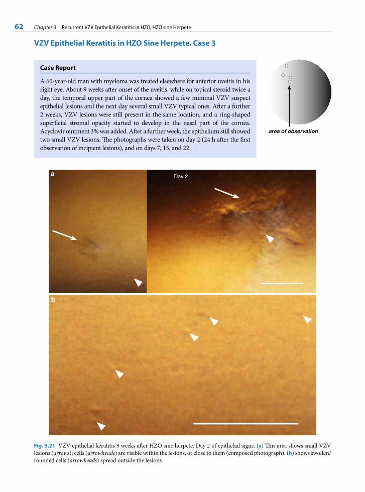
a Day 2
b
Fig. 3.21 VZV epithelial keratitis 9 weeks after HZO sine herpete. Day 2 of epithelial signs. (a) This area shows small VZV lesions (arrows); cells (arrowheads) are visible within the lesions, or close to them (composed photograph). (b) shows swollen/rounded cells (arrowheads) spread outside the lesions
Case Report
A 60-year-old man with myeloma was treated elsewhere for anterior uveitis in his right eye. About 9 weeks after onset of the uveitis, while on topical steroid twice a day, the temporal upper part of the cornea showed a few minimal VZV suspect epithelial lesions and the next day several small VZV typical ones. After a further 2 weeks, VZV lesions were still present in the same location, and a ring-shaped superficial stromal opacity started to develop in the nasal part of the cornea. Acyclovir ointment 3% was added. After a further week, the epithelium still showed two small VZV lesions. The photographs were taken on day 2 (24 h after the first observation of incipient lesions), and on days 7, 15, and 22.
area of observation

VZV Epithelial Keratitis in HZO sine Herpete (Case 3, cont.) 63
VZV Epithelial Keratitis in HZO sine Herpete (Case 3, cont.)
Day 7 Day 15
Day 22
ba
c
Fig. 3.22 VZV epithelial lesions captured on days 7, 15, and 22. (a) With fluorescein sodium, surface plaques (arrows) stain yellow; the brightly green staining indicates diffusion of green stained tear fluid into the surroundings. (b) A larger VZV lesion showing heaped-up swollen/rounded cells (arrowhead). (c) Of these two lesions (arrows), the larger (upper) one shows surface debris staining yellow with fluorescein and red with rose bengal; the smaller (lower) one is located below an apparently intact surface. (The arrows are placed in corresponding locations)
Addendum
Later on, the patient recalled ipsilateral headache (but no rash) in connection with the initial (anterior uveitis) eye symptoms. During the following 5 years, the ring-shaped stromal opacity slowly expanded centripetally. A mild anterior uveitis was waxing and waning, and a diffuse stromal opacity persisted. No recurrences of epithelial keratitis were observed.


H. M. Tabery, Varicella-Zoster Virus Epithelial Keratitis in Herpes Zoster Ophthalmicus DOI: 10.1007/978-3-642-14487-5_4, © Springer-Verlag Berlin Heidelberg 2011
In Case 1, a patient with classic HZO rash, the situa-tion was complicated by basal epithelial edema, ante-rior uveitis, elevated IOP, and basement membrane dystrophy. The epithelium showed a figure standing out against a background consisting of regularly arranged swollen basal epithelial cells. Since this fig-ure neither belonged to the edema, nor showed fea-tures compatible with basement membrane dystrophy, it seemed to be an expression of VZV epithelial infection.
Case 2 was unique in that the epithelium showed a large number of small clear cysts spread over the surface. The cysts, many containing one or two swollen/rounded cells, were indistinguishable from
those often seen in recurrent erosions (own obser-vations), but neither their distribution over the sur-face nor their dynamics belonged to that picture. The mechanism behind their development in this patient with classic HZO rash remains unknown. Later on, after the cysts had disappeared, the epithe-lium showed small lesions compatible with VZV infection.
In Case 3, the ocular surface showed well-discernible conjunctival lesions concurrently with VZV typical corneal ones. Although in HZO the conjunctiva is often injected, conjunctival lesions are rarely seen. Whether in this case a concurrent HIV infection was a predisposing factor remains open.
Chapter 4
Three Rare Cases of Ocular Surface Involvement in Acute HZO

66 Chapter 4 Three Rare Cases of Ocular Surface Involvement in Acute HZO
a b cDay 1
Fig. 4.1 a–c Survey of an arborizing figure (arrow) seen against the background of corneal epithelial edema. It shows (a) with-out staining, many swollen/rounded cells; (b) with fluorescein sodium, surface elevations (dark in the green stained tear film) and small cysts (brightly green dots); and (c) with rose bengal, some damaged surface cells/cell debris staining red. The areas in frames (b and c) are shown at higher magnification in Fig. 4.3, opposite page. (The arrows indicate corresponding locations)
Case 1: HZO, Epithelial Edema, and (presumed) VZV Epithelial Keratitis
Case Report
A 69-year-old woman with corneal basement membrane dystrophy and a history of recurrent erosions presented with a fresh (about 24 h) HZO rash in the right half of the forehead and the right upper lid. The eye was only slightly injected, there was no corneal involvement, and the anterior chamber was quiet.
A week later, she presented again because of blurred vision for 24 h. The eye was injected, the cornea showed epithelial edema all over the surface and several keratic precipitates. There was a mild anterior uveitis, and the IOP was elevated (36 mmHg). In addition to the epithelial edema, the nasal upper corneal quadrant showed an arborizing figure that opti-cally differed from its surroundings. She was treated with topical steroid, cycloplegic, and acetazolamide p.o.
The photographs were taken at presentation (referred to as day 1) and on day 2.
area of observation

HZO, Epithelial Edema, and (presumed) VZV Epithelial Keratitis (Case 1, cont.) 67
HZO, Epithelial Edema, and (presumed) VZV Epithelial Keratitis (Case 1, cont.)
ba
Fig. 4.2 (a) Outside the arborizing figure (Fig. 4.1), the epithelium shows a regular pattern of swollen basal cells (arrowhead) and a few small bullae (arrow). (b) In the area of the arborizing figure, against the background of edematous basal epithelial cells, are visible swollen/rounded cells (arrowheads) that diverge from the picture of epithelial edema
ba
Fig. 4.3 Areas in frames in Fig. 4.1 (opposite page) shown at higher magnification. (a) Elevated parts of the arborizing figure appear dark in the green stained tear film. Cystic spaces stain green with fluorescein sodium (arrow). (b) In the tear film stained red with rose bengal, the figure appears pale. Within the figure are visible swollen/rounded cells (arrowheads) and cysts (arrow) and outside it the regular pattern of swollen basal epithelial cells. (The arrows are placed in corresponding locations)

68 Chapter 4 Three Rare Cases of Ocular Surface Involvement in Acute HZO
HZO, Epithelial Edema, and (presumed) VZV Epithelial Keratitis (Case 1, cont.)
Fig. 4.4 (Right) An arborizing figure, protruding (dark) in the green stained tear film and showing small cystic spaces (green dots). The area in frame is shown in Fig. 4.5 (below)
24 h later, the eye was white, the epithelial edema had almost dis-appeared, and the IOP was 24 mmHg. An arborizing figure was still present in approximately the same location as before.
Fig. 4.5 (Below) The area in frame in Fig. 4.4 (right) at higher magnifica-tion. Both (a) with fluorescein sodium and (b) without staining are visible swollen/rounded cells (arrowheads). In (a) the green staining additionally visualizes a cystic space (arrow). (The markers are placed in correspond-ing locations)
ba
Day 2

HZO, Epithelial Edema, and (presumed) VZV Epithelial Keratitis (Case 1, cont.) 69
HZO, Epithelial Edema, and (presumed) VZV Epithelial Keratitis (Case 1, cont.)
Addendum
The figure disappeared within 5 days. It left a subepithelial shadow that disappeared within the next 3 weeks. During the following year, the steroid was slowly tapered but each attempt to withdraw the treatment resulted either in a mild anterior uveitis or in an episcleritis in the nasal part of the eye. Finally, 14 months after onset, the steroid was stopped. After a further 6 weeks, the patient presented again because of repeated episodes of redness in the nasal part of the eye. At that occasion, the upper nasal part of the cornea showed a few small VZV-compatible epithelial lesions that disappeared within a month. They left subepithelial opacities (Fig. 4.6, below). A year later, when last seen, the eye was quiet and the cornea showed no sequelae.
Comment
In the context of HZO, it was probable that the arborizing figure was relatable to it. Out of the context, it would be difficult to diagnose it as caused by VZV, mainly because of the absence of the VZV typical light-reflecting surface plaques. The reasons for it remain speculative. The lesion left a transitory subepithelial opacity compati-ble with a sequela of VZV infection.The second episode, about 15 months after the first one, was compatible with a recurrence that, similarly to the first one, left transitory subepithelial opacities.
Fig. 4.6 A subepithelial opacity (arrow) developing after a second episode of VZV epithelial keratitis occurring 15.5 months after acute HZO. Many abnormal cells (arrowheads) located about the level of the epithelial basement membrane are visible in the right part of the picture and, at higher magnification, in the inset

70 Chapter 4 Three Rare Cases of Ocular Surface Involvement in Acute HZO
Day 1
Fig. 4.7 Day 1. Many clear epithelial cysts (arrows) spread over the whole cornea (inset, composed photograph) 4 days after the onset of HZO. In some is visible a rounded structure (arrowheads), probably a swollen/rounded cell. (The arrows are placed in corresponding locations.) Cf. also Fig. 4.9 (opposite page)
Case Report
A 79-year-old healthy woman with a history of thoracic HZ thirty years previously presented with a HZO typical rash involving the left part of the scalp, forehead, and the left upper lid. The duration of symptoms was 4 days. The left eye
was injected, and the cornea showed large num-bers of small cysts. She was treated with acyclovir p.o. (800 mg five times a day for a week.) The pho-tographs were taken on day 1 (at presentation), and on days 3 and 8.
Case 2: HZO and Corneal Epithelial Cysts
ba Day 3
Fig. 4.8 Day 3. (a) Epithelial cysts (arrows) are still present 6 days after onset. (b) The upper temporal quadrant shows a small light-reflecting intraepithelial lesion (arrows, placed in corresponding locations)

HZO and Corneal Epithelial Cysts (Case 2, cont.) 71
HZO and Corneal Epithelial Cysts (Case 2, cont.)
Day 3
Fig. 4.9 Day 3. Many of the cysts (arrows) contain rounded structures (arrowheads); the area in frame is shown at higher magnification in the inset to the right
Addendum
Three weeks after the onset of HZO all corneal epithe-lial changes were gone without sequelae. The cornea showed no other changes except for those relatable to keratoconjunctivitis sicca (diagnosed in both eyes after the HZO episode).
Day 8
Fig. 4.10 On day 8 (11 days after the onset of HZO), two small lesions (arrows) were present in the temporal part of the cornea; all epithelial cysts had disappeared
Comment
The corneal epithelial involvement in this patient was exceptional. The small lesions shown in Figs. 4.8b and 4.10 were per se not obviously diag-nostic of a VZV corneal epithelial infection but, in the context of HZO, compatible with it. The cysts developed during a HZO episode, but the mecha-nism behind them remains obscure. Such cysts are very common in recurrent erosion syndrome (own observations), yet in this patient there was no pathology relatable to basement membrane dystrophy.

72 Chapter 4 Three Rare Cases of Ocular Surface Involvement in Acute HZO
Case 3: HZO, VZV Epithelial Keratitis, and VZV Conjunctival Lesions
ba
dc
Day 1
Fig. 4.11 VZV ocular surface lesions in a patient with HIV infection. The inset in (a) shows two conjunctival lesions located close to the corneal limbus. The lesion indicated by the short arrow is shown at higher magnification in (a), and that indicated by the long arrow in (b). (c) shows another conjunctival lesion situated farther away from the limbus. In (d) is visible a concur-rently present corneal epithelial lesion; it contains swollen/rounded cells (arrowheads) (a–c, fluorescein sodium, blue filter)
Case Report
A 28-year-old HIV-positive man had right-sided headaches for a week, rash for 4 days, and redness in the right eye for 2 days. The eye was injected, the cornea showed a few small VZV lesions, and several lesions were present on the conjunctiva close to the limbus. He was treated with acyclovir
p.o. (800 mg five times a day). Two days later, there were several VZV lesions on the cornea, and conjunctival lesions were still present. He did not return for a follow-up visit. The photo-graphs were taken on day 1 (at presentation) and on day 3.

HZO, VZV Epithelial Keratitis, and VZV Conjunctival Lesions (Case 3, cont.) 73
HZO, VZV Epithelial Keratitis, and VZV Conjunctival Lesions (Case 3, cont.)
Day 3
Fig. 4.12 VZV epithelial keratitis after 2 days of acyclovir treatment p.o.; the epithelium shows several light-reflecting surface plaques (straight arrows), and in the left upper corner is visible an incipient lesion (dark, bowed arrow)


H. M. Tabery, Varicella-Zoster Virus Epithelial Keratitis in Herpes Zoster Ophthalmicus DOI: 10.1007/978-3-642-14487-5_5, © Springer-Verlag Berlin Heidelberg 2011
In corneal epithelial keratitis caused by (recurrent) HSV respective (HZO-related) VZV infections, the incipient manifestations resemble: subsurface cell swell-ing and rounding which results in volume increase causing surface elevations and disruptions.
In the further development the two infections diverge, mainly in that HSV lesions show a strong propensity to surface ulcerations while VZV lesions do not; instead, they show light-reflecting surface plaques which give them, or their parts, a whitish/grayish appearance. Well-developed HSV lesions consist of a central area, either filled with swollen/rounded cells or partly cell devoid, surrounded by fairly well defined edges show-ing uplifted grayish surface cells; such organization is not encountered in VZV lesions.
HSV and VZV epithelial keratitis additionally clearly differ in the treatment response. In HSV, topical appli-cation of an antiviral drug arresting the virus replica-tion (acyclovir) results in prompt morphological changes. In VZV, on the other hand, the same treat-ment has no discernible effect on the morphology. (In VZV, the same applies to antiviral treatment p.o.; this has not been studied in HSV.)
HSV and VZV infections seem to differ from each other also in the duration of virus shedding, as judged from the development of new lesions. In human,
a follow-up of the natural course of HSV epithelial keratitis is not possible, but a development of new lesions immediately after the treatment is stopped (usually after 7–10 days) is rare. In VZV, on the other hand, new lesions often continue to appear during sev-eral weeks (³5 weeks, personal observation), and that regardless of if treated or not treated with antiviral drug (acyclovir or valacyclovir).
Unless very early after onset, the differential diag-nosis is often clear before staining but when in doubt, application of fluorescein sodium is helpful. In both HSV and VZV, elevated areas appear dark in the green stained tear film, and both infections may show protruding swollen/rounded cells and surface disrup-tions. The difference is that (a) in fresh HSV lesions, the brilliantly green staining caused by fluorescein diffusion often extends far beyond the lesions’ edges and rapidly transforms the whole area into a green fleck while in VZV the diffusion is slower and limited to the lesions and their immediate surroundings, and that (b) pooling of the green stained tear fluid within lesions occurs only in HSV; in VZV, surface plaques show yellow (adherent) staining with fluorescein. With rose bengal, diseased/damaged surface cells stain red in both HSV and VZV; VZV surface plaques stain vividly.
Chapter 5
Comparison of HSV and VZV Epithelial Keratitis

76 Chapter 5 Comparison of HSV and VZV Epithelial Keratitis
Swollen Epithelial Cells; Surface Ulceration (HSV)
HSV VZV
Fig. 5.1 Both HSV and VZV corneal epithelial infections show swollen/rounded cells, individual (arrowheads) or grouped (arrows)
HSV VZV

Subsurface Changes, Surface Elevations 77
Comment
In the further development, the morphology of HSV and VZV diverges. Surface depressions/erosions occur in HSV but not in VZV lesions. Conversely, light-reflecting surface plaques occur in VZV but not in HSV (cf. Fig. 5.11).
Subsurface Changes, Surface Elevations
HSV VZV
Fig. 5.3 Similarity between incipient stages of HSV and VZV corneal epithelial infections. In both develop foci (long arrows) containing swollen/rounded cells (arrowheads) located in subsurface cell layers. Volume increase caused by cell swelling and rounding results in surface elevations (appearing dark in the green stained tear film). Such HSV and VZV lesions are practi-cally indistinguishable from each other. Surface disruptions, here visualized with fluorescein sodium in HSV (green staining, short arrow), occur also in VZV (cf. Fig. 5.10). (The markers in the VZV pair of photographs are placed in corresponding locations)
Fig. 5.2 (Opposite page) In this small HSV lesion, the center is filled with bright swollen/rounded cells (white arrowhead) and surrounded by dull appearing ones (black arrowhead); the superficial cell layer is missing in the center and uplifted in the surroundings; and there is a well visible margin between the two areas (arrow). Such organization does not occur in VZV infections. VZV lesions also contain swollen/rounded cells (arrowheads), but they are not erosive and lack well-defined edges. (The red fleck visualizes a damaged surface cell stained with rose bengal)
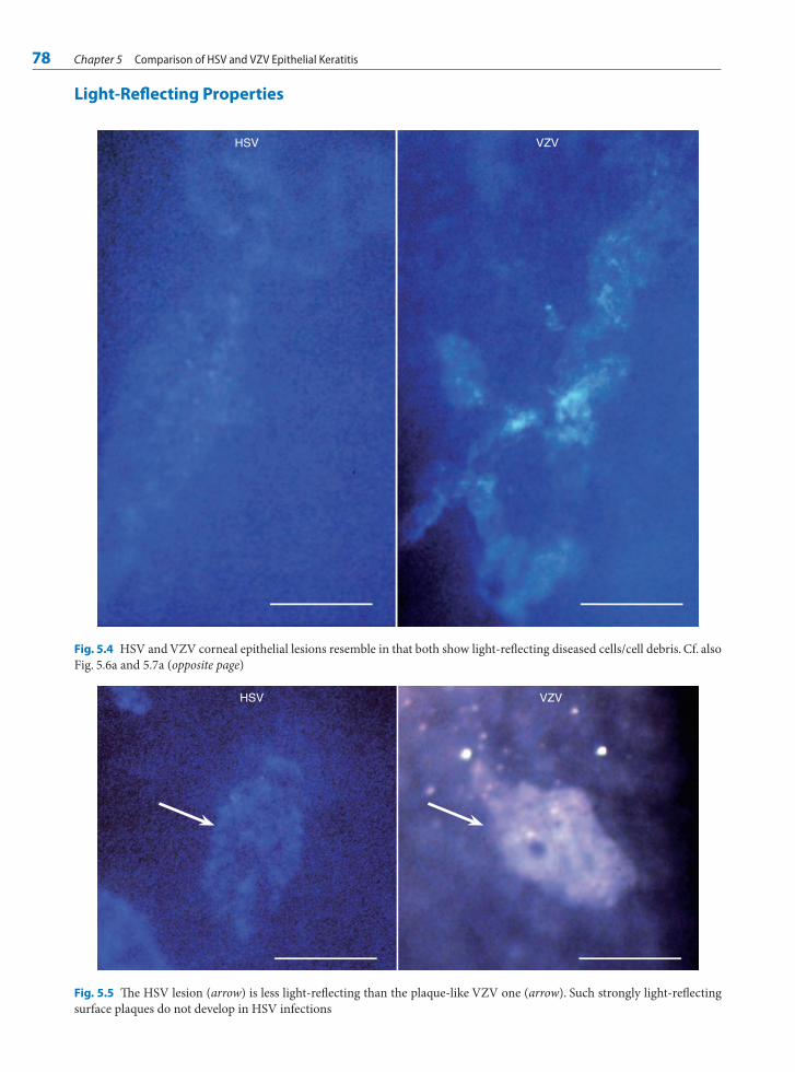
78 Chapter 5 Comparison of HSV and VZV Epithelial Keratitis
Light-Reflecting Properties
HSV VZV
Fig. 5.4 HSV and VZV corneal epithelial lesions resemble in that both show light-reflecting diseased cells/cell debris. Cf. also Fig. 5.6a and 5.7a (opposite page)
HSV VZV
Fig. 5.5 The HSV lesion (arrow) is less light-reflecting than the plaque-like VZV one (arrow). Such strongly light-reflecting surface plaques do not develop in HSV infections

Light-Reflecting Properties and Staining Features 79
Light-Reflecting Properties and Staining Features
VZV
ba
Fig. 5.7 This part of a larger VZV corneal epithelial lesion shows (a) at random distributed light-reflecting cells/cell debris (arrows). (b) Because of diffusion of the green stained tear fluid, the lesion appears larger. Damaged surface cells/cell debris (arrows) stain red with rose bengal; in places, the staining is confluent. (The arrows are placed in corresponding locations)
HSV
ba
Fig. 5.6 In this HSV corneal epithelial lesion, (a) light-reflecting cells/cell debris (arrowhead) are located at the lesion’s edges; the central part appears dark (cf. also Fig. 5.4, opposite page); (b) by diffusion, tear fluid stained green with fluorescein has reached the edges of the lesion; some damaged/diseased surface cells (arrowhead) stain red with rose bengal. (The arrowheads are placed in corresponding locations)

80 Chapter 5 Comparison of HSV and VZV Epithelial Keratitis
Various Aspects of an HSV Lesion
HSVa
b
c
Fig. 5.8 a–c Various aspects of a fresh corneal epithelial HSV lesion
(a) The left part of the picture shows many swollen/rounded cells (arrow-heads); in the island-like area (long arrow) the superficial layers seem still preserved. The right part of the picture shows smaller foci (short arrows); the center of the right one (bowed arrow) seems devoid of superficial cells
(b) Many of the swollen/rounded cells (arrowheads) are light-reflect-ing. The area indicated by the long arrow appears dark
(c) Immediately after the applica-tion of fluorescein sodium, the tear film is stained green. The area indi-cated by the long arrow is protrud-ing (dark)
(The green fluorescence is not visi-ble in the right part of the picture because of a shift of illumination.)
(The arrows in (a–c) are placed in corresponding locations)

Various Aspects of a VZV Lesion 81
Various Aspects of a VZV Lesion
VZVa
b
c
Fig. 5.9 Various aspects of a corneal epithelial VZV lesion
(a) The lesion shows swollen/rounded cells (arrowheads) and a dark (light obstructing) area (arrow)
(b) The dark area corresponds to a strongly light-reflecting surface plaque (arrow). Also the adjacent areas are light-reflecting
(c) The light-reflecting surface plaque (arrow) stains yellow with (adherent) fluorescein and so do the adjacent ones. At the edges is visible incipient fluorescein diffusion (green staining)
(The arrows in (a–c) are placed in corresponding locations)
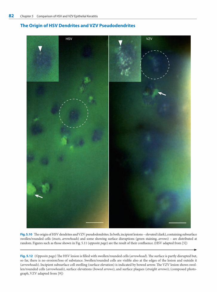
82 Chapter 5 Comparison of HSV and VZV Epithelial Keratitis
The Origin of HSV Dendrites and VZV Pseudodendrites
VZVHSV
Fig. 5.10 The origin of HSV dendrites and VZV pseudodendrites. In both, incipient lesions – elevated (dark), containing subsurface swollen/rounded cells (insets, arrowheads) and some showing surface disruptions (green staining, arrows) – are distributed at random. Figures such as those shown in Fig. 5.11 (opposite page) are the result of their confluence. (HSV adapted from [5])
Fig. 5.12 (Opposite page) The HSV lesion is filled with swollen/rounded cells (arrowhead). The surface is partly disrupted but, so far, there is no erosion/loss of substance. Swollen/rounded cells are visible also at the edges of the lesion and outside it (arrowheads). Incipient subsurface cell swelling (surface elevation) is indicated by bowed arrow. The VZV lesion shows swol-len/rounded cells (arrowheads), surface elevations (bowed arrows), and surface plaques (straight arrows); (composed photo-graph, VZV adapted from [9])
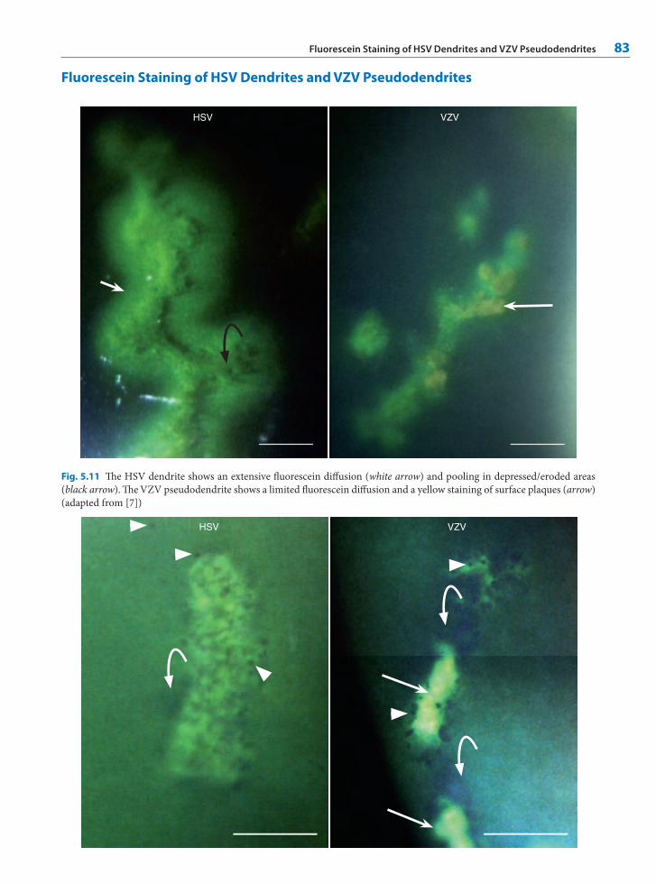
Fluorescein Staining of HSV Dendrites and VZV Pseudodendrites 83
Fluorescein Staining of HSV Dendrites and VZV Pseudodendrites
HSV VZV
VZVHSV
Fig. 5.11 The HSV dendrite shows an extensive fluorescein diffusion (white arrow) and pooling in depressed/eroded areas (black arrow). The VZV pseudodendrite shows a limited fluorescein diffusion and a yellow staining of surface plaques (arrow) (adapted from [7])

84 Chapter 5 Comparison of HSV and VZV Epithelial Keratitis
Rose Bengal Staining of HSV Dendrites and VZV Pseudodendrites
HSV
VZV
Fig. 5.13 This part of a HSV dendrite shows many diseased/damaged surface cells staining red with rose bengal. To the right, the heavy rose bengal staining reminds of a VZV surface plaque because the green fluorescein staining of the in-between areas seems absent; this phenomenon is caused by a shift of illumination. For comparison with a typical VZV plaque, cf. Fig. 5.14 (opposite page). In the VZV lesion, the heavy rose bengal staining shows a configuration reminding of HSV. The green fluo-rescein staining visible in the lesion’s surroundings is caused by penetration of the dye into an epithelium of poor quality

Rose Bengal Staining of HSV Dendrites and VZV Pseudodendrites 85
HSV
VZV
Fig. 5.14 In HSV, this rose bengal staining of diseased surface cells/cell debris is not caused by an active infection; it is a rare sequela that occurred during healing after 11 days of topical acyclovir treatment in a patient with KCS. It is shown here because of the resemblance to surface plaques caused by VZV. The VZV pair of photographs shows typical dense rose bengal staining of a well-developed VZV surface plaque; the left photograph additionally shows fluorescein diffusion into the sur-roundings. (The arrows are placed in corresponding locations in each pair of photographs)
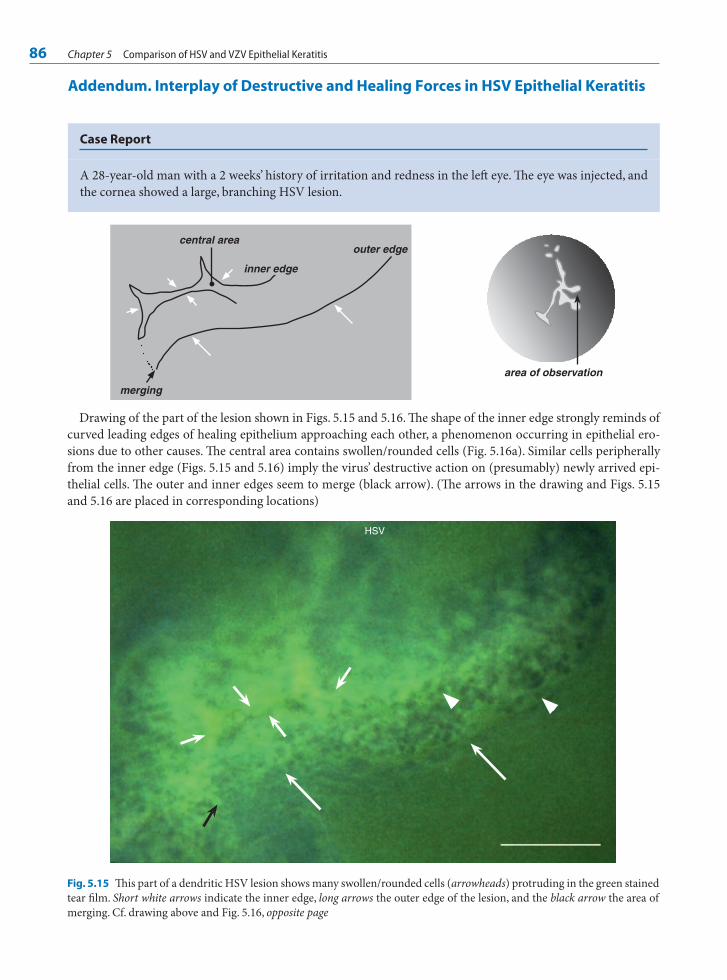
86 Chapter 5 Comparison of HSV and VZV Epithelial Keratitis
Addendum. Interplay of Destructive and Healing Forces in HSV Epithelial Keratitis
HSV
Fig. 5.15 This part of a dendritic HSV lesion shows many swollen/rounded cells (arrowheads) protruding in the green stained tear film. Short white arrows indicate the inner edge, long arrows the outer edge of the lesion, and the black arrow the area of merging. Cf. drawing above and Fig. 5.16, opposite page
Case Report
A 28-year-old man with a 2 weeks’ history of irritation and redness in the left eye. The eye was injected, and the cornea showed a large, branching HSV lesion.
area of observation
inner edge
outer edgecentral area
merging
Drawing of the part of the lesion shown in Figs. 5.15 and 5.16. The shape of the inner edge strongly reminds of curved leading edges of healing epithelium approaching each other, a phenomenon occurring in epithelial ero-sions due to other causes. The central area contains swollen/rounded cells (Fig. 5.16a). Similar cells peripherally from the inner edge (Figs. 5.15 and 5.16) imply the virus’ destructive action on (presumably) newly arrived epi-thelial cells. The outer and inner edges seem to merge (black arrow). (The arrows in the drawing and Figs. 5.15 and 5.16 are placed in corresponding locations)

Interplay of Destructive and Healing Forces in HSV Epithelial Keratitis (cont.) 87
Interplay of Destructive and Healing Forces in HSV Epithelial Keratitis (cont.)
a
b
HSV
Fig. 5.16 The same area as in Fig. 5.15 captured in different illumination modes. The curved edges within the lesion (short white arrows) strongly remind of leading edges of healing epithelium. The area between them and that between them and the outer edge of the lesion (long arrows), both contain swollen/rounded cells (arrowheads). Merging of the outer and inner edges is visible in (a) (black arrow)

88 Chapter 5 Comparison of HSV and VZV Epithelial Keratitis
Final Remark
An explanation of mechanisms behind the morphol-ogy of corneal epithelial lesions in (recurrent) HSV and (HZO-related) VZV infections is challenging because it visualizes a complex, ongoing process involv-ing several events: the primary one (virus-caused dam-age of the individual cell); secondary changes (impact on the epithelial architecture); and the epithelial reac-tion to the disturbance (healing response). The com-parison of the two infections is instructive in that it points to two different modes of interplay between destructive and reparative forces, each one own to the respective virus.
In human HSV corneal epithelial infection, the natu-ral course of the disease cannot be followed because an effective treatment is available. Still, the dynamics of the disease can be reasonably well derived from the typical compositions of the lesions since they are com-patible with a wound that, for some time, resists heal-ing. Of the two forces involved, only the noxious one is clearly visible: partly as cell swelling and rounding, in this context indicating virus-infected epithelial cells, and partly as cell-devoid areas indicating destruction of all epithelial layers. The contribution of reparative forces can be reasonably deduced. In mechanical inju-ries, it is well-known that healing occurs by epithelial reorganization and sliding of epithelial sheets to cover the wound. Most probably, this natural tendency is not arrested in HSV infections but the reparative attempts are unsuccessful because the leading edges of the heal-ing epithelium are continuously infected and destructed (cf. Figs. 5.15 and 5.16). This concept would account for the appearance of lesions that seem to have kept their shapes, are narrow, and contain swollen cells, and that even after two or three weeks after the onset of symptoms. Without a supply of fresh cells from the surroundings, such lesions could hardly exist; at the same time, epithelial movements toward the lesions probably are the force that restricts their lateral
enlargement. The dramatic effect on the morphology of a topically applied antiviral drug (acyclovir) impli-cates in situ replication of HSV virus as the cause of persistence of the infection within the epithelium.
In VZV corneal epithelial infection, the noxious action of the virus on individual cells manifests, simi-larly to HSV, as cell swelling and rounding; contrary to HSV, however, cell damage does not result in a loss of substance (surface ulcerations). This type of distur-bance of the epithelial architecture is lacking an experi-mental model for comparison, and thus the interpretation cannot but rely on the morphology itself in combination with the dynamic features of the dis-ease. The morphology of VZV-caused lesions implies a process involving cell infection, elimination of dam-aged cells by their transportation to the surface, and their substitution by fresh ones. Their dynamics reveal rapid changes of the lesions’ shapes and locations, a course of events well compatible with ongoing, suc-cessful repair that keeps pace with virus damage in conjunction with ongoing virus shedding from the trigeminal ganglion causing new damage in new loca-tions. Hence, in contrast to HSV, focal replication of VZV within the corneal epithelium seems to play an insignificant role, if any. This concept is further sup-ported by clinical observations such as the well-known difficulties to prove living VZV in eye swabs by cell cul-ture (although VZV DNA can be proven by PCR); the equally well-known fact that (contrary to HSV infec-tions) topically applied steroids have no harmful effect; and the absence of detectable morphological changes relatable to an arrest of VZV replication by acyclovir (an effect so promptly reflected in HSV infections).
Taken together, it seems that VZV shed from the trigeminal ganglion infects and damages corneal epi-thelial cells but shows a poor ability to produce infec-tious progeny, for reasons unknown. Perhaps the virus is too slow to replicate and the majority of infected cells are shed from the surface before a complete, infectious virus is produced or released.

H. M. Tabery, Varicella-Zoster Virus Epithelial Keratitis in Herpes Zoster Ophthalmicus DOI: 10.1007/978-3-642-14487-5, © Springer-Verlag Berlin Heidelberg 2011
1. Tabery HM (1992) Dual appearance of fluorescein staining in vivo of diseased human corneal epithelium. A non-contact photomicrographic study. Br J Ophthalmol 76:43–44
2. Tabery HM (1995) Morphology of herpes simplex dendritic keratitis. A non-contact photomicrographic study in vivo in the human cornea. Herpes 2:55–57
3. Tabery HM (1997) Micropunctate staining of the human corneal surface: microerosions or cystic spaces? A non-con-tact photomicrographic in vivo study. Acta Ophthalmol Scand 75:134–136
4. Tabery HM (1998) Toxic effect of rose bengal dye on the living human corneal epithelium. Acta Ophthalmol Scand 76:142–145
5. Tabery HM (1998) Early epithelial changes in recurrent her-pes simplex virus keratitis. A non-contact photomicrographic study in vivo in the human cornea. Acta Ophthalmol Scand 76:349–352
6. Tabery HM (2000) Epithelial changes in early primary her-pes simplex virus keratitis. Photomicrographic observations in a case of human infection. Acta Opthalmol Scand 78:706–709
7. Tabery HM (2000) Morphology of epithelial keratitis in her-pes zoster ophthalmicus. A non-contact photomicrographic study in the human cornea. Acta Ophthalmol Scand 78:651–655
8. Tabery HM (2001) Healing of recurrent herpes simplex cor-neal epithelial lesions treated with topical acyclovir. A non-contact photomicrographic in vivo study in the human cornea. Acta Ophthalmol Scand 79:256–261
9. Tabery HM (2002) Corneal epithelial keratitis in herpes zoster ophthalmicus: ‘delayed’ and ‘sine herpete’. A non-con-tact photomicrographic in vivo study in the human cornea. Eur J Ophthalmol 12:267–275
10. Tabery HM, Holm O (1987) Photography in vivo of epithelial lesions in the human cornea. Acta Ophthalmol (Kbh) 65:513–514
11. Tabery HM (2009) Herpes simplex virus epithelial keratitis: in vivo morphology in the human cornea. Springer, Berlin, Heidelberg
Bibliography.


H. M. Tabery, Varicella-Zoster Virus Epithelial Keratitis in Herpes Zoster Ophthalmicus DOI: 10.1007/978-3-642-14487-5, © Springer-Verlag Berlin Heidelberg 2011
AAcyclovir, 17, 18, 22, 30, 43, 44, 51–53, 62, 70, 72, 75, 88
BBars, xiBasement membrane, 39
dystrophy, 39, 65
CCell(s), 1
abnormal, 1, 12, 13, 28, 29, 36, 38, 69basal, 65, 67confluence, 2culture, 1, 2damaged/diseased, 9debris, 20, 78, 79, 85dull, 77fusiform, 14, 52, 61inflammatory, 13–15, 38, 52, 61rounded, 49swelling, 1swollen/rounded, 2–4, 20, 24, 32, 34, 36, 40, 41,
46–51, 54, 56, 57, 60, 62, 63, 66–68, 76, 77, 80–82, 86, 87
Cyst(s), 5, 26, 41, 49, 54, 65, 66, 70–71Cytopathic effect (CPE), 1
DDendrites, 8, 82–84
EEdema
epithelial, 10, 44, 52, 53, 66, 67stromal, 46, 52, 54
Effect, 1cytopathic, 1–3
Endothelium, 14, 15, 38, 52, 61Episcleritis, 69Epithelium, 86
edema, 65healing, 86–88
FFluorescein, xi, 4, 5, 8, 10, 11, 19–22, 25, 27, 32, 33, 35, 37,
39, 41, 46–48, 50, 52, 54, 56, 63, 66–68, 72, 75, 77, 79, 80, 83, 85
adherent, 7, 9, 18, 26, 81diffusion, 7, 18dynamics, 5–6
Forces, 88destructive, 88reparative, 88
HHerpes, ix
simplex, ixzoster, ix
Herpes simplex virus (HSV), ixrecurrent, 75, 88
Herpes zoster ophthalmicus (HZO), ixsine herpete, 43, 53, 62
Human immunodeficiency virus (HIV), 65, 72
KKeratitis, ix
epithelial, ix
LLesion(s), 4
conjunctival, 65, 72dendritic, 86dynamics, 17–41, 88incipient, 20, 33, 34, 41, 47, 49, 50, 54, 57, 73, 82migration, 54recurrent, 43, 44, 46, 48, 50–52shapes, 17–29
Lines, 18wavy, 18, 21
Lymphoma, 53
MMacroglobulinemia, 52Myeloma, 62
Index

92 Index
OOpacities, 1, 12
stromal, 52, 62subepithelial, 1, 12, 13, 69
PPhotomicrography, xiPrecipitates, 1, 46
keratic, 1, 46, 52–54, 56, 58, 61, 66Pseudodendrites, 1, 8, 18, 19, 22, 43, 45, 53, 61, 82, 83
delayed, 43
RRose bengal, xi, 5, 7–11, 18–20, 22, 23, 25–27, 32, 33, 35, 37,
39, 41, 52, 54, 56, 63, 66, 67, 75, 79, 85toxic effect, 56
SSequela, 1, 11Spaces, 21
cystic, 21, 27, 67, 68
Surface, 1debris, 1disruptions, 1, 4, 77elevations, 1, 24, 35, 47, 50, 54, 77, 82erosions, 1, 11, 77, 86plaques, 1, 7, 18, 19, 25, 26, 35, 36, 43, 46–50, 60, 63, 73,
75, 77, 78, 81–85ulcerations, 1, 75, 88
TTreatment, 75
UUveitis, 1
anterior, 1, 13–15, 30, 38, 44, 46, 52–54, 62, 66, 69
VValacyclovir, 46, 52Varicella-zoster, ixVaricella-zoster virus (VZV), ix